
Jordan Kazakov and Moishe Liberman
Technical Aspects
Echoendoscopes can be categorized as radial and linear (or convex). Radial echoendoscopes provide circumferential view of the airway or the gastrointestinal tract. Linear echoendoscopes provide a view in a plane that is parallel to the long axis of the scope.
The linear endobronchial ultrasound (EBUS) scope is available with an optical system that has a forward oblique viewing angle of 35 degrees or 45 degrees to the long axis of the scope or with a forward viewing optical system. The EBUS scope has an optical field of view of 80 degrees or 100 degrees. The ultrasound transducer (UST) of the linear EBUS scope has a frequency range between 5 and 12 MHz, and a scanning range between 50 and 60 degrees, which is parallel to the long axis of the scope. The depth of view is 2 to 50 mm. Presently, 21- and 22-gauge (G) needles are available for use with the linear EBUS scope. Recent studies by Nakajima et al. and Yarmus et al. showed no difference in the diagnostic yield between 21G and 22G needles although the first study suggested improved preservation of the histologic structure with the 21G needle.
There is no dedicated radial US bronchoscope, instead, there are radial US probes (diameter between 1.8 and 2.6 mm) that can be inserted via the working channel of the bronchoscope and passed distally in the bronchi. The radial US has a frequency range of 12 to 30 MHz and 360-degree circumferential scanning range, which is perpendicular to the long axis of the scope.
The linear and radial endoscopic ultrasound (EUS) scopes have optical system with 55-degree forward oblique viewing angle and 100-degree field of view or forward viewing with 140-degree field of view. The linear UST has frequency range of 5 to 12 MHz, with a scanning range of 180 degrees or 120 degrees, which are parallel to the long axis of the scope. The radial UST has frequency range of 5 to 10 MHz, with a scanning range of 360 degrees, which are perpendicular to the long axis of the scope. The depth of the field is 3 to 100 mm. 19, 22, and 25G needles are available for the EUS scopes (Fig. 3.1).

Figure 3.1 From left to right: Endoscope, radial EUS, linear EUS, bronchoscope, and linear EBUS.
Patient Preparation
Routine preprocedure screening is not recommended; instead, focused history, examination, and testing based on the clinical suspicion is advised. Pregnancy test may be indicated in women in childbearing age. Although endoscopy is not contraindicated in pregnancy, adequate shielding should be utilized when fluoroscopy is used, and adequate preparation should be made in cases when general anesthesia may be required.
Patients should fast at least 2 hours after consuming clear liquids and 6 hours after consuming light meals before administration of sedation. They are advised to continue their cardiac, antihypertensive, pulmonary, antiepileptic, psychiatric drugs, and oral birth control medication. Oral hypoglycemic agents should be withheld on the morning of the procedure until resumption of normal diet. Pills can be taken with small amount of water. Patients on insulin should be advised to take half of their usual morning dose.
Anticoagulation should be managed based on the risk on the thrombotic risk and the estimated risk of bleeding. The anticoagulant is indicated as prophylaxis against development of new thrombi or embolism (mechanical valves and atrial fibrillation) and as treatment of thrombus-related problems (deep vein thrombosis (DVT) and pulmonary embolus (PE)).
 Warfarin should be withheld 5 days before elective procedures. Rapid reversal may be achieved by withholding warfarin and administering vitamin K or if quick reversal is indicated, fresh frozen plasma or prothrombin complex should be administered. Bridging with enoxaparin or heparin is indicated if the thrombotic risk is high (e.g., mechanical valve). Warfarin can be restarted 12 to 24 hours after the procedure with or without bridging. Full anticoagulant effect will be achieved in 4 to 6 days.
Dabigatran should be stopped 1 to 2 days before the procedure if the creatinine clearance is more than 50 mL/min and 3 to 5 days if the creatinine clearance is less than 50 mL/min. The peak anticoagulant effect is achieved 2 to 3 hours after administration. Rivaroxaban and apixaban have elimination half-life and rapid onset of action similar to dabigatran but are less dependent on renal function.
Unfractionated heparin should be stopped 4 to 5 hours before the procedure. The last dose of low-molecular-weight heparin should be 24 hours before the procedure. The anticoagulation effect starts 1 hour after administration and peaks in 3 to 5 hours.
Antiplatelet agents such as clopidogrel and aspirin–dipyridamole (Aggrenox) should be held 7 days before the planned procedure. Clopidogrel is mandatory for 6 weeks after bare-metal stent placement, 3 to 6 months after myocardial infarction, and at least 12 months after drug-eluting stent. Aspirin alone does not increase significantly the risk of bleeding and does not need to be stopped.
Prophylactic antibiotics are not recommended for patients undergoing bronchoscopy, EBUS-, and EUS-guided lung mediastinal and hilar biopsy despite minimal risk of bacteremia. Prophylactic antibiotics are indicated in patients undergoing EUS– fine needle aspiration (FNA) of cystic lesions along the GI tract, including the mediastinum.
Patients undergoing endoscopic procedures with moderate and deep sedation must have continuous monitoring before, during, and after the administration of sedatives. A combination of opioid and benzodiazepine is usually used for moderate sedation. Topical anesthetics like lidocaine, benzocaine, and tetracaine are used for pharyngeal anesthesia, and topical anesthesia of the vocal cords and the airways. Topical anesthetics are associated with serious adverse effects such as aspiration, anaphylactoid reactions, and methemoglobinemia. Deep sedation utilizing propofol alone or propofol in combination with other sedative hypnotics may be used but necessitates a dedicated person with no other procedure-related responsibilities for observation and monitoring of the patient. Furthermore there is a requirement for specific training and credentialing for the personnel involved in medication administration and patient monitoring. General anesthesia or assistance of an anesthesia specialist should be considered in the following scenarios: ASA physical class III, IV, and V; complex endoscopic procedures, prior adverse reactions to sedation and anesthesia, intolerance to standard medication utilized for moderate sedation, inadequate response to moderate sedation, and patients with anatomic variations predictive of increased risk for airway obstruction and difficult intubation.
 INDICATIONS
EBUS and EUS are indicated in diagnostic and therapeutic procedures. Diagnostic procedures include: Staging of nonsmall lung cancer, staging of esophageal cancer, diagnostic evaluation of mediastinal, pulmonary, and endobronchial lesions. Echoendoscopes may also be utilized to guide photodynamic therapy, brachytherapy, and airway recanalization.
Echoendoscopy is a great tool for the diagnosis and staging of lung cancer. EBUS and EUS may be utilized in the determination of all three characteristics—local tumor (T), lymph node metastasis (N), and distant metastasis (M) in the seventh edition of the TNM staging system. The laminar structure of the airway wall and invasion of the airway wall by tumor can be visualized with EBUS. EBUS can also be helpful in differentiating infiltration of the airway wall from external compression with sensitivity, specificity, and accuracy higher than chest computed tomography (CT). It may be used to measure the distance between the carina and the proximal end of tumors involving the bronchial wall. Tumor invasion of the mediastinum can be assessed with EUS. This modality is highly accurate in ruling out mediastinal invasion by lung cancer and in diagnosing great vessels involvement but has high false-positive rate for mediastinal fat involvement. Mediastinal lymph node involvement should be assessed in patients with known or suspected lung cancer who have either one of the following clinical scenarios: Normal mediastinum (by CT and positron emission tomography (PET)) and a central tumor, discrete mediastinal lymph node involvement on CT or PET uptake, and PET activity in a mediastinal lymph node and normal appearing nodes by CT. Utilizing needle technique (EBUS and/or EUS) for mediastinal lymph node staging is recommended over surgical staging as a best first test in patients with high suspicion of N2,3 involvement. Endoscopic needle techniques are also useful for detection of metastases to the liver, adrenal glands, and the celiac lymph nodes. The combination of EBUS and EUS was complementary with pooled median sensitivity of 91% and specificity of 100% in a population with 33% prevalence of cancer. The median negative predictive value (NPV) was 96%.
Echoendoscopy plays a major role in the locoregional staging of esophageal cancer and has essentially replaced chest CT as the locoregional staging modality of choice. Nevertheless, chest CT and PET are used for evaluation of distant metastatic disease. Although the overall accuracy of EUS for tumor (T) and node (N) staging is up to 90%, the accuracy may be lower in patients with early superficial esophageal cancer—67% for T1 lesions. EUS identifies the depth of invasion and defines the T staging based on the involvement of the mucosa (T1), muscularis propria (T2), adventitia (T3), and surrounding structures (T4). The measurement of the maximal thickness of the esophageal mass may help in predicting the extraesophageal extension. EUS may overstage esophageal cancer when invasion of the mediastinum is detected since the tumor may not be reliably differentiated from the surrounding inflammation. EUS combined with FNA carries high sensitivity, specificity, and accuracy for detection of metastatic disease in the lymph nodes. The number of the involved lymph nodes is utilized in the nodal staging of esophageal tumors. Although celiac nodal metastases are considered regional nodal disease in the 2010 TNM staging system, their presence may portend poor prognosis. EUS may also be utilized in the detection of metastatic disease in the adrenal glands, liver, peritoneum, and the lung. Restaging of esophageal cancer after neoadjuvant chemoradiotherapy remains a contentious issue. Two recent studies suggest that EUS may not be useful in the evaluation of the response after neoadjuvant treatment.
EBUS and EUS may be utilized for mediastinal staging of extrathoracic malignancies like breast, kidney, and colon. Also, they provide an alternative approach to transbronchial biopsies and transthoracic needle aspiration for evaluation of peripheral lung masses.
EUS-guided biopsy is helpful in diagnosing lymphomas. The sensitivity of EBUS– trans-bronchial needle aspiration (TBNA) varies between 57% and 90.9% with specificity of 100%. In a recent study by Moonim et al. new diagnosis of lymphoma was made correctly in 88%. The sensitivity for relapse was 100%. The sensitivity of EBUS–TBNA in subtyping lymphomas into high-grade non-Hodgkin, low-grade non-Hodgkin, and Hodgkin lymphomas were 90%, 100%, and 79%, respectively. EBUS–TBNA diagnosis was adequate for clinical management in 84%. EUS-guided biopsy correctly diagnosed 79% of lymphomas and subclassification was successful in 67% in a study by Ribeiro et al.
EBUS-guided TBNA is superior to conventional TBNA using a standard 19G needle as diagnostic approach in patients with hilar and mediastinal lymphadenopathy and suspected of having sarcoidosis in a study by Tremblay et al. In the same study the diagnostic yields were 100% in patients with stage I disease and 87.5% in patients with stage II disease after expert pathologic review of the samples. This yield is comparable with the yield of mediastinoscopy. A prospective study by Annema et al. evaluated 51 patients with suspected sarcoidosis including 36 patients who underwent a nondiagnostic bronchoscopy. EUS–FNA utilizing 22G needle demonstrated noncaseating granulomas in 41 of 50 patients (82%).
Bronchogenic and paraesophageal cysts can be visualized with EBUS and EUS. Aspiration of these cysts should be undertaken with caution because it may lead to infection and mediastinitis.
 CONTRAINDICATIONS
EBUS and EUS, in general, are very safe procedures. There are few absolute contraindications: High-grade proximal airway obstruction, esophageal perforation, uncorrectable bleeding diathesis, high-grade respiratory failure, and a hemodynamically unstable patient.
 SURGERY
The procedure starts with successful insertion of the scope in the trachea (EBUS) or the esophagus (EUS). Using either scope, the viewing angle is oblique to the long axis of the scope, which creates difficulty in terms of visualization of the tip of the endoscope. One should take into consideration the fact that the tip of the scope is distal and posterior to the visual field. For example, when the trachea is intubated the tip of the scope is distal and posterior when the vocal cords are in full view. Thus, slight forward flexion well above the vocal cords will be needed and just the anterior commissure and the anterior part of the vocal cords will be in view. Similar consideration should be made when inserting the EUS scope in the esophagus. There will be slight resistance from the upper esophageal sphincter, which can be penetrated with light sustained pressure. Sustained resistance indicates improper positioning of the scope or anatomic barrier (diverticulum, mass, or stenosis) and further advancement may lead to serious injuries.
Once the scope is in the trachea or the esophagus, it can be advanced directly to the area of interest or systematic evaluation may be needed depending on the clinical situation. Gentle flexion of the scope will bring the tip of the scope in contact with the mucosa. The contact can be enhanced by inflation of the balloon at the tip of the scope with normal saline. When in contact, the scope should be rotated to identify vascular landmarks, which together with anatomical landmarks will help for the proper classification of the mediastinal and hilar lymph nodes and other structures of interest. Color Doppler can be used to further identify vascular structures. Ultrasound signs for malignant involvement of the lymph nodes include size more than 1 cm in the short axis, round shape, distinct borders, heterogeneous echogenicity, hypoechogenicity, presence of coagulation necrosis sign, and the lack of central hilar structures.
When the biopsy target is identified, the needle can be placed in the working channel of the endoscope. Careful manipulation of the needle is of highest importance to minimize the risk for damaging the scope. Providers should be familiar with the particular needle that is available in their institution. The scope should be in relaxed position when the needle is advanced through the working channel. Once the needle is locked safely in place, the sheath should be adjusted using the sheath adjuster knob while visualizing the distal end of the sheath on the endoscopic image. The next step is gentle flexion and visualization of the target again. When the target is in view the assistant should hold the scope close to the mouth to minimize movement of the tip when the needle is deployed. If possible, the space between the cartilaginous rings should be used to penetrate the tracheal wall. Once the needle is inside the target, the needle stylus is withdrawn partially if no suction is needed or completely if suction is applied. The stylus should be used to clean the inside of the needle before withdrawal. Application of suction may cause bloody samples. The needle is moved back and forth in the longest possible tract. The movement is repeated 15 to 20 times. Finally, the needle is removed from the working channel and the stylus is utilized to push the sample on a slide glass for smear or in a container for cellblock preparation.
The optimal number of needle passes per node is four. The incremental diagnostic yield plateaus after the third pass resulting in a cumulative sensitivity after three passes above 90%. Rapid on-site cytopathologic evaluation (ROSE), where available, may improve the diagnostic yield.
Targets
Imaging: http://www.radiologyassistant.nl/en/p4646f1278c26f/mediastinum-lymph-node-map.html
The lymph nodes are categorized according to the Mountain–Dressler regional lymph node classification.
Station 1: Low cervical, supraclavicular, and sternal notch lymph nodes. The cricoid cartilage serves as the upper border and the upper border of the manubrium is the lower border. The midline of the trachea separates 1R and 1L.
Station 2: Upper paratracheal lymph nodes. The upper border of the manubrium serves as the upper border. The lower border on the right is the intersection of the caudal border of the innominate (left brachiocephalic) vein with the trachea. The lower border on the left is the upper border of the aortic arch. The left border of the trachea separates 2R and 2L (Figs. 3.2 and 3.3).
Station 3: Prevascular and prevertebral lymph nodes. The upper border of the manubrium serves as the upper border and the level of the carina serves as the lower border. The prevascular (3A) nodes occupy the space between the posterior surface of the sternum and the anterior surface of the superior vena cava on the right and the left carotid artery on the left. The prevertebral lymph (3P) nodes are located behind the trachea and anterior to the vertebral bodies (Fig. 3.4).
Station 4: Lower paratracheal nodes. The upper border is the intersection of the caudal border of the innominate (left brachiocephalic) vein with the trachea on the left and the cephalic border of the aortic arch on the right. The lower border on the left is the caudal border of the azygos vein. The lower border on the right is upper border of the left main pulmonary artery. The left border of the trachea separates 4R and 4L (Figs. 3.5 and 3.6).

Figure 3.2 Lymph node station 2L seen through EUS. The aortic arch and the left subclavian artery are seen distal to the node.

Figure 3.3 Lymph node 2R seen with EUS. Innominate vein seen distal to the node.

Figure 3.4 Lymph node station 3P seen with EUS.

Figure 3.5 Stations 4 and 5 seen with EBUS. Aorta and pulmonary artery are seen distal from 4L.
Station 5: Subaortic lymph nodes. These lymph nodes occupy the space lateral to ligamentum arteriosum. The lower border of the aortic arch serves as superior border and the upper rim of the left main pulmonary artery serves as the lower border (Fig. 3.7).
Station 6: Para-aortic lymph nodes. These nodes occupy the space anterior and lateral to the ascending aorta and the aortic arch and limited between the upper and the lower borders of the aortic arch (Figs. 3.8and 3.9).
Station 7: Subcarinal lymph nodes. This station occupies the space below the carina, medial of the mainstem bronchi and above the left atrium. The distal end of bronchus intermedius and the distal end of the left mainstem bronchus serve as the lower border of this station (Figs. 3.10 and 3.11).
Station 8: Paraesophageal lymph nodes. These lymph nodes are located around the esophagus. The distal ends of bronchus intermedius and left main bronchus serve as the upper border; and the diaphragm is the distal border (Figs. 3.12 and 3.13).
Station 9: Pulmonary ligament lymph nodes. These nodes are located within the pulmonary ligament. They are limited from the inferior pulmonary vein superiorly and the diaphragm inferiorly (Fig. 3.14).
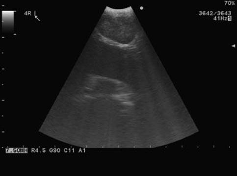
Figure 3.6 Station 4R seen with EBUS. SVC seen distal from 4R.
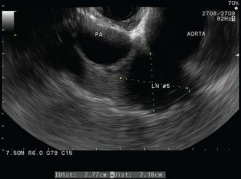
Figure 3.7 Station 5 seen with EUS lateral (distal) to ligamentum arteriosum.

Figure 3.8 The aortic arch with the left subclavian artery seen with EUS. Lymph nodes 5 and 6 seen lateral (distal) to the vessels.

Figure 3.9 Lymph node station 6 lateral (distal) to the aortic arch and the left subclavian artery.
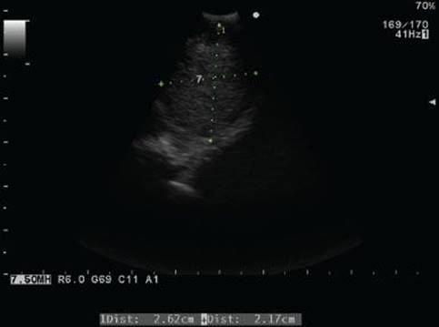
Figure 3.10 Station 7 seen with EBUS.

Figure 3.11 Lymph node 7 seen with EUS. Part of the left atrium is seen anterior and distal to the node.

Figure 3.12 Lymph node station 8L seen with EUS.

Figure 3.13 Station 8R seen with EUS.
Station 10: Hilar lymph nodes. These lymph nodes are located anterior and lateral to the mainstem bronchi. The lower rim of the azygos vein and the upper rim of the left main pulmonary artery serve as the superior border on the right and left, accordingly (Figs. 3.15 and 3.16).
Station 11: Interlobar lymph nodes. These nodes are located in the space between the origin of the lobar bronchi. 11S is located between the upper lobe bronchus and bronchus intermedius. 11I is located between the middle lobe bronchus and the lower lobe bronchi. 11L is located between the left upper and left lower bronchi (Figs. 3.17 and 3.18).
Station 12: Lobar lymph nodes. These nodes are adjacent to the lobar bronchi (Figs. 3.19 and 3.20).
Station 13: Segmental lymph nodes. These nodes are adjacent to segmental bronchi.
Station 14: Intrapulmonary lymph nodes.
Abdominal lymph nodes: Lymph nodes in the gastrohepatic, para-aortic, and celiac trunk areas (Figs. 3.21 and 3.22).
Liver: Segments I, II, and part of segment III are accessible for biopsy via the esophagus (Fig. 3.23).
Adrenal glands: The left and the right adrenal glands can be visualized and biopsied utilizing an EUS scope (Fig. 3.24).

Figure 3.14 Station 9L lymph node seen with EUS. The pleura is seen as a shiny white line just distal to the node.
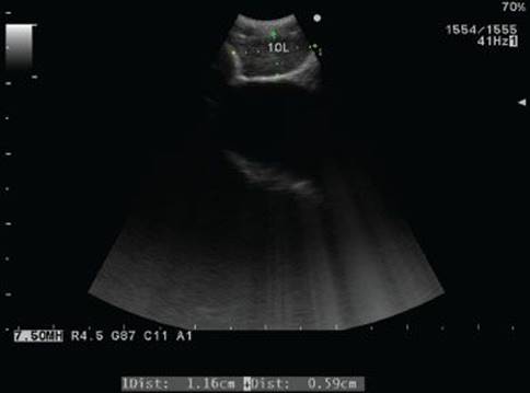
Figure 3.15 Lymph node station 10L seen with EBUS. Left pulmonary artery is seen distal to the node.

Figure 3.16 Station 10R lymph node seen with EBUS.
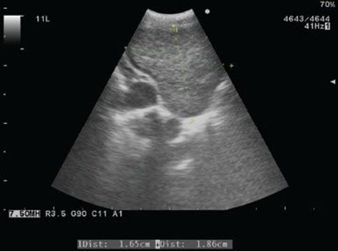
Figure 3.17 Station 11L lymph node seen with EBUS. A branch of the pulmonary artery is seen distal to the node.
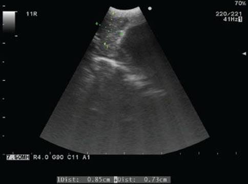
Figure 3.18 Station 11R seen with EBUS. The lymph node is seen inferior to the right pulmonary artery.
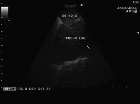
Figure 3.19 Lymph node 12L seen with EBUS. Left hilar mass is seen distal to the node.

Figure 3.20 Station 12R lymph node is seen with EBUS.
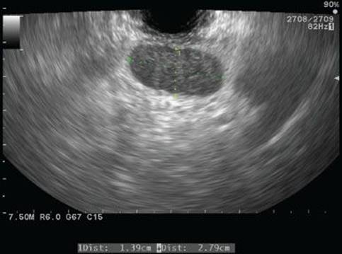
Figure 3.21 Celiac axis lymph node seen with EUS.
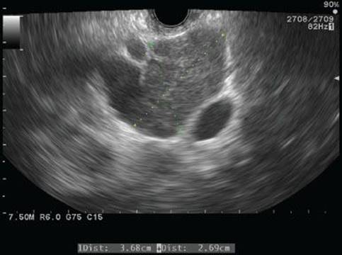
Figure 3.22 Lymph node in the gastrohepatic ligament seen with EUS. The liver is seen superior (right) to the lymph node.
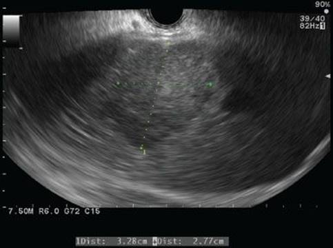
Figure 3.23 Liver mass seen with EUS.
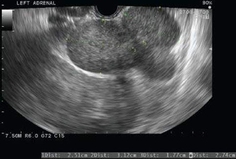
Figure 3.24 Left adrenal gland seen with EUS. The left kidney can be seen inferior and lateral to the nodule within the gland.
 COMPLICATIONS
A recent report by the Japanese Society for Respiratory Endoscopy encompassing 7,345 cases of EBUS–TBNA biopsies in 210 facilities observed a complication rate of 1.23% with hemorrhage being the most common, followed by infection, and pneumothorax.
Bleeding—could originate from the mucosa, laceration of the airway or esophageal wall, and perforation or injury to the surrounding vascular structures during biopsy. In the majority of cases, the bleeding is minor and selflimited. Inadvertent injury to the vascular structures could occur but rarely results in significant bleeding. Local hematoma is the most frequent complication.
Infection—possible infectious complications include mediastinitis, pneumonia, pericarditis, cyst infection, and sepsis. There are rare reports of transmission of hepatitis C, hepatitis B, and Strongyloides by contaminated endoscopic equipment.
Mechanical complications—care should be exercised when the scope is advanced, especially when the scope has oblique view. The tip of the scope can easily perforate the esophageal wall or the membranous wall of the trachea. Abdominal and distal thoracic lymph nodes’ evaluation may be very challenging when there is partial obstruction of the esophagus from esophageal tumor. Initial evaluation with white light endoscope is mandatory to assure distal lumen and assess the diameter of the stenotic area. Laceration of the mucosa may occur.
Pneumothorax—may occur after biopsy of lung mass or inadvertent injury to the lung.
Other complications include bronchospasm, laryngospasm, vocal cord injury, respiratory failure, arrhythmia, and stroke.
 CONCLUSIONS
EBUS and EUS are safe and versatile tools that have revolutionized the diagnosis and staging of thoracic malignancies. EBUS and EUS are now widely used and their role will only increase in future.
Recommended References and Readings
Banerjee S, Shen B, Baron TH, et al. Antibiotic prophylaxis for GI endoscopy. Gastrointest Endosc. 2008;67(6):791–798.
Brugge WR, Lee MJ, Carey RW, et al. Endoscopic ultrasound staging criteria for esophageal cancer. Gastrointest Endosc. 1997;45(2):147–152.
Detterbeck FC, Postmus PE, Tanoue LT. The stage classification of lung cancer: Diagnosis and management of lung cancer, 3rd ed: American College of Chest Physicians evidence-based clinical practice guidelines. Chest.2013;143(5 suppl):e191S–e210S.
Douketis JD, Spyropoulos AC, Spencer FA, et al. Perioperative management of antithrombotic therapy: Antithrombotic Therapy and Prevention of Thrombosis, 9th ed: American College of Chest Physicians Evidence-based Clinical Practice Guidelines. Chest. 2012;141(2 suppl):e326S–e350S.
Edge SB, Byrd DR, Compton CC, et al. (eds). American Joint Committee on Cancer Staging Manual. 7th ed. New York, NY: Springer; 2010.
Ernst A, Silvestri GA, Johnstone D, et al. Interventional pulmonary procedures: Guidelines from the American College of Chest Physicians. Chest. 2003;123(5):1693–1717.
Lee HS, Lee GK, Lee HS, et al. Real-time endobronchial ultrasound-guided transbronchial needle aspiration in mediastinal staging of non-small cell lung cancer: How many aspirations per target lymph node station? Chest.2008;134:368–374.
Ribeiro A, Pereira D, Escalon MP, et al. EUS-guided biopsy for the diagnosis and classification of lymphoma. Gastrointest Endosc. 2010;71(4):851–855.
Sarkaria IS, Rizk NP, Bains MS, et al. Post-treatment endoscopic biopsy is a poor-predictor of pathologic response in patients undergoing chemoradiation therapy for esophageal cancer. Ann Surg.2009;249(5):764–767.
Silvestri GA, Gonzalez AV, Jantz MA, et al. Methods for staging non-small cell lung cancer: Diagnosis and management of lung cancer, 3rd ed: American College of Chest Physicians evidence-based clinical practice guidelines. Chest. 2013;143(5 suppl):e211S–e250S.
Steinfort DP, Conron M, Tsui A, et al. Endobronchial ultrasound-guided transbronchial needle aspiration for the evaluation of suspected lymphoma. J Thorac Oncol. 2010;5:804–809.
Tremblay A, Stather DR, Maceachern P, et al. A randomized controlled trial of standard vs endobronchial ultrasonography-guided transbronchial needle aspiration in patients with suspected sarcoidosis. Chest.2009;136(2):340–346.
Wahidi MM, Jain P, Jantz M, et al. American College of Chest Physicians consensus statement on the use of topical anesthesia, analgesia, and sedation during flexible bronchoscopy in adult patients. Chest.2011;140(5):1342–1350.
Wilson W, Taubert KA, Gewitz M, et al. Prevention of infective endocarditis: Guidelines from the American Heart Association: A guideline from the American Heart Association Rheumatic Fever, Endocarditis, and Kawasaki Disease Committee, Council on Cardiovascular Disease in the Young, and the Council on Clinical Cardiology, Council on Cardiovascular Surgery and Anesthesia, and the Quality of Care and Outcomes Research Interdisciplinary Working Group. Circulation. 2007;116(15):1736–1754.