
Antonio D’Andrilli, Federico Venuta, and Erino A. Rendina
Introduction
Resection and reconstruction of the pulmonary artery (PA) associated with lobectomy is progressively gaining diffusion in lung cancer surgery as a safe and effective therapeutic option that may allow radical resection in a number of patients for whom standard lobectomy is technically not feasible and a pneumonectomy (PN) would be otherwise required.
The right and the left PAs can be involved to various extents. In case of limited, marginal infiltration of the arterial wall, a simple tangential resection with direct suture can be sufficient to achieve a radical exeresis. This technique is generally regarded as a variation of a standard resection and therefore is not considered in this chapter. Larger defects up to 30% to 40% of the vessel circumference may require reconstruction by a patch (of biologic or synthetic material). In case of more extended infiltration a sleeve resection and reconstruction by end-to-end anastomosis, or by the interposition of a prosthetic conduit has to be performed.
The first case of tangential resection and direct suture repair of the PA for lung cancer was reported by Allison in 1952. Later, further descriptions of PA reconstructive procedures were made by Thomas, Perelman, and Wurning between 1956 and 1967. Gundersen in 1967 reported for the first time the successful results of sleeve resection and anastomotic reconstruction of the PA in two patients with left upper lobe tumor.
In 1971 Pichlmeier and Spelberg described four cases of combined bronchial and vascular sleeve resections without complications.
However, during the following two decades, reconstructive procedures of the PA have achieved limited acceptance among the thoracic surgeons due to concern about technical difficulties and increased risk of complications.
The series of 37 PA sleeve resections published by Vogt-Moykopf in 1986 proved that this intervention was feasible with acceptable morbidity rate and good long-term prognosis, but only recently lobectomy associated with PA resection and reconstruction has been demonstrated to be an advantageous alternative to PN.

 Indications and Preoperative Evaluation
The indication for a sleeve resection for lung cancer is well established: A tumor arising at the origin of a lobar bronchus and/or at the origin of the lobar branches of the PA, but not infiltrating as far as to require PN (Fig. 40.1A,B,C). In addition, a sleeve resection may be indicated when N1 nodes infiltrate the bronchus and/or the PA from the outside, as is often the case in the left upper lobe tumors requiring a combined reconstruction of the bronchus and the PA. From a functional point of view, sleeve lobectomy (SL) is strictly indicated in patients who cannot withstand PN, but recent experiences have shown that the advantages of sparing lung parenchyma are evident even in patients without cardiopulmonary impairment.
It is difficult to establish the correct indication for a reconstructive procedure of the PA preoperatively. Computed tomography (CT) with contrast medium is the most used diagnostic tool. Angiography and magnetic resonance imaging (angio-MRI) of the blood vessels can provide useful information to define the pattern of infiltration, but the decision is usually made intraoperatively.
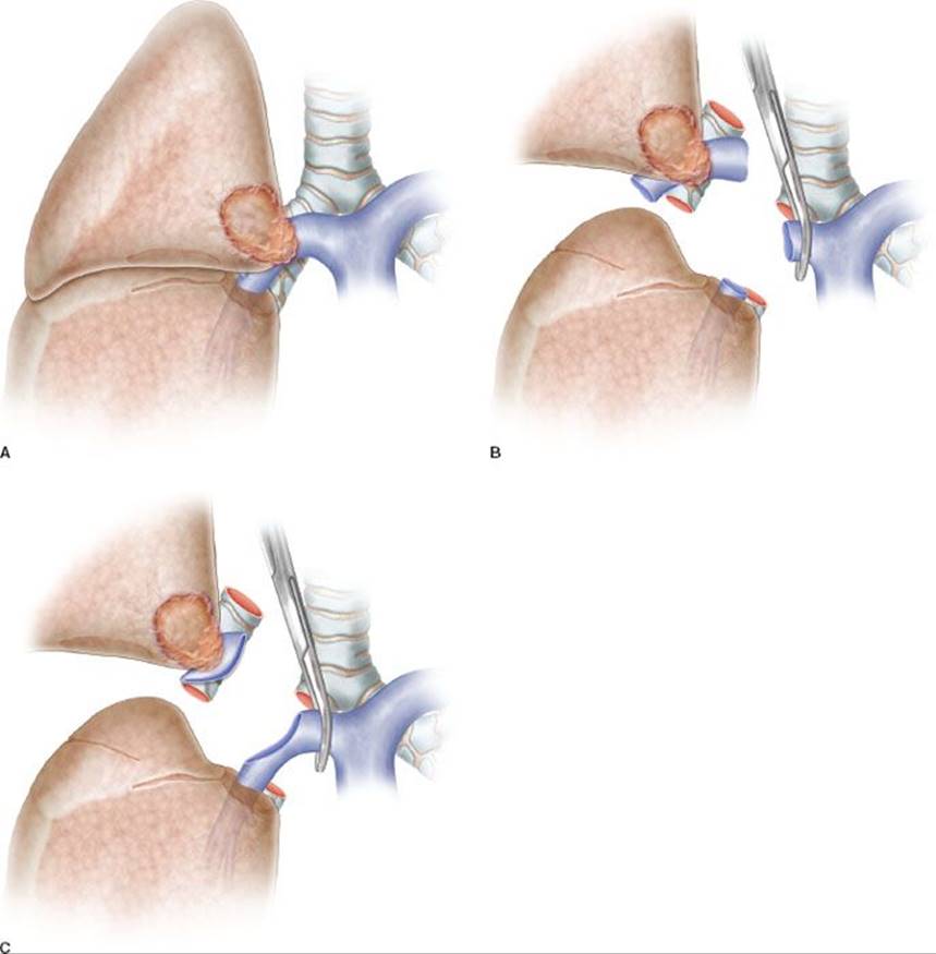
Figure 40.1 A: Partial resection of the pulmonary artery requiring a patch reconstruction. B: Hilar tumor with extensive infiltration of the pulmonary artery requiring a sleeve resection. C: Sleeve resection of the pulmonary artery requiring a reconstruction by end-to-end anastomosis.
PA infiltration degree and extension is not always clearly indicated at preoperative imaging. Sometimes discrepancies between radiologic evidences and intraoperative findings may be responsible of wrong indications, since the preoperative study may overestimate or underestimate the vascular involvement.
Oncologically, the primary goal is in every case the complete resection of the tumor with free resection margins. The decision to perform PN or SL is based on both oncologic and physiologic considerations. There is evidence that PN, especially right PN, is a disease itself, with severe impairment of lung function and quality of life after surgery, and therefore the latter intervention should be avoided whenever possible.
 SURGERY
The first step of the operation consists of achieving full control of the proximal portion of the PA. Although division of the superior pulmonary vein can facilitate the exposure of the PA, transection of this vessel should be postponed until the feasibility of the procedure has been ascertained.
The resection phase begins once the main and distal PA, the bronchus, and both pulmonary veins have been duly prepared. The superior pulmonary vein is generally divided first. Clamping of the proximal PA is then performed after systemic heparinization. In the past we used to clamp the inferior pulmonary vein to obtain backflow control. Actually, we prefer to clamp the PA distally to the tumor infiltration.
The dose of intravenous heparin represents the only intraoperative management modification adopted by the authors over time. We actually prefer to administer 1,500 to 2,000 units (about 25 units/kg) instead of a dose between 3,000 and 5,000 units used in the past. Heparin dose has been reduced to prevent postoperative oozing, especially from the lymphadenectomy sites. We have observed that, with progressive reduction of mean clamping time due to increased experience, this dose proves effective in avoiding the risk of thrombosis. Heparin is not reversed by protamine after declamping, once the vascular reconstruction has been completed.
Resection and Reconstruction Phase
Partial Resection and Patch Reconstruction
Patch reconstruction technique can be applied to a variety of conditions. These may range from limited infiltration of the origin of segmental arteries to larger resection of the PA involving less than one-half of the vessel circumference (Fig. 40.2A). In case of more extended infiltration a sleeve resection with end-to-end anastomosis or conduit interposition has to be performed (Fig. 40.1A,B,C).
Various materials have been proposed and used for patch reconstruction, including synthetic or biologic options, with the latter being preferred because of the higher biocompatibility.
One biologic option is represented by the venous patches including azygos and saphenous vein patches. Azygos vein patches present adequate characteristics, but are available only on the right side and provide limited amount of tissue. Other venous patches generally require separate surgical procedures for tissue harvesting.
Among the biologic materials, the authors recommend the use of the autologous or bovine pericardium. In particular, they prefer the autologous pericardium because it has a number of advantages: It shows adequate thickness and resistance, it is cost free and available on both sides of the chest, and it has superior biocompatibility if compared with the bovine pericardium. Moreover, it provides an amount of tissue that is sufficient also for large defect repair and does not require a separate surgical procedure for its harvesting. However, fresh autologous pericardium has some technical limits, since it has a tendency to shrink and curl making more difficult the adaptation and suturing of the patch to the vascular wall.
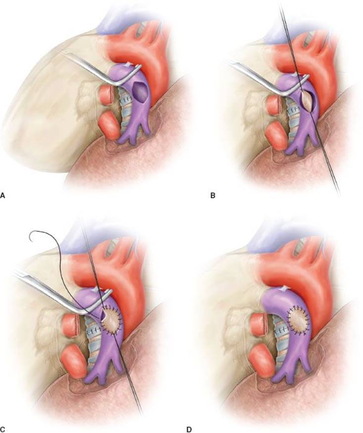
Figure 40.2 Partial resection and patch reconstruction of the pulmonary artery. A: Left pulmonary artery after partial resection of the vascular wall. An oval defect is visible before reconstruction. B: Patch reconstruction: The patch is secured to the vascular wall of the pulmonary artery by two stay sutures. C:Patch is sutured (running suture) to the vascular wall. D: Completed patch reconstruction.
Conversely, bovine pericardium displays limited elasticity and exhibits even and stiff edges that considerably reduce the pitfall of harvesting, trimming, and suturing the autologous pericardium.
To improve technical features of the autologous pericardium, over the last decade, the authors have devised and employed an intraoperative method of fixation of the patch by a glutaraldehyde-buffered solution. The glutaraldehyde preservation of the pericardium minimizes its tendency to retract and curl, thus allowing an easier vascular reconstruction, and reducing the risk of bleeding from the patch suture related to the elastic recoil of the autologous pericardium. Harvesting of the autologous pericardium is performed anteriorly to the phrenic nerve, leaving open the pericardial defect.
The patch is appropriately trimmed and secured to the arterial wall by two stay sutures at the opposite sides of the vascular defect (Fig. 40.2B). Suturing is done with 5-0 or 6-0 monofilament nonabsorbable material proceeding from top-to-bottom “artery first,” and then continuing from bottom-to-top “patch first,” while the assistant grasps and stretches the patch (Figs. 40.2C,D and 40.3). The inferior stay suture is not tied: It is used only to keep the patch in place and is removed when the suture line reaches its level.
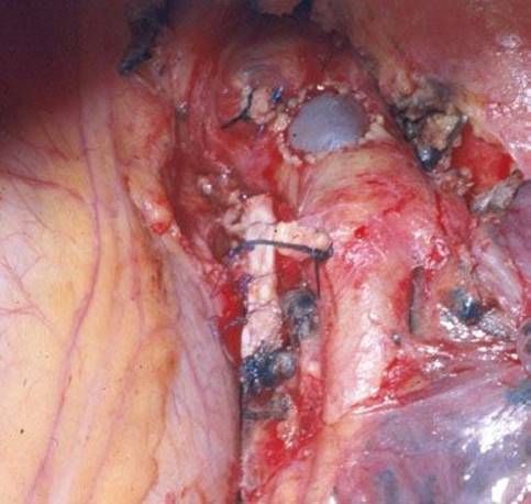
Figure 40.3 Intraoperative picture showing a completed patch reconstruction of the left pulmonary artery.
In patients requiring also a bronchial anastomosis, the PA patch reconstruction is generally performed first, to reduce clamping time.
Check of the suture line after residual lung re-expansion is mandatory, especially if fresh autologous pericardium is used for reconstruction, since retraction of the patch margins may result in bleeding sites when tension changes are applied on the PA axis.
Sleeve Resection and Reconstruction by End-to-end Anastomosis
Sleeve resection of the PA is always required when half or more of the vessel circumference is infiltrated by the tumor (Figs. 40.1B,C and 40.4A). In every case, after resection, frozen section histology should be made on vascular margins and if tumor infiltration persists, PN has to be performed without hesitation.
Transection of the artery proximally and distally to the tumor has to ensure regular and even margins to allow subsequent proper placement of sutures. In addition, regular borders facilitate, during reconstruction, the correction of the caliber discrepancy that is usually found between the two vascular stumps after resection. Due to the elasticity of the arterial wall, caliber discrepancy never represents a technical problem.
In patients requiring a concomitant bronchial anastomosis, the PA reconstruction is generally postponed, since exposure of the bronchial stumps is better when the artery is divided. Moreover, after completion of the bronchial anastomosis the bronchial axis is shortened thus reducing tension on the vascular anastomosis. Distance between the two vascular ends can be further reduced by elevating the lower lobe while suturing. If the distance appears excessive before reconstruction, a prosthetic conduit interposition is indicated. The anastomosis is performed by running suture using 5-0 or 6-0 monofilament nonabsorbable material (Figs. 40.4B,C and 40.5).
The posterior (mediastinal) portion of the anastomosis is performed first, then the suture is completed in the anterior portion, which is the easiest part of the reconstruction. Once the anastomosis has been completed, suture is tied after removal of the distal clamp to restore backflow and to allow air drainage through the untied part of the suture line.
It is important to test the arterial axis and suture line after re-expansion of the residual lobe. Reinflation of the lower lobe elevates the hilum and can produce rotation and kinking of the PA. This may distort suture line and reopen bleeding sites that are not visible when the artery is stretched downward by the atelectatic lower lobe.
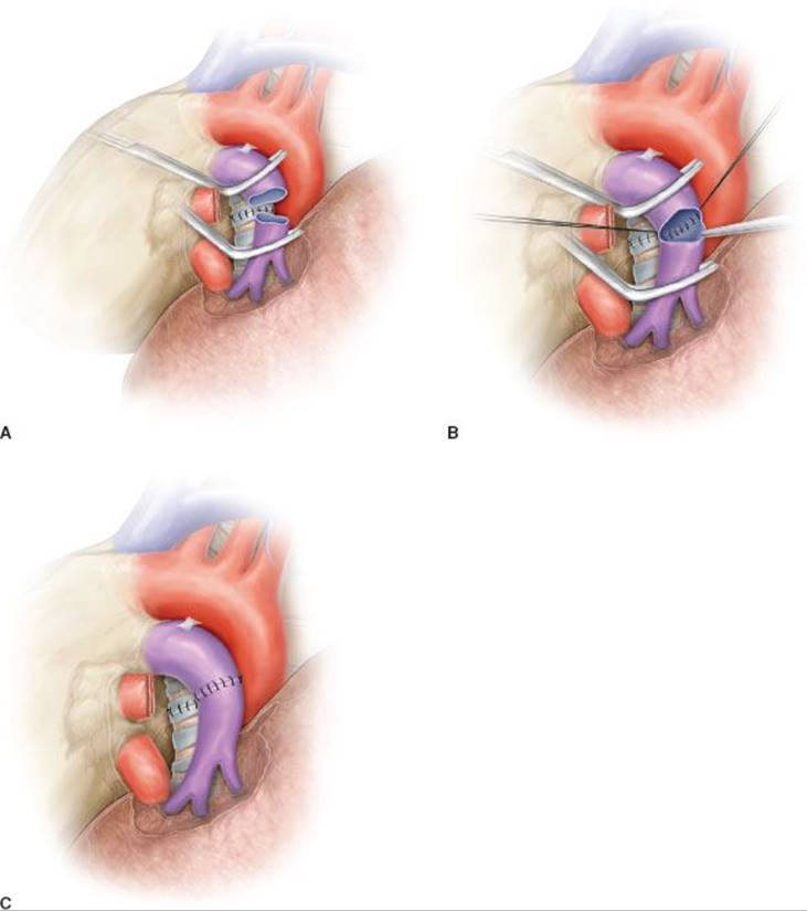
Figure 40.4 Sleeve resection and end-to-end anastomosis of the pulmonary artery. A: Sleeve resection of the left pulmonary artery. The PA is clamped proximally and distally and the two vascular stumps are visible before reconstruction. B: End-to-end anastomosis by running suture. C: Completed end-to-end anastomosis.
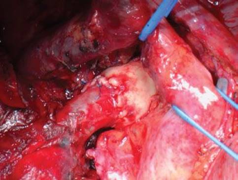
Figure 40.5 Intraoperative picture showing a completed anastomotic reconstruction of the right pulmonary artery.
Sleeve Resection and Reconstruction by a Prosthetic Conduit
In some patients after sleeve resection of the PA an excessive distance between the two vascular stumps may result. This condition could produce a high tension on the anastomosis. Such technical situation may occur, usually on the left side, in those cases requiring resection of a long segment of the PA without associated bronchial sleeve resection, because the lobar bronchus is not involved. In these cases the vascular reconstruction cannot be performed by a direct end-to-end anastomosis and a prosthetic conduit interposition is required (Fig. 40.6A,B).
Although the need for a vascular conduit is not a frequent condition, various materials and different techniques have been proposed for such reconstructive procedure.
Biologic materials are generally preferred because of higher biocompatibility and lower risk of thrombosis. The authors have reported the successful use of the autologous and the bovine pericardium. Intraoperatively, the pericardial leaflet is trimmed to a rectangular shape and wrapped around a chest tube or a syringe of appropriate diameter and sutured longitudinally (Figs. 40.6B and 40.7). In our initial experience this suture was performed manually with a 6-0 monofilament nonabsorbable material (Fig. 40.7). More recently we have described a technical alternative with a mechanical suture using a linear stapler for the conduit construction. The creation of a 1 to 2 cm conduit is so accomplished. When the autologous pericardium is employed the epicardial surface is oriented inside the conduit lumen.
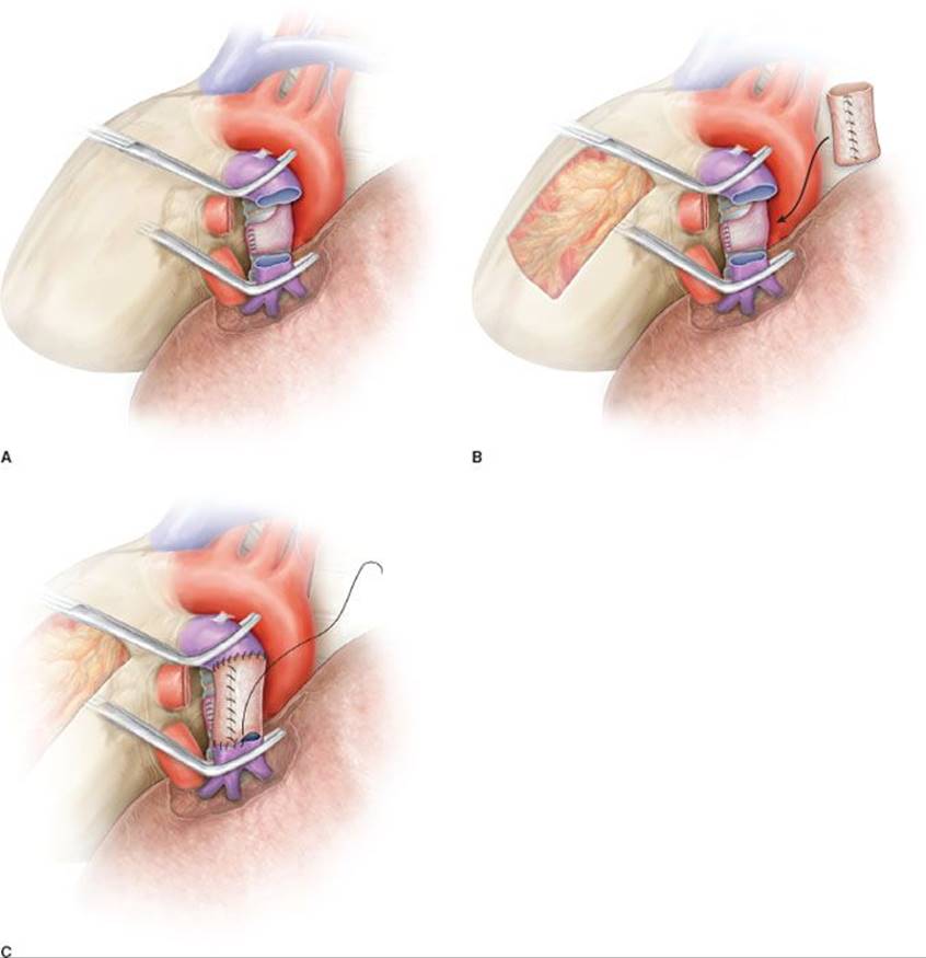
Figure 40.6 Sleeve Resection of the pulmonary artery and anastomotic reconstruction with pericardial conduit interposition. A: Sleeve resection of the left pulmonary artery. The distance between the two PA stumps is excessive and does not allow direct end-to-end anastomosis. B:The autologous pericardial conduit has been created and is ready for reconstruction. The pericardial defect is visible. C: The distal anastomosis is performed by a running suture. The proximal anastomosis has been completed.
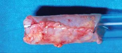
Figure 40.7 Autologous pericardial conduit. The pericardial leaflet has been wrapped around a chest tube and sutured longitudinally.
A very interesting alternative for conduit reconstruction is represented by the pulmonary vein of the resected lobe when the extraparenchymal portion of this vessel is free from tumor. This original technical option has been first described by Cerezo et al. in 2009. The superior pulmonary vein is closed proximally by a thoracoabdominal (TA) stapler (30 mm) and is ligated more distally at the extralobar origin of its branches. Then it is sectioned proximally and distally between sutures, so that a 1 to 2 cm conduit is obtained (Figs. 40.8A,B and 40.9).
The venous conduit is an ideal substitute for PA replacement since it has adequate thickness and structural similarity with the arterial wall. It is advisable to tailor the length of the biologic conduit on the basis of the resected arterial segment, because the elasticity of the two tissues is comparable.
The proximal anastomosis is performed first with running 5-0 monofilament suture. The distal anastomosis is then performed with the same technique, after the conduit length has been checked (Fig. 40.9).
Care must be taken to avoid lengthening of the reconstructed PA, which may cause kinking of the vessel, impaired blood flow, and therefore thrombus formation.
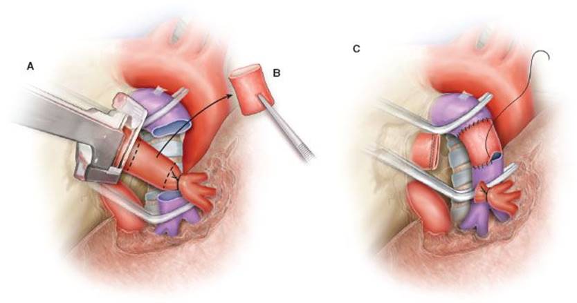
Figure 40.8 Sleeve resection of the PA and reconstruction by a pulmonary vein conduit. A: The pulmonary vein is closed proximally by a TA stapler and ligated more distally at the extralobar origin of its branches. Pulmonary vein is then cut proximally and distally between sutures, thus creating a 1- to 2-cm conduit. B: Pulmonary vein conduit has been created. C: Proximal and distal anastomotic sutures of the venous conduit with the PA stumps.
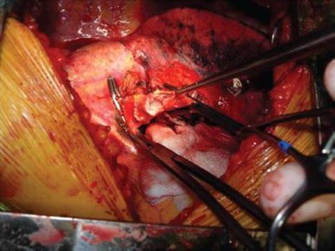
Figure 40.9 Intraoperative picture showing the reconstruction of the left pulmonary artery by the interposition of a pulmonary vein conduit.
For the final success of the reconstruction, it is fundamental to avoid tension on the anastomosis. Tension release can be improved by sectioning the inferior pulmonary ligament and, on the right side, by opening the pericardium around the inferior pulmonary vein.
Arterial Mobilization
Left PA
In case of left upper lobe tumors, the left PA can be isolated extrapericardially if the neoplastic infiltration is found only distally near the interlobar fissure. However, in many patients the artery may be involved close to its origin and therefore the pericardium has to be opened to obtain its proximal clamping. The pericardium is generally incised longitudinally behind the phrenic nerve to confirm that the origin of the left PA is free from tumor and to allow its preparation. The vessel is then encircled by an umbilical tape.
The aortopulmonary window must be carefully dissected paying attention to avoid injury to the recurrent laryngeal nerve. Dissection between the posterior mediastinal pleura and the descending aorta must be then performed to allow exposure and preparation of the main bronchus.
The interlobar fissure is approached, once complete control of the proximal PA has been obtained. Dissection in this area can be safer after proximal clamping of the PA. At this stage exposure of the arterial branches to the superior segment and to the anterior basal segment of the lower lobe has to confirm that the vasculature to the lower lobe is free from tumor and can be spared.
In case of primary tumors of the lower lobe or metastatic hilar nodes infiltrating the inferior aspect of the interlobar artery, the preparation of the proximal PA is generally easier since it is not involved by tumor.
Right PA
Proximal control of the PA is generally obtained extrapericardially and can be facilitated with the anterior and medial retraction of the superior vena cava (SVC). For anatomical reasons, proximal tumors in this area can be unresectable due to invasion of the SVC and right atrium.
Alternatively, when proximal infiltration of the artery is close to the pericardial reflection, the PA can be isolated and clamped transpericardially between the SVC and the ascending aorta.
Distally, it is essential to verify the integrity of the arterial branches to the middle lobe and to the superior segment of the lower lobe. The superior pulmonary vein should not be divided unless the feasibility of the vascular reconstruction has been demonstrated or the patient can functionally tolerate PN.

 Complications and Perioperative Management
The good final outcome in every reconstructive procedure of the PA is principally the result of a meticulous surgical technique. If the operation has been correctly performed, the occurrence of specific perioperative complications may be expected in a low rate of patients. The two main technical complications include bleeding and thrombosis of the reconstructed vessel.
Postoperative bleeding may be related to leakage from suture line of the anastomotic or patch reconstruction. Since the PA is a low-pressure vessel, oozing may not be identified intraoperatively, but it can occur later, once lung re-expansion has been completed, because of modification in PA axis. This can distort suture line and reopen bleeding sites. Such phenomenon may explain unexpected blood loss up to 1,000 mL beginning in the second or third postoperative day with spontaneous resolution within 24 to 48 hours, as observed also in the authors’ experience.
Intraoperative accurate check of suture line even after lung reinflation may help to reduce the risk of this complication.
The occurrence of bronchoarterial fistula after PA reconstruction is rare, but generally represents a catastrophic event resulting in death due to fatal hemorrhage. It is usually associated with a concomitant bronchial sleeve resection and can be effectively prevented by the interposition of a vascularized flap between the two structures. The authors routinely use an intercostal muscle flap, which has an excellent vascularization provided by the intercostal artery. This may favor the preservation of the air tightness at bronchial level even in case of small anastomotic dehiscence, minimizing the risk of PA erosion. Alternatively, the use of mediastinal fat pad, pericardial, or pleural flap has been reported by others.
Lower lobe re-expansion after PA reconstruction may also promote the occurrence of thrombosis because of kinking and folding over on itself produced by the reposition of the reconstructed vessel.
After patch reconstruction of the PA associated with a bronchial sleeve resection, the bronchial axis is shortened while the artery length remains stationary. This discrepancy may produce angulation of the vessel axis, thus determining impairment of blood flow and consequent thrombosis. In such circumstances, after intraoperative check, it is better to cut the distorted segment away and perform an end-to-end anastomosis. The current authors have experienced postoperative thrombosis of the reconstructed vessel only in one patient undergoing patch repair who required reoperation and completion PN. No other case of PA thrombosis was observed over a 30-year period.
In the postoperative period, it is advisable to administer a low-dose anticoagulation therapy (6,000 to 8,000 units/day low-weight heparin subcutaneously) for 1 week. This pharmacologic management contributes to reduce the risk of thrombosis.
However, thrombosis represents a rare event if a patch or anastomotic reconstruction is performed. Differently, higher rates of this complication have been reported after tangential resection with direct suture repair (Read, Wada).
 RESULTS
The evaluation of short-term and long-term results of reconstructive procedures of the PA for lung cancer has been limited over time by the small number of published studies and the different patients’ characteristics of the series reported. Only few data are available in the literature to assess the reliability of this operation and these generally consider heterogeneous populations with prevalence of tangential direct suture repair with respect to true reconstructions (patch, end-to-end anastomosis, and conduit).
Studies Focusing on PA Reconstruction
The authors have published in 2009 the long-term results of their 20-year experience including 105 patients undergoing patch (55 patients) or anastomotic reconstruction (direct end-to-end anastomosis in 47 patients and conduit interposition in three) of the PA for lung cancer. The vascular reconstruction was associated with lobectomy in 102 cases and with a PN under cardiopulmonary bypass in three patients. Tumor stage ranged from IB to III and induction chemotherapy was administered in 23 patients. Patches were made by autologous pericardium in 47 patients and by bovine pericardium in eight. Conduits were made by autologous pericardium in two patients and bovine pericardium in one. In 65 cases the PA reconstruction was associated with a bronchial sleeve resection.
The postoperative morbidity rate was 28.5%, with air leaks resulting the most frequent complication (9.5%). Procedure-related major complications occurred in two patients (1.9%). There was one case of PA thrombosis requiring completion PN and one massive hemoptysis leading to death in a patient undergoing combined bronchovascular reconstruction. The operative mortality was 0.95%. No evidence of PA obstruction or impaired blood flow was observed in the remaining patients by contrast CT during follow-up.
Overall 5-year survival rate including all stages was 44%. The disease-free survival at 5 years was 43.1%. Survival rate at 5 years according to pathologic stage was 75% for stage IB, 53% for stage II, 31% for stage IIIA, and 19% for stage IIIB. These results are in line with those reported in literature for patients not receiving vascular reconstruction. Stage III patients (IIIA and IIIB) presented a significantly worse survival rate at 5 years if compared with patients at stage I and II (25% vs. 60%; p = 0.004).
Surprisingly, patients undergoing isolated PA reconstruction had a significantly worse 5-year survival with respect to patients receiving combined bronchovascular reconstruction (23% vs. 55%; p = 0.01). However, there was no significant difference in disease-free survival between these two groups indicating that the incidence of local recurrence was not higher after isolated vascular reconstruction.
Univariate analysis of overall survival showed significant differences (worse prognosis) for stages III versus stage I and II, induction versus noninduction therapy, PA reconstruction alone versus combined bronchovascular reconstruction, nonsquamous versus squamous histologic type, N2 versus N0 to 1, and presence of complications and recurrence versus none. A trend toward significance (p = 0.05) was found for patch reconstruction versus sleeve resection. At multivariate analysis, only the diagnosis of adenocarcinoma, N2 disease, administration of induction therapy, onset of recurrence, and isolated PA reconstruction confirmed negative effects on outcome.
Univariate and multivariate analyses for disease-free survival did not confirm a negative effect of isolated PA reconstruction.
Another large experience focusing on PA reconstruction associated with lobectomy has been published in 2009 by Alifano. This series included a total of 93 patients (87 with lung cancer), 88 of whom received a tangential resection with direct suture of the PA. Two patch reconstructions and three sleeve resections with end-to-end anastomosis were performed in the remaining patients. Postoperative mortality rate was 5.4%. Nonlife-threatening complication incidence was 27%. Overall 5-year survival in lung cancer patients was 34.9%, with no significant difference between isolated PA and combined bronchovascular reconstructions. The presence of peritumoral and/or intratumoral neoplastic vascular emboli resulted in the most significant adverse prognostic factor (p = 0.006). No significant difference in survival was found according to nodal status (N0 vs. N+) and to histologic type. However, results of this experience cannot be compared with those of ours because of different inclusion criteria, since 95% of patients received only a direct suture PA repair, which was an exclusion criterion in our study.
Some other studies have appeared in the last decade focusing on PA resection and reconstruction for lung cancer. Results from these series have contributed to prove the feasibility and safety of this operation.
Comparison Between Parenchymal Sparing Resections and PN
When evaluating the reliability of parenchymal sparing operations with respect to PN at short and long term, literature data usually analyze results of PA reconstructions together with those of bronchial reconstructions. Therefore a definitive conclusion on the role of PA reconstructive procedures in lung cancer surgery cannot be achieved.
An interesting meta-analysis by Ma, including 12 papers published between 1996 and 2006, has compared early and long-term outcome of SL with those of PN. A total of 2,984 patients have been included in this analysis, of whom 21% undergoing SL and 79% undergoing PN. Two hundred and two patients underwent PA resection and reconstruction in association (164 patients) or not (38 patients) with a bronchial sleeve resection. The evaluation of surgical morbidity including results from eight studies showed a pooled incidence of 31.3% in the SL group and of 31.6% in the PN group without statistically significant difference. Similar results were observed limiting the analysis to studies reporting a larger experience (more than 50 patients) of SL (30.8% complication rate in SL group and 33.6% in PN group). The mean postoperative complication rate reported after PA reconstruction (32.4%) was similar to that reported after bronchial SL and PN. Overall postoperative mortality presented a pooled incidence of 3.5% in the SL group and of 5.7% in the PN group, but this difference did not reach statistical significance. However, when considering only studies with larger number (over 50) of SL, mortality rate was significantly lower in the SL group (3%) than in the PN group (5.7%).
Overall 5-year survival rate extracted from 10 studies was 50.3% after SL and 30.6% after PN, showing a statistically significant difference. The median overall survival was 60 months for the SL group and 28 months for the PN group. This result may have been partially influenced by the higher rate of stage III patients included in the PN group in most studies. However, when considering survival according to pathologic N status (from the few studies reporting this data), pooled 1-year and 5-year survival rate of patients with N0 or N1 disease are significantly higher after SL. In patients with pN2, a slight statistically significant advantage has been observed in 1-year survival for the SL group, while no significant difference was shown for 5-year survival.
Data regarding locoregional recurrences have been reported in only six of the studies considered in this meta-analysis. The pooled locoregional recurrence rate was 16.1% in the SL group and 27.8% in the PN group, but this difference did not reach statistical significance. If considering only studies with larger number of reconstructive procedures a significantly lower incidence of local recurrence was found in the SL group in comparison with the PN group (14.5% vs. 28.7%).
These results in terms of local recurrence were conflicting with respect to those of a previous meta-analysis published by Ferguson. This analysis showed similar survival results with a better long-term prognosis after SL, but the incidence of local recurrence appeared higher in comparison with PN.
The most recent meta-analysis assessing the role of parenchymal sparing operations in comparison with pneumonectomy appeared in 2012 by Shi et al. This included 19 studies published between 1996 and 2011. A total of 3,878 patients have been analyzed, 33.9% of whom undergoing SL and 66.1% undergoing PN. The distribution of patients according to tumor stage was significantly different between the two operations, with a predominance of stage I and II patients in the SL group and of stage III patients in the PN group. PA reconstructions in association or not with bronchoplasty were considered, but the number of patients receiving vascular procedures was not specified. This meta-analysis showed a significantly lower pooled postoperative mortality in patients undergoing SL as compared with those receiving PN (2.91% vs. 5.86%). Differently there was no significant difference in the pooled incidence of postoperative complications between bronchovascular reconstructions and PN (32.88% vs. 27.06%).
The pooled locoregional recurrence rate after SL was lower (14.4%) than that reported after PN (26%), but this difference did not reach statistical significance. The analysis of survival at 1, 3, and 5 years extracted from those studies reporting this data showed a statistically significant difference (combined risk difference and hazard ratio) in favor of the SL group. However, these results could have been strongly influenced by the higher rate of early stage patients included in the SL group.
Bronchovascular Reconstructions after Induction Therapy
Special concern has been expressed by many thoracic surgeons when considering bronchovascular reconstructive procedures after induction therapy, due to the higher risk of perioperative complications and mortality. Although only few authors perform sleeve resection routinely after neoadjuvant therapy, it has been proven in our experience, that even complex parenchymal sparing operations can be performed safely after oncologic treatment without increased morbidity and mortality rates, observing long-term results comparable to those of the standard procedures.
In 2013, we have published the long-term results of our experience comparing SL with PN after induction chemotherapy. A total of 39 patients undergoing bronchial and/or vascular reconstruction associated with lobectomy were analyzed and compared with 39 patients undergoing PN over a 14-year period. A PA sleeve resection was performed in 18 patients and it was associated with bronchial sleeve resection in six of them.
Postoperative complications occurred in 28.2% of patients receiving bronchovascular reconstruction and in 33.3% of the PN group, without statistically significant difference between the two surgical options. Complications related to the reconstructive procedure occurred in one patient: A late stenosis of the bronchial anastomosis was observed and it was successfully treated by laser and stenting. Postoperative mortality rate in the PN group was 2.6%, whereas there was no mortality in the SL group. Difference in postoperative mortality was not significant (p = 0.3).
The tumor recurrence rate was 20.5% in the SL group (locoregional in two patients, distant in six) and 30.8% in the PN group (locoregional in one patient, distant in 11), but this difference was not significant. In particular there was no significant difference between the two groups if considering locoregional recurrence rate only.
 CONCLUSIONS
Lobectomy associated with sleeve resection of the PA represents a viable and safe therapeutic option in lung cancer surgery. When this operation is performed with correct indications and adequate technical experience, it may provide functional advantages of standard lobectomy associated with oncologic radicality of PN, even after induction therapy. Results of our experience and other literature data support the choice of this technique also in patients without cardiopulmonary impairment.
Recommended References and Readings
Alifano M, Cusumano G, Strano S, et al. Lobectomy with pulmonary artery reconstruction: Morbidity, mortality and long term survival. J Thorac Cardiovasc Surg. 2009;137:1400–1405.
Allison PR. Course of thoracic surgery in Groningen. Quoted by Jones PH. Lobectomy and bronchial anastomosis in the surgery of bronchial carcinoma. Ann R Coll Surg Engl. 1959; 25:20–38.
Cerezo F, Cano JR, Espinosa D, et al. New technique for pulmonary artery reconstruction. Eur J Cardiothorac Surg. 2009;36(2):422–423.
Cerfolio RJ, Bryant AS. Surgical technique and results for partial or circumferential resection of the pulmonary artery for patients with non-small cell lung cancer. Ann Thorac Surg. 2007;83:1971–1977.
Chunwei F, Weiji W, Xinguan Z, et al. Evaluation of bronchoplasty and pulmonary artery reconstruction for bronchogenic carcinoma. Eur J Cardiothorac Surg. 2003;23:209–213.
D’Andrilli A, Ibrahim M, Venuta F, et al. Glutaraldehyde preserved autologous pericardium for patch reconstruction of the pulmonary artery and superior vena cava. Ann Thorac Surg. 2005; 80:357–358.
Deslauriers J, Grégoire J, Jacques LF, et al. Sleeve lobectomy versus pneumonectomy for lung cancer: A comparative analysis of survival and sites or recurrences. Ann Thorac Surg. 2004;77(4):1152–1156.
Fadel E, Yldizeli B, Chapelier A, et al. Sleeve lobectomy for bronchogenic cancers: Factors affecting survival. Ann Thorac Surg. 2002;74:851–859.
Ferguson MK, Lehman AG. Sleeve lobectomy or pneumonectomy: Optimal management strategy using decision analysis techniques. Ann Thorac Surg. 2003;76(6):1782–1788.
Kim YT, Kang CH, Sung SW, et al. Local control of disease related to lymph node involvement in non-small cell lung cancer after sleeve lobectomy compared with pneumonectomy. Ann Thorac Surg.2005;79:1153–1161.
Lausberg HF, Graeter TP, Tscholl D, et al. Bronchovascular versus bronchial sleeve resection for central lung tumors. Ann Thorac Surg. 2005;79(4):1147–1152.
Ludwig C, Stoelben E, Olshewski M, et al. Comparison of morbidity, 30-day mortality and long-term survival after pneumonectomy and sleeve lobectomy for non-small cell lung carcinoma. Ann Thorac Surg.2005;79:968–973.
Ma Z, Dong A, Fan J, et al. Does sleeve lobectomy concomitant with or without pulmonary artery reconstruction (double sleeve) have favourable results for non-small cell lung cancer compared with pneumonectomy? A meta-analysis. Eur J Cardiothorac Surg. 2007;32:20–28.
Maurizi G, D’Andrilli A, Anile M, et al. Sleeve lobectomy compared with pneumonectomy after induction therapy for non-small cell lung cancer. J Thorac Oncol. 2013;8:637–643.
Nagayasu T, Matsumoto K, Tagawa T, et al. Factors affecting survival after bronchoplasty and broncho-angioplasty for lung cancer: Single institutional review of 147 patients. Eur J Cardiothorac Surg.2006;29:585–590.
Okada M, Yamagishi H, Satake S, et al. Survival related to lymph node involvement in lung cancer after sleeve lobectomy compared with pneumonectomy. J Thorac Cardiovasc Surg. 2000;119(4 Pt 1):814–819.
Read RC, Ziomek R, Ranval TJ, et al. Pulmonary artery sleeve resection for abutting left upper lobe lesions. Ann Thorac Surg. 1993;55:850–854.
Rendina EA, Venuta F, Ciriaco P, et al. Bronchovascular sleeve resection. Technique, perioperative management, prevention and treatment of complications. J Thorac Cardiovasc Surg. 1993;106(1):73–79.
Rendina EA, Venuta F, Ricci P, et al. Protection and revascularization of bronchial anastomoses by the intercostal pedicle flap. J Thorac Cardiovasc Surg. 1994;107(5):1251–1254.
Rendina EA, Venuta F, De Giacomo T, et al. Safety and efficacy of bronchovascular reconstruction after induction chemotherapy for lung cancer. J Thorac Cardiovasc Surg. 1997;114:830–837.
Rendina EA, Venuta F, Degiacomo T, et al. Sleeve resection and prosthetic reconstruction of the pulmonary artery for lung cancer. Ann Thorac Surg. 1999;68:995–1002.
Rendina EA, Venuta F, De Giacomo T, et al. Sleeve resection after induction therapy. Thorac Surg Clin. 2004;14:191–197.
Shrager JB, Lambright ES, Mc Grath CM, et al. Lobectomy with tangential pulmonary artery resection without regard to pulmonary function. Ann Thorac Surg. 2000;70:234–239.
Shi W, Zhang W, Sun H, et al. Sleeve lobectomy versus pneumonectomy for non-small cell lung cancer: A meta-analysis. World J Surg Oncol. 2012;10:265.
Takeda S, Maeda H, Koma M, et al. Comparison of surgical results after pneumonectomy and sleeve lobectomy for non-small cell lung cancer: Trends over time and 20-year institutional experience. Eur J Cardiothorac Surg.2006;29(3):276–280.
Venuta F, Ciccone AM, Anile M, et al. Reconstruction of the pulmonary artery for lung cancer: Long term results. J Thorac Cardiovasc Surg. 2009;138:1185–1191.
Vogt-Moykopf I, Frits TH, Meyer G, et al. Bronchoplastic and angioplastic operation in bronchial carcinoma: Long-term results of a retrospective analysis from 1973 and 1983. Int Surg. 1986;71:211–220.
Wada H, Okubo K, Hirata T, et al. Evaluation of cases with combined bronchoplasty and pulmonary arterioplasty for the treatment of lung cancer. Lung Cancer. 1995:13:113–120.