
Robert J. McKenna Jr
 INDICATIONS/CONTRAINDICATIONS
Sleeve lobectomies performed by video assisted thoracic surgery (VATS) are a natural evolution of VATS procedures from simple procedures, such as wedge resections, to advanced procedures. The indications for VATS sleeve lobectomies are the same as the indications for open sleeve lobectomies: Centrally located tumors that require resection of a bronchus or artery to obtain clear margins around a tumor. Contraindications for VATS sleeve lobectomies include inability to assess if the patient needs a pneumonectomy or sleeve resection, difficulty with the anastomosis, or surgeon’s discomfort. I usually perform vascular sleeve resections via thoracotomy because of the danger if a vascular clamp slips off the artery.
 PREOPERATIVE PLANNING
Preoperative planning includes the standard workup for evaluation of pulmonary tumors. A chest CT scan with contrast helps to determine the relationship of the tumor to the pulmonary vessels, although intraoperative dissection is the ultimate determinate. A PET scan is done to evaluate for nodal or distant metastatic disease. Pulmonary function test determines that the patient is physiologically operable, although the lobe to be removed is often nonfunctional or minimally functional because the function of a lobe for sleeve lobectomy is usually reduced because the tumor has obstructed the lumen of the bronchus.
 Surgery (Figures 41.1–41.8)
The patient receives antibiotics and DVT prophylaxis. Preoperative bronchoscopy is done for biopsy and to determine where the bronchus needs to be cut to obtain clear margins. If the mass has the typical brown–purple appearance of a carcinoid, a biopsy can be carefully obtained, but carcinoid tumors can be very bloody. If the bronchial mucosa is edematous or if the bronchial margin of the tumor is unclear, then multiple biopsies should be obtained to determine where the bronchus should be transected for the sleeve resection.
Mediastinoscopy is performed on the same day as the sleeve lobectomy. A complete node dissection is performed; this is only possible with the use of the video mediastinoscope. All the tissue from the superior vena cava to the trachea, from the pulmonary artery to the innominate artery, is removed and sent to pathology as level 2 and 4 lymph nodes. The dissection continues in the subcarinal space with blunt dissection on the posterior wall of the pulmonary artery, left main stem bronchus, right main stem bronchus, and the esophagus. This removes all the subcarinal nodes and mobilizes the right main stem bronchus in preparation for the sleeve resection.
Positioning
The patient is intubated with a double-lumen endotracheal tube. It should be contralateral to the side of the tumor (e.g., right-sided tube for a left-sided tumor). The patient is then placed in the lateral decubitus position with a slight posterior tilt.
Incisions
The incisions for a sleeve lobectomy are seen in Figure 41.1. Incision 1 is 2 cm long. It is made directly over an interspace and is tunneled posteriorly so instruments pass easily through the incision and directly into the major fissure. The incision is made as far inferiorly and medially as possible, usually in about the 6th intercostal space and one space below the mammary fold. A 5-mm re-usable trocar with a 5-mm, 33-degree thoracoscope pass through the 8th intercostal space in the posterior axillary line. Incision 3 (the utility incision) is a 4-cm incision that goes anteriorly from the edge of the latissimus muscle. It is made directly up from the superior pulmonary vein. That location is determined by pressing on the chest wall while looking with the thoracoscope up at the chest wall and then down at the hilum with the chest wall to pick the correct interspace. The 4th incision is made 4 fingerbreadths below the tip of the scapula and slightly posteriorly. It should be made directly over an interspace so that instruments pass easily into the chest.
Technique for Right Upper Lobe Sleeve Lobectomy
Level 10 nodes
 Retract lung posteriorly and inferiorly
Dissect along SVC, azygos vein, superior aspect of hilum
Remove all fate and level 10 nodes
Superior Pulmonary Vein
Retract lung posteriorly
Identify right upper lobe (RUL) vein and middle lobe vein
Mobilize RUL vein
Staple vein with vascular stapler passed through incision 4
Minor Fissure
Retract lung posteriorly
Stapler through incision 1 completes minor fissure. Anvil of stapler pointed at the confluence of the RUL and right middle lobe (RML) veins and the staple cartridge pointed to the minor fissure
Second firing of the fissure with staple cartridge in minor fissure and the anvil placed above the pulmonary artery
Anterior Trunk of the Artery
Retract lung posteriorly and inferiorly
Mobilize anterior trunk and staple with vascular stapler through incision 4
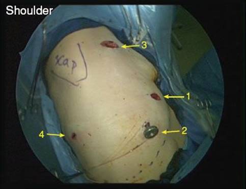
Figure 41.1 Incisions for a right upper lobe sleeve lobectomy.

Figure 41.2 Right-angle clamp surrounds the right upper lobe vein.

Figure 41.3 The anvil of the stapler is placed just above the artery to position the stapler to complete the minor fissure.

Figure 41.4 Vascular stapler is positioned around the anterior trunk to transect the artery.
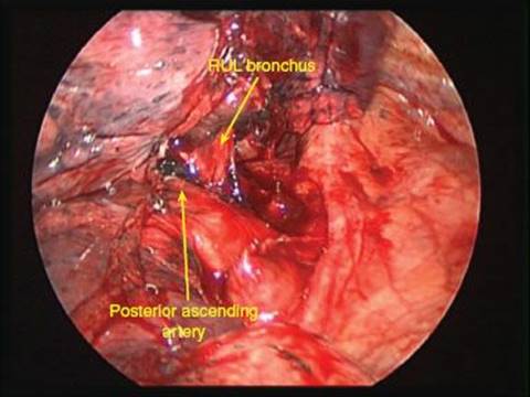
Figure 41.5 Posterior ascending artery.

Figure 41.6 The Yankauer suction in the subcarinal space elevates the bronchi to expose the main stem and the bronchus intermedius.

Figure 41.7 Stay suture on intermediate bronchus.
Posterior Ascending Artery
Retract lung posteriorly
Clip posterior ascending artery at its origin
Major Fissure
Metzenbaum scissors through incision 1 dissects between posterior ascending artery and the inferior border of the RUL bronchus until the scissors feel vertebral bodies
Through incision 1, stapler with 4.8-mm staples completes the remainder of the fissure
Sleeve Resection
Place umbilical tape around the pulmonary artery to retract it inferiorly to better expose right main stem bronchus.
Lift the RUL away from the artery and cut any remaining attachments on the medial border of the right main stem and the intermediate bronchus (BI).
Through the utility incision, a no.15 blade on the long handle cuts the main stem and then the BI. Place a stay suture at the junction of the cartilage and the membranous portion of the BI.
Through the utility incision, suture the main stem bronchus to the BI with 3-0 Vicryl on an SH needle. The membranous portion is done first. The sides are done next. Finally, the top surface of the anastomosis is done. The last few sutures are better placed with an SH-1 needle to fit into the nearly closed anastomosis.

Figure 41.8 Suturing main stem bronchus to intermediate bronchus.
 POSTOPERATIVE MANAGEMENT
At the end of the operation, the double-lumen tube is replaced with a large-bore single-lumen tube. Flexible bronchoscopy assesses the anastomosis and saline lavage clears any bloody secretions. There should be a low threshold for a therapeutic bronchoscopy during the postoperative period because, after a bronchus has been completely transected, the bronchial cilia do not work normally to help remove mucus so patients do not clear secretions as well as they did before the operation. In the past, I performed routine bronchoscopy on postop day 1 or 2, but if the bronchial tree has been cleaned well intraoperatively, that is not necessary unless the patient develops fever, large air leak, atelectasis, or hypoxia.
Patients undergo standard postoperative care following lobectomy. They are infrequently sent to an intensive care unit. Chest drainage system is placed to water seal. Chest tubes are removed when there is no air leak and the chest drainage is <400 mL per day. Starting on the day of the operation, patients are ambulated in the halls four times per day. Pain is controlled with Oxycodone p.o. or Dilaudid IV. Epidural catheters and PCA pumps are infrequently used.
 RESULTS
The first reported bronchial sleeve resection was performed in 1947 at the Brompton Hospital in London, England, by Sir Clement Price Thomas. Whether sleeve resection is radical enough and indicated for patients who could tolerate pneumonectomy continues to be debated, although many recent reports have suggested that sleeve resection can achieve adequate curability rates. However, sleeve lobectomy has a definite role in the surgical management of lung cancer for patients whose pulmonary reserve is considered inadequate to permit pneumonectomy, and should be used anytime it is possible to achieve a margin-negative (R0) resection.
Santambrogio reported the first case report of a VATS sleeve lobectomy in 2002 and in 2008 the author reported 13 patients who underwent VATS sleeve lobectomy. Schmid et al. reported on a combined robotic and VATS approach for a minimally invasive right upper sleeve lobectomy.
 CONCLUSIONS
Complex procedures, such as VATS sleeve lobectomy, are now possible with minimally invasive surgery. The morbidity and mortality are comparable to that for a sleeve lobectomy performed by a thoracotomy. Patient recovery is expedited by the use of minimally invasive surgery.
Recommended References and Readings
He J, Shao W, Cao C, et al. Long-term outcome of hybrid surgical approach of video-assisted minithoracotomy sleeve lobectomy for non-small-cell lung cancer. Surg Endosc. 2011;25(8):2509–2515.
Li Y, Wang J. Video-assisted thoracoscopic surgery sleeve lobectomy with bronchoplasty. World J Surg. 2013;37(7):1661–1665.
Mahtabifard A, Fuller CB, McKenna RJ Jr. Video-assisted thoracic surgery sleeve lobectomy: A case series. Ann Thorac Surg. 2008; 85(2):S729–S732.