
John C. Wain
 INDICATIONS/CONTRAINDICATIONS
Carinal resection and reconstruction without pulmonary resection that involves restitution of the carina by suturing the right and left mainstem bronchi together and approximating these to the end of the distal trachea is termed neocarinal reconstruction. This method of reconstruction was one of the first approaches used for tumors involving the distal trachea and tracheal carina. However, in the past it was felt that this technique was less frequently applicable for reconstruction following carinal resection because after approximation of the right and left mainstem bronchi the neocarina is fixed in the middle mediastinum by the aortic arch, making approximation to the trachea challenging. In the modern era, with earlier identification of neoplastic lesions involving solely the tracheal carina, which do not require resection of significant lengths of trachea or mainstem bronchi, neocarinal reconstruction is performed more frequently (Fig. 42.1).
Carinal resection with neocarinal reconstruction is applicable most commonly to low-grade malignant or benign airway tumors and occasionally for very well localized early nonsmall cell lung cancers. Inflammatory lesions in the distal trachea or mainstem bronchi are rarely localized enough to be managed using this type of reconstruction. While the absolute limits of airway resection that can be managed by near carinal reconstruction are variable, certainly more than 3 to 4 cm of proximal tracheal involvement and more than 1 to 1.5 cm of either mainstem bronchial involvement would preclude this option for airway reconstruction.
 PREOPERATIVE PLANNING
All patients who are candidates for carinal resection and possible neocarinal reconstruction should undergo a complete preoperative physiologic evaluation. Assessment of pulmonary and cardiac function is important given the potential duration and physiologic stress related to the operative procedure. Complete pulmonary function tests including spirometry lung volumes and diffusion capacity are mandatory. However, with a localized lesion that does not require prior resection, it is infrequent that pulmonary function parameters would limit the patient’s candidacy for resection. On the other hand, a prolonged operative procedure with intermittent ventilation and possible hypoxia increases the risk of cardiac stress and thus a low threshold for cardiac stress testing is indicated for patients with risk factors for coronary artery disease.

Figure 42.1 Carinal tumor. This lesion arising on the tracheal carina is amenable to resection without lung resection and neocarinal reconstruction.
Preoperative imaging should include a chest radiograph and a chest CT scan. If the patient has a seemingly localized nonsmall cell lung cancer, accurate staging including positron emission tomography (PET) scanning and brain MRI imaging is mandatory. Some controversy exists about the validity of classifying mediastinal lymph nodes involved by a carinal carcinoma as N2 disease. However, in most cases if there is histologically confirmed metastasis to level 4 or level 2 mediastinal lymph nodes, carinal resection is contraindicated.
Bronchoscopy is an essential and integral part of the preoperative evaluation. Rigid bronchoscopy is the only methodology that allows accurate measurements of the extent of airway involvement. The feasibility of neocarinal reconstruction following carinal resection can only be determined by this modality. In addition, accurate assessment of the histology of the tumor is required. Adenoid cystic carcinomas typically have submucosal or perineural extension to a degree far beyond the gross mucosal disease. Thus, the likelihood of neocarinal resection is extremely limited for this type of low-grade airway malignancy. Rigid bronchoscopy also allows for debulking of central tumors and potential relief of obstructive pneumonitis to optimize the patient’s preoperative status.
 SURGERY
Anesthetic Technique
Ideally, anesthesia for carinal resection and reconstruction should allow extubation at the end of the procedure. The goals of maintaining adequate anesthesia and gas exchange while allowing for surgical exposure require close cooperation between the anesthesiologist and the surgeon. In these cases, an extra-long, flexible, armored single-lumen endotracheal tube is used. This is placed in the trachea proximal to the carina at the beginning of the operative procedure. After dissection and isolation of the carinal structures, depending upon the operative approach, transection of the mainstem bronchi is usually performed prior to transection of the trachea. This allows intubation of the left mainstem bronchus across the operative field for maintenance of ventilation and anesthesia during the remainder of the resection and in the initial part of the reconstruction. Once the anastomotic sutures are placed and the initial approximation of the airway is completed, gentle hand ventilation through the proximal endotracheal tube, which has been guided carefully across the anastomosis by the surgeon into the left mainstem bronchus or, alternatively, ventilation with the endotracheal tube proximal to the anastomosis are two options to be used while the anastomotic sutures are being tied. At times, alternative techniques may be required. High-frequency jet ventilation of the right lung concomitant with ventilation of the left lung using an endotracheal tube passed across the operative field may be useful to maintain oxygenation. In occasional circumstances, independent lung ventilation with cross-field ventilation of both mainstem bronchi may be required to provide both adequate ventilation and oxygenation. A final alternative, extracorporeal membrane oxygenation (ECMO) may be employed, but is usually avoided because of concerns of pulmonary hemorrhage resulting from intraoperative manipulation of the lung.
Surgical Approach
Right posterolateral thoracotomy, median sternotomy, or right anterolateral thoracotomy can be used to approach the tracheal carina. The right posterolateral thoracotomy through the fourth or fifth interspace provides optimal exposure of the carinal structures, particularly if there is significant pleural or posterior mediastinal disease or tumor extension. This approach, however, does not provide optimal access for mobilization of the proximal airway or for the possibility of left hilar release, although the latter is an uncommon requirement for carinal resection without lung resection. Finally, visualization using the right posterolateral thoracotomy incision may be hampered due to ongoing ventilation of the right lung, although in most cases gentle retraction on the right lung, even with active expansion of that lung, will allow adequate exposure of the carina. Median sternotomy provides optimal anterior exposure of the tracheal carina in addition to providing the potential for both laryngeal and bilateral hilar release, if required. In practice, however, for neocarinal reconstruction bilateral hilar release is rarely necessary. Median sternotomy does eliminate the problem of a partially ventilated lung in the operative field. Right anterolateral thoracotomy can be used for carinal exposure, but in the setting of carinal resection without lung resection, may not offer any specific advantage over posterolateral thoracotomy or median sternotomy.
Cervical mediastinoscopy can be an important initial step in the planned operative procedure. Unless there is an indication of mediastinal nodal involvement from preoperative imaging, it is best performed at the time of the planned carinal resection. Mediastinoscopy not only allows direct assessment of mediastinal lymph nodes, but it also provides the opportunity to widely dissect in the anterior tracheal plane from the level of the cricoid cartilage onto the origin of both mainstem bronchi. This approach assures optimal airway mobilization, with preservation of lateral segmental blood supply and is particularly helpful if a right posterior lateral thoracotomy incision is used for exposure.
Surgical Technique
Wide preparation of the operative field should be performed. For a right posterior lateral thoracotomy, this would include preparation posteriorly, medial to the scapula to allow a high posterior lateral thoracotomy incision. Some authors have advocated preparation of the right arm into the operative field and preparation of the anterior neck should a laryngeal release be required. As previously mentioned, however, this is unlikely to be needed in the case of neocarinal resection and reconstruction. The use of cervical mediastinoscopy as an initial step in the operative procedure also makes this less likely to be required. Alternatively, if median sternotomy is performed, the patient is sterilely prepared from the submental space to the infraumbilical region and laterally to allow access to the anterior aspect of both hemithoraces. This field then allows all the potential alternatives for tension reduction including laryngeal release and bilateral hilar release. It also allows the possibility of omental mobilization should there be deemed an indication for its use.
The blood supply to the trachea is segmental. Blood vessels enter in the midlateral position of the cartilaginous wall on either side of the trachea. The distal trachea is supplied by branches originating from the bronchial arteries arising on the descending thoracic aorta. Although the anatomy can be variable, most commonly on the right side there is a branch from the right intercostal bronchial artery supplying the right mainstem bronchus and lower trachea and two branches supplying the left mainstem bronchus. There can also be branches arising from the brachiocephalic artery to provide segmental vessels to the lower trachea. Most typically, the right bronchial artery and/or intercostal bronchial artery must be divided for the resection to be performed. Thus, preservation of the remaining blood supply of the distal trachea and of the left mainstem bronchus is critically important. The anterior plane of dissection on the surface of the airway is safe. On the left side, however, dissection posterior to the left mainstem bronchus should be meticulously avoided to preserve the bronchial arterial blood supply.
Intraoperative bronchoscopy through the single-lumen endotracheal tube may help facilitate identifying the proximal and distal extent of the tumor. Transillumination with the flexible bronchoscope at the proposed sites of airway division allows marking in the operative field. Confirmation of the feasibility of resection can therefore, be made intraoperatively based on the length of involved airway. The length of distal trachea to be resected should be less than 3 cm and the length of mainstem bronchi to be resected should be no more than 1 cm or on the left side no more than 1.5 cm. Circumferential dissection of the airway should only be carried out at the planned points of resection and in the region of the intervening airway. With regard to local lymph nodes, those nodes that are adjacent to the airway to be resected can be resected en bloc with the specimen. Excessive lymph node dissection is to be avoided because of potential compromise of the blood supply, especially adjacent to the proximal tracheal segment.
Initial division of the left mainstem bronchus is preferred. This is can be done as a partial transection of the airway (through the membranous wall from a posterolateral thoracotomy or through the cartilaginous wall from median sternotomy approach). After inspection of the distal bronchial segment to confirm that it is uninvolved, a 2-0 Vicryl stay suture is placed into a midlateral position in the cephalad (superior) bronchial wall. The remainder of the bronchial wall is divided. The right mainstem bronchus is then transected in a similar fashion and a 2-0 Vicryl stay suture is also placed in the cephalad (superior) bronchial wall (Fig. 42.2). The distal trachea is then transected at the marked site. As with the main bronchi, partial transection of the trachea initially allows mucosal inspection to confirm the completeness of resection and facilitates placement of 2-0 Vicryl stay sutures in the midlateral position on either side of the trachea. After completion of the tracheal transection, the specimen is removed. Intraoperative pathologic evaluation of the proximal and distal main bronchial resection margins is imperative.
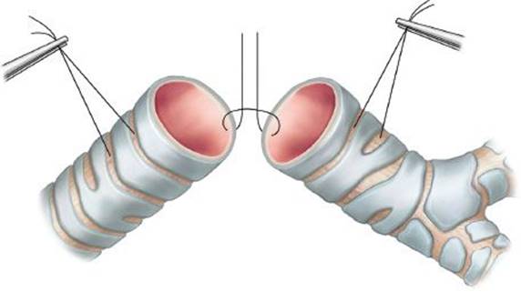
Figure 42.2 Creating the neocarina. Initial placement of suture near the cartilaginous/membranous junction to approximate the medial walls of the right and left main bronchi.
Although there are a variety of anastomotic techniques, the most useful involves interrupted suture of 4-0 Vicryl placed in a concentric fashion 3 to 4 mm apart and 3 to 4 mm from the cut edge of the airway. Inspection of the cut edge of the airway can confirm adequate vascularity by identifying bleeding from the submucosal plane. The neocarina is created first. This is done by approximating the right and left main bronchi together with interrupted 4-0 Vicryl sutures along the medial aspects of the main bronchi. While the remainder of the sutures are placed in such a fashion that the knots are tied on the outside of the airway, for creation of the neocarina the knots of the absorbable suture material can be placed within the airway along the spur of the neocarina. The first suture typically is placed at the cartilaginous member disjunction from both mainstem bronchi, which facilitates proper orientation. Three to five sutures are required to create the neocarina encompassing approximately 1/3 of the cartilaginous wall of the airway (Fig. 42.2). The remainder of the anastomotic sutures are then placed between the distal trachea and the now syncytial main bronchi. A mattress suture incorporating the distal tracheal segment and neocarina both anteriorly that is at the cartilaginous walls and posteriorly at the membranous walls is helpful in maintaining an airtight closure. From a right posterior lateral thoracotomy approach the anterior mattress suture is placed first, followed by the remaining cartilaginous and then membranous wall sutures. From a median sternotomy approach, the posterior mattress sutures are placed first, followed by anastomotic sutures in the membranous and then cartilaginous wall (Fig. 42.3).
After placement of the anastomotic sutures flexion of the neck is to be performed and gentle traction on the stay sutures on the proximal tracheal segment and the main bronchi can assess the degree of tension on the anastomosis. In general, as previously mentioned, the main bronchi are limited in their cephalad motion by the aortic arch. Occasionally, hilar release may facilitate additional proximal mobility of the main bronchi. More importantly, accurate neck flexion and mobilization in the anterior pretracheal plane will allow the proximal tracheal segment to devolve to the neocarinal segment. After approximation of the two ends of the airway with minimal tension, the stay sutures are tied down. The anastomotic sutures are then tied, typically in the reverse order of their initial placement (Fig. 42.4). Following this, intraoperative bronchoscopy is mandatory to assure appropriate orientation and to clear distal secretions. Ventilation by an endotracheal tube in the proximal tracheal segment with the neocarina under a fluid level assures the airtight nature of the anastomosis. Gaps, particularly along the membranous wall of the neocarina can be reinforced with interrupted 4-0 Vicryl sutures.

Figure 42.3 Approximating the distal trachea to the neocarina. A mattress suture encompassing the distal trachea and both main bronchi is useful at both the anterior and posterior ends of the neocarinal suture line. The anterior suture is shown here.

Figure 42.4 Completed neocarina. The lateral stay sutures are tied first to approximate the airways and reduce tension when the anastomotic sutures are tied. Note the now tied mattress suture in the anterior midline of the anastomosis.
In all cases the anastomosis should be wrapped with vascularized tissue. Adjacent pericardial fat tissue pedicled on a branch of the internal mammary artery can be used for circumferential reinforcement of the airway anastomosis. In addition, this interposes a layer of tissue between the anastomotic site and the adjacent anterior vascular structures. Posteriorly the esophagus, generally a well-vascularized structure, will buttress the membranous wall aspect of the anastomosis. In cases where postoperative radiation therapy may be indicated based on pathologic findings (local nodal metastasis from nonsmall cell lung cancer or adenoid cystic carcinoma), then a more robust anastomotic wrap consisting of intercostal muscle flap, easily obtained via a posterior lateral thoracotomy or omentum can be used to encompass the site of the anastomosis.
At the conclusion of procedure the incision is closed in standard fashion. The patient’s neck is maintained in flexion by a nonabsorbable suture extending from the presternal fascia at the level of the sternal angle to the submental space. The patient is carefully awakened from the anesthetic and extubated in the operating room. In cases where there may be a question of upper airway obstruction or strider, placement of the laryngeal mask airway will allow assessment of the larynx and vocal cords. Flexible bronchoscopy at this stage is quite useful to clear secretions from the distal airway and gain a final inspection of the airway anastomosis.
 POSTOPERATIVE MANAGEMENT
Postoperative management of the patient undergoing carinal resection and neocarinal reconstruction is reasonably straightforward. Excellent bronchopulmonary hygiene including chest physiotherapy and, as needed, flexible bronchoscopy are crucial. Appropriate pain control, typically managed by epidural anesthesia, is important. Perioperative antibiotic therapy should be tailored to the patient’s own lower respiratory tract flora. The duration of antibiotic therapy should be determined by the patient’s clinical status and the amount and nature of any airway secretions they produce.
There is a normal postsurgical inflammatory state, which increases alveolar capillary permeability. This is rather minimal after neocarinal resection as compared to other forms of carinal resection involving lung resection. Nonetheless, fluid restriction is an important component of the postoperative management of these patients. Intravenous fluid should be maintained to achieve a urine output of 0.5 mL/kg/hr.
An additional important element in postoperative management is prevention and control of aspiration. The head of the bed should remain elevated at all times and oral intake should be restricted in the first 1 to 3 days following surgery. Assessment of vocal cord mobility and evaluation of swallowing function by qualified speech pathologist may be necessary if the patient has a persistently abnormal postoperative voice.
Routine surveillance bronchoscopy should be performed prior to discharge. Typically this is 4 to 6 days postoperatively.
 COMPLICATIONS
Anastomotic complications are the most serious complication following carinal resection and neocarinal reconstruction. They are typically related to necrosis from poor airway blood supply or excessive anastomotic tension. The incidence can be minimized by restricting neocarinal reconstruction to those patients with very localized carinal neoplasms. The extent of resection is the most critical factor. In addition, excessive dissection along the left lateral aspect of the trachea and posterior to the left mainstem bronchus are common causes of interference with the blood supply to the remaining airway segments. In the event of what appears to be excessive tension in the operating room, if this cannot be alleviated by the previously mentioned maneuvers, an alternative type of airway reconstruction should be considered.
Any indication of ischemia or necrosis at bronchoscopy postoperatively requires close observation. A chest CT scan should be obtained for anyone with abnormal airway mucosa identified at the time of a therapeutic or surveillance bronchoscopy. Because of the complexity of the reconstruction, options of transanastomotic intubation are relatively limited. In most cases, empiric therapy with antibiotics and maintenance of cervical flexion for an extended period of time will allow for healing by secondary intention. Hyperbaric oxygen therapy may have a benefit in facilitating healing of partially ischemic airway. However, healing by secondary intention is likely to lead to anastomotic stenosis, which can be treated by dilation. Repeat resection is typically not possible for the neocarina patient without some loss of pulmonary parenchyma. The best treatment is avoidance of the complication by careful attention to intraoperative technique.
Other postoperative complications are similar to those seen with routine pulmonary resections and include atrial fibrillation, deep venous thrombosis, and pneumonia. The management of these complications is similar to that for other patients undergoing lung resection. However, the occurrence of pneumonia should always raise the specter of a possible anastomotic complication and deserves bronchoscopic evaluation in all cases should it occur.
 RESULTS
In the absence of the anastomotic complication, the results of carinal resection and neocarinal reconstruction are excellent. The vast majority of these patients have very localized tumors and complete resection with negative margins and a good technical result is curative in the majority of cases.
Recommended References and Readings
Grillo HC, Bendixen HH, Gephart T. Resection of the carina and lower trachea. Ann Surg. 1963;158:899–893.
Grillo HC. Carinal reconstruction. Ann Thorac Surg. 1982;34:356–373.
Hardin CA, Fitzpatrick MJ. Primary carcinoma of the carina and bifurcation successfully treated by resection and reconstruction. Surgery. 1959;46:534–540.
Lanuti M, Mathisen DJ. Carinal resection. Thorac Surg Clin. 2004; 14:199–209.
Macchiarini P, Altmayer M, Go T, et al. Technical innovations of carinal resection for non-small cell lung cancer. Ann Thorac Surg. 2006;82:1989–1997.
Minnich DJ, Mathisen D. Anatomy of the trachea, carina and bronchi. Thorac Surg Clin. 2007;17:571–585.
Mitchell JD, Mathisen DJ, Wright CD, et al. Clinical experience with carinal resection. J Thorac Cardiovasc Surg. 1999;117:39–53.
Pearson FG, Todd TR, Cooper JD. Experience with primary neoplasms of the trachea and carina. J Thorac Cardiovasc Surg. 1984;88:511–518.
Weder W, Inci I. Carinal resection and sleeve pneumonectomy. Thorac Surg Clin. 2014;24:77–83.