
Michael Lanuti
 INDICATIONS/CONTRAINDICATIONS
Indications
The most common resection of the carina involves the entire right lung and carina because of the frequency of bronchogenic carcinoma relative to other primary tracheal neoplasms. Right carinal pneumonectomy should be considered in patients who harbor nonsmall cell lung cancer, usually originating from the right upper lobe orifice and extending to the lateral aspect of the lower trachea (Fig. 44.1). The neoplasm should not extend beyond 4 cm from the distal trachea as documented by computed tomography (CT) imaging and bronchoscopy. Primary airway tumors such as squamous cell carcinoma or adenoid cystic carcinoma may also be considered provided resection is feasible. Positive microscopic resection margins may be acceptable with adenoid cystic carcinoma since adjuvant radiation improves long-term results. The alternative indication for right carinal sleeve is a positive bronchial margin after standard pneumonectomy.
Contraindications
Absolute contraindications to right carinal pneumonectomy include insufficient pulmonary reserve, impaired cardiac function including pulmonary hypertension, and airway involvement in excess of 4 cm. Great caution should be applied in patients in whom extensive airway resection is undertaken where the trachea is to be anastomosed to the left main bronchus. The incidence of anastomotic problems significantly increases when the extent of resection is 4 cm or greater. The left main bronchus is tethered in its cephalad migration by the aortic arch and is therefore relatively immobile. With extensive resections, left-sided anastomoses are subject to excessive tension and greater potential for devascularization of the region. A history of prior mediastinal irradiation, although no longer an absolute contraindication to surgery, requires special operative considerations, including the use of an omental flap to wrap the anastomosis. Finally, in patients with NSCLC, the presence of N2 (stage IIIA) or N3 (stage IIIB) disease (American Joint Committee on Cancer 7th edition) should serve as a relative contraindication to surgery given the poor long-term outcome demonstrated in these cases. The need for superior vena cava (SVC) resection in these cases does not necessarily preclude surgery.

Figure 44.1 Right carinal pneumonectomy. A: Tumor extending from bronchus intermedius to lateral wall of the trachea where planned airway division is delineated by dotted lines. B: The trachea and left mainstem bronchus are anastomosed in an end-to-end fashion.
 PREOPERATIVE PLANNING
All patients in whom carinal sleeve pneumonectomy is being considered should undergo CT chest and PET to permit proper evaluation of lung parenchyma, mediastinum, and to identify nodal or extraregional disease. Invasive mediastinal staging (endobronchial ultrasound [EBUS] or mediastinoscopy) should be performed in these patients to rule out pathologic N2/N3 disease despite a negative PET or anatomically normal lymph nodes.
This author favors the use of mediastinoscopy at the time of formal resection and reserves EBUS for diagnostic purposes in the preoperative period. Mediastinoscopy followed by a delayed carinal resection can be associated with scar tissue along the pretracheal plane that may compromise tracheal mobility at the time of resection. The degree of extraluminal tumor invasion can also be ascertained at the time of mediastinoscopy. The bronchoscopic appearance of the tumor will dictate candidacy for sleeve pneumonectomy. The degree of invasion should always be documented by biopsies to understand the limits of resection. If the tumor extends beyond 4 cm of the lower trachea or beyond 1.5 cm of the left mainstem bronchus, tension on the airway anastomosis can be expected. All patients need routine pulmonary function tests to determine adequate pulmonary reserve. A stair climb test of 2 to 3 flights (12 to 18 m) can also be used as a crude test for assessment of pulmonary and cardiac reserve. If the patient has borderline pulmonary function, one should consider a quantitative ventilation and perfusion scan and cardiopulmonary exercise testing (CPET) to measure oxygen consumption (VO2). Echocardiogram should also be routinely obtained to assess left and right ventricular function and to rule out significant valvular heart disease or pulmonary hypertension.
 SURGERY
Anesthestic Technique
Every effort is made to design an anesthetic plan that permits extubation at the end of the procedure.
This technique should include placement of an epidural catheter preoperatively and the use of total intravenous anesthesia. This strategy relies on short-acting hypnotics, narcotics, and paralytic agents to permit adequate respiratory drive upon completion of the surgery. Because of their size and inflexibility, double-lumen endotracheal tubes often present difficulties in these procedures. As such, an extra long, flexible, armored, single-lumen endotracheal tube is used, which can be advanced into the left mainstem bronchus to provide one-lung ventilation as indicated. As surgical resection proceeds, the left mainstem bronchus is intubated across the operative field. A plan of intermittent ventilation is then used to allow precise placement of anastomotic sutures. As the end-to-end tracheobronchial anastomosis is reapproximated and the sutures are tied, the original endotracheal tube is advanced into the bronchus, allowing uninterrupted ventilation. Alternative ventilatory techniques, such as high-frequency (jet) ventilation and independent lung ventilation, are options about which both surgeon and anesthesiologist should be knowledgeable. Cardiopulmonary bypass, although feasible, should be discouraged during carinal resection and reconstruction because of the incremental morbidity associated with the procedure and the potential for tumor dissemination. Autologous blood recovery systems “Cell Saver” should be avoided in this context.
Positioning
After successful fiberoptic placement of an extra long, flexible, armored, single-lumen endotracheal tube down the left mainstem bronchus, the patient is placed in the maximally flexed left lateral decubitus position. After the patient is sterilely draped, cross-field ventilation tubing can be secured with the ability to monitor exhaled carbon dioxide.
Surgical Technique
The procedure is performed through a standard right posterolateral thoracotomy via the fourth intercostal space where the fourth intercostal muscle can be harvested on a posterior vascular pedicle for buttressing the airway anastomosis. The pleural space and mediastinum should be inspected to confirm resectability. The blood supply to the trachea is predominantly segmental, and every effort should be made to avoid interruption. After development of the pretracheal plane and opening the pericardium, tapes are placed around the mobilized trachea and right and left mainstem bronchi (Fig. 44.2). The azygos vein can be divided to improve exposure around the carina. Careful handling of the tissues is crucial, especially when encircling the trachea to avoid injury to the left recurrent laryngeal nerve. Lateral dissection proximal and distal to the proposed lines of transection should be limited to 1 to 2 cm to avoid devascularization of the airway. An extensive lymph node dissection beyond removal of immediately adjacent or clearly involved nodes is avoided in a similar manner. Some degree of lymph node sampling must be accepted in patients who have bronchogenic carcinoma. When neoplastic resections are performed, frozen section analysis of resected airway margins by competent pathologists must be available. Limited tumor involvement of adjacent structures (i.e., superior vena cava, esophagus) does not preclude resection if primary repair of the adjacent mediastinal structure can be accomplished. If more than 4 cm of trachea will have to be removed, the surgeon must be aware of excessive anastomotic tension, and question the feasibility of surgery.
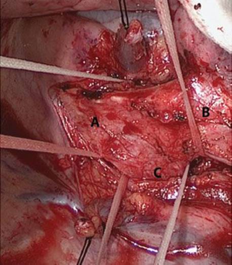
Figure 44.2 View of the trachea from right thoracotomy. The azygos vein has been divided to facilitate dissection and mobilization of the carina. A: Distal trachea, B: Right mainstem bronchus, C: Left mainstem bronchus.
The left main bronchus is usually divided first (after placing traction sutures) followed by the transection of the distal trachea while left lung ventilation is assured via cross-field intubation of the left main bronchus. Traction sutures are also placed on the divided trachea. Pathologically negative airway margins should be confirmed on frozen section. Next, the right pulmonary artery and veins are encircled and ligated (often intrapericardially) with vascular staplers. The specimen is removed from the field (Fig. 44.3). Before re-establishing airway continuity, the patient’s neck must be flexed so that the trachea is delivered down to the left main bronchus, which is fixed underneath the aorta and thus relatively immobile. Significant reduction in anastomotic tension is provided by mobilization of the hilum using an inferior hilar release on either side. The release is best accomplished before commencing airway resection. After division of the inferior pulmonary ligament, a U-shaped incision is made in the pericardium beneath the hilum with intrapericardial division of the raphe, extending between the inferior pulmonary vein and the inferior vena cava.
Airway Anastomosis
Although there are several ways to perform the airway anastomosis (Fig. 44.4), the current author prefers the technique where interrupted sutures (3-0 or 4-0 Vicryl [polyglactin]; Ethicon, Inc, Somerville, New Jersey) are used with knots tied outside of the airway (Fig. 44.5). Sutures are inserted at 2- to 3-mm intervals, and size disparity between the trachea and left main bronchus is equalized by stretching the bronchial lumen to the size of the tracheal one. The sutures are generally tied after they have all been inserted and after the previously inserted traction sutures have been pulled together (Fig. 44.6). Once completed (Fig. 44.7), the anastomosis is covered with either a pleural flap, pericardial fat pad, intercostal muscle or a combination of flaps. An intercostal muscle flap can be difficult to place completely around the anastomosis (too bulky), and therefore should be used as an onlay flap to cover part of the anastomosis. The site of reconstruction should finally be checked for air leaks.

Figure 44.3 Perspective via right thoracotomy. The specimen has been removed en bloc (right lung, carina, small part of trachea, and left mainstem bronchus) from the field. Ventilation is performed across the operative field via the left mainstem bronchus or via a high-frequency jet catheter.

Figure 44.4 Tractions sutures are placed 1 to 2 cm from the cut edge of the airway on the cartilaginous portion. The airway anastomosis is performed with a running suture along the membranous wall (this author prefers interrupted suture for the entire reconstruction). A high-frequency jet catheter is visualized within the lumen of the endotracheal tube.
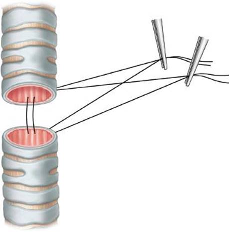
Figure 44.5 Interrupted suture technique for airway anastomosis: Each succeeding suture is placed anterior to the previous suture and clipped in a fan-like arrangement to the operative drapes. All sutures are typically placed with knots on outside and subsequently tied after all are inserted.
 POSTOPERATIVE MANAGEMENT
Postoperative care begins in the operating room with a flexible surveillance bronchoscopy to study the anastomosis and permit adequate clearance of retained blood and secretions. Extubation on completion of the procedure remains paramount to the integrity of the reconstruction. Vigorous pulmonary toilet maneuvers, including chest physiotherapy and, if necessary, repeated flexible bronchoscopy, should be pursued aggressively. The administration of intravenous fluid should be minimized to avoid volume overload and possible pulmonary edema. Before discharge from the hospital, all patients should undergo flexible surveillance bronchoscopy (∼7 days postoperatively) to evaluate the airway anastomosis. Any sign of early ischemia or necrosis must be observed closely. Since atrial arrhythmias are common after carinal pneumonectomy, one may consider early prophylactic use of calcium channel blockers or β-blockers. If amiodarone is considered as a prophylactic antiarrhythmic agent, one should be aware of the possibility of subacute pulmonary toxicity.

Figure 44.6 Prior to tying down the anastomotic sutures, the traction sutures are first tied to bring the airway together and reduce tension. The indwelling single-lumen endotracheal tube can be advanced beyond the anastomosis with cuff inflated to provide ventilation at this time.

Figure 44.7 Completed anastomosis.
 COMPLICATIONS
 Postpneumonectomy pulmonary edema
Atrial arrhythmia
Anastomotic dehiscence
Bronchopleural fistula
Vocal cord dysfunction
Occasionally, a condition resembling adult respiratory distress syndrome (ARDS)appears within 36 to 72 hours following surgery, and it remains a leading cause of mortality after carinal resection. Early presentation has been with hypoxia, tachypnea, and a “ground glass” appearance on chest radiography. The cause of this disorder remains unclear. Fluid overload, barotrauma, lymphatic interruption, occult microaspiration, and pulmonary injury secondary to a systemic inflammatory response have been postulated as causative factors. In addition to conservative therapy, one should optimize ventilatory settings, hemodynamics, pulmonary toilet, nutrition, and antibiotic coverage. If anastomotic healing is deemed abnormal on the first surveillance bronchoscopy, the patient should be observed in the hospital. Significant mortality is associated with postoperative anastomotic complications. These complications range from necrosis and separation and mucosal slough (early) to stenosis, excessive granulation tissue, and recurrent episodes of obstructing pneumonia (late). Bronchopleural fistula must be treated with drainage and antibiotic coverage. Early airway separation can initially be managed with careful intubation beyond the anastomosis, followed by internal stenting with a custom made silastic stent or T tube. Late stenosis is typically treated with repeated endobronchial dilatations, although reresection is possible, with acceptable results. The best treatment for anastomotic complications is prevention through meticulous surgical technique and sound judgment. Injury to the recurrent laryngeal nerves is possible and unilateral vocal cord palsy should expeditiously managed with Restylane injection vocal cord medialization to avoid aspiration.
 RESULTS
While carinal resections are technically demanding, excellent clinical outcomes can be achieved in high-volume centers. Despite earlier reports of unacceptable postoperative mortality, contemporary data from experienced centers have yielded mortality rates between 3% and 12%. The principle cause of perioperative mortality is noncardiogenic pulmonary edema or pneumonia with subsequent ARDS and early anastomotic complications. The major reasons for improvement in the early mortality rates include careful patient selection, advances in anesthetic technique, and vastly improved postoperative ICU care. The overall survival for patients with malignant histologies undergoing carinal sleeve resection is approximately 25% to 50% at 5 years. The need for postoperative mechanical ventilation and development of anastomotic complications are predictors of decreased overall survival on univariate analysis. Nodal status and degree of endobronchial tumor extension are predictors for decreased disease-free survival on multivariate analysis. Survival at 5 years in patients with involved mediastinal lymph nodes (N2) is 7% versus 32% for patients who are N0/N1.
 CONCLUSIONS
Accurate selection of patients, a meticulous adherence to surgical precision, and optimal postoperative patient care have become mandatory to reduce the risk of the most serious complications, such as noncardiogenic pulmonary edema and suture dehiscence. With carinal resection for bronchogenic carcinoma, contemporary studies suggest that there are reasonable survival rates in the absence of involved mediastinal lymph nodes or distant metastatic disease. The role of neoadjuvant therapy for bronchogenic carcinoma involving the carina deserves further investigation; this type of therapy should be used with caution because of the deleterious effects on anastomotic healing.
Recommended References and Readings
Dartevelle P, Macchiarini P. Carinal resection for bronchogenic cancer. Semin Thorac Cardiovasc Surg. 1996;8(4):414–425.
Deslauriers J, Gregoire J, Jacques LF, et al. Sleeve pneumonectomy. Thorac Surg Clin. 2004;14(2):183–190.
Lanuti M, Mathisen DJ. Carinal resection. Thorac Surg Clin. 2004; 14(2):199–209.
Mitchell JD, Mathisen DJ, Wright CD, et al. Resection for bronchogenic carcinoma involving the carina: Long-term results and effect of nodal status on outcome. J Thorac Cardiovasc Surg. 2001; 121(3):465–471.