
Harald C. Ott and Douglas J. Mathisen
Introduction
Resection of central bronchogenic carcinomas involving the carina remains one of the most challenging operations in thoracic surgery, and is still performed by only a few centers in carefully selected patients. Nevertheless, several groups have reported case series with acceptable mortality and good long-term survival (1–3). Patients presenting with right upper lobe tumors involving the distal lateral wall of the trachea or the right mainstem bronchus, but sparing the right bronchus intermedius are a unique subgroup, and may be candidates for a parenchyma-sparing strategy such as a right upper lobectomy with concomitant carinal resection and reconstruction. Patients with compromised lung function and limited exercise tolerance would benefit from an approach sparing the right middle and lower lobe. While preserving lung parenchyma is desirable, reconstruction of the carina in this setting is complicated by the size mismatch of distal trachea, bronchus intermedius, and left mainstem bronchus. In addition, there is the concern regarding two anastomoses resulting in anastomotic tension, and a tenuous blood supply of the fully mobilized airways. Several different approaches have been described, and their applicability depends on the extent of airway involvement. If only a small portion of the bronchus intermedius is involved, lobectomy and wedge tracheobronchoplasty with primary closure of the airway wall has been described (4). If the inferior wall of the right mainstem bronchus is not involved, continuity between the trachea and the left mainstem bronchus can be preserved and reconstruction via Nohl-Oser or Sato tracheobronchoplasty can be accomplished (5,6). If there is more extensive involvement of the right mainstem bronchus and the distal trachea, carinal resection and reconstruction may be necessary (2). End-to-end anastomosis of the left mainstem bronchus to the distal trachea with reimplantation of the bronchus intermedius to distal trachea or left mainstem bronchus can be accomplished safely and avoids size mismatch between the three airway lumina (3,7). Lastly, preservation of continuity between distal trachea and left mainstem bronchus and reimplantation of the bronchus intermedius in the distal trachea with primary closure of the remaining defect has been described (8). Herein we describe our technique of carinal right upper lobectomy, and discuss technical pitfalls, and management of potential complications (Fig. 45.1).
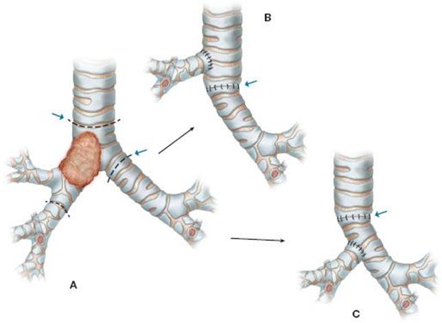
Figure 45.1 A: Tumor involving right upper lobe, mainstem and distal trachea allows point of transection. B: Reimplantation into side of trachea. Arrow trachea to left mainstem anastomosis. C: Reimplantation into side of left mainstem. Arrow trachea to left mainstem anastomosis.
Patient Selection
Given the substantial perioperative risk, careful patient selection is of utmost importance. Our routine preoperative workup includes complete staging by PET scan, and brain MRI. We consider nodal disease of more than two N2 stations to be a contraindication for surgical resection, unless the patient is young and otherwise healthy. We perform a direct examination of the tracheobronchial tree via rigid bronchoscopy to assess the precise extent of disease into distal trachea, carina, and right mainstem bronchus before proceeding with surgical resection. In terms of lung function, ideally even marginal patients have to be able to at least theoretically tolerate a right pneumonectomy. However, carinal right upper lobectomy is possible and probably indicated for patients who can tolerate only a right upper lobectomy. Quantitative VQ scan is therefore, an important part of our preoperative workup.
 SURGERY
We perform bronchoscopy in every patient at the time of planned resection to confirm tumor extent and location in relation to distal trachea, carina, mainstem bronchi, and bronchus intermedius. We then place the patient in supine position with hyperextension of the neck. We perform a standard mediastinoscopy in all patients to assess mediastinal lymph nodes, and to free the pretracheal plane extending to carina and left and right mainstem bronchus. This step provides us with additional mobility of trachea and left mainstem bronchus necessary to allow reconstruction under minimal tension. After completion of mediastinoscopy, a long cuffed single-lumen tube is advanced into the left mainstem bronchus under bronchoscopic guidance. The cuff is inflated and the right lung is isolated. The patient is then positioned in left lateral decubitus position, and prepped and draped for standard posterolateral thoracotomy. In these challenging cases, we routinely remove the fifth rib to maximize exposure. We divide the inferior pulmonary ligament and free the entire lung from any adhesions. We subsequently perform a complete hilar release to maximize mobility of the right middle and lower lobe (Fig. 45.2). After careful examination of vascular and airway anatomy, we divide the azygos vein, and dissect the trachea at this level circumferentially. We pay attention to stay close to the trachea to avoid injury of the left recurrent laryngeal nerve. It is most at risk in encircling the distal trachea. To determine the optimal level of division it helps to pull the endotracheal tube back into the trachea, transilluminate using a flexible bronchoscope, and use a TB needle to mark the precise location and proximal extent of the tumor. At this stage, we then mark the proximal and distal tumor extent by placing superficial 4-0 silk sutures on the outside of the airway. Before proceeding with a right upper lobectomy in usual fashion, we perform a full hilar release (Fig. 45.2). We release the inferior pulmonary ligament dissecting the pleura anteriorly and posteriorly. We also circumferentially incise the pericardium around the pulmonary veins. These maneuvers maximize mobility of the right middle and lower lobe, and help to minimize tension in the reconstruction that follows. Care must be taken not to interfere with the blood/lymphatic supply of the right bronchus intermedius. We try to preserve a pedicle posteriorly along the bronchus intermedius and divide the pericardium inside this pedicle. In preparation for cross-field ventilation, we place a single long wire armed cuffed endotracheal tube into the field and pass ventilator tubing to the anesthesiologist. After division of pulmonary arterial branches, and the upper lobe vein, we divide the bronchus intermedius using a scalpel blade at the level of the distal marking suture. We then divide the distal trachea proximal to the tumor and the left mainstem bronchus just distal to the carina. After en bloc removal of the now completely dissected right upper lobe, we directly intubate the left mainstem bronchus and initiate cross-field ventilation of the left lung (Fig. 45.3). We dissect 1-mm margins of the distal trachea, left mainstem bronchus, and bronchus intermedius to confirm negative margins using frozen sectioning. 2-0 Vicryl traction sutures are then placed on both sides just anterior to the cartilagino-membranous junction in the distal trachea, and left mainstem bronchus (Fig. 45.4). We perform the first anastomosis between left mainstem bronchus and trachea using interrupted 4-0 Vicryl sutures with knots on the outside (Fig. 45.5). After completion of the posterior wall, we advance the endotracheal tube into the left mainstem bronchus, which allows continuous ventilation while we complete the anterior portion of the anastomosis. To accommodate the second anastomosis between lateral wall of the distal trachea and the bronchus intermedius, we choose a location at least two rings proximal to the first anastomosis to prevent ischemia of the distal tracheal segment (Fig. 45.1). We then perform an elliptical incision in the cartilaginous portion of the lateral tracheal wall and place 2-0 Vicryl traction sutures on distal trachea superior and inferior to the opening, and on the bronchus intermedius (Fig. 45.6). We then perform a side-to-end anastomosis between lateral tracheal wall and bronchus intermedius using 4-0 Vicryl sutures. If visualization and mobilization of both anastomoses is challenging, we prioritize on perfect approximation and try to reduce traction trauma by placing the posterior wall knots on the inside of the airway lumen. In our experience this has not negatively affected healing of the anastomosis, short- or long-term outcomes. If it appears that the anastomosis between bronchus intermedius and lateral wall of the distal trachea would be under an excessive amount of tension, anastomosis of the bronchus intermedius to the left mainstem bronchus has been described (Fig. 45.1). Attention has to be paid to allow for a long enough tissue bridge between the two anastomoses to avoid ischemia of the airway segment between the two suture lines. After completion of the anastomosis, we like to approximate well-perfused tissue such as an intercostal muscle flap or a pericardial fat pad to reduce the risk of anastomotic ischemia and fistula formation. If we use an intercostal muscle flap, we do not wrap it circumferentially around the anastomosis to avoid contraction, and possible constriction of the anastomosis. Before emergence from anesthesia and extubation in the operating room, we place a chin stitch using 0 Ethibond to remind the patient of neck flexion. The stitch is typically removed after we confirm well-healing anastomoses at a follow-up flexible bronchoscopy on postoperative day 7.
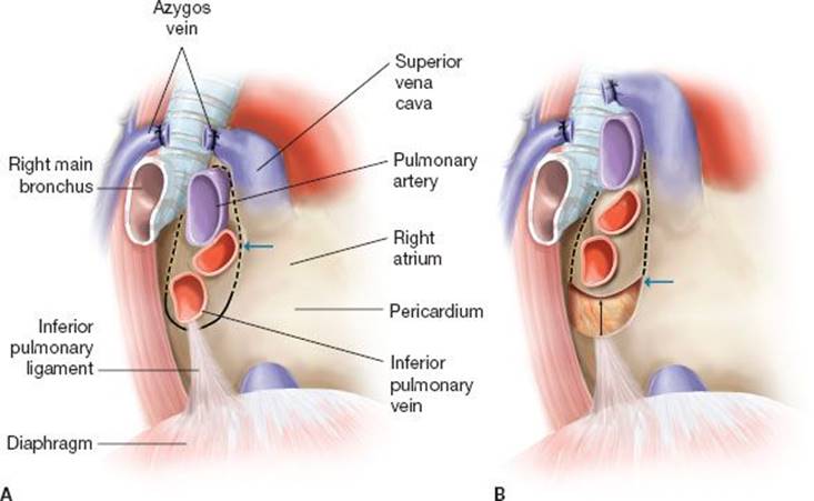
Figure 45.2 A: Location of incision in pericardium for hilar release. B: Amount of relaxation achieved.
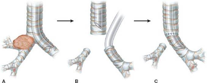
Figure 45.3 A: Tumor involving right upper lobe, mainstem and carina. B: Resection of trachea, left mainstem and bronchus intermedius (note oral endotracheal tube pulled back and intubation of left mainstem for cross field ventilation). C: Completed trachea to left mainstem anastomosis.
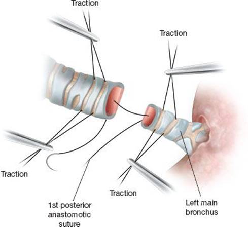
Figure 45.4 Traction sutures and first posterior anastomotic suture.
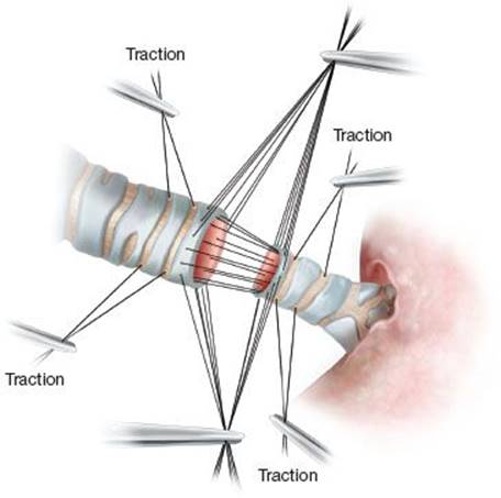
Figure 45.5 Placement of traction sutures and all anastomotic sutures.
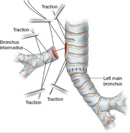
Figure 45.6 Traction sutures placed proximally in trachea and distally in bronchus intermedius. First suture placed posteriorly knots on outside as shown.
If conversion to pneumonectomy is required, careful closure of the lateral defect in the trachea or left main bronchus is mandatory. Primary closure is preferred. A patch may be needed if the airway lumen is compromised. We have had one experience using cryopreserved aortic allograft, which remains our first choice. At the time of repair, airtightness has to be confirmed, and a muscle buttress has to be placed over the patch to provide the support of vascularized tissue.
 COMPLICATIONS
Perioperative management after carinal lobectomy is similar to the management of other high-risk lung resections. During the case, we carefully monitor airway pressures to avoid barotrauma of the left lung. Extubation in the OR is desirable, prudent fluid resuscitation helps to prevent pulmonary edema. Postpneumonectomy pulmonary edema is a potentially lethal complication. It is best avoided by careful avoidance of intraoperative barotrauma and fluid overload. When signs of postoperative acute respiratory distress syndrome (ARDS) present, it has to be treated aggressively with diuresis, antibiotics, and early intubation. Inhaled nitric oxide has been helpful in selected patients.
Early mobilization and aggressive pulmonary toilet helps to prevent atelectasis and pneumonia. Injury of the left recurrent nerve can occur during circumferential dissection of the distal trachea. We try to minimize this risk by keeping the dissection to the left of the trachea to a minimum and staying strictly close to the trachea. If injury occurs, vocal cord injection in the early postoperative period will allow for vocal cord apposition, and help the patient to cough, and clear secretions.
Both anastomoses can suffer from short-term complications such as bleeding, dehiscence, and bronchopleural fistula formation. Long-term complications include granulation tissue and stricture formation. Early anastomotic failure is caused by technical error, or tissue ischemia related to extensive dissection and excess tension on the anastomosis. Large air leaks in the early postoperative period and mediastinal emphysema on postoperative chest radiographs are concerning signs for anastomotic dehiscence. We routinely perform flexible bronchoscopies after completion of both airway anastomoses to ensure proper tissue apposition and to assess the quality of the tissue. We usually perform a routine bronchoscopy on postoperative day seven. If airway tissues at any given point in time do not appear well perfused, or it appears that there is partial dehiscence of the anastomosis without air leak into the surrounding tissue we have had good experience with hyperbaric oxygen treatments twice daily for several days may be of value to help healing (9).
Whenever there are concerns for anastomotic breakdown in the early postoperative period, repeat flexible bronchoscopy is required to perform thorough assessment of the suture lines. If there is any evidence for anastomotic leak in the early postoperative period, immediate surgical repair is necessary to avoid the risk of mediastinitis, pulmonary arterial fistula, and empyema formation, while broad-spectrum antibiotic coverage is initiated. Conversion to a pneumonectomy with resection of the middle and lower lobe may be necessary. Primary closure of the resulting defect with pedicled muscle coverage is preferable.
Late occurrence of bronchopleural fistula can be managed similar to other lung resections with drainage, pleural window, and coverage with repair after the infection has been cleared. Granulation tissue at the site of the anastomosis can be resected during flexible bronchoscopy. Stricture formation as a consequence of tissue ischemia occurs most commonly at the reimplantation site of the bronchus intermedius. Balloon dilatation via flexible bronchoscopy can improve luminal diameter but usually has to be repeated several times since, recurrence is common. Especially dilation of a stricture at the site of the second anastomosis must be performed with caution because of the proximity of the pulmonary artery and risk of massive bleeding.
 CONCLUSIONS
Carinal right upper lobectomy is a parenchyma-sparing alternative to carinal pneumonectomy for patients presenting with central tumors involving the right mainstem bronchus and the distal trachea. Carinal reconstruction between the three airway lumina is technically challenging due to the difference in diameters. We therefore, routinely anastomose the left mainstem bronchus to the distal trachea in an end-to-end fashion. Mediastinoscopy and complete hilar release allow for mobilization of the right middle and lower lobe and allow us to perform a tension-free separate anastomosis of the bronchus intermedius into distal trachea or left mainstem bronchus. Postoperative management includes epidural analgesia, extubation in the OR, and early mobilization with aggressive pulmonary toilet. If there are concerns for anastomotic complications, early bronchoscopic evaluation is prudent to guide decision between nonsurgical (hyperbaric oxygen, antibiotics) and surgical (reexploration, anastomotic repair vs. pneumonectomy) management. Late-stage complications can be managed using standard endoscopic techniques.
Recommended References and Readings
de Perrot M, Fadel E, Mercier O, et al. Long-term results after carinal resection for carcinoma: Does the benefit warrant the risk? J Thorac Cardiovasc Surg. 2006;131:81–89.
Kamiyoshihara M, Nagashima T, Takise A, et al. Wedge tracheobronchoplasty technique for primary pulmonary carcinoma of the right upper bronchus. Clin lung cancer. 2010;11:426–428.
Kaya SO, Sevinc S, Ceylan KC, et al. One-stoma carinoplasty: Right upper sleeve lobectomy with hemicarinectomy for resection of right-tracheobronchial-angle tumors. Tex Heart Inst J. 2013; 40:435–438.
Macchiarini P, Altmayer M, Go T, et al. Technical innovations of carinal resection for nonsmall-cell lung cancer. Ann Thorac Surg. 2006;82:1989–1997; discussion 1997.
Mitchell JD, Mathisen DJ, Wright CD, et al. Clinical experience with carinal resection. J Thorac Cardiovasc Surg. 1999;117:39–52; discussion 52–33.
Nohl-Oser HC, Fenn A, Dottori V. Tracheobronchoplastic procedure in the treatment of advanced carcinoma of the right upper lung. Ann Thorac Surg. 1982;33:396–399.
Porhanov VA, Poliakov IS, Selvaschuk AP, et al. Indications and results of sleeve carinal resection. Eur J Cardiothorac Surg. 2002; 22:685–694.
Sato M, Hasegawa S, Shoji T, et al. Tracheobronchoplasty for resection of lung cancer arising from a tracheal bronchus. Ann Thorac Surg. 2002;73:310–312.
Stock C, Gukasyan N, Muniappan A, et al. Hyperbaric oxygen therapy for the treatment of anastomotic complications after tracheal resection and reconstruction. J Thorac Cardiovasc Surg. 2014;147:1030–1035.