
Cameron D. Wright
 INDICATIONS/CONTRAINDICATIONS
Left carinal resections are quite rare, likely due to the long length of the left main bronchus (LMB), thus eliminating lung cancers that extend toward the carina from requiring carinal resection like those on the right often do. Rare tumors that involve a long length of LMB and the distal trachea are the most common cancers that require left carinal pneumonectomy. Very rarely a local recurrence involves the LMB stump after left pneumonectomy and is resectable. The preoperative evaluation should eliminate those with metastatic disease or extensive mediastinal invasion. Most important in those with lung cancer is to not operate on patients with N2 disease since the outcome of resection of T4N2 lung cancers are so poor. Since the left lung is removed usually the patient should be able to tolerate that physiologically.
 PREOPERATIVE PLANNING
Careful assessment of the preoperative imaging is performed to decide upon an approach for the individual patient. A left carinal resection can be performed via a left thoracotomy or a median sternotomy. A thoracotomy approach is favored if the lung resection is going to be difficult due to adhesions or tumor involvement of other structures that also need to be resected. If any significant amount of trachea is involved then it is usually easier to operate from the front. Patients with a very deep chest (from an anterior–posterior perspective) are harder to approach from the front. Operating from a sternotomy allows ready access for cannulation if cardiopulmonary bypass is under consideration (though it is rarely needed). In general I prefer the median sternotomy approach. Patients are carefully padded, sequential compression devices are placed on the legs and antibiotics are administered. Bronchoscopy is always performed to delineate the extent of the tumor and confirm the plan for resection based upon the imaging.
 SURGERY
Median Sternotomy Approach
A long wire-reinforced single-lumen endotracheal tube is placed with the aid of bronchoscopy in the right main bronchus. A median sternotomy is performed and the aorta is separated from the pulmonary artery. The left pleura is opened and the chest explored to confirm absence of metastatic disease. The posterior pericardium is opened under the aorta and the distal trachea, right and left main bronchi dissected out (Fig. 46.1). The left pulmonary artery is dissected out along its course and the left recurrent laryngeal nerve carefully preserved. The left pulmonary artery is divided followed by the two pulmonary veins. The view of the airway is enhanced if a sling is paced around the aorta for careful retraction. The endotracheal tube is pulled back into the trachea and the trachea and proximal right main bronchus divided. A short sterile endotracheal tube is used to intermittently ventilate the right lung as needed. Alternatively a small catheter can be used for jet ventilation. The specimen is freed up from under the aortic arch and removed. The anterior aspect of the airway over the trachea and right main bronchus is bluntly dissected free in the pretracheal plane to enhance mobility. 2-0 Vicryl lateral stay sutures are placed about two rings deep at 3 and 9 o’clock in the trachea and right main bronchus. Then circumferential 4-0 Vicryl sutures are placed about 4 mm apart and 4 mm deep with the knots to be tied on the outside. Each suture is clipped to the drapes in successive fashion so as to organize this sizable number of sutures. When all sutures are placed the short endotracheal tube is removed from the right main bronchus and the long endotracheal tube re-advanced into the proximal right main bronchus. The chin is flexed by the anesthesiologist and the lateral 2-0 stay sutures are tied. The 4-0 sutures are then tied starting first on the anterior aspect where there is strong cartilage followed last by the membranous wall sutures (Fig. 46.2). The anastomosis is then checked for any air leaks by deflating the endotracheal cuff and pulling the tube back into the proximal trachea and reinflating it and delivering 20 to 30 cm of airway pressure. The anastomosis is wrapped with a pedicle of pericardial fat and thymus. The chest is then closed in the routine fashion. The patient is extubated at the end of the procedure after a control bronchoscopy to visualize the anastomosis and clear the airway of secretions.
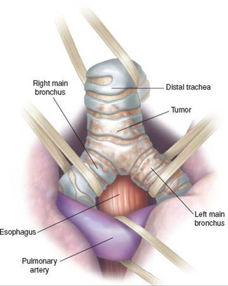
Figure 46.1 Median sternotomy approach. View of carina between the aorta and the pulmonary artery. The view is enhanced if a tape is placed around the aorta and the left pulmonary artery is divided.
Thoracotomy Approach
A long wire-reinforced single-lumen endotracheal tube is placed with the aid of bronchoscopy in the right main bronchus. A left posterolateral thoracotomy on top of the fifth rib is performed (Fig. 46.3). The chest is explored to confirm resectability and the absence of metastatic disease. The aorta is encircled by tapes to retract and aid exposure of the aortopulmonary window. The distal trachea is encircled as well to pull the carina into the operative field. Care is taken to avoid both recurrent laryngeal nerves. The anterior surface of the trachea and right main bronchus are bluntly dissected free in the areolar pretracheal plane to enhance mobility to reduce tension on the anastomosis. The hilar blood vessels are usually divided first followed by division of the trachea and proximal right main bronchus (Fig. 46.4). Ventilation of the right lung is usually carried out by a separate short flexible endotracheal tube that is connected in a sterile fashion to the anesthesia circuit after the airway is divided. The indwelling long endotracheal tube is pulled back within the trachea to unclutter the operative field. Alternatively jet ventilation with a small catheter can be carried out. 2-0 Vicryl lateral stay sutures are placed about two rings deep at 3 and 9 o’clock in the trachea and right main bronchus. Then circumferential 4-0 Vicryl sutures are placed about 4 mm apart and 4 mm deep with the knots to be tied on the outside. Each suture is clipped to the drapes in successive fashion so as to organize this sizable number of sutures. When all sutures are placed the short endotracheal tube is removed from the right main bronchus and the long endotracheal tube readvanced into the proximal right main bronchus. The chin is flexed by the anesthesiologist and the lateral 2-0 stay sutures are tied. The 4-0 sutures are then tied starting first on the anterior aspect where there is strong cartilage followed last by the membranous wall sutures (Fig. 46.2). The anastomosis is then checked for any air leaks by deflating the endotracheal cuff and pulling the tube back into the proximal trachea and reinflating it and delivering 20 to 30 cm of airway pressure. The anastomosis is wrapped with a pedicle of pericardial fat and thymus. The chest is closed in layers in the routine fashion. The patient is extubated at the end of the procedure after a control bronchoscopy to visualize the anastomosis and clear the airway of secretions.
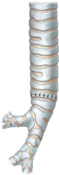
Figure 46.2 End to end anastomosis of the trachea to the right main bronchus. The lateral 2-0 stay sutures are tied first followed by the circumferential 4-0 sutures starting anteriorly and moving posteriorly.
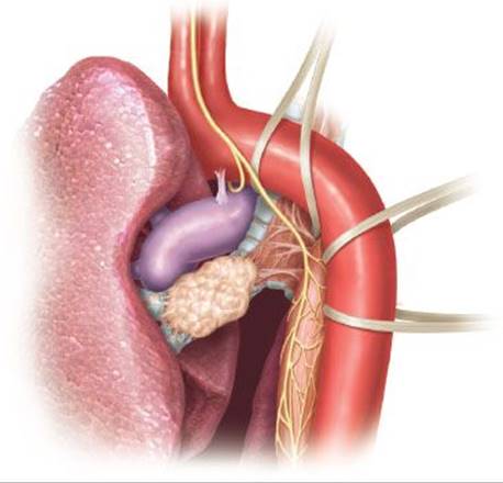
Figure 46.3 Left thoracotomy approach. The view is enhanced if a tape (s) is placed around the aorta and around the distal trachea (taking care not to injure the recurrent nerves). The ligamentum arteriosum should be divided.
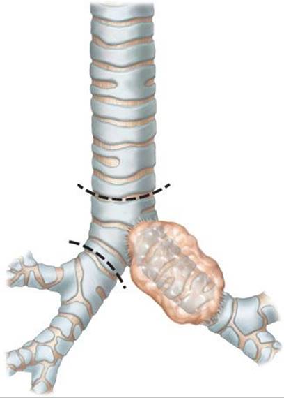
Figure 46.4 Typical cancer in the left main bronchus that requires left carinal pneumonectomy. The dashed lines show the division points of the trachea and right main bronchus.
 POSTOPERATIVE MANAGEMENT
The primary concern is adequate secretion clearance after a carinal resection. Adequate pain control, early mobilization, and moisturizing nebulizers are important keys to recovery. Bedside bronchoscopy is often necessary in the early postoperative period. Intravenous fluids are cautiously administered so as to avoid fluid overload.
 COMPLICATIONS
Any of the typical complications of lung resection can occur after carinal resection. The two feared complications specific to carinal resection are anastomotic complications and postpneumonectomy pulmonary edema (if a carinal pneumonectomy was done). Anastomotic complications are mostly related to tension, but also may be related to preoperative radiation, excessive dissection causing ischemia, or faulty anastomotic technique. The incidence of anastomotic complications varies from 11% to 17% in the various series. Mitchell et al. examined the issue of tension and found that resections longer than 4 cm were associated with high rates of anastomotic complications.
Postpneumonectomy pulmonary edema is a rare but catastrophic complication of pneumonectomy and seems to be more common with complex carinal pneumonectomies rather than simple pneumonectomy. The incidence varies from 0% to 14% in different series. The pathophysiology of this condition is still not understood and does not appear to be a simple matter of fluid overload but rather one of creation of a capillary leak. Barotrauma during the resection is currently the most popular hypothesis of postpneumonectomy pulmonary edema. Current recommendations include limiting perioperative fluid administration, minimizing peak and mean airway pressures during operation, balancing the mediastinum after chest closure, and minimizing pulmonary artery pressures. If postpneumonectomy pulmonary edema occurs the treatment is largely supportive.
 RESULTS
The 5-year survival after carinal resection varies from 27% to 51% for nonsmall cell lung cancer and is largely influenced by the N2 status. In the absence of N2 disease the 5-year survival varies from 38% to 53%. The operative mortality of recent large series of carinal resections varies from 4% to 15% with a trend toward lower mortality. These results suggest this operation is certainly worthwhile in carefully selected patients.
 CONCLUSIONS
Left carinal resection, alone or in combination with pneumonectomy is a rarely done procedure. The most common indication is an airway tumor involving most of the LMB extending to the tracheal carina. The optimal approach is not clear and both left thoracotomy and median sternotomy offer reasonable exposure. Standard techniques of airway resection and reconstruction are used. Early morbidity and mortality are low in centers of excellence. The long-term results are good in the absence of N2 disease for nonsmall cell lung cancer.
Recommended References and Readings
Jiang F, Xu L, Yuan J, et al. Carinal resection and reconstruction in surgical treatment of bronchogenic carcinoma with carinal involvement. J Thorac Oncol. 2009;4:1375–1379.
Mitchell JD, Mathisen DJ, Wright CD, et al. Clinical experience with carinal resection. J Thorac Cardiovasc Surg. 1999;117:39–53.
Moreno P, Lang G, Taghavi S, et al. Right-sided approach for management of left main bronchial stump problems. Eur J Cardiothorac Surg. 2011;40:926–930.
Parissis H, Young V. Carinal surgery: Experience of a single center and review of the current literature. J Cardiothorac Surg. 2010; 5:51–55.
Rea F, Marulli G, Schiavon M, et al. Tracheal sleeve pneumonectomy for non small cell lung cancer (NSCLC): Short and long-term results in a single institution. Lung Cancer. 2008;61: 202–208.
Yamamoto K, Miyamoto Y, Ohsumi A, et al. Results of surgical resection for tracheobronchial cancer involving the tracheal carina. J Thorac Cardiovasc Surg. 2007;55:231–239.