
Olaf Mercier, Marc de Perrot, and Shaf Keshavjee
 INDICATIONS/CONTRAINDICATIONS
Bronchopleural fistula (BPF) remains a life-threatening complication of pneumonectomy occurring between 2% and 13% of such procedures. While early postpneumonectomy BPF requires immediate surgical repair with the help of a vascularized tissue patch, late BPF (i.e., weeks to years after pneumonectomy) requires emergent positioning of a chest tube draining the empyema cavity followed by definitive therapy of both empyema and BPF. Open-window thoracotomies (Clagett window), which are the most widely used procedures, have the advantage of treating empyema and BPF at the same time. Even though it is an effective and technically simple procedure allowing a small BPF to potentially close with regular dressings, it raises the problem of a second stage chest wall closure, outpatient management of dressings, and large BPF closure. The use of vascularized flaps (omentum, muscles) to close the BPF and fill the cavity through a redo thoracotomy may be considered. The transsternal transpericardial approach offers good exposure to the tracheobronchial bifurcation allowing bronchial closure or carinal resection in good conditions with healthy surrounding tissue. It allows dissection of the healthy tracheobronchial tree at a distance from infected tissue and without opening the contralateral pleura. Usually, this approach is considered as a salvage procedure when initial attempts at repair of BPF through rethoracotomy have failed, the rethoracotomy is contraindicated, or the stump length does not allow direct closure and carinal resection is required. Debilitated and malnourished patients should not be selected for this procedure. Previous sternotomy is not a contraindication. Some authors have reported transsternal transpericardial BPF repair in patients who had coronary artery bypass grafts highlighting that it was easier to go through postoperative adhesions than through infected tissue. However, previous mediastinal surgery may remain a relative contraindication.
 PREOPERATIVE PLANNING
As soon as the diagnosis of late BPF is suspected, chest cavity drainage should be instituted promptly to avoid life-threatening aspiration. A flexible bronchoscopy is useful to confirm the diagnosis of BPF, assess the contralateral bronchial tree, clear it from secretions, and above all assess the length of the bronchial stump. Indeed, the bronchial stump length would determine the technique of bronchial repair between direct reclosure and carinal resection. If pneumonectomy is performed for cancer, the preoperative workup includes a chest CT scan screening for a possible recurrence at the level of the bronchial stump or the remaining lung. Given that transsternal surgery deals with bronchial closure alone, the empyema and pleural cavity need to be treated properly prior to the surgery. Hence, broad spectrum antibiotic therapy should be started before and be continued after the procedure. Open-window thoracostomy may be considered if the empyema is not controlled by antibiotics and drainage.
 SURGERY
A variety of anesthesia techniques can be used to ventilate the patient in the presence of a BPF including double-lumen tubes (left- and right-sided), long armored single-lumen tube inserted into the left main bronchus, or endobronchial blocker in the pneumonectomy stump. Our preference is to use the long armored single-lumen bronchial tube when possible. The positioning of the tube is controlled by flexible bronchoscopy. A peripheral arterial line is inserted as well as two peripheral venous lines for volume expansion if necessary. A venous central line may be useful for CVP monitoring and prolonged postoperative antibiotic therapy. An O2 saturation probe should be adequately placed and should work accurately. A nasogastric tube is placed for localization of the esophagus during the surgery.
Positioning
The patient is placed in a supine position, both arms along the body for a standard median sternotomy. An inflatable roll placed behind the shoulders can be used to improve exposure, particularly if a carinal resection is anticipated. The skin is prepped and draped from the chin down to the thighs. Safety requires a groin exposure, as femoral vessels access may be needed to institute cardiopulmonary assistance in case of complications. The operating surgeon stands on the right side and the assistant is on the left.
Technique
A median sternotomy is performed exposing the pericardium. Care must be paid not to open the contralateral pleura, which could be behind the sternum because of the mediastinal shift and the compensatory hypertrophy of the remaining lung. The pericardium is opened longitudinally from the ascending aorta to the diaphragm and suspended with silk stitches. The left innominate vein is dissected and thymic veins divided before retracting the sternum to avoid any tear. The superior vena cava (SVC) is freed from the pericardial reflection, mobilized and encircled with a Penrose drain to aid retraction laterally to the right to expose the ascending aorta. The ascending aorta is then dissected from the right and main pulmonary arteries up to the aortic arch allowing its lateral retraction to the left (Fig. 48.1). The space between the SVC and the ascending aorta is then exposed and the posterior pericardium is opened. The right pulmonary artery is encircled and retracted inferiorly or reresected to improve the exposure (Fig. 48.2). The lower trachea and carina is exposed taking care to avoid injury to the left laryngeal nerve. Complete mobilization of the ascending aorta and the pulmonary artery allows safe dissection of the carina and the left main bronchus even after left pneumonectomy and significant left mediastinal shift. The bronchial stump is dissected at the level of the carina, isolated and divided. The new stump must always be divided to ensure that the edges heal adequately. It is closed preferentially with a TA stapler or sewn with interrupted sutures (Fig. 48.3). The nasogastric tube helps to localize the esophagus behind the main bronchus to avoid injury. The distal stump should be totally excised without opening the infected pleural cavity if possible. If it proves impossible to remove the distal stump, an alternative is to destroy the distal bronchial mucosa with cautery, close it with interrupted sutures and leave it in situ. A vascularized tissue pedicle of thymus or pericardial fat pad is used to cover the new bronchial stump. Omentum may be used—particularly if the patient had received high-dose radiation therapy to the mediastinum in the past. For this, the sternal incision is extended toward the upper abdomen and an omental pedicle is harvested based on the right gastroepiploic vessel (Fig. 48.4).
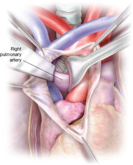
Figure 48.1 After opening of the sternum and the pericardium, superior vena cava and ascending aorta are dissected, mobilized, and retracted giving exposure to the right pulmonary artery and the posterior pericardium in front of carina and distal trachea.
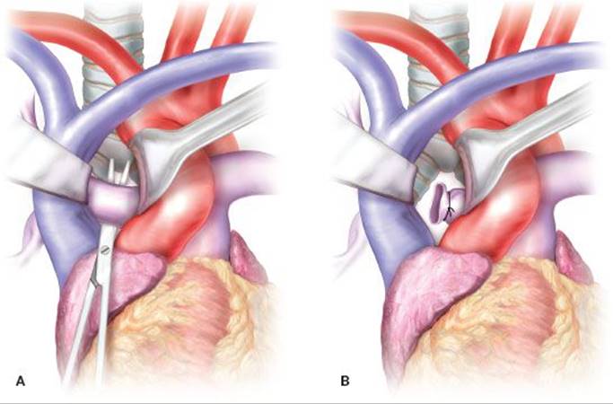
Figure 48.2 A: The right pulmonary artery is dissected and retracted allowing dissection of the lower trachea and bronchial bifurcation after opening of the posterior pericardium. B: Reresecting the right pulmonary arterial stump could allow safer mobilization and give more room for bronchial dissection and resection.
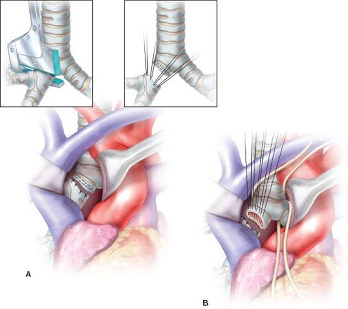
Figure 48.3 In case of long bronchial stump, the main bronchus is divided at the level of its origin and stapled (A) or sutured (B). The distal affected stump could be either removed or be left in place after ablating the mucosa with cautery depending on the difficulty of resection. If not removed, the distal affected stump should be closed proximally with interrupted sutures. A vascularized tissue flap of thymus or pericardial fat pad should be sutured in place to cover the new bronchial suture.
In case of a short bronchial stump (i.e., less than 1 cm), a carinal resection with tracheobronchial anastomosis can be performed through the sternotomy and transpericardial approach. The affected stump is removed and the contralateral main bronchus is anastomosed to the trachea, which has been mobilized using a sharp dissection. During the anastomosis, the single-lumen transtracheal tube is pulled back a few centimeters and ventilation is carried out using a sterile tubing and single-lumen tube inserted intermittently into the bronchus across the surgical field (Fig. 48.5). The inflatable roll placed behind the shoulders is deflated before proceeding with the anastomosis to reduce the tension on the suture lines. After completion of the anastomosis, usual mechanical ventilation through the endotracheal tube is resumed (Fig. 48.6). The trachea is anastomosed to the bronchus using a posterior running suture of 4-0 PDS and interrupted sutures of 3-0 or 4-0 PDS anteriorly. Thymus, pericardial fat pad, or omentum is used to buttress and protect the anastomosis as mentioned previously. Before closing the chest, the absence of anastomotic air leak is checked using normal saline. One retrosternal and one pericardial tube are inserted before closing the sternum and the skin in a usual manner.
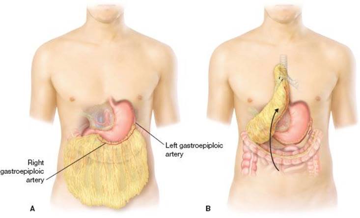
Figure 48.4 In patients that have received radiation therapy, use of an omental flap has been shown to reduce bronchial fistula recurrence. The sternal incision is extended to the upper abdomen and an omental pedicle flap is harvested based on the right gastroepiploic artery (A) the omentum is then brought substernally to the level of the carina in the mediastinum and sutured to the new bronchial stump. B: Dimension of the omentum flap could be modified to perfectly fill and obliterate any space in the posterior mediastinal area. The pericardium is left open.
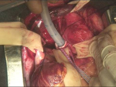
Figure 48.5 Ventilation is performed with a sterile standard endotracheal tube across the operating field. The endotracheal tube is inserted directly into the left main bronchus after resection of the carina.
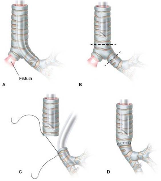
Figure 48.6 When the affected bronchial stump is too short for direct closure (A), carinal resection is required (B). Anastomosis between the trachea and the left main bronchus can be performed safely using the transsternal transpericardial approach (C,D). During the anastomosis, the lung is intermittently ventilated using an endotracheal tube placed across the field (C).
 POSTOPERATIVE MANAGEMENT
As mechanical ventilation has a detrimental role in tracheobronchial suture healing and promotes acquired pneumonia, patients should be extubated as soon as possible after the surgery. Physiotherapy is started the day of surgery and continued on a daily basis. Active mobilization and continuous respiratory exercise are encouraged. Pain control does not differ from other sternotomies, which are generally better tolerated than thoracotomies and do not require the use of epidural analgesia. All these measures aim at reducing the risk of atelectasis, bronchial obstruction, and pneumonia. Thromboembolic disease prophylaxis consists of prescription of subcutaneous heparin and intermittent compression stocking devices. Chest tubes are removed as soon the amount drained is lower than 100 mL per day, usually at postoperative day 2 or 3. Alimentation increases gradually from the day of surgery keeping in mind that the left laryngeal nerve may be injured. Any doubt concerning the left laryngeal nerve requires laryngeal examination and careful feeding.
The last main issue is the treatment of the associated empyema, which is the main risk factor for BPF recurrence. Prolonged intravenous antibiotic therapy adapted to pneumonectomy cavity bacterial cultures combined with debridement of the infected chest cavity. Chest cavity debridement can be done by packing the infected pleural space through an open-window thoracostomy if necessary.
 COMPLICATIONS
Intraoperative Bleeding
Retraction of the pulmonary artery to access the carina and the main bronchi can cause bleeding from the arterial stump or actual tearing of the vessel. Reresection of the right pulmonary artery stump can reduce this risk and improve the exposure to tracheobronchial bifurcation. Care must be paid to avoid excessive traction on the pulmonary artery. Major bleeding from the pulmonary artery in this location may require the use of cardiopulmonary bypass to be repaired.
Respiratory Failure
Respiratory failure is the most common postoperative complication potentially requiring prolonged ventilator support. Preventive measures have to be maximized because such respiratory complications can seriously compromise the postoperative results. These measures consist of avoiding aspiration, selecting patients with good nutritional and clinical status, checking selective ventilation during the surgery, and controlling the sepsis preoperatively. Early tracheostomy may be of value in reducing the length of mechanical ventilation by shortening the weaning time.
Bronchopleural Fistula Recurrence
Recurrence rate ranges from 0% to 30% and represents the main risk factor for postoperative mortality. Lack of sepsis control in the postpneumonectomy cavity favors recurrent fistula. The lack of use of an open thoracostomy and a short bronchial stump have both been reported as risk factors for recurrence. Indeed, ongoing infection close to the newly sutured bronchial stump prevents effective natural healing. If conservative treatment of empyema is chosen, its efficacy needs to be closely assessed to move quickly to a salvage open thoracostomy if conservative therapy is failing to limit the risk of a recurrent or new fistula or a new respiratory complication. Covering the stump with vascularized tissue significantly decreases the risk of recurrence. An omental flap, with its enhanced healing qualities, is particularly useful in preventing this complication. A short bronchial stump should not be reresected and closed with direct suturing as the resultant excessive tension on the suture line is a major risk factor for fistula. A limited carinal resection while a bit more challenging, is a safer procedure that can achieve good result in these patients.
Other Complications
Common complications after thoracic surgery such as atrial fibrillation, pulmonary embolism, and myocardial infarction had been reported after transsternal repair of bronchial fistula occurring in less than 2% of cases. Even though these complications are rare; they have serious consequences since patients have a single remaining lung.
 RESULTS
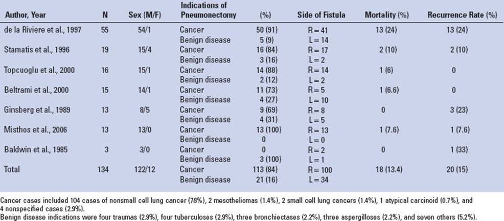
The results of seven case series including 134 transsternal transpericardial repairs of postpneumonectomy BPF between 1985 and 2013 are summarized in Table 48.1. There were mainly men and fistula occurred in majority on the right side. Complication rates were reported between 0% and 24% mainly due to fistula recurrence. Use of vascularized flap covering the suture, better selection of operable patients, and better control of sepsis have decreased the complication rate. Taken together, overall recurrence rate was 15%. Hence, the transsternal procedure allowed bronchial healing in 85% of patients with such a debilitating postpneumonectomy complication. Interestingly, half of the patients had undergone previous attempts at closure of the BPF and the transsternal approach was used as a salvage procedure. Postoperative mortality varied between 0% and 24%.
TABLE 48.1 Results of Transsternal Transpericardial Postpneumonectomy Bronchopleural Fistula Repair from the Literature from 1985 to 2013
 CONCLUSIONS
Transsternal transpericardial approach is a safe and effective method to treat recalcitrant postpneumonectomy BPF. Postoperative mortality and morbidity can be prevented with careful patient selection. This approach has the advantage of accessing late BPF through an area that is free from inflammation and adhesions. It also enables safe carinal resection if the bronchial stump is too short to allow direct closure.
Recommended References and Readings
Beltrami V, Angelici A, Bertagni A, et al. Transsternal approach to closure of bronchopleural fistulas after pneumonectomy. A fifteen cases report. Lung Cancer. 2000;29(1):43–47.
de la Riviere AB, Defauw JJ, Knaepen PJ, et al. Transsternal closure of bronchopleural fistula after pneumonectomy. Ann Thorac Surg. 1997;64(4):954–957.
Ginsberg RJ, Pearson FG, Cooper JD, et al. Closure of chronic postpneumonectomy bronchopleural fistula using the transsternal transpericardial approach. Ann Thorac Surg. 1989;47(2):231–235.
Ginsberg RJ, Saborio DV. Management of the recalcitrant postpneumonectomy bronchopleural fistula: The transsternal transpericardial approach. Semin Thorac Cardiovasc Surg. 2001;13(1):20–26.
Misthos P, Kakaris S, Sepsas E, et al. Surgical management of late postpneumonectomy bronchopleural fistula: The transsternal, transpericardial route. Respiration. 2006;73(4):525–528.
Saito H, Tatsuzawa T, Kikkawa H, et al. Transpericardial bronchial closure with omentopexy for postpneumonectomy bronchopleural fistula. Ann Thorac Surg. 1989;47(2):312–313.
Stamatis G, Martini G, Freitag L, et al. Transsternal transpericardial operations in the treatment of bronchopleural fistulas after pneumonectomy. Eur J Cardiothorac Surg. 1996;10(2):83–86.
Topcuoglu MS, Kayhan C, Ulus T. Transsternal transpericardial approach for the repair of bronchopleural fistula with empyema. Ann Thorac Surg. 2000;69(2):394–397.