
Francis C. Nichols
 INDICATIONS
Bronchopleural fistula (BPF) is defined as an abnormal communication between a bronchus and the pleural cavity. Persistent air leaks originating from the lung parenchyma are not BPF and are properly designated alveolar-pleural fistula. BPF remains one of the most dreaded complications following pulmonary resection. BPF is uncommon occurring in 1% to 16% of patients undergoing pulmonary resection. Its incidence is greater following pneumonectomy than after lobectomy or segmental resection. Development of BPF has been significantly associated with the presence of benign disease, decreased preoperative FEV1, right pneumonectomy, and prolonged chest tube duration. Management of BPF continues to be a significant challenge particularly following pneumonectomy, and its presence has been associated with high morbidity and mortality. Management of postpneumonectomy BPF will be the focus of this chapter.
Fundamental to the prevention of BPF is careful attention to surgical details. Devascularization of the bronchus can occur following overly aggressive dissection. During pulmonary resection, leaving an excessively long bronchial stump predisposes to pooling of contaminated secretions and eventual stump dehiscence. There is increased risk of BPF when pulmonary resection is undertaken following radiation therapy or when pre-existing empyema is present. When such adverse conditions are present, the author and his colleagues prefer to reinforce the bronchial stump with viable tissue, most commonly an extrathoracic skeletal muscle such as serratus anterior.
Because the onset of infection is often insidious and occurs days to years following pulmonary resection, BPF diagnosis can be difficult. Patient presentation ranges from the nondescript findings of feeling chronically ill and having failure to thrive to the more obvious fever, chills, cough, purulent sputum, and chest discomfort. For pneumonectomy patients, a drop in the air–fluid level within the ipsilateral hemithorax especially when associated with expectoration of serosanguinous fluid is pathognomonic of BPF. Computed tomography (CT) can be helpful in evaluating the size and location of loculated air–fluid collections. Bronchoscopy should be performed inspecting the bronchial stump carefully for small BPF. The bronchial stump should be thoroughly irrigated to remove fibrin and mucus and gently probed though care taken to avoid inadvertent perforation.
 PREOPERATIVE PLANNING
Initial management of a patient with postpneumonectomy BPF depends on the clinical presentation. If the patient is acutely ill with respiratory distress and contralateral pneumonia, initial management starts with placing the patient in the lateral decubitus position, pneumonectomy side down until drainage of the pneumonectomy space is obtained. Chest tube placement in the pneumonectomy space should quickly be performed resulting in drainage of the cavity, stopping contralateral spillage, and beginning control of the infection. Appropriate resuscitation is undertaken and respiratory support provided as needed. Broad spectrum antibiotics begun and further adjusted when antimicrobial sensitivities become available. If a large BPF is present, mechanical ventilation may be required. A double-lumen endotracheal tube or long single-lumen tube carefully placed in the remaining lung’s mainstem bronchus is required to bypass the fistula, ventilate and decrease contamination of the remaining lung. Rarely is emergent surgery required. The author’s preferred treatment of postpneumonectomy BPF is a modification of the original Clagett procedure. The modification includes transposition of well-vascularized muscle to cover the bronchial stump to prevent recurrent fistula formation. Knowing which chest wall muscles were divided or otherwise utilized at the time of initial pulmonary resection is important. Usually the serratus anterior muscle is intact despite a previous thoracotomy. Other possible muscles for transposition include the pectoralis major and cephalic portion of the previously transected latissimus dorsi. Other options include the rectus abdominis and omentum. With the modified Clagett procedure, no attempt is made to completely fill the pleural cavity with either muscle or other autogenous tissue. Instead, the chest open chest cavity is filled with antibiotic solution at the time of skin closure.
Nutritional status is important in this group of patients who are often debilitated and malnourished from chronic infection. If necessary, nutritional supplementation including liberal use of enteral feedings should be considered in all of these patients.
 SURGERY
There are three basic principles to treatment: (1) Adequate pleural drainage, (2) closure of the fistula, and (3) obliteration of the residual pleural space. The author and his colleagues’ management of postpneumonectomy BPF has evolved over time. Our most common approach is two staged with each stage often containing multiple individual operative procedures. The first stage, again most commonly accomplished in a series of operations, consists of open drainage, closure of the BPF, muscle transposition, and debridement. Only after successful completion of the first stage is the second stage undertaken, which specifically includes obliteration of the pleural space with antibiotic solution and definitive closure of the chest wall.
All procedures begin with a team briefing. Surgeons, anesthesiologists, and operating room personnel are in attendance. Details of the airway management individualized for each specific patient are a major focus of the briefing. Patient identification, site marking, procedure verification, and final anesthetic evaluation are completed. Appropriate intravenous access is established. Epidural catheters are not utilized because of the risk of contamination. Instead, postoperative pain management most commonly is via a patient-controlled analgesia (PCA) pump. Following intubation, a urinary catheter and lower extremity sequential compression devices are placed. Subcutaneous injection of 5,000 units of unfractionated heparin is administered to reduce the risk of deep venous thrombosis (DVT) and pulmonary embolism.
Positioning
As long as the BPF is open, all procedures are done with isolation of the remaining lung via a long single-lumen endotracheal tube or double-lumen tube. All procedures are done in an open fashion, in a lateral decubitus position with the pneumonectomy side up. The exception being if the rectus abdominis muscle or omentum is harvested whereby the patient is in a supine position for that particular portion of the procedure.
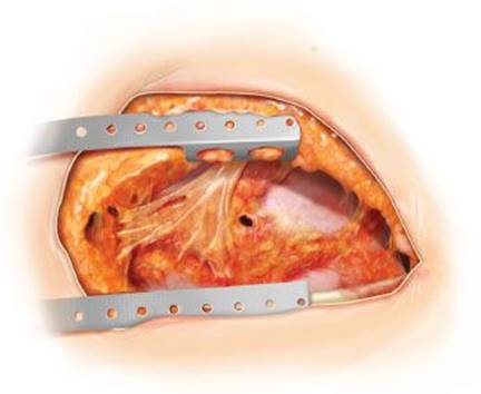
Figure 49.1 Open pleural drainage. The entire previous thoracotomy incision is reopened. A small bronchopleural fistula is seen.
First Stage
Open Pleural Drainage, Closure of the Fistula, Muscle Transposition, Chest Cavity Debridement
Technique
 Entire ipsilateral posterolateral thoracotomy is reopened (Fig. 49.1).
Chest cavity is debrided, appropriate specimens are sent for microbiologic and histopathologic testing.
BPF is sought by filling pleural cavity with saline and observing for escape of air bubbles from the mediastinum with positive pressure ventilation. This testing requires appropriate adjustment of the double-lumen tube or repositioning of the long single-lumen endotracheal tube.
Small BPFs do not need to be closed at the time of initial debridement but instead can be repaired later when the pleural cavity is cleaner (usually at the second or third operation).
If a large significant fistula is present, direct closure at the time of initial thoracotomy and debridement is preferred to prevent loss of ventilation and minimize ongoing contralateral contamination (Fig. 49.2A).
A long bronchial stump if present is mobilized into the mediastinum, redivided close to the carina with either staples (TA or TX 30 4.8-mm stapler) or sutured with interrupted 3-0 or 4-0 nonabsorbable monofilament suture (Fig. 49.2B).
This dissection can be hazardous because of proximity of the bronchus to the pulmonary artery and esophagus.
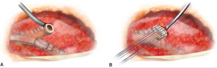
Figure 49.2 A: A double-lumen endotracheal tube is present in the contralateral bronchus protecting the remaining lung from ongoing contamination and allowing adequate ventilation. The bronchopleural fistula has been debrided and resected back to near the carina. B: The now appropriately shortened bronchial stump is securely closed with interrupted 3-0 or 4-0 nonabsorbable monofilament sutures.
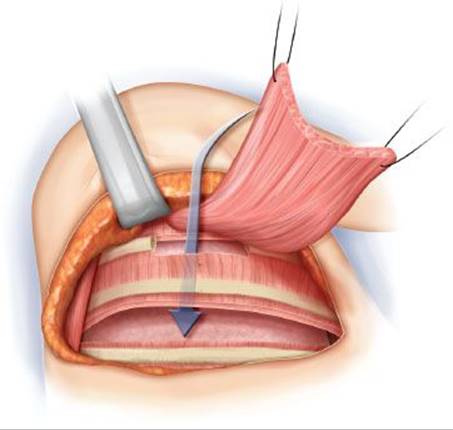
Figure 49.3 After elevating the previously divided latissimus dorsi muscle, serratus anterior muscle is mobilized. A portion of the ipsilateral third rib anteriorly is removed, the muscle is transposed tension free into the debrided pleural cavity to cover the bronchus now appropriately shortened bronchial stump is securely closed with interrupted 3-0 or 4-0 nonabsorbable monofilament sutures.
This dissection into the mediastinum should be kept very close to the bronchial wall at all times. This can be accomplished by placing an Allis clamp on the cartilage of the bronchial remnant, which permits traction on the bronchial remnant while sharply dissecting the bronchus circumferentially down to the carina. Literally hugging the bronchial cartilage allows for safe dissection of the bronchial remnant and avoidance of the adjacent pulmonary artery stump.
A short bronchial stump should be mobilized and completely opened and its margins freshened. If possible, it is then closed with interrupted 3-0 or 4-0 nonabsorbable monofilament suture (Fig. 49.2B).
If the BPF is small and not initially closed, or if the bronchus is successfully closed, the chest cavity is further sharply debrided followed by pulsatile irrigation. The chest cavity is then packed with debridement antibiotic solution (DABS) (20 mg gentamicin and 500,000 units of polymyxin B per liter of 0.9% sodium chloride solution) lightly saturated gauze rolls (Fig. 49.3).
All bronchial stump closures are reinforced with intrathoracic transposition of intrathoracic skeletal muscle.
Serratus anterior, latissimus dorsi (even if previously divided), and pectoralis major muscle are most frequently utilized in that order (Fig. 49.3).
Occasionally rectus abdominis muscle or omentum is transposed.
The transposed muscle is secured in place with 3-0 or 4-0 nonabsorbable monofilament suture (Fig. 49.4A).
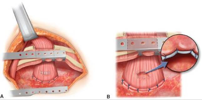
Figure 49.4 A: The bronchus has been successfully closed with interrupted suture. The serratus muscle flap then completely covers the repaired bronchus and is secured in place with interrupted 3-0 or 4-0 nonabsorbable monofilament suture. B: When the bronchus cannot primarily be repaired, the muscle is circumferentially sewn to the margin of the open bronchus with interrupted 4-0 nonabsorbable monofilament suture. An airtight closure is obtained.
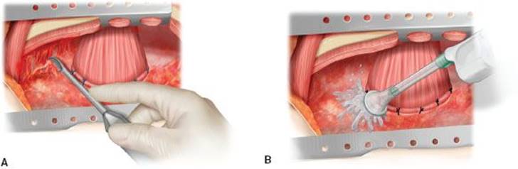
Figure 49.5 A,B: The open pleural cavity is sharply debrided and pulse irrigated during one of many return trips to the operating room.
When the bronchus cannot be primarily closed, the transposed muscle is circumferentially sutured around the perimeter of the open bronchus with 3-0 or 4-0 nonabsorbable monofilament suture obtaining an airtight closure (Fig. 49.4B).
The patient then undergoes over a period of 1 to 2 weeks a series of sharp pleural cavity debridements, hand-held pulsatile irrigation, and packing of the pleural cavity with gauze rolls lightly saturated with DABS (Fig. 49.5A,B).
The debridements and dressing changes take place in the operating room every 48 hours for 7 to 10 days. This allows for proper adhesion of the muscle flap to the bronchial stump.
Once the bronchus is closed, most commonly after the second or third pack change most patients do not require mechanical ventilation for the serial wound debridements. Monitored conscious sedation may be utilized.
Once the transposed muscle is sufficiently adhered, the pack change can often take place in the patient’s hospital room utilizing appropriately monitored conscious sedation (Fig. 49.6).
The ultimate goal for this first stage is a clean wound, with closed bronchus, an adherent muscle flap, and pink and healthy granulation tissue covering the parietal pleura, mediastinal surfaces, and wound edges.
Second Stage
Obliteration of the Pleural Space and Closure of the Chest Wall
Technique
Once the fistula has remained closed, the muscle flap adherent and growing into place, the pleural cavity clean and covered with healthy granulation tissue, the wound is ready for closure. Final pleural space tissue cultures are not used to determine the timing of chest closure, but may be obtained to guide post-closure systemic antibiotic therapy.
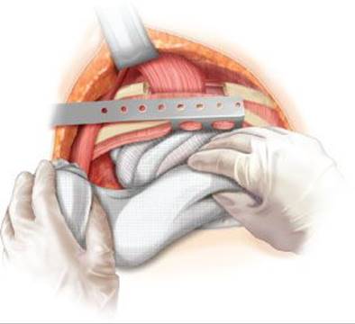
Figure 49.6 The pleural cavity is packed with multiple gauze rolls lightly saturated with debridement antibiotic solution (DABS).
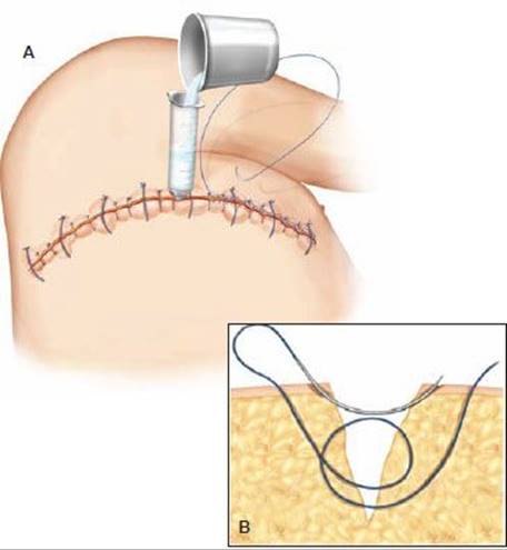
Figure 49.7 A: After the bronchopleural fistula is known to be successfully closed and the pleural cavity is clean and covered with healthy pink granulation tissue, the pleural cavity is filled with DABS and the chest wound closed with interrupted no. 1 nonabsorbable monofilament “Arnold sutures” (inset B), interrupted nonabsorbable monofilament 3-0 vertical mattress sutures, and closely spaced running 4-0 nonabsorbable monofilament suture.
The chest wall is closed ideally in layers by separation of the soft tissue planes, mobilization of adjacent muscles and at least partial reapproximation with absorbable suture, and watertight closure of the skin with a combination of heavy and lighter both interrupted and running nonabsorbable monofilament sutures (Fig. 49.7A,B).
Prior to final skin closure, the pleural cavity is filled with DABS solution.
Direct apposition of the skin (dermis to dermis) is of utmost importance.
Skin and subcutaneous tissues are first approximated by placing initially untied no. 1 nonabsorbable monofilament “Arnold sutures” every 6 to 8 cm (Fig. 49.7B).
Several interrupted 3-0 nonabsorbable monofilament vertical mattress sutures are evenly spaced between the “Arnold sutures,” which are then tied.
Meticulous dermis to dermis approximation is accomplished with closely spaced running 4-0 nonabsorbable monofilament suture.
Cyanoacrylate tissue adhesive is applied over both the incision and where the no. 1 and 3-0 sutures penetrate the skin.
The wound is then covered with occlusive petrolatum gauze, large gauze pads held loosely in place with paper tape, and finally a long wide elastic wrap, which keeps both the dressings atraumatically in place and the DABS within the pleural cavity. Without the elastic wrap, the DABS pushes against the previously elevated subcutaneous flaps.
 POSTOPERATIVE MANAGEMENT
BPF patients should go to a unit skilled in the management of patients who have undergone complex general thoracic surgery. Most of our patients following the initial first stage procedure are successfully managed in our general thoracic surgical progressive care unit (PCU). Only rarely are these patients kept intubated and managed in our general thoracic surgical intensive care unit (ICU). Following the initial procedure, even when the BPF remains open, the majority of these patients can remain successfully extubated. Following subsequent first stage procedures, depending on patient-specific circumstances, the patients are managed in the PCU or on the general thoracic surgical floor. Standard orders should not be routinely implemented without consideration of specific individualized patient needs; nevertheless, we strive to follow unit-specific care pathways. These pathways routinely contain patient monitoring, supplemental oxygen therapy, intravenous fluids, and medications including appropriate pain management, DVT prophylaxis, wound care, chest physiotherapy by trained respiratory therapists, physical therapy, diet, patient and family education, social services, and when appropriate discharge planning.
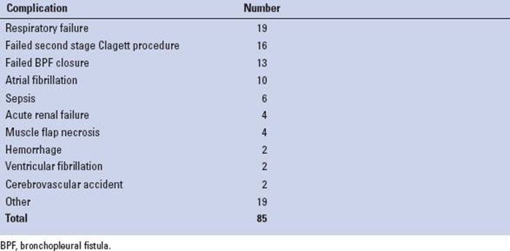
TABLE 49.1 Complications in 84 Postoperative Patients with Postpneumonectomy Empyema Including 55 Patients with Bronchopleural Fistula
Following second stage definitive chest closure, the care pathway is continued and adjusted accordingly. Wound care most commonly involves adjustments to the elastic wrap as necessary. The occlusive petrolatum gauze and large gauze pads changed every 24 to 48 hours and along with the elastic wrap continued for several weeks. The patient may shower starting in 5 to 7 days. Removal of the monofilament sutures is individualized to the patient’s wound status but routinely remain for several weeks. In general, the no. 1 sutures remain for at least 2 to 4 weeks, the 3-0 sutures removed in 4 to 6 weeks, and the 4-0 running suture removed after 6 weeks. The no. 1 sutures are removed earliest since if left too long they may fistulize to the underlying obliterated pleural cavity.
 COMPLICATIONS
The incidence of intraoperative complications during repair of postpneumonectomy BPF is rare. In a recent review from our institution involving 84 consecutive patients with postpneumonectomy empyema 55 of whom had a BPF, complications occurred in 46 patients (55%; Table 49.1). Tracheostomy was necessary in 12 patients, all of whom had BPF repair. Operative mortality occurred in 4 of 55 patients with BPF (7.2%). In two of the deaths, cerebral metastases were ultimately found. The remaining two deaths were attributed to respiratory and acute renal failure in one patient and ventricular fibrillation in another.
 RESULTS
Our institution’s longstanding experience and multiple reported series addressing the management of postpneumonectomy BPF reinforces the success we have had utilizing the modified Clagett procedure in this difficult group of patients. Very good results can be obtained in more than 80% of patients using this approach. Utilizing the modified Clagett procedure for BPF closure, Pairolero et al., from our institution, reported 24 of 28 patients (85.7%) with a successfully closed BPF and well-healed chest wall without signs or symptoms of infection on long-term follow-up. Four patients developed recurrent BPF in the early postoperative period. Three of the four patients had early respiratory failure requiring prolonged mechanical ventilation and all died. The remaining patient was found to have recurrent cancer near the bronchial stump, and no further attempt at closure was undertaken. Our most recent series of 84 patients with postpneumonectomy empyema revealed that these patients have 1 to 25 separate operative procedures (median, 7); moreover, this number did not vary with or without the presence of a BPF. Median hospitalization was 30 days (range, 2 to 425). We had follow-up in all patients with a median of 1.5 years and range of 28 days to 22 years. Seven patients were discharged from the hospital with open chest wounds and having daily dressing changes. One open wound was eventually successfully closed. However, six of these patients never had chest wall closure because of recurrent cancer or other life-threatening medical problems. All six patients were managed with a chronic open pleural window. The chest remained closed and well-healed in 68 of 74 patients (89.5%). Importantly, the BPF in all patients was eventually successfully closed although in 18% of our patients, the BPF had to be reclosed at least one additional time. Similarly, the second stage of the Clagett procedure resulted in a healed chest wall without drainage in nearly 90% of patients who were closed.
Reasons for failure of the second stage of the modified Clagett procedure are not entirely clear. Patients with recurrent cancer or other major life-threatening medical conditions are not candidates for wound closure, something that occurred in 8% of our patients. In addition, patients who experienced late failure were usually malnourished and immunocompromised, emphasizing the importance of optimal nutrition in these patients.
 CONCLUSIONS
BPF is an uncommon but still dreaded and challenging complication following pneumonectomy. While certain risk factors are associated with an increased incidence of BPF, fundamental to prevention is meticulous surgical technique. For over 35 years, the modified Clagett procedure has been successfully employed in our practice in the management of BPF. The modified Clagett procedure is safe and successful in the majority of patients with postpneumonectomy empyema and associated BPF. Some patients with postpneumonectomy BPF will not be candidates for chest wall closure because of recurrent cancer, respiratory failure, and other life-threatening medical conditions. This small subset of patients might be best managed with a chronic open pleural window.
Recommended References and Readings
Allen MS, Deschamps C, Trastek VF, et al. Bronchopleural fistula. Chest Surg Clin N Am. 1992;2:823–837.
Arnold PG. Space obliterating skin suture. Plast Reconstr Surg. 1997; 100:1056–1058.
Arnold PG, Pairolero PC. Intrathoracic muscle flaps. A 10-year experience in the management of life-threatening infection. Plast Reconstr Surg. 1989;84:92–99.
Cerfolio RJ. The incidence, etiology, and prevention of postresectional bronchopleural fistula. Semin Thorac Cardiovasc Surg. 2001; 13:3–7.
Deschamps C, Allen MS, Miller DL, et al. Management of postpneumonectomy empyema and bronchopleural fistula. Semin Thorac Cardiovasc Surg. 2001;13:13–19.
Deschamps C, Bernard A, Nichols FC, et al. Empyema and bronchopleural fistula after pneumonectomy: Factors affecting incidence. Ann Thorac Surg. 2001;72:243–248.
Miller JI. Overview: Postresectional bronchopleural fistula. Semin Thorac Cardiovasc Surg. 2001;13:37–28.
Pairolero PC, Arnold PG, Piehler JM. Intrathoracic transposition of extrathoracic skeletal muscle. J Thorac Cardiovasc Surg. 1983; 86:809–817.
Pairolero PC, Arnold PG, Trastek VT, et al. Postpneumonectomy empyema. The role of intrathoracic muscle transposition. J Thorac Cardiovasc Surg. 1990;99:958–968.
Pairolero PC, Deschamps C, Allen MS, et al. Postpneumonectomy empyema. Chest Surg Clin N Am. 1992;2:813–822.
Puskas JD, Mathisen DJ, Grillo HC, et al. Treatment strategies for bronchopleural fistula. J Thorac Cardiovasc Surg. 1995;63:771–775.
Stafford EG, Clagett OT. Postpneumonectomy empyema: Neomycin instillation and definitive closure. J Thorac Cardiovasc Surg. 1972;63:771–775.
Wright CD, Wain JC, Mathisen DJ, et al. Postpneumonectomy bronchopleural fistula after sutured bronchial closure: Incidence, risk factors, and management. J Thorac Cardiovasc Surg. 1996; 112:1367–1371.
Zaheer S, Allen MS, Cassivi SD, et al. Postpneumonectomy empyema: Results after the Clagett procedure. Ann Thorac Surg. 2006;82:279–287.