
Mark S. Allen
Introduction
Tansini originally described the use of muscle flaps in 1906 when he described using the latissimus dorsi muscle to reconstruct the chest wall after a mastectomy. The technique did not gain much popularity until improvements in anesthesia, antibiotics, and vascular techniques permitted safe and reliable harvest of the chest wall muscles. In both World War II and the Korean War advances in the understanding of the pathophysiology of chest trauma lead to the development of better techniques to treat chest wall injuries and severe infections of the pleural space. In 1950, Campbell revisited the idea of using the latissimus dorsi for chest wall reconstruction. His description was ignored for almost 20 years until Pairolero and Arnold began using the chest wall musculature as flaps to treat chest wall and pleural space problems. Under the guidance of Dr. Maurice J. Jurkiewicz, at Emory University in Atlanta, Georgia, many plastic surgeons were taught the use of muscle flaps to treat these often-difficult problems and the techniques now have become the standard of care.
 INDICATIONS/CONTRAINDICATIONS
The indications for the use of a muscle flap vary considerably depending on the pathology and the clinical situation. Muscle flaps are used to bring well-vascularized tissue to an area that has been damaged by radiation or infection. For example, in patients with radiation necrosis of the chest wall after external beam irradiation for breast cancer, a muscle flap can provide new vascularized tissue to cover the reconstruction of the chest wall. Muscle flaps can also provide coverage for an area with impaired healing. In the case of a bronchopleural fistulae with an infected bronchial stump, a muscle flap transposed through the chest wall into the pleural cavity can cover the newly closed bronchial stump and even though the edges of the bronchial closure may not stay approximated, the presence of a muscle flap covering the stump will prevent leakage of air or purulent material into the pleural cavity. Muscle flaps can also be placed on bronchial stumps prophylactically as in the patient who has had high-dose radiation therapy prior to resection.
Another major indication for the use of muscle flaps in thoracic surgery is to act as space filler. A typical example is in a patient who has a space after a lobectomy and a persistent air leak. By placing a muscle flap into the pleural space, the empty pleural space is obliterated allowing the air leak to heal. Muscle flaps can also be used to fill infected spaces, such as in the patient with an empyema whose decortication does not permit the lung to expand to fill the pleural space. Using a muscle flap in this circumstance will obliterate the potential space and bring well-vascularized tissue into the area to assist in healing the infection.
Another use for muscle flaps is to separate tissue. The classic example is using a muscle flap to separate the trachea and esophagus after a tracheoesophageal fistula repair. The interposed muscle will prevent the adjacent suture lines on the trachea and esophagus from coming together to reform the fistula.
 PREOPERATIVE PLANNING
The use of muscle flaps to control infection, obliterate dead space, or separate tissue is not a commonly used technique. It is only necessary when other less traumatic techniques have not been successful or the patient has a unique problem. For most empyema the treatment is antibiotics and early drainage, before significant fibrosis of the pleural contaminants occurs. Early drainage will cure the vast majority of patients. When empyemas are treated late a decortication may be necessary. To perform a decortication there must be a plane between the lung and the fibrotic peel or excessive bleeding and air leakage will occur. There must also be a lung underneath the fibrous peel that can expand to fill the pleural space. If either of these conditions does not exist, decortication will be unsuccessful and some sort of space filler will be needed. When there is a bronchopleural fistula present this must be closed and reinforcement with a muscle flap is an excellent means to prevent reopening of the bronchial stump.
Most patients who present with the need for a muscle flap to correct some chest wall or pleural pathology have significant deficiencies in nutrition and have been sedentary for quite some time. Both of these problems should be addressed prior to undertaking reparative surgery of the magnitude that involves a muscle flap. Enteral feeding and physical rehabilitation can be undertaken well in advance of the reconstructive operation. While the rehabilitation is ongoing, infection should be controlled by antibiotics and any collections of purulent material drained. This may require an open window into the pleural space and repeated dressing changes before definitive reconstruction is undertaken. If a large bronchopleural fistula is present this should be closed and the infection drained before reconstruction is done.
The choice and use of muscular flaps should be made preoperatively in conjunction with a qualified plastic surgeon. It is possible for a thoracic surgeon to raise a simple muscle flap without the assistance of a plastic surgeon, but if the initial muscle flap fails, it sure is helpful to have had the plastic surgeon involved from the beginning, so the secondary options have been thought out. The size and location of the defect should be well known in advance. With current imaging technology, the three-dimensional configuration of the defect can be determined allowing for selection of the appropriate reconstructive technique. The thoracic surgeon should be well aware of previous operations that may have damaged the blood supply to various muscles of the chest wall that therefore make them nonviable options. Examples include the use of the left internal mammary artery would make the use of a left rectus muscle based on the superior epigastric vessel a poor choice, or a prior thoracotomy may have divided the latissimus dorsi muscle on the ipsilateral side, making its use less desirable.
Patients can usually be positioned in the lateral decubitus position for easy access to the pleural cavity as well as harvesting the muscles of the chest wall. If a skin graft is going to be necessary, the lateral thigh can be prepped into the field as well.
 SURGERY
Pectoralis Major Muscle Flap
The pectoralis muscle, along with the serratus anterior, pectoralis minor, and subclavius, is one of the four muscles that keep the pectoral girdle attached to the chest wall. It originates from the medial portion of the clavicle and from along the lateral sternal border and upper costal cartilages. The muscle converges laterally and inserts on the intertubercular groove of the proximal humerus. Its main function is to facilitate adduction and internal rotation of the humerus. The anterior axillary fold is made up from its lateral muscle fibers. The vascular supply to the pectoralis major is mainly via the thoracoacromial artery with some perforators that come through the chest wall from the internal mammary artery (Fig. 50.1). The thoracoacromial artery arises from the second portion of the axillary artery and then courses laterally. It divides into four branches as it passes through the clavipectoral fascia: Clavicular, pectoral, acromial, and humeral branches. Detailed knowledge of the course of these vessels is important when harvesting the muscle for use.
The pectoralis major muscle is usually used as an advancement flap to cover an infected sternum but can occasionally be used to fill pleural cavity defects, by detaching the insertion on the humerus (Fig. 50.2). To harvest the muscle as an advancement flap to cover a sternal defect or infection the muscle is raised from medial to lateral off the chest wall. Perforators from the internal mammary artery should be ligated. Do not free too much of the subcutaneous tissue off the anterior surface since this may interfere with the skin’s blood supply and impair healing of the incisions. The muscle will usually reach to cover a midline defect, but occasionally the insertion onto the humerus needs to be divided via a second incision to allow greater mobility.

Figure 50.1 Axillary artery. Cut away view of the right axillary artery and its branches showing the principal blood supply to the pectoralis major muscle (thoracoacromial artery), the latissimus dorsi muscle (thoracodorsal artery), and serratus anterior muscle (circumflex scapular artery).
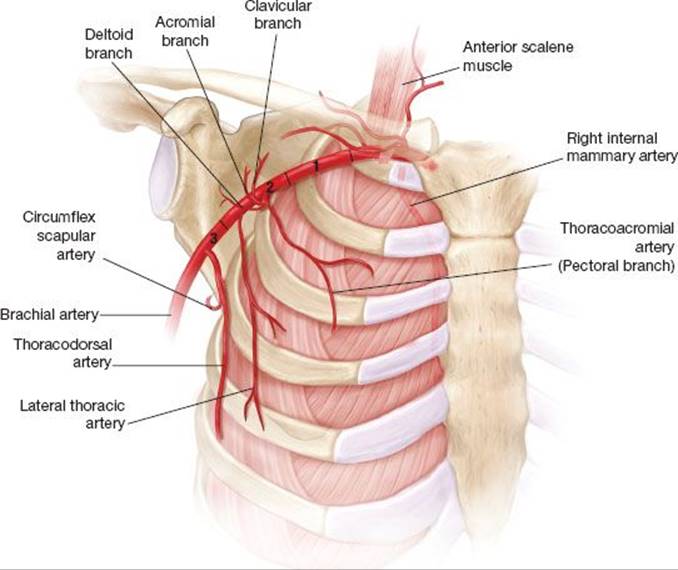
Figure 50.2 Pectoralis major muscle flap. Lateral view of elevation of the pectoralis major flap.
The pectoralis major can also be used as a turnover flap, especially if coverage of the lower portion of the sternum is necessary. In this configuration the muscle flap is based medially on the perforators from the internal mammary artery. When the ipsilateral internal mammary artery has been used to revascularize the heart this muscle flap cannot be used. The insertion on the humerus is detached and the muscle is “turned over” and used to cover the defect. Disadvantages of this configuration include basing the muscle flap on its secondary blood supply, the necessity of raising large subcutaneous flaps, and cosmetic distortion of the anterior axillary fold.
The pectoralis major can also be placed into the pleural cavity, basing the muscle flap on its primary arterial supply from the acromialdorsal artery. The use of this muscle for this purpose is a second or third choice since it only provides a small volume of muscle and cannot reach deep into the pleural cavity. The other muscles of the chest wall, the latissimus dorsi and serratus anterior are much better suited for use inside the chest cavity.
Latissimus Dorsi Muscle Flaps
The latissimus dorsi muscle is the workhorse for reconstruction of chest wall defects. It is a large, broad-based muscle with a reliable blood supply and relatively easy to harvest. It originates from multiple areas, including T7 to T12 spinous processes, thoracolumbar fascia, posterior iliac crest, lower four ribs, external oblique, and the scapula (Fig. 50.3). It thickens as it approaches its insertion onto the intertubercular groove of the humerus. The dominant blood supply is from the thoracodorsal artery a branch of the subscapular artery. In about 2% to 5% of patients the thoracodorsal artery will arise directly from the axillary artery. The thoracodorsal artery gives off branches to the serratus anterior muscle just after it enters the latissimus dorsi muscle, allowing both of the muscles to be harvested at once, if necessary. There are some minor secondary arterial supplies to the muscle, most notably branches from the 8th to 11th intercostal artery that can allow the inferior aspect of a latissimus dorsi muscle that has been transected during a previous thoracotomy to be elevated and used to repair posterior defects. Unfortunately, after a standard posterior lateral thoracotomy where the latissimus has been transected there is not much muscle supplied by the primary arterial source left to use for reconstruction.
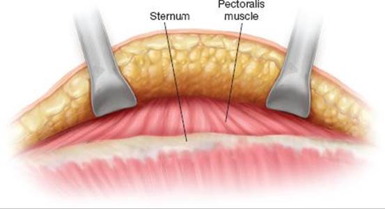
Figure 50.3 Latissimus dorsi muscle flap. Posterior view of chest to demonstrate the anatomy of the latissimus dorsi muscle.
The latissimus dorsi muscle can be harvested from a lateral decubitus position, so it can be done in the same position needed for a lateral thoracotomy (Fig. 50.4). The muscle can be raised with a skin paddle attached if skin coverage is needed for chest wall reconstruction. The muscle is typically detached from its insertions on the spine, iliac crest, and ribs and then rotated on its vascular access to the required location. If it is going to be placed into the pleural cavity, it is often raised in conjunction with the serratus anterior muscle. A portion of chest wall is removed, usually one rib for about 10 to 15 cm and the muscle(s) are passed through the hole in the chest wall and into the pleural space. Care must be taken not to constrict the muscle as it passes into the chest. Removal of a portion of the second or third rib usually allows the muscle to pass into the chest without undue torsion. The muscles’ arterial and venous supply must not be impaired or the flap will necrose and the operation will be a failure.

Figure 50.4 Latissimus dorsi muscle flap inserted after rotation. Latissimus dorsi muscle elevated and rotated onto the anterior chest to cover a defect.

Figure 50.5 Serratus anterior muscle flap. Elevated serratus anterior muscle flap that has been passed through a partially resected third rib to cover a bronchial stump.
Serratus Anterior Muscle Flaps
The serratus anterior muscle flap is probably the easiest flap to elevate and the most useful since it is not typically cut during a standard thoracotomy and the harvesting can be done through a standard posterior lateral thoracotomy incision. The serratus muscle has its origin from 9 or 10 slips from ribs one through nine or ribs one through eight, and its insertion is on the medial border of the scapula between the superior and inferior angles and along the thoracic vertebrae. Its blood supply, as mentioned above, is from the thoracodorsal vessels, which is a branch of the artery that supplies the latissimus dorsi muscle as well. There are some secondary blood supplies that can support only a small portion of the muscle. The long thoracic nerve innervates the serratus muscle, which is a branch from the brachial plexus. These anatomic arrangements lead to the major morbidity of taking the serratus muscle, and that is of a winged scapula. Patients should be clearly warned of this known postoperative finding prior to surgery. The muscle is harvested by detaching its origins off of the chest wall, and the insertion can also be detached if extra length is needed, but this is rarely necessary. As in the case of the latissimus dorsi muscle for use inside the pleural cavity, a portion of the second or third rib is removed and the muscle is passed through this chest wall defect into the area of the muscle (Fig. 50.5). Its primary use is to cover a bronchial stump after a pneumonectomy or cover closure of a bronchopleural fistula.
Rectus Abdominis Muscle Flap
The rectus abdominis muscle is usually used for breast reconstruction but can also be used to repair defects in the lower sternum or lower chest or even to fill the inferior aspect of the pleural cavity. It is a vertical strap-like muscle that begins at the xiphoid process and the fifth through eighth cartilages and inserts on the pubic symphysis. Its blood supply is dual. Superiorly, the superior epigastric artery supplies the muscle and inferiorly it is supplied by the inferior epigastric vessel, a branch of the external iliac artery (Fig. 50.6). The muscle flap can be based on either of these arteries although basing it on the inferior vessel can restrict its mobility into the chest cavity. The muscle cannot usually be based on the superior epigastric vessel if one of the internal mammary arteries on the ipsilateral side has been used to revascularize the heart. The muscle can be harvested through a variety of incisions, including a midline incision or transverse incision. It can also be harvested with or without a skin flap, depending on whether or not skin reconstruction is needed on the chest wall. If the muscle is based on the superior epigastric artery, the deep inferior epigastric artery is ligated, and the flap then can be rotated or turned over to cover the central portion of the chest or passed through the chest wall to fill a cavity in the anterior-inferior aspect of the pleural space.

Figure 50.6 Rectus abdominal muscle flap. Supine patient with the right rectus abdominis elevated based on the superior epigastric artery in preparation to cover a lower sternal defect.
Free Flaps
Due to modern microvascular techniques, virtually any muscle in the body now can be transferred with free vascular anastomotic techniques to supply the area around the chest or intrathoracic area. The donating vessels can be the intercostal vessels or the internal mammary artery as necessary. Obviously, these flaps are much more complicated than local rotation flaps, but occasionally there are no local flaps available and one must resort to a free flap type of reconstruction.
Intercostal Muscle Flap
For completeness sake, the intercostal muscle flap should be mentioned. These muscles can be harvested during the opening of a posterior lateral thoracotomy by carefully preserving the artery and vein associated with the intercostal muscle. In dividing it anteriorly, a small thin paddle of muscle can be harvested. It is occasionally useful in the chest cavity for coverage of an anastomosis or repair of the esophagus or even to cover a bronchus. This flap, however, is not as robust as a serratus muscle or latissimus dorsi muscle and should be used with caution in that it oftentimes becomes ischemic as the chest is closed.
 POSTOPERATIVE MANAGEMENT
Typically, the postoperative care after a chest wall or pleural procedure employs standard postoperative management techniques, including routine management of chest tubes and pain control. If there are subcutaneous drains placed where a large flap has been raised, these can be removed after the drainage reaches an acceptable limit. When patients have artificial material for the reconstruction, such as a Gore-Tex patch or methyl methacrylate reconstruction of the chest wall, drains should be removed earlier rather than later to reduce the chance of infection of the artificial material. Occasionally binders are used to try to reduce the risk of a seroma formation. These should be used with caution, so that they do not compress the vasculature of any muscle flap reconstruction. Postoperative physical therapy is also indicated in patients who had a muscle flap to try and regain full mobility and increase the strength of the affected area where the muscle has been harvested. As mentioned above, these patients are typically nutritionally depleted, thus nutritional intervention is often necessary.
 COMPLICATIONS
Complications after chest wall muscle flaps really are based on the viability of the flap, which depends on several factors, including the amount of tissue raised, the vascular supply of the tissue, avoiding any compression, and not putting the flap under tension. If there is a question about the viability of the flap, patients should be re-explored and the flap examined to determine its viability since a nonviable flap will promote infection and increase the difficulties that the initial problem caused. Flaps that are not viable should be debrided back to viable tissue and then a secondary plan established to try and remedy the problem. As mentioned above, a winged scapula after a serratus anterior muscle has been harvested is an expected outcome and not truly a complication. Patients should just be made aware of this to help manage their expectations.
 RESULTS
The largest series in the literature (Table 50.1) comes from Arnold and Pairolero with over 500 chest wall reconstructions performed over an 18-year period from the Mayo Clinic. In their series, they describe approximately 611 muscle flaps, 355 pectoralis major flaps, 141 latissimus dorsi flaps, and 151 other flaps, including rectus abdominis and serratus anterior. Patients from all age ranges were included, and the chest wall problems were a result of chest wall resections, infected median sternotomies, radiation-induced necrosis, or a combination of the above. The postoperative mortality rate was 3%, and over 80% had excellent results with healed wounds at the time of their last follow-up, an average of 57 months after surgery. These authors demonstrate the feasibility, safety, and usefulness of performing muscle flap reconstructions in patients who have had otherwise incurable problems.
Another large series comes from the Memorial Sloan-Kettering Cancer Center via Chang et al., who described 113 patients who underwent a total of 157 musculocutaneous or muscle flap reconstructions over a 10-year period. The most common diagnoses in their series were from breast cancer and sarcoma. As mentioned above, only a small percentage required free tissue transfer, and most patients could be treated with only one muscle flap harvest. Their flap rate loss of 4% typifies the experience that should be achievable and, as in the Pairolero and Arnold series, over 80% of the patients had a healed wound without problems.
For patients who developed an infected median sternotomy, Nahai et al., from Emory University, have reported an excellent approach to management using muscle flaps. Based on the pioneering work of Jurkiewicz, they reported on 211 patients who developed significant sternal wound infections. Using muscle flaps or omentum they were able to achieve a healed incision in all surviving patients with an overall mortality rate of 5.3%. Of the 377 flaps used, 212 were pectoralis major turnover or advancement and 145 rectus abdominis flaps. A one-stage procedure was attempted in 139 patients and was successful in 132 (94.9%). The complications they experienced included persistent drainage in 11.8%, hematoma in 9%, osteomyelitis in 4.7%, and flap necrosis in 0.8%. Their paper succinctly describes the techniques required to manage this difficult problem.
TABLE 50.1 Outcome of Operations Using Muscle Flaps

In 2003, Ascherman et al. updated their series from Columbia Medical Center by describing 114 patients with a sternal infection who were managed by muscle flaps. Updates from their 1994 series of 74 patients included leaving the subcutaneous drains in longer to reduce the seroma rate, the use of larger sutures further away from the skin edges to decrease the dehiscence rate, and a tailored dissection of the pectoralis flap reduced the incidence of hematoma formation. They used a pectoralis muscle flap in all patients and in the vast majority (107 of the 114) used a one-stage treatment protocol; debridement and flap placement at one operation. They reported a 7.9% mortality rate and 16.7% had postoperative morbidity. The most common complications were partial dehiscence or skin-edge necrosis in 5.3% each, seroma formation in 3.5%, and recurrent infection in 0.9%. They felt that this technique of a one-stage debridement and placement of pectoralis advancement flap was “quick, and the relative morbidity and mortality [were] relatively low in this patient population frequently afflicted with numerous comorbidities.”
In 2006, Zaheer et al. reported on their use of muscle flaps to treat postpneumonectomy empyema. They described the management of 84 patients who had postpneumonectomy empyema, 66% of those had an associated bronchopleural fistula. Muscle flap was used to cover or reinforce the closure in 70% of the patients with eventual successful closure in all patients. They then were able to successfully close the chest cavity by filling it with antibiotic solution (second stage of the Clagett procedure) in 68 of the 75 patients who were attempted. This paper highlights the use of muscle flaps to manage difficult problems in thoracic surgery.
 CONCLUSIONS
Muscle flaps can be a very useful technique in the thoracic surgeon’s armamentarium. They are an excellent way to bring new viable tissue into infected or necrotic areas. They are also excellent as a space-filling device and as a preventative measure to prevent breakdown of a suture line or recurrence of a fistula.
Recommended References and Readings
Allen M. Radionecrosis and infection of the chest wall and sternum. In: Patterson G, Cooper J, Deslauriers J, eds. Pearson’s Thoracic and Esophageal Surgery. Philadelphia, PA: Churchill Livingston Elsevier; 2008:1243–1252.
Arnold P, Pairolero P. Chest-wall reconstruction: An account of 500 consecutive patients. Plast Reconstr Surg. 1996;98:804–810.
Bakri K, Mardini S, Evans K, et al. Workhorse flaps in chest wall reconstruction: The pectoralis major, latissimus dorsi, and rectus abdominis flaps. Semin Plast Surg. 2011;25:43–54.
Botianu P, Botianu A. Thoracomyoplasty in the treatment of empyema: Current indications, basic principles, and results. Pulm Med. 2012;2012:1–6.
Walsh M, Bruno A, Onaitis M, et al. The role of intrathoracic free flaps for chronic empyema; annals of thoracic surgery. 2011;91:865–868.
Zaheer S, Allen M, Cassivi S, et al. Postpneumonectomy empyema: Results after the Clagett procedure. Ann Thorac Surg. 2006;82:279–287.