
Kyle Cologne, M.D., Tina J. Hieken, M.D.
1 Which of the following statements regarding splenic anatomy is true?
A The splenic ligaments are all avascular.
B The tail of the pancreas is often contained in the splenorenal ligament.
C The average weight of the adult spleen is 300 g.
D The first branches of the splenic artery are the short gastric arteries.
E Accessory spleens are most commonly found in the greater omentum.
Ref.: 1-3
Comments
Although the majority of the splenic ligaments are in fact avascular, the gastrosplenic ligament contains the short gastric vessels. Additionally, in the case of portal hypertension, other splenic ligaments may become vascularized. The tail of the pancreas may be injured during splenectomy because it often lies within the splenorenal ligament. The average weight of the adult spleen is 150 g (range, 75 to 300 g). The first branches of the splenic artery are the pancreatic branches and then the short gastric, the left gastroepiploic (which may also give rise to the short gastric arteries), and the terminal splenic branches. The splenic artery divides into segmental branches that enter the trabeculae of the spleen. There are two types of anatomy of the splenic artery: the distributive and magistral types. The distributive subtype is much more common (70% of individuals) and is characterized by a short splenic artery trunk and multiple long branches entering the spleen. Conversely, the magistral type has a long main trunk that divides relatively near the hilum. Accessory spleens are common, especially in patients with hematologic disorders, and are found in 15% to 35% of patients. In decreasing order of frequency, they are found in the splenic hilum, the gastrosplenic ligament, the splenocolic ligament, the splenorenal ligament, the greater omentum and mesentery, and the left pelvis along the left ureter or by the left testis or ovary, but they have been identified anywhere within the peritoneal cavity.
Answer
B
2 Which statement regarding the segments and function of the spleen is true?
A The white pulp consists of lymphatic sheaths surrounding vessels and is where B-lymphocyte precursors mature before migrating to the red pulp.
B The white pulp usually constitutes 50% of the normal spleen.
C The major function of the red pulp is to store old or defective erythrocytes for future use.
D The marginal zone is the zone usually absent except in the presence of lymphoma.
E The spleen can act as a large reservoir of platelets, erythrocytes, and other lymphatic cells.
Ref.: 1, 4
Comments
The white pulp of the spleen consists of lymphatic sheaths that usually surround splenic blood vessels. The white pulp contains plasma cells and lymphocytes and functions as the immune center of the spleen. The lymphatic sheaths contain mostly T cells, as well as some B-cell follicles that can be either primary or secondary (after stimulation by antigen). The white pulp makes up approximately 25% of a normal spleen. The red pulp is the largest component of the spleen and represents a network of sinuses that filter the blood. Here, red cells are removed from the circulation and destroyed. The junction between the red pulp and white pulp is the marginal zone, which allows additional antigen presentation and contributes further to the lymphatic functions of the spleen. The spleen serves as a large reservoir. As much as 30% of an individual’s platelets reside in the normal spleen. In disease states such as portal hypertension, splenic sequestration can trap an even larger proportion of the body’s circulating cells, including as much as 90% of the total number of platelets.
Answer
E
3 A 49-year-old woman with Felty’s syndrome undergoes successful splenectomy. Several years after surgery, examination of her peripheral blood smear would reveal which one of the following to be true?
A Howell-Jolly bodies, which are suggestive of the presence of an accessory spleen
B Stippling, spur cells, and target cells because of the lack of filtration
C High levels of properdin and tuftsin
D No change in the level of antibodies needed to clear organisms as in the presplenectomy state
E Red blood cells undergoing maturation more quickly
Ref.: 1, 2
Comments
Howell-Jolly bodies are abnormal cytoplasmic inclusions within red blood cells. They are seen in individuals who have undergone splenectomy because normally they are removed by a functioning spleen, and thus their absence would suggest the presence of an accessory spleen. Stippling, spur cells, and target cells are all functionally altered erythrocytes that are normally cleared from the circulation by the spleen and thus are commonly seen following splenectomy. Properdin and tuftsin are important opsonins manufactured in the spleen. Properdin helps initiate the alternative pathway of complement activation, which is particularly useful for fighting encapsulated organisms. Tuftsin enhances the phagocytic activity of granulocytes. Asplenic individuals lack the ability to produce these substances. The spleen is the initial site of IgM synthesis in response to bacteria. Without this primary defense mechanism, asplenic individuals require increased levels of antibodies to clear organisms relative to the presplenectomy state. Erythrocytes do not undergo maturation more quickly after splenectomy. As part of its “pitting” function, the spleen removes cytoplasmic inclusions (particles such as nuclear remnants [Howell-Jolly bodies], insoluble globin precipitates [Heinz bodies], and endocytic vacuoles) from within circulating red blood cells. Felty’s syndrome is an uncommon disorder marked by splenomegaly, neutropenia, and rheumatoid arthritis. Patients may have thrombocytopenia and anemia, with a predisposition to infections. Splenectomy in patients with Felty syndrome is beneficial in correcting the anemia and neutropenia associated with this syndrome.
Answer
B
4 A 55-year-old individual has a history of a splenectomy following a motor vehicle accident as a child. The patient now comes to the physician about other complaints. This patient:
A Has decreased susceptibility to infection with malarial parasites and Bartonella species.
B Is likely to require vaccination against Haemophilus influenzae, Streptococcus pneumoniae, and meningococci.
C Is subject to an increased risk for infection and should have been vaccinated at least 1 month before splenectomy.
D Requires revaccination every 10 years.
E Should be vaccinated with the newest 14-valent vaccine against S. pneumoniae.
Ref.: 1, 2, 5
Comments
The spleen has the ability to clear unopsonized bacteria and microorganisms for which the body has no antibodies. It can clear organisms contained within erythrocytes (such as malaria and Bartonellaspecies); thus, an asplenic individual is more susceptible to these infections. Individuals who have undergone splenectomy are more susceptible to infection with encapsulated organisms, especially H. influenzae, S. pneumoniae, and meningococci. If preoperative vaccination is possible, administration should be undertaken at least 2 weeks before splenectomy for optimal antibody response. For patients undergoing emergency splenectomy, vaccines should be given 2 weeks postoperatively or at the time of hospital discharge if the patient is deemed unlikely to comply with follow-up care. The recommended reimmunization interval for pneumococcal and meningococcal vaccines is 5 years, not 10 years. The benefit of revaccination for H. influenzae is unclear. A 23-valent pneumococcal vaccine is now used for immunization.
Answer
B
5 A 5-year-old child with sickle cell anemia who underwent splenectomy several months earlier following a sequestration crisis is now being evaluated because of several days of fever and fatigue after exposure to a sick contact at school. When he arrives at the emergency department, he is hypotensive, tachycardic, and lethargic. Which of the following is true about his current condition?
A The incidence is lower when splenectomy is performed for hematologic disease.
B The incidence is highest within 6 months of splenectomy.
C Patients require lifelong antibiotic prophylaxis, usually with penicillin, especially those older than 5 years.
D Mortality rate approaches 90%.
E Complications include peripheral gangrene, deafness, and endocarditis.
Ref.: 1, 2, 5
Comments
Overwhelming post-splenectomy infection (OPSI) is a life-threatening disorder that must be recognized promptly and treated appropriately. Typically, it manifests as a prodromal phase of 1 to 2 days of nonspecific symptoms such as sore throat, malaise, myalgias, diarrhea, vomiting, fevers, and chills. This situation can progress rapidly to hypotension, disseminated intravascular coagulation, respiratory distress, and death. Mortality rates may exceed 50%, and it is often complicated by severe sequelae such as those in choice E. The incidence of OPSI is higher following splenectomy performed for hematologic disorders and in younger patients. OPSI may occur at any time following splenectomy, although the incidence is highest within the first 2 years after splenectomy. Daily prophylactic penicillin therapy for asplenic patients remains controversial. The main reason for prophylaxis is to decrease the incidence of OPSI. The duration of and need for therapy have been contested, although many advocate prophylaxis for 3 to 5 years, especially in younger children undergoing splenectomy.
Answer
E
6 A 43-year-old man has thrombocytopenia, ecchymoses, and a history of melena. His primary doctor suspects that he might have idiopathic thrombocytopenia purpura (ITP). Which of the following is true about this condition?
A It is characterized by a low platelet count, mucosal hemorrhage, normal bone marrow, and an enlarged spleen.
B It is caused by splenic overproduction of IgM, which attacks the platelet membrane and causes platelet destruction.
C The bone marrow often hypertrophies to counteract the increased platelet destruction.
D It affects young men more commonly than women.
E Diagnosis requires exclusion of other causes of thrombocytopenia.
Ref.: 1, 2
Comments
Idiopathic thrombocytopenia purpura is a disorder of increased platelet destruction caused by autoantibodies to platelet membrane components. This results in platelet phagocytosis in the spleen, and the bone marrow does not adequately compensate for this increased destruction. Although ITP is characterized by a low platelet count, mucosal hemorrhage, and relatively normal bone marrow (not hyperactive), the spleen is not enlarged. The autoantibodies are IgG antibodies, not IgM, directed against the platelet fibrinogen receptor. The mechanism underlying the use of intravenous IgG for the treatment of ITP is that IgG saturates the fibrinogen receptors so that they will not bind and thus destroy platelets. This autoimmune disorder affects women more commonly than men. A diagnosis of ITP requires exclusion of other potential causes of thrombocytopenia such as drugs, myelodysplasia, thrombotic thrombocytopenia purpura (TTP), systemic lupus erythematosus, lymphoma, and chronic disseminated intravascular coagulation.
Answer
E
7 The patient described in Question 6 undergoes a complete work-up and ITP is diagnosed. Which of the following about the treatment of ITP is true?
A Platelet transfusions are best given before ligation of the splenic artery.
B Initial medical therapy includes steroid therapy with the possible addition of intravenous IgG.
C Initial response rates to medical therapy in adults are as high as 75%, with permanent cure from medical therapy being achieved in greater than 50%.
D Spontaneous resolution is rare in children.
E Splenectomy is indicated if ITP does not improve after 1 year of steroid therapy or if thrombocytopenia recurs following steroid taper.
Ref.: 1, 2
Comments
Initial therapy for idiopathic thrombocytopenia purpura is medical and consists of high-dose corticosteroids, usually prednisone, 1 mg/kg/day. The goal of therapy is to induce remission and achieve platelet counts higher than 100,000/mm3. This is effective initially in approximately 75% of patients, usually within 1 week, but up to 3 weeks of therapy may be required. Treatment is generally initiated when platelet counts fall to less than 20,000 to 30,000/mm3 or for individuals with platelet counts of less than 50,000/mm3 and mucous membrane bleeding or significant risk factors for bleeding. Although the initial response to treatment is good, only 15% to 25% of patients achieve a lasting response. If platelet counts remain low despite steroid therapy, intravenous IgG is indicated at doses of 1 g/kg for 2 days. In most cases, this increases platelet counts within 3 days. In contrast to adults, 70% to 80% of children will experience spontaneous permanent remission. Splenectomy is considered if platelet counts remain below 10,000/mm3 after 8 weeks of therapy, regardless of whether bleeding is present. Splenectomy is also recommended for those who experience a relapse after initial success with glucocorticoid treatment or who have significant morbidity from continued high-dose steroids. Intracranial bleeding in patients with ITP is usually managed by prompt administration of intravenous IgG followed by splenectomy. Finally, women in their second trimester of pregnancy are offered splenectomy if they have platelet counts of less than 10,000/mm3 or bleeding with counts of less than 30,000/mm3 despite appropriate medical therapy. Platelet transfusion during splenectomy should be withheld until after ligation of the splenic artery, if possible, to prevent platelet consumption.
Answer
B
8 A 17-year-old girl is evaluated for fever, purpura, hemolytic anemia, and hematuria with renal insufficiency. Focal neurologic defects soon develop, and head computed tomography (CT) demonstrates an intracranial hemorrhage. Which of the following is true regarding this condition?
A Splenectomy is curative in most patients and should be considered after a trial of steroids.
B It is caused by hyaline membranes that form within arterioles and capillaries and resultant platelet aggregation.
C Plasmapheresis is considered as a last resort when other therapy fails.
D Administration of platelets can result in clinical improvement, thereby allowing splenectomy to be delayed.
E Even with rapid progression, the prognosis is generally good because of the high success of medical therapy with splenectomy for salvage.
Ref.: 1, 2
Comments
This patient has thrombotic thrombocytopenic purpura. The disease is characterized by occlusion of arterioles and capillaries by hyaline deposits of aggregated platelets and fibrin. First-line therapy is plasmapheresis. Fresh frozen plasma and high-dose corticosteroids may be used to control bleeding. Splenectomy is not curative and is considered only for salvage therapy. Mortality rates in patients with TTP can approach 50%, mostly from intracranial hemorrhage or renal failure. The disease can have a rapidly fulminant course. Most long-term survivors of TTP have undergone splenectomy. Platelet transfusion does not control the bleeding; therapy should be focused on high-volume plasmapheresis.
Answer
B
9 A 10-year-old boy is found to have an abnormal complete blood count notable for an elevated mean cell hemoglobin concentration, an elevated red cell distribution width, and reticulocytosis. On examination of his peripheral smear, the red blood cells exhibit a lack of central pallor and loss of the usual biconcave shape, and the cells are fairly uniform in size and shape. Which of the following is true regarding this condition?
A It is usually transmitted as an autosomal recessive disorder and causes a membrane abnormality that results in decreased osmotic fragility.
B Splenectomy can decrease the incidence of secondary complications such as jaundice, pigmented gallstones, and anemia.
C Surgery should be delayed until after the age of 10 because of the risk for overwhelming post-splenectomy sepsis in younger patients.
D It is less severe than other heredity membrane disorders, including pyruvate kinase deficiency, sickle cell anemia, thalassemia, and elliptocytosis, and less likely to require splenectomy than these conditions.
E It is associated with a small spleen.
Ref.: 1, 2, 6
Comments
Hereditary spherocytosis (HS) is generally inherited as an autosomal dominant disease, although up to 25% of cases in the United States are inherited in an autosomal recessive manner. It is the most common hereditary hemolytic disorder in persons of northern European descent, with an incidence of approximately 1 in 5000 or less. HS results from deficiency of an erythrocyte cytoskeletal membrane protein, most commonly spectrin. Lack of spectrin produces spherical erythrocytes that are small and rigid with increased osmotic fragility and results in increased destruction of erythrocytes as they pass through the trabeculae of the spleen. The clinical manifestations are variable. Anemia may develop, as well as jaundice, splenomegaly, and pigmented gallstones from hemolysis. Splenectomy can decrease these secondary complications but should be delayed until after 6 years of age, if possible, to preserve immunologic function in young children (who are at greatest risk for overwhelming post-splenectomy infection). Cholecystectomy may be required in patients with symptomatic cholelithiasis but otherwise mild HS. Splenectomy is clearly indicated for patients with severe anemia. The hereditary disorders listed in choice D are often less severe than those in HS and much less likely to require splenectomy. Splenomegaly is a prominent feature of HS.
Answer
B
10 A 47-year-old woman with a history of a radical mastectomy 20 years previously has had long-standing lymphedema of her upper extremity on the treated side. She is now complaining of a reddish blue nodule on her arm and dyspnea on exertion. Which one of the following is not true regarding her condition?
A The lymphatic system is a collection of small lymphatic vessels that parallel the major blood vessels and may contain red blood cells, bacteria, and proteins.
B The lymphatic system has valves.
C Her reddish blue nodule probably represents a benign condition related to long-standing lymphedema.
D Extrinsic factors such as muscle contraction, arterial pulsation, and respiratory movement aid in the movement of lymph flow.
E Measurement of the protein content of edema fluid can be used to assess the lymphatic function of her arm.
Ref.: 7-9
Comments
This patient has a lymphangiosarcoma following mastectomy complicated by untreated chronic lymphedema, or Stewart-Treves syndrome. This malignant tumor is rare and typically occurs in patients with long-standing lymphedema. It is characterized by a rapid and aggressive course and a tendency to metastasize to the lungs early, as suggested by her history of dyspnea. Treatment often involves multimodality therapy, and amputation of the limb may be necessary. The lymphatic system begins as a network of valveless capillaries in the superficial dermis that drain into a secondary system of valved vessels in the deep or subdermal layer, which then drain into major lymphatic channels that parallel the major blood vessels. Intradermal lymphatics can be evaluated by the intradermal injection of blue dye. Lymphangiography is rarely done as a mapping technique for patients with lymphedema because it may worsen the symptoms. Proteins, red blood cells, and lymphocytes that make their way into the extracellular fluid readily enter the lymphatic vessels. Measurement of the protein content of edema fluid in an extremity can be used to assess the status of lymphatic function. The protein content should be less than 1.5 mg/dL; higher values suggest declining lymphatic return.
Answer
C
11 A 23-year-old woman is seen with left supraclavicular adenopathy. She has no history of fever, chills, night sweats, or weight loss. CT of the chest and abdomen shows no other findings. Bone marrow biopsy is negative. Biopsy of the lymph node discloses nodular sclerosing Hodgkin’s disease. Which of the following statements regarding further surgical intervention for this patient is not true?
A Staging laparotomy (or laparoscopy) for Hodgkin’s disease, when indicated, includes thorough abdominal exploration, splenectomy with splenic hilar lymphadenectomy, bilateral wedge and core needle liver biopsies, bilateral retroperitoneal lymph node sampling, bone marrow biopsy, and oophoropexy for female patients.
B Staging laparotomy (or laparoscopy) has largely been supplanted by CT and positron emission tomography (PET) for assessing the extent of disease.
C Eighty percent of patients undergoing splenectomy will have evidence of Hodgkin’s involvement of the spleen.
D The spleen is the only site of intra-abdominal disease in approximately one half of patients with Hodgkin’s disease found to have splenic involvement.
E Except for patients with early-stage Hodgkin disease, who may be treated with radiation therapy alone, most patients receive systemic chemotherapy.
Ref.: 1, 2
Comments
Staging laparotomy (or laparoscopy) for Hodgkin lymphoma is now largely of historical interest because PET and CT have all but replaced the need for operative staging. The components of a staging laparotomy (laparoscopy) are listed in choice A. When splenectomy is performed, approximately 40% of patients will be found to have splenic involvement. In one half of these patients with splenic involvement, there will be no other site of disease in the abdominal cavity or pelvis.
Answer
C
12 Which of the following descriptions of the extent of Hodgkin’s disease is paired with the correct clinical stage?
A Bilateral involvement of the axillary lymph nodes with no subdiaphragmatic disease is considered stage I.
B Epigastric lymph node and liver hilar lymph node involvement is stage III if there is no disease above the diaphragm.
C The presence of positive left cervical and right mediastinal nodes denotes stage IV disease because of involvement of the contralateral side.
D Splenic involvement in the presence of mediastinal lymph node involvement represents stage III disease.
E Bone marrow involvement represents stage II disease but carries a poor prognosis.
Ref.: 1, 2
Comments
According to the Ann Arbor classification, Hodgkin’s disease is staged as follows: stage I—one or two contiguous areas of lymph node involvement on the same side of the diaphragm; stage II—two noncontiguous areas on the same side of the diaphragm; stage III—involvement of lymph node groups on both sides of the diaphragm (the spleen is considered a lymph node for this classification); and stage IV—involvement of the liver, bone marrow, lungs, or any other non–lymph node tissue, exclusive of the spleen. A superscript E signifies extranodal involvement adjacent to the involved lymph nodes. In addition, patients are subcategorized as being asymptomatic (A) or having constitutional symptoms (B) if they have had fever (>38° C), night sweats, or 10% weight loss within 6 months.
Answer
D
13 Hairy cell leukemia is diagnosed in a 58-year-old man with pancytopenia and palpable splenomegaly. He is referred to your office for a second opinion after another surgeon did not offer him a splenectomy. Which of the following is true regarding his condition?
A It is a B-cell lymphoma characterized by cytoplasmic protrusions that first invade the thymus and then the spleen secondarily.
B The mainstay of treatment is methotrexate chemotherapy.
C It is associated with a two- to threefold risk for the development of a second malignancy, including prostate, skin, and lung cancers.
D Splenectomy may be palliative but is infrequently done because of the lack of sustained response.
E The 5-year survival rate is less than 20%.
Ref.: 1, 2
Comments
Hairy cell leukemia is a clonal disorder of B lymphocytes that involve the blood and bone marrow (not the thymus). It usually affects elderly men and is characterized by filamentous cytoplasmic projections on lymphocytes and splenomegaly. Pancytopenia is common because of bone marrow replacement by leukemic cells. It is associated with a two- to threefold risk for a second solid tumor, most commonly prostate, skin, lung, or gastrointestinal tract adenocarcinoma. As many as 10% of affected patients have an indolent course requiring no specific therapy. Survival after medical treatment with purine analogues (cladribine) is generally good (80% at 5 years). Splenectomy is now reserved for patients who fail medical management or have bleeding complications from thrombocytopenia. Splenectomy results in improvement of the pancytopenia in 40% or more of patients and may be sustained for many years.
Answer
C
14 A 32-year-old woman comes to the emergency department complaining of pain in her foot and calf. She reports that her left leg has been swollen for the last 15 years. She has a temperature of 101.5° F and reports that she had a splinter removed from her leg 1 week earlier. Her left lower extremity is swollen from the foot to the inguinal ligament, and she has erythema of the foot and calf. In addition to cellulitis, what is the most likely underlying diagnosis?
A Chronic venous insufficiency
B Deep venous thrombosis
C Lymphedema praecox
D Meige’s disease
E Milroy’s disease
Ref.: 7, 8, 10
Comments
Chronic venous insufficiency is usually bilateral and marked by signs of venous stasis such as hemosiderin deposits and possibly ulceration. Deep venous thrombosis would be manifested acutely and would be unlikely to have such a long-standing history. Swelling of an extremity from a pathologic condition of the lymphatic system is classified as primary or secondary lymphedema. Primary lymphedema is an uncommon condition and is not related to any extrinsic process. Primary lymphedema is divided into three groups, depending on age at diagnosis. With onset before completion of the first year of life it is called congenital lymphedema (or Milroy’s disease if associated with a family history). Lymphedema praecox refers to the onset of primary lymphedema before the age of 35. It is the most common form of primary lymphedema and affects women four times more commonly than men. Onset is usually in puberty, and 70% of cases are unilateral, with the left side more commonly affected than the right. Lymphedema tarda (or Meige’s disease) refers to swelling of the legs that occurs after the age of 35. It is the least common form of primary lymphedema. Secondary lymphedema can be the result of multiple disease processes, including but not limited to infection, trauma, filariasis, lymph node dissection, and exposure to radiation. The mainstay of lymphedema treatment is conservative and nonoperative. The goals of therapy are prevention of infection and reduction of subcutaneous fluid volume with the use of decongestive massage therapy, pneumatic compression devices, and fitted elastic stockings. Diuretics are not used routinely but may be useful in women with premenstrual fluid retention. Recurrent lymphangitis is common following injury, and streptococci are the usual offending organisms. Penicillin is appropriate therapy. Rarely, a protein-losing enteropathy attributed to lymphatic obstruction of the small bowel develops in patients with lymphedema. Various surgical procedures have been described for the treatment of lymphedema, including removal of skin, subcutaneous tissue, and fascia, followed by split-thickness skin graft reconstruction (the Charles operation); excision of strips of skin and subcutaneous tissue, followed by primary closure; and creation of buried dermal flaps. All these procedures are associated with significant failure rates.
Answer
C
15 A 56-year-old African-American man with a history of lung disease is referred to your office for evaluation of an abnormality seen in his spleen on abdominal CT. He also has a history of sarcoidosis. Which of the following is not true about splenic involvement in his case?
A One fourth of patients have splenomegaly from granulomatous involvement of the spleen.
B Not all patients who have splenomegaly experience thrombocytopenia.
C Splenic rupture can occur as a result of granulomatous involvement of the spleen.
D Caseating granulomas are the hallmark of sarcoidosis.
E Patients with active sarcoidosis may have elevated levels of angiotensin-converting enzyme, which may be secreted by cells within the granuloma.
Ref.: 2
Comments
Sarcoidosis is a disease that is characterized by noncaseating granulomas. One quarter of patients will have granulomatous involvement of the spleen, although bilateral lung involvement is even more common. Granulomas may also be found in the liver. Splenic involvement can lead to splenomegaly, but of those affected, only 20% have hypersplenism (increased hemolytic function of the spleen resulting in a deficiency of one or more peripheral blood elements, hypercellularity of the bone marrow, and splenomegaly). Thrombocytopenia usually resolves following splenectomy. Complications of granulomatous involvement of the spleen include splenic rupture, anemia, and neutropenia. Epithelial cells within the sarcoid granulomas may produce angiotensin-converting enzyme, thereby resulting in elevated serum levels of this enzyme.
Answer
D
16 A 61-year-old heavy smoker undergoes CT of the abdomen after a motor vehicle accident. The scan reveals a 4-cm irregular hypodensity in the spleen without other associated lymphadenopathy or masses. Which of the following statements is true regarding primary and metastatic tumors of the spleen?
A Vascular neoplasms, including hemangiomas and angiosarcomas, are the most common primary tumors of the spleen.
B With the exception of lymphomas, the spleen is rarely a site of metastatic involvement from primary tumors.
C Splenectomy is often curative for primary tumors of the spleen.
D A laparoscopic approach to splenectomy for malignancy is associated with inferior outcomes.
E Most splenic metastases are symptomatic.
Ref.: 1, 11
Comments
Not infrequently, lung cancer metastasizes to the spleen. Other common primary tumors that may metastasize to the spleen include breast cancer and melanoma, as well as ovarian, gastric, and colon cancers. The most common primary splenic tumors are vascular in origin and include hemangiomas (benign) and angiosarcomas (malignant). The latter may be associated with environmental exposure to vinyl chloride or thorium dioxide. Most splenic metastases are asymptomatic and are found at autopsy in about 7% of cancer patients. Occasionally, secondary tumors in the spleen may cause symptomatic splenomegaly or splenic rupture. By the time that metastases are detected, splenectomy is rarely curative, but it may be palliative in appropriately selected symptomatic patients or be reasonable therapy for isolated splenic metastases. The laparoscopic approach to splenectomy is appropriate for most splenic tumors.
Answer
A
17 A 41-year-old man is referred to you with chronic abdominal pain after abdominal CT demonstrates a 7-cm solitary splenic cyst. This is proposed as the source of his symptoms. Which of the following is true about splenic cysts?
A A large majority of splenic “cysts” are actually pseudocysts that are mostly post-traumatic in origin.
B Echinococcal parasitic cysts should be suspected when a solitary simple cyst has a calcified wall.
C Treatment should be considered for cysts larger than 10 cm or for those producing symptoms.
D True congenital splenic cysts may have an epithelial lining that secretes carcinoembryonic antigen (CEA) and CA 19-9. These cysts should be removed to prevent malignant transformation.
E All can be treated effectively by percutaneous aspiration under image guidance.
Ref.: 1, 11
Comments
The most commonly diagnosed splenic cysts are actually pseudocysts and are usually post-traumatic in origin. Echinococcal parasitic cysts are calcified but have internal daughter cells, which gives them a complex appearance on imaging studies. The diagnosis of echinococcal or hydatid cysts can be confirmed by serologic testing. When excising these cysts, just as for echinococcal cysts of the liver, care must be taken to avoid spillage into the abdominal cavity because of the possibility of anaphylactic shock or dissemination of parasites. Cysts can be sterilized by the injection of 3% sodium chloride, alcohol, or 0.5% silver nitrate. Treatment should be considered for symptomatic cysts or those larger than 5 cm (not 10 cm). True congenital splenic cysts may in fact secrete tumor markers such as CEA or CA 19-9. They are, however, benign and have no malignant potential. Splenic pseudocysts or simple cysts may be aspirated percutaneously, but not all cysts can be treated effectively in this way; the success rate for elimination and resolution of symptoms is higher with splenic cystectomy or partial or total splenectomy.
Answer
A
18 A 23-year-old sustains blunt force trauma in a high-speed motor vehicle accident. Abdominal CT demonstrates a subcapsular hematoma involving 60% of the surface area of the spleen without obvious injury to the hilum. The patient has a heart rate of 110 beats/min and blood pressure of 105/60 mm Hg. Correct statements regarding the management of this patient include which of the following?
A A Kehr’s sign is a contraindication to nonoperative management given the high associated severity of injury.
B Diagnostic peritoneal lavage is not sensitive for the detection of splenic injury.
C For patients with moderate to severe splenic injuries managed nonoperatively, follow-up abdominal CT is indicated in 2 to 3 days.
D Angiography is required in all patients with splenic injury to exclude unsuspected areas of active hemorrhage not seen on CT.
E Nonoperative management may be pursued, provided that the patient is hemodynamically stable, the injury can be clearly classified by imaging, and transfusion requirements remain less than 6 units of packed red blood cells.
Ref.: 1, 12
Comments
This patient has a grade III splenic injury (see Table 26-1 online at www.expertconsult.com). Kehr’s sign (pain referred to the left shoulder) does correlate highly with splenic injury but does not mandate operative management. Diagnostic peritoneal lavage is perhaps too sensitive in detecting significant splenic injury and has largely been replaced by ultrasound. Follow-up scans are recommended to exclude any vascular blush not seen on initial imaging because of sampling error with larger cuts or subsequent lysis of clot. Angiography, with possible angioembolization, should be pursued if a vascular blush appears on CT in patients with grade III and higher injuries or in patients with any grade of injury if frank hemorrhage from the splenic artery is seen. It is not required for all patients. Criteria for nonoperative management include hemodynamic stability, documented CT classification of injury, absence of additional injuries necessitating surgery, and transfusion of 2 or fewer units of red blood cells (not 6 units). The success of nonoperative management is reported to be 70% to 90% for children and 40% to 50% for adults treated in trauma centers. The differing success rates may be related to both anatomic considerations and mechanisms of injury.
E-TABLE 26-1 American Association for the Surgery of Trauma Splenic Injury Scale (1994 Revision)
|
Grade |
Type |
Injury Description |
|
I |
Hematoma |
Subcapsular, <10% surface area |
|
Laceration |
Capsular tear, <1 cm in parenchymal depth |
|
|
II |
Hematoma |
Subcapsular, 10%-50% surface area; intraparenchymal, <5 cm in diameter |
|
Laceration |
Capsular tear, 1-3 cm in parenchymal depth and does not involve a trabecular vessel |
|
|
III |
Hematoma |
Subcapsular, >50% surface area or expanding; ruptured subcapsular or parenchymal hematoma |
|
Laceration |
Intraparenchymal hematoma >5 cm or expanding; >3 cm in parenchymal depth or involving trabecular vessels |
|
|
IV |
Laceration |
Laceration involving segmental or hilar vessels and producing major devascularization (>25% of spleen) |
|
V |
Laceration |
Completely shattered spleen |
|
Vascular |
Hilar vascular injury that devascularizes the spleen |
Adapted from Moore EE, Cogbill TH, Jurkovich GJ, et al: Organ injury scaling: spleen and liver (1994 revision), J Trauma 38:323–324, 1995.
Answer
C
19 A 14-year-old girl is involved in a high-speed car accident and has a mildly distended abdomen, a seat belt sign, and a positive focused abdominal sonography for trauma (FAST) examination. She is tachycardic and found on CT to have a splenic injury; she fails nonoperative management over the next day because of transfusion requirements. At the time of exploration, which of the following is most correct?
A Eighty percent of the spleen can be sacrificed at splenorrhaphy before total splenectomy should be performed.
B Although available techniques for splenorrhaphy include argon beam coagulation, fibrin glue, and mattress suturing with pledgets, mesh wrap should not be done because of the increased risk for infection.
C Splenorrhaphy is especially advantageous in the setting of pancreatic or hollow viscus injury because it decreases the occurrence of subphrenic abscess.
D Splenorrhaphy may be attempted safely in patients with severe head injuries.
E Splenorrhaphy is best used only for grades I and II injuries.
Ref.: 1, 12
Comments
Splenorrhaphy is a useful tool to allow preservation of the spleen. Only one third of the spleen is required for retention of its immunologic benefit; injuries requiring sacrifice of more than two thirds of the spleen are best treated by splenectomy. Available tools for splenorrhaphy include all those listed in choice B. The use of a mesh wrap has not contributed to an increased incidence of infection following splenorrhaphy, even in the setting of associated hollow viscus injury. It is true that splenorrhaphy decreases the incidence of abscess formation following pancreatic or hollow viscus injury. In unstable patients or those with severe head injuries, expeditious splenectomy should be performed if needed instead of partial splenectomy or splenorrhaphy because the latter tends to be more time-consuming. There is no grade restriction for performing splenorrhaphy after injury, and it may be done as long as one third of the spleen remains viable for continued immunologic function.
Answer
C
20 After failing medical therapy, a 46-year-old woman with ITP is referred to you for splenectomy. She is very interested in a laparoscopic procedure and was told by her hematologist that she is a good candidate. Review of her CT scan shows a normal-sized spleen and normal splenic vascular anatomy. Which of the following is true about laparoscopic splenectomy?
A Operative mortality rates are the same regardless of the underlying disease type.
B Laparoscopic splenectomy has similar success rates as open splenectomy, except when performed for ITP.
C The rate of conversion from laparoscopic to open splenectomy is 0% to 20%.
D Laparoscopic splenectomy can be considered for spleen sizes up to 35 cm.
E Laparoscopic splenectomy results in a higher incidence of splenosis than does the open approach.
Ref.: 1, 2
Comments
Laparoscopic splenectomy is increasingly being selected as the technique when elective splenectomy is indicated. The operative morbidity and mortality rates after splenectomy are higher for patients with malignant hematologic disease than for those with benign disease. The risk for postoperative portal venous thrombosis is greatest for patients with myeloproliferative disorders. For idiopathic thrombocytopenic purpura, laparoscopic splenectomy has success rates similar to those of open splenectomy. Regardless of the surgical approach, when splenectomy is performed for hematologic disease, a careful search for accessory spleens must be performed. Their appearance may mimic that of a lymph node, and they may more easily be palpated than visualized, thus giving rise to concern that the laparoscopic approach may overlook some accessory spleens. The conversion rate to an open procedure is reported to range from 0% to 20%. Conversion is usually secondary to bleeding, but extensive adhesions, obesity, and splenomegaly may also be factors. Spleens up to 20 to 25 cm in size are amenable to laparoscopic splenectomy. A splenic size of 35 cm is generally too large for a laparoscopic approach. The laparoscopic approach does not result in a higher incidence of splenosis (autotransplantation and subsequent growth of splenic fragments from an injured spleen that may remain functional and occasionally cause pain or symptoms related to a mass effect).
Answer
C
21 A 41-year-old with chronic myelogenous leukemia and massive splenomegaly underwent splenectomy after successful bone marrow transplantation. Which of the following is true?
A The most common site of postoperative bleeding is the splenic hilum.
B Nasogastric tube decompression is recommended for 1 to 2 days postoperatively in all patients to prevent gastric expansion and disruption of the ligated short gastric vessels.
C There is a higher risk for thromboembolism and pulmonary embolism following splenectomy than with other types of intra-abdominal surgery.
D Injury to the tail of the pancreas occurs in 5% to 10% of patients undergoing splenectomy.
E Seventy-five percent of postoperative infections after splenectomy are respiratory in nature.
Ref.: 1-3
Comments
For chronic myelogenous leukemia, splenectomy has not been shown to improve survival when done before bone marrow transplantation. Thrombosis and thromboembolic events are reported in 2% to 4% of patients after splenectomy. There is an increased incidence of deep venous thrombosis, pulmonary embolism, and splenic venous thrombosis in postsplenectomy patients, particularly those with myeloproliferative disorders. The mesenteric, portal, and renal veins are particularly at risk. The most common site of bleeding following splenectomy is from the diaphragmatic portion of the splenic bed, not the splenic hilum. Nasogastric tube decompression, although theoretically of benefit to prevent gastric distention and disruption of short gastric vessels, is no longer done routinely. Injury to the tail of the pancreas is reported in 2% of patients after splenectomy and may be secondary to either devascularization or direct injury (such as when pancreatic tissue is included with ligation of the splenic vessels). Respiratory infections account for approximately one half of postoperative infections in patients after splenectomy.
Answer
C
22 A 66-year-old morbidly obese woman has fever, tachycardia, abdominal pain, and bacteremia after an infected lap band was removed 4 weeks earlier. CT of the abdomen demonstrates a splenic fluid collection with rim enhancement (Figure 26-1). All of the following may be part of the appropriate treatment of this patient except:
A Broad-spectrum antibiotics
B Splenectomy
C Percutaneous drainage of the splenic fluid collection
D Open or laparoscopic splenotomy
E Echocardiography
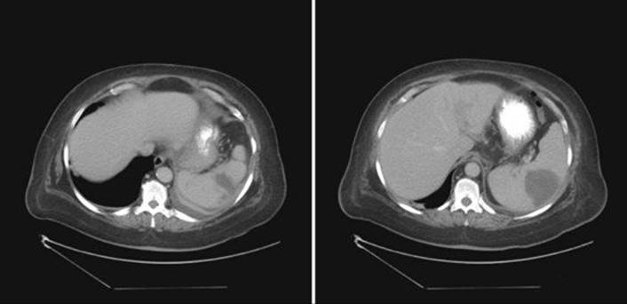
Figure 26-1 Abdominal computed tomographic scans of a morbidly obese female.
Ref.: 1, 11
Comments
The images show a hypodense fluid collection with rim enhancement within the spleen. Splenic abscesses are rare but are associated with high mortality and complication rates because of delayed detection and treatment. Patients often have vague symptoms and signs such as fever and nonspecific abdominal or chest pain. Less than one half of patients complain of left upper quadrant pain. One half of patients will have positive blood cultures. The majority of splenic abscesses arise from hematogenous spread from a distant focus such as endocarditis, pyelonephritis, or direct inoculation from intravenous drug use, but spread from a contiguous infected organ or adjacent intra-abdominal infection or secondary infection of a post-traumatic hematoma can occur as well. The most commonly cultured organisms are gram-positive aerobes, but gram-negative or polymicrobial infections can also occur, as can infection with mycobacteria and fungi. Broad-spectrum antibiotics are begun empirically until culture data are available. Splenectomy is effective treatment. However, in the interest of splenic preservation, image-guided percutaneous drainage has gained popularity over the past decade. The success rate for treatment of unilocular abscesses in this fashion exceeds 75%. Thus, splenectomy may be reserved for failure of nonoperative management or for patients with multiple abscesses in whom percutaneous drainage is not feasible. Splenotomy would not be beneficial for this patient. Echocardiography to evaluate for valvular vegetations is prudent in patients with positive blood cultures and splenic abscess.
Answer
D
References
1 Beauchamp RD, Holzman MD, Fabian TC, et al. The spleen. In Townsend CM, Beauchamp RD, Evers M, et al, editors: Sabiston textbook of surgery: the biological basis of modern surgical practice, ed 18, Philadelphia: WB Saunders, 2008.
2 Sebastian ML, Marohn MR. Splenectomy for hematologic disorders. In Cameron JL, editor: Current surgical therapy, ed 9, Philadelphia: CV Mosby, 2008.
3 Hoyt DB, Coimbra R. Splenectomy and splenorrhaphy. In Fisher JE, Bland KE, editors: Mastery of surgery, ed 5, Philadelphia: Lippincott Williams & Wilkins, 2007.
4 Griffin J, Arif S, Mufti A, editors. Crash course immunology and haematology, ed 2, Toronto: CV Mosby, 2003.
5 Mourtzoukou EG, Pappas G, Peppas, et al. Vaccination of asplenic or hyposplenic adults. Br J Surg. 2008;95:273-280.
6 Gonzalez G, Eichner R. Spherocytosis, hereditary: treatment & medication. Available at. Besa E, editor. eMedicine Clinical Knowledge Base, Hematology, 2009. www.emedicine.medscape.com/article/206107-overview. Accessed July 19
7 Pipinos I, Baxter T. The lymphatics. In Townsend CM, Beauchamp RD, Evers M, et al, editors: Sabiston textbook of surgery: the biological basis of modern surgical practice, ed 18, Philadelphia: WB Saunders, 2008.
8 Park JE, Manson PN, Deune EG. Lymphedema. In Cameron JL, editor: Current surgical therapy, ed 9, Philadelphia: CV Mosby, 2008.
9 Liem TK, Moneta GL. Venous and lymphatic disease. In Brunicardi FC, Andersen DK, Billiar TR, et al, editors: Schwartz’s principles of surgery, ed 9, New York: McGraw-Hill, 2010.
10 Rutherford RB, editor. Vascular surgery, ed 6, Philadelphia: WB Saunders, 2005.
11 McIntyre T, Zenilman ME. Cysts, tumors, and abscesses of the spleen. In Cameron JL, editor: Current surgical therapy, ed 9, Philadelphia: CV Mosby, 2008.
12 Haider AH, Cornwell EEIII. Splenic salvage procedures: therapeutic options. In Cameron JL, editor: Current surgical therapy, ed 9, Philadelphia: CV Mosby, 2008.