
Daniel J. Deziel, M.D.
1 Which of the following vascular relationships is not an important consideration during resection of the head of the pancreas?
A Arterial supply of the pancreatic head from the splenic artery
B Confluence of the splenic vein and superior mesenteric vein dorsal to the pancreatic neck
C Absence of ventral portal vein branches dorsal to the pancreatic neck
D Origin of the right hepatic artery from the superior mesenteric artery
E Origin of the middle colic artery from the superior mesenteric artery
Ref.: 1-4
Comments
The relationship of the pancreas to neighboring organs and to critical vascular structures is of great surgical significance. The arterial supply to the head of the gland is derived from both the gastroduodenal and the superior mesenteric arteries via the anterior and posterior pancreaticoduodenal arcades. For the most part, the head of the pancreas and the duodenum have a shared blood supply, so they must generally be resected together. However, techniques for “duodenal-sparing” resection of the pancreatic head or “pancreatic-sparing” duodenectomy are appropriate in select circumstances.
The body and tail of the pancreas receive their blood supply mainly from multiple branches of the splenic artery, which also connect with superior mesenteric sources. Variations in major arteries—such as the origin of the right hepatic artery from the superior mesenteric artery and the origin of the middle colic artery from the superior mesenteric artery or dorsal pancreatic artery—place these vessels in close proximity to the head and neck of the pancreas, where they are subject to injury during pancreatectomy. The junction of the splenic vein and superior mesenteric vein to form the portal vein lies behind the neck of the pancreas. Usually, these vessels do not have large anterior tributaries in this area, but appropriate caution must nonetheless be exercised when developing this plane during pancreatic operations.
Answer
A
2 Endoscopy demonstrates a 1-cm submucosal nodule with central umbilication in the second portion of the duodenum. This finding is usually associated with which of the following?
A Peptic ulceration
B Increased risk for pancreatic cancer
C Islet cell hyperplasia
D Absence of symptoms
E Intussusception
Ref.: 1, 2, 4
Comments
A heterotopic pancreas is pancreatic tissue located at sites other than the normal location of the gland. Ectopic pancreatic tissue has been described at many anatomic locations but is typically found in the stomach, the duodenum, or a Meckel diverticulum. Theories of origin include metaplasia (the favored theory) and transplantation. Histologic findings range from those of a rudimentary structure to a fully formed gland. Most heterotopic rests contain ducts, and both endocrine and exocrine elements may be present. This entity is not uncommon, being described in 1% to 2% of autopsies. It is usually asymptomatic. When symptoms occur, they are related to the location of the ectopic site and include obstruction (as a result of intussusception), ulceration, and bleeding. Although malignancy has been reported, there is no evidence that heterotopic pancreatic tissue is predisposed to cancer. The typical gross appearance is a submucosal nodule, often with central umbilication. Resection is indicated for symptomatic lesions and is appropriate diagnostically for incidental lesions discovered during operations for other reasons.
Answer
D
3 The embryologic ventral pancreas forms which area of the fully developed gland?
A Superior head
B Uncinate process
C Neck
D Body
E None of the above because it regresses
Ref.: 1, 3, 4
Comments
The pancreas is formed from two outpouchings of the primitive gut. The dorsal pancreas originates from the duodenum, and the ventral pancreas begins as a bud from the hepatic diverticulum, which itself is an outpouching of the duodenum. Other outgrowths from the hepatic diverticulum mature into the liver, gallbladder, and bile ducts. During normal fetal development, the ventral pancreas rotates along with the primitive gut and fuses with the dorsal component. The ventral pancreas constitutes the uncinate process and the inferior portion of the head of the gland in the fully developed state, and the dorsal pancreas forms the remainder of the gland. Abnormalities in this developmental process result in recognized congenital anomalies that can be clinically important. An understanding of this embryologic development is also important to recognizing the relationship of the pancreas to adjacent vascular structures during pancreatic operations.
Answer
B
4 The uncinate process of the pancreas is adjacent and dorsal to which of the following?
A Splenic vein
B Inferior vena cava
C Superior mesenteric artery
D Left renal vein
E Fourth portion of the duodenum
Ref.: 1-4
Comments
The pancreas can be divided into various parts: head, uncinate, neck, body, and tail. The uncinate process is the portion of the gland that extends to the left, dorsal to the portal vein and superior mesenteric artery and ventral to the aorta and inferior vena cava. The uncinate process is located caudad and ventral to the left renal vein and cephalad to the distal duodenum. Understanding the extent and location of the uncinate is important during resection of the head of the pancreas. The blood supply of the uncinate is derived from numerous short branches of the superior mesenteric artery and portal vein. When performing pancreaticoduodenectomy, these branches must be carefully controlled to prevent bleeding and avoid injury to the superior mesenteric artery or portal vein.
Answer
C
5 What is the recommended treatment of duodenal obstruction caused by an annular pancreas?
A Endoscopic division of the associated duodenal web
B Gastrojejunostomy
C Duodenoduodenostomy
D Surgical division of the annular tissue
E Pancreaticoduodenectomy
Ref.: 1, 2, 4
Comments
An annular pancreas is a congenital anomaly involving a band of pancreatic tissue encircling the second portion of the duodenum. The annular tissue appears to originate from the embryologic ventral pancreas. Causal theories include abnormal fixation of the ventral pancreatic primordium before gut rotation, failure of involution of part of the ventral pancreas, and the development of heterotopic pancreatic tissue in the duodenum. Approximately one half of these cases are diagnosed in infants and the remainder in adults, with a peak during the fourth decade of life. Most patients are asymptomatic. Clinical findings are obstruction in infants and children and obstruction, ulceration, or pancreatitis in adults. Associated anomalies include duodenal stenosis or atresia and Down syndrome. Treatment of symptomatic patients consists of surgical bypass by duodenoduodenostomy or duodenojejunostomy. Gastrojejunostomy can also alleviate obstruction but risks marginal ulceration. Resection or division of the annular band is not advised because it risks the development of a pancreatic fistula and may fail to relieve the obstruction.
Answer
C
6 Which of the following developmental anomalies best characterizes pancreas divisum?
A Aplasia of the dorsal pancreatic anlage
B Aplasia of the ventral pancreatic anlage
C Incomplete rotation of the ventral pancreatic anlage
D Failed fusion of the ventral and dorsal pancreatic parenchyma
E Failed fusion of the ventral and dorsal pancreatic ducts
Ref.: 1, 4, 5
Comments
See Question 7.
Answer
E
7 The diagnosis of pancreas divisum is usually made by which of the following?
A Laparoscopic exploration
B Endoscopic ultrasound (EUS)
C Computed tomography (CT)
D Endoscopic retrograde cholangiopancreatography (ERCP)
E Genetic testing
Ref.: 4
Comments
Pancreas divisum currently refers to congenital variations of the pancreatic ducts that result from failed or incomplete fusion of the embryologic ventral and dorsal ductal systems. (Historically, the term may also refer to the rare failure of parenchymal fusion.) There may be complete separation of the ducts, an absent or minimal ventral duct, or only a few meager connections between the systems. As a consequence, most of the pancreatic duct drainage is through the dorsal duct joining the duodenum at the minor papilla. Any existing ventral ducts (Wirsung) drain only the uncinate process and the caudal head of the gland rather than the bulk of the gland at the major papilla, as when normally developed. Some variation of pancreas divisum is present in about 10% of the population. In some individuals, it is clinically significant if the relatively stenotic minor papilla imposes an obstruction to ductal flow. This can potentially result in recurrent abdominal pain, acute pancreatitis, or even chronic pancreatitis. The diagnosis is usually made by ERCP, and cannulation of the minor papilla may be required to image the dorsal duct. Magnetic resonance cholangiopancreatography (MRCP) might also demonstrate this ductal anatomy.
Answer
D
8 Which of the following is appropriate treatment of a patient with pancreas divisum, chronic abdominal pain, a dilated dorsal pancreatic duct, and an enlarged, calcified pancreatic head?
A Pancreaticoduodenectomy
B Endoscopic dorsal sphincterotomy
C Operative dorsal sphincterotomy
D Endoscopic or operative ventral sphincterotomy
E Splanchnic nerve ablation
Ref.: 1, 4
Comments
The vast majority (95%) of individuals with pancreas divisum are asymptomatic. Whether there is a true relationship between the anatomic diagnosis of pancreas divisum and any clinical symptoms that may be present is often difficult to determine. Symptomatic patients with pancreas divisum require thorough evaluation of the nature of their symptoms and for any other causes of abdominal pain or pancreatitis. When it is reasonable to suspect that a stenotic lesser papilla is the cause of recurrent abdominal pain or recurrent acute pancreatitis, therapeutic considerations include endoscopic treatments (dilation, stenting, sphincterotomy) or operative sphincterotomy/sphincteroplasty, which may be combined with cholecystectomy and sphincteroplasty of the major papilla. Occasionally, there are patients (as described in this question) with established findings of chronic pancreatitis and pancreas divisum. Sphincter operations are not successful in this setting. Rather, surgical treatment involving resection or decompression of the pancreatic head may be indicated.
Answer
A
9 Which of the following is not characteristic of pancreatic acinar cells?
A Zymogen granules
B Carbonic anhydrase
C Golgi apparatus
D Rough endoplasmic reticulum
E Contractile proteins
Ref.: 4, 5
Comments
The twofold function of the exocrine pancreas—to secrete bicarbonate-rich fluid and to synthesize digestive enzymes—is accomplished by two cell types. Acinar cells, which elaborate and secrete digestive enzymes, are designed for protein synthesis. They contain abundant rough endoplasmic reticulum, Golgi apparatus, and secretory zymogen granules. Contractile proteins are also abundant near the apical membrane of the cell and facilitate exocytosis of the enzyme bundles into the ductal lumen. The centroacinar cells are part of the ductal system. They secrete bicarbonate and therefore contain carbonic anhydrase, which dissociates carbonic acid into bicarbonate and hydrogen ion:

Some ductal cells also contain synthetic and secretory organelles for the production of mucoproteins.
Answer
B
10 The bicarbonate concentration of pancreatic secretions is:
A Primarily increased by cholecystokinin (CCK)
B Primarily decreased by secretin
C Independent of acinar cell secretion
D Reciprocally related to the chloride concentration
E Reciprocally related to the sodium concentration
Ref.: 1, 3, 5
Comments
The centroacinar cells secrete a bicarbonate-rich solution by an active transport mechanism, primarily in response to secretin. Cholecystokinin is the primary stimulant of enzyme secretion from the acinar cells. The bicarbonate and chloride contents of pancreatic juice are reciprocally related. As ductal flow rates increase, the bicarbonate concentration increases and the chloride concentration decreases. This is the result of two processes: (1) changes in passive exchange of intraductal bicarbonate for intracellular chloride and (2) changes in the relative contribution of acinar cell secretion. Acinar cells secrete fluid high in chloride in addition to digestive enzymes. In contradistinction to anion concentrations, the concentrations of sodium and potassium in pancreatic duct secretions remain relatively constant despite the flow rate and are similar to their concentrations in plasma.
Answer
D
11 Normally, activation of pancreatic trypsinogen involves which of the following?
A Pancreatic amylase
B pH greater than 7.0
C Lysosomal hydrolase
D Pancreatic enterokinase
E Duodenal enterokinase
Ref.: 1-3, 5
Comments
The pancreatic acinar cells secrete digestive enzymes for fats, carbohydrates, and proteins. Except amylase, these enzymes are secreted in inactive forms to protect the pancreas from autodigestion. Activation of the proenzyme trypsinogen to trypsin is the primary event that leads to activation of the other various proteases and phospholipases. It occurs in the duodenum via the action of enterokinase. Trypsinogen activation can also occur in acidic environments (pH <7.0). With acute pancreatitis, intraglandular activation can take place when the inactive enzymes are exposed to lysosomal hydrolases.
Answer
E
12 Which pancreatic islet cell type produces a hormonal peptide to stimulate glycogenolysis and gluconeogenesis?
A Alpha cell
B Beta cell
C Delta cell
D F cell
E PP cell
Ref.: 1-3, 5
Comments
See Question 13.
Answer
A
13 Pancreatic delta cells secrete which inhibitory peptide?
A Bombesin
B Glucagon
C Somatostatin
D Insulin
E Pancreatic polypeptide
Ref.: 1-3, 5
Comments
The endocrine pancreas is composed of various cells located in the islets of Langerhans, approximately 1 million of which are interspersed with the acinar and ductal elements throughout the gland. The hormonal peptides produced by the islets effect a wide range of metabolic and physiologic actions. The primary function of the endocrine pancreas is to regulate glucose homeostasis. Beta cells, which are the most numerous, produce insulin. Insulin promotes glucose transport, stimulates protein synthesis, and inhibits glycogenolysis and lipolysis. Alpha cells secrete glucagon, which counterbalances insulin by stimulating hepatic glycogenolysis, gluconeogenesis, ketogenesis, and lipolysis. Glucagon also inhibits intestinal motility and gastric acid and pancreatic exocrine secretion. Somatostatin, produced by delta cells, has a broad range of inhibitory effects on the gastrointestinal tract, including inhibition of secretion of other pancreatic peptides; inhibition of gastric, biliary, intestinal, and pancreatic exocrine secretions; and inhibition of gastrointestinal motility. PP cells are the source of pancreatic polypeptide. Pancreatic polypeptide inhibits pancreatic exocrine secretion and biliary and gut motility. Clinically, deficiency of pancreatic polypeptide has been linked to diabetes following resection of the pancreatic head or chronic pancreatitis. Because postprandial secretion of pancreatic polypeptide is dependent on vagal innervation, it has been used to assess the completeness of vagotomy.
Answer
C
14 Which is the principal cell type located at the center of the islets of Langerhans?
A Alpha cell
B Beta cell
C Delta cell
D F cell
E Varies according to the location of the islet in the pancreas
Ref.: 1, 3
Comments
Each islet of Langerhans is composed of an average of 3000 cells, with the major types as listed earlier and discussed in the preceding Comments. Beta cells are located at the core and make up about 70% of the islet. The other cell types are located at the periphery of the islet. This cellular anatomy has potential functional implications that are as yet not well understood. The distribution of cell types within the islet varies in different areas of the gland. Islets in the uncinate process derived from the embryologic ventral pancreas contain PP cells but few alpha cells. Islets in the body and tail of the gland have abundant alpha cells but no PP cells.
Answer
B
15 Which of the following statements is true regarding blood flow to the pancreas?
A Islet cells receive a greater proportion of pancreatic blood flow than do the exocrine elements.
B CCK and secretin regulate secretion by altering blood flow.
C Fragile anastomotic networks predispose the gland to ischemia.
D The blood supply to the islet cells is independent of the acinar supply.
E Pancreatic blood flow is highly sensitive to changes in systemic blood pressure.
Ref.: 3-5
Comments
The microcirculation of the pancreas is complex and has important correlations with the endocrine and exocrine functions of the gland. The rich anastomotic supply from various sources makes pancreatic ischemia unusual. The islets receive a disproportionately large amount of total pancreatic blood flow (10% to 25%) relative to their mass (1% to 2%). Both the islets and exocrine tissue have arteriolar blood supply. The acinar tissue is also perfused by blood that drains from the islets, a mechanism referred to as the islet-acinar or insuloacinar portal system. This system is the structural basis for endocrine regulation of exocrine function. Insulin receptors are present on acinar cells, and the density of receptors is higher on acini located near the islets. Because the islets themselves often have a central-to-peripheral pattern of perfusion, insulin from the centrally located beta cells can influence the other peripheral islet cell types. In addition, some islets are apparently perfused in a peripheral-to-central pattern. CCK and secretin have relatively little effect on blood flow and thus exert their stimulatory effects independently. Pancreatic blood flow is maintained relatively constant despite changes in arterial pressure.
Answer
A
16 Which of the following events occurs in acinar cells with acute pancreatitis?
A Accelerated extrusion of zymogen granules
B Impaired synthesis of zymogen granules
C Fusion of lysosomes and zymogen granules
D Fusion of mitochondria and zymogen granules
E Impaired protein synthesis
Ref.: 3-5
Comments
The pathogenesis of pancreatitis involves intrapancreatic activation of digestive enzymes that are normally secreted in inactive form. This results in “autodigestion” of the gland. Although the mechanisms by which the various causes of clinical pancreatitis lead to this state are incompletely understood, experimental observations have identified certain derangements in acinar cell biology that may be the underlying common pathway to pancreatic injury. The primary defects involve blocked extrusion of zymogen granules containing inactive digestive enzymes and alterations in intracellular transport that result in fusion of zymogen granules with lysosomes to form large cytoplasmic vacuoles. This sequence results in co-localization of digestive enzymes and lysosomal hydrolases. Lysosomal enzymes, such as cathepsin B, activate trypsinogen and initiate a cascade of intracellular digestive enzyme activation. Amino acid uptake and protein synthesis are not impaired during this process.
Answer
C
17 The mechanism of alcohol-induced acute pancreatitis is thought to involve all of the following except:
A Pancreatic ductal obstruction
B Pancreatic exocrine hypersecretion
C Hypertriglyceridemia
D Acetaldehyde toxicity
E Genetic defect in lysosomal membranes
Ref.: 1, 3
Comments
Ethanol is the prevalent etiologic factor in acute pancreatitis. There are several contributory mechanisms by which alcohol-induced pancreatic injury occurs. Ethanol causes pancreatic ductal hypertension by increasing ampullary resistance and by intraductal deposition of stone proteins. Concomitantly, ethanol stimulates gastric acid secretion and increases pancreatic exocrine secretion via release of secretin. The combination of ductal obstruction with stimulated secretion may result in enzyme extravasation. Acetaldehyde, the metabolic product of ethanol, injures acinar cells by increasing membrane permeability and disrupting the microtubule structure. The elevated levels of serum triglycerides induced by alcohol are a source of cytotoxic free fatty acids. Alcohol also impairs normal trypsin inhibition and reduces pancreatic blood flow. All of these effects may contribute to intraglandular enzyme activation and the development of acute alcoholic pancreatitis.
Answer
E
18 Hyperamylasemia is diagnostic of acute pancreatitis when associated with which of the following laboratory findings?
A Hyperlipasemia
B Increased urinary amylase levels
C Amylase-creatinine clearance ratio (ACCR) greater than 5%
D Hypocalcemia
E None of the above
Ref.: 1-4
Comments
The diagnosis of acute pancreatitis is based on signs and symptoms, supported by biochemical findings and morphologic abnormalities seen on imaging studies such as CT. No biochemical feature is pathognomonic of acute pancreatitis. Hyperamylasemia, hyperlipasemia, and elevations in urinary amylase levels and the ACCR are typical of acute pancreatitis but are not specific or sensitive, and they can occur with other abdominal and extra-abdominal disorders. Hypocalcemia may occur as a consequence of pancreatitis, but it is also nonspecific. There is no absolute level of serum amylase or lipase that is diagnostic of acute pancreatitis. Marked elevations are more indicative of pancreatitis but are not themselves diagnostic. Both amylase and lipase levels may be elevated in a number of conditions that can be confused with acute pancreatitis, such as acute cholecystitis, perforated peptic ulcer, and intestinal infarction. Moreover, severe pancreatitis can occur without substantial elevations in these serum enzymes.
Answer
E
19 A patient with abdominal pain is found to have a serum amylase level of 1200 IU/L, a normal urinary amylase level, and an ACCR of less than 2%. Based on these findings, the probable diagnosis is which of the following conditions?
A Acute pancreatitis
B Chronic pancreatitis
C Renal failure
D Choledocholithiasis without pancreatitis
E Macroamylasemia
Ref.: 4
Comments
Elevations in serum and urinary amylase levels and in the ACCR, as determined by the following equation, are typical of acute pancreatitis.

where U = urine, S = serum, amy = amylase, and cr = creatinine. Elevation of the amylase-creatinine clearance ratio above the normal 2% to 5% range is not specific for pancreatitis, but a normal ratio in the presence of hyperamylasemia suggests that the hyperamylasemia is the result of something other than pancreatitis. Serum and urinary amylase levels and the ACCR may be normal in patients with chronic pancreatitis or elevated during an acute exacerbation. Renal disease may be associated with low urinary amylase levels and an elevated ACCR. Common duct stones may produce hyperamylasemia without true pancreatitis. The urinary amylase level is elevated, although the ACCR may be normal. With macroamylasemia, amylase forms complexes with serum proteins too large for glomerular filtration. The serum amylase level is therefore elevated, but urinary amylase levels and the ACCR are low. The diagnosis can be confirmed by electrophoresis. Abdominal pain has been reported in more than one half of patients with macroamylasemia, although the biochemical abnormality is probably not etiologically related to the pain. Hyperamylasemia predominantly caused by salivary amylase may also be associated with a low urinary amylase level and ACCR because the salivary isoenzyme is cleared more slowly by the kidneys than the pancreatic isoenzyme.
Answer
E
20 Which of the following is an unfavorable prognostic factor in patients with acute alcoholic pancreatitis?
A Initial white blood cell count higher than 16,000/mm3
B Elevated serum triglycerides during the initial 48 hours
C Serum amylase level higher than 1200 IU/L on admission
D Serum lipase level more than three times normal
E Serum blood urea nitrogen (BUN) level elevated more than 2 mg/dL during the initial 48 hours
Ref.: 1, 3, 4
Comments
Several systems have been devised to gauge the severity of acute pancreatitis. These systems involve multiple clinical, biochemical, and sometimes radiologic criteria. The most widely used system in the United States, developed by Ranson, was based on retrospective analysis and subsequent prospective verification. The Ranson criteria include 11 parameters determined at the time of admission or during the subsequent 48 hours. Patients with three or more criteria have more severe disease and are at increased risk for septic complications and death. The criteria reflect the patient’s underlying status, the severity of the retroperitoneal inflammatory process, and the effects on renal and respiratory function. The Ranson criteria were originally developed for alcoholic pancreatitis and have been modified somewhat for gallstone pancreatitis. For example, a rise in the serum BUN level of more than 2 mg/dL is one of the 10 criteria for gallstone pancreatitis, but the rise must be more than 5 mg/dL to meet the criteria for alcoholic pancreatitis (a subtle point). Other physiologic scoring systems, such as the Acute Physiology, Age, and Chronic Health Evaluation II (APACHE II), are also useful prognostically, although they are not designed specifically for acute pancreatitis.
Answer
A
21 What is the leading cause of death from acute pancreatitis?
A Hemorrhage
B Pseudocyst rupture
C Secondary pancreatic infection
D Biliary sepsis
E Renal failure
Ref.: 6
Comments
Formerly, death from acute pancreatitis often occurred early in the course of the disease as a result of the acute effects of hypovolemia and inadequate resuscitation. In the current era, about 80% of deaths are attributed to secondary pancreatic infection, which develops in approximately 10% of patients with acute pancreatitis. Fatal pancreatic sepsis typically progresses to multisystem organ failure, and deaths occur later in the course of the disease. To have an impact on this disease, therapeutic efforts have therefore focused on the prevention and early diagnosis of pancreatic infection and on more effective methods of surgical therapy.
Answer
C
22 Which of the following complications of acute pancreatitis is associated with the highest mortality rate?
A Peripancreatic abscess
B Infected pancreatic pseudocyst
C Infected pancreatic necrosis
D Sterile pancreatic necrosis
E Bile duct obstruction
Ref.: 4, 6
Comments
Retroperitoneal infection is a serious, often fatal complication of acute pancreatitis. The early literature pertaining to the local infectious sequelae of pancreatitis may be confusing because of nonselective use of the term pancreatic abscess to describe infectious complications, which vary in severity. Pancreatic abscess best describes a localized collection of drainable pus in or around the pancreas. Pancreatic abscess and infected pseudocyst can be treated effectively by external drainage, and the anticipated mortality rate for each is about 5%. Pancreatic necrosis is a manifestation of severe pancreatitis. When accompanied by infection, it has been associated with a mortality rate that may exceed 40%, which is higher than that for noninfected necrosis. Infected pancreatic necrosis is treated by operative débridement and open or closed retroperitoneal drainage. Patients with sterile necrosis may require operative intervention as well but are generally treated nonoperatively with intensive support as long as their condition permits.
Answer
C
23 A 45-year-old man is admitted with severe alcoholic pancreatitis. Forty-percent pancreatic necrosis is estimated on CT. Which of the following statements best describes the current use of antibiotics for this patient?
A Systemic antibiotics are not indicated unless his condition deteriorates.
B Systemic antibiotics are indicated for coverage of gut-derived bacteria.
C Systemic antibiotics are indicated for coverage of gut-derived bacteria and fungal organisms.
D Nonabsorbable antibiotics are indicated for gut decontamination.
E Systemic antibiotics are not indicated if enteric feeding can be tolerated.
Ref.: 7
Comments
The risk for infected pancreatic necrosis is related to the clinical severity and duration of disease and to the extent of necrosis. Strategies to decrease secondary pancreatic infection focus on patients at higher risk. Unfortunately, controlled trials of systemic antibiotics for prophylaxis against secondary infection have yielded conflicting results. These differences are probably because of numerous factors, including heterogeneity in the severity of disease, patient characteristics, and concomitant therapy among those studied, as well as to differences in study methodologies. Current practice favors systemic antibiotics for patients with severe disease and more extensive (>30%) necrosis based on studies demonstrating fewer septic complications and perhaps decreased mortality. However, not all studies have shown benefit, and the risk for subsequent infection with multiresistant bacterial or fungal organisms may be increased, particularly if prophylactic antibiotic use is prolonged. Because the gut is typically the source of the offending organisms, the use of nonabsorbable enteral antibiotics for selective gut decontamination has had some appeal. The effect of this measure remains unclear, and it is not typically used. Enteric feedings are beneficial to maintain the gut mucosal barrier to bacterial translocation. However, the efficacy of enteric feeding alone for prevention of secondary pancreatic infection has not been demonstrated.
Answer
B
24 Which of the following types of antibiotics does not achieve adequate levels in the pancreas?
A Imipenem
B Third-generation cephalosporins
C Metronidazole
D Aminoglycosides
E Fluoroquinolones
Ref.: 8
Comments
Early studies of antibiotic prophylaxis in patients with acute pancreatitis showed no efficacy, in part because they involved individuals with mild pancreatitis and in part because they used antibiotics that did not achieve adequate therapeutic levels in the pancreas and retroperitoneum. Aminoglycosides, first-generation cephalosporins, and aminopenicillins do not adequately penetrate the pancreas. Drugs with penetration include the other choices listed, as well as piperacillin and mezlocillin.
Answer
D
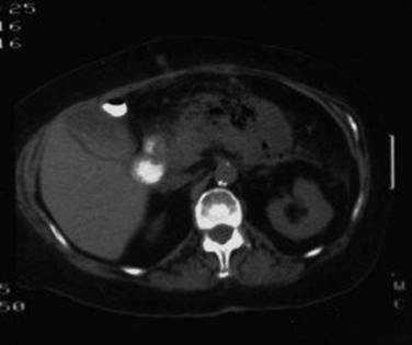
25 An alcoholic patient has acute pancreatitis with five of the Ranson criteria. He gradually improves over a 14-day hospitalization, but then a pulse of 120 beats/min, a temperature of 39° C, and abdominal distention develop. CT is performed and the results are shown below. The next most appropriate therapy is which of the following measures?
A Antibiotics
B Percutaneous catheter drainage
C Peritoneal lavage
D Endoscopic cyst gastrostomy
E Operative drainage
Ref.: 4, 6
Comments
Pancreatic infection complicating acute pancreatitis should be suspected in any patient who fails to improve following supportive medical therapy or improves but then demonstrates deterioration. Pancreatic infection occasionally occurs early during the chronologic course of the disease, but it typically occurs later, as in the patient described. CT is the best method for imaging the pancreas. The results of CT in this patient demonstrate air in the pancreas, which is characteristic of pancreatic infection. The technique of dynamic pancreatography can identify ischemic areas of pancreas and is useful for evaluating patients who may have pancreatic necrosis. Dynamic pancreatographyis performed by serially imaging the pancreas after bolus injection of an intravenous contrast agent. Percutaneous needle aspiration of fluid collections or necrotic areas found on CT can be performed to identify the presence of infection and guide therapeutic decisions about the need for drainage. When pancreatic infection is present, operative drainage and débridement are indicated. Interest has focused on the selection of closed or open methods of operative drainage. Minimal-access operative approaches are also used to drain and débride pancreatic necrosis in the hope of lowering morbidity in these ill patients. Percutaneous catheters can drain thin fluid but are usually inadequate for the management of infected pancreatic necrosis. Peritoneal lavage has been used early in the course of patients with severe acute pancreatitis. Endoscopic cyst gastrostomy may be appropriate for some patients with pancreatic pseudocysts. These latter two modalities have no role in the management of infected pancreatic necrosis.
Answer
E
26 Acute gallstone pancreatitis is diagnosed in a 54-year-old man. Which of the following is considered standard treatment?
A Urgent (within 24 hours) cholecystectomy and common bile duct exploration
B Urgent ERCP and subsequent laparoscopic cholecystectomy
C Initial supportive therapy with cholecystectomy performed during the same admission
D Initial supportive therapy with cholecystectomy performed in 6 to 8 weeks
E Initial supportive therapy with cholecystectomy performed only if symptoms recur
Ref.: 9
Comments
Gallstone pancreatitis is related to the passage of stones through the ampulla of Vater. Patients with smaller gallstones have an increased risk for the development of this manifestation. Cholecystectomy is indicated because gallstone pancreatitis is a recurrent problem in 30% to 50% of patients if surgery is not performed. The traditional controversy has involved the timing of surgery. Proponents of immediate intervention have found a higher incidence of choledocholithiasis but have not demonstrated that this approach is safer than delayed surgery or that it is necessary for most patients. Most surgeons advise initial nonoperative therapy until the patient’s signs and symptoms subside (most do within 2 to 3 days), followed by elective cholecystectomy with intraoperative imaging of the common bile duct by cholangiography or intraoperative ultrasonography during the same hospitalization.
The role of urgent endoscopic retrograde cholangiopancreatography and endoscopic sphincterotomy for the management of biliary pancreatitis has been controversial. The vast majority (97%) of patients with gallstone pancreatitis have mild pancreatitis that improves rapidly. ERCP finds common duct stones in only a small percentage of patients and is not indicated routinely. Some trials comparing urgent ERCP and sphincterotomy with traditional treatment have suggested benefit in patients with severe pancreatitis, but this has not been consistently observed. ERCP is indicated for patients with concomitant obstructive jaundice and biliary sepsis. Less invasive methods of duct imaging, such as magnetic resonance cholangiopancreatography or EUS, might be useful in patients with an intermediate risk for choledocholithiasis but is not necessary for most with biliary pancreatitis. For the small proportion of patients with severe biliary pancreatitis, early cholecystectomy should be avoided. Treatment in this group is directed at resolution of the pancreatitis and its complications. When the pancreatitis has subsided, delayed cholecystectomy is indicated.
Answer
C
27 Which of the following is the preferred nutritional support for a patient with severe pancreatitis?
A Nasogastric feeding
B Feeding via percutaneous endoscopic gastrostomy
C Nasojejunal feeding
D Parenteral amino acids and glucose
E Parenteral amino acids, glucose, and lipids
Ref.: 10
Comments
Nutritional support is a critical component of the successful management of patients with severe pancreatitis. Mortality is reduced by positive nitrogen balance. Direct delivery of nutrients into the jejunum is the preferred route. Enteral jejunal feeding does not stimulate pancreatic exocrine secretion and helps maintain the intestinal mucosal barrier. Jejunal feeding is associated with a lower risk for infection and shorter hospital stay than parenteral nutrition. Moreover, enteric feeding avoids catheter-related sepsis and other complications of central venous lines. Feeding into the stomach does stimulate the pancreas and is not usually tolerated because of retrogastric inflammation and delayed gastric emptying. Nasojejunal tubes may require radiologically guided or endoscopic placement.
If nutritional goals cannot be met within a few days of initiation, parenteral nutritional support may also be necessary. Intravenous lipids are not detrimental and prevent essential fatty acid deficiency.
Answer
C
28 In North America, chronic pancreatitis is most commonly related to chronic alcohol ingestion. Which of the following is the second most common cause?
A Gallstones
B Drugs
C Infection
D Malnutrition
E Idiopathic
Ref.: 3
Comments
In the Western world, alcohol use accounts for about 75% of cases of chronic pancreatitis. Approximately 20% of cases are considered idiopathic. In parts of Africa and Asia, protein malnutrition is an important etiologic factor. Other, less common causes of chronic pancreatitis include pancreatic duct obstruction (secondary to stenosis or pancreas divisum), hyperparathyroidism, trauma, cystic fibrosis, and hereditary causes. Unlike acute pancreatitis, calculous biliary disease is not a typical cause of chronic pancreatitis. Certain infections (particularly viral) and drugs are among the many factors that can produce acute, rather than chronic pancreatitis.
Answer
E
29 With regard to the histologic characteristics of chronic pancreatitis, all but which of the following is observed?
A Increased interstitial connective tissue
B Loss of acinar cells
C Loss of islet cells
D Neural hypertrophy
E Damaged perineurium
Ref.: 4
Comments
Chronic pancreatitis is characterized on histologic examination by the loss of exocrine acinar cells and a marked increase in interstitial fibrous connective tissue. The islets of Langerhans are preserved and constitute a relatively greater proportion of the pancreatic tissue. Hyperplasia of islet cells is also seen. The sizes and number of nerves are increased, but the protective perineural sheath is damaged, and nerves are found in proximity to inflammatory foci. There appear to be selective increases in certain peptidergic nerves. These histologic observations may be related to the cause of pain in chronic pancreatitis.
Answer
C
30 Pain is the predominant clinical manifestation of chronic alcoholic pancreatitis. Most patients also have which of the following associated manifestations?
A Clinical diabetes mellitus
B Hypoglycemia
C Steatorrhea
D Subclinical fat malabsorption
E Hepatic cirrhosis
Ref.: 1-3
Comments
Recurrent or persistent abdominal pain is the predominant symptom of chronic pancreatitis. Patients usually have varying degrees of nausea, anorexia, and weight loss. Mechanisms that may contribute to pain include ductal obstruction, parenchymal hypertension, acute inflammation, and perineural inflammation. About two thirds of patients have abnormal glucose tolerance test results and subclinical fat malabsorption, whereas overt diabetes is present in perhaps 30% to 50% and frank steatorrhea in only 10% to 15%. Endocrine and exocrine insufficiency progresses during the course of the disease. Diabetes mellitus may be related to impaired insulin release because the islet cells themselves are relatively preserved. Despite the common etiologic factor of ethanol, most patients with chronic pancreatitis do not have hepatic cirrhosis.
Answer
D
31 Which of the following would not be appropriate for the management of steatorrhea in a patient with chronic pancreatitis?
A Restriction of fat to 75 g/day
B Encapsulated pancreatic enzymes
C Encapsulated pancreatic enzymes and a proton pump inhibitor
D Nonencapsulated pancreatic enzymes
E Nonencapsulated pancreatic enzymes and a proton pump inhibitor
Ref.: 3, 4
Comments
Gross steatorrhea and diarrhea occur when pancreatic exocrine function is reduced to about 10% of normal. Therapy involves limitation of fat intake and administration of adequate amounts of exogenous pancreatic enzyme preparations to provide at least 10% of normal lipolytic activity in the duodenum at the time that the food substrate is present. Various commercial formulations of pancreatic enzymes are available. Nonencapsulated forms may improve the malabsorption but can be ineffective because of inactivation in the stomach when the pH falls below 4. The addition of H2 blockers may then be useful. Enteric-coated preparations release their enzymes at a pH above 5. Therefore, they are useful for patients whose gastric pH remains low to ensure that the enzyme is not released until it reaches the duodenum. The use of encapsulated forms with H2 blockers is counterproductive because the enzyme is released in the stomach and is then inactivated if the pH falls. In addition, enteric-coated preparations are microspheres of varying sizes, and the larger ones do not empty into the duodenum until after the food substrate does.
Answer
C
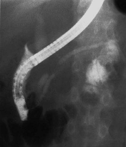
32 A 58-year-old woman with jaundice underwent ERCP (results shown below) as part of her diagnostic work-up. On the basis of this radiograph, what diagnosis is considered the most likely?
A Chronic pancreatitis
B Pancreatic cancer
C Cholangiocarcinoma
D Pancreas divisum
E Ectopic pancreas
Ref.: 1, 2
Comments
This ERCP study shows the classic “double-duct sign:” dilation of the biliary system above an area of abrupt narrowing and abrupt termination of the main pancreatic duct. These findings place the primary abnormality in the geographic location of the pancreatic head, and it is not uncommon for a pancreatic neoplasm to involve both ducts. Chronic pancreatitis may cause biliary obstruction, but the obstruction in the biliary systems is usually more distal. Likewise, there are no coexistent changes in this patient, such as irregular beading of the pancreatic duct, to suggest that chronic pancreatitis is present. Cholangiocarcinoma may be responsible for the stenosis in the biliary system, but cholangiocarcinomas rarely become large enough to involve the pancreatic duct. With pancreas divisum, injection of the major papilla opacifies only a short, tapering ventral duct draining the caudal portion of the pancreatic head and uncinate process. Injection of the minor papilla demonstrates the dorsal duct draining the major portion of the gland.
Answer
B
33 A 45-year-old nondiabetic patient with chronic alcoholic pancreatitis and intractable abdominal pain has a 10-mm pancreatic duct. Which of the following choices constitutes the best treatment?
A Sphincteroplasty
B Lateral pancreaticojejunostomy
C Caudal (tail) pancreatectomy
D Total pancreatectomy
E Continued nonoperative therapy
Ref.: 1, 4, 11
Comments
Pain is the primary indication for surgery in patients with chronic pancreatitis. Selection of the best operation for a particular patient must include consideration of the anatomy of the gland, preexisting endocrine or exocrine dysfunction, compliance and the rehabilitative capacity of the patient, postoperative endocrine or exocrine deficiency, and the likelihood of postoperative pain relief. Patients with a dilated duct (>6 mm) are candidates for ductal drainage, with lateral pancreaticojejunostomy being the best choice of these procedures. It is important to achieve adequate decompression of the enlarged pancreatic head and uncinate process during drainage procedures. Variations such as the Frey or Beger procedure are intended to accomplish this. Sphincteroplasty does not play a role in the management of patients with established chronic pancreatitis.
Patients with small ducts disease are treated by resection if surgery is necessary. Resection of the pancreatic head in properly selected patients has generally yielded better long-term results for pain relief than has tail resection. The head of the pancreas is often enlarged and bulky in chronic pancreatitis and has been considered to be the “pacemaker” of the disease. A number of operative techniques are available for resection of the pancreatic head. Total or nearly total (95%) resections have higher long-term morbidity and mortality rates related to postoperative endocrine insufficiency. Although endocrine and exocrine function tends to deteriorate over time in patients with chronic pancreatitis, some evidence suggests that pancreaticojejunostomy halts or delays this decline better than nonoperative therapy.
Answer
B
34 Ascites develops in a 60-year-old patient with acute pancreatitis. Paracentesis demonstrates that the ascitic fluid amylase level is higher than the serum amylase level and that the fluid protein level is higher than 3 g/dL. Which of the following best explains the ascites?
A Pancreatic duct leak
B Secondary bacterial peritonitis
C Portal vein thrombosis
D Underlying pancreatic cancer
E Resuscitative fluid overload
Ref.: 1, 2
Comments
Pancreatic ascites can be differentiated from ascites of other causes by the characteristic high amylase and protein content of the peritoneal fluid. Pancreatic ascites and pleural effusion are the results of a disruption in the pancreatic duct, usually consequent to pancreatitis. The ascites may resolve with conservative management consisting of paracentesis (thoracentesis), total parenteral nutrition, and administration of a somatostatin analogue to inhibit pancreatic exocrine secretion. Otherwise, an operation may eventually be required for internal drainage of the pancreatic duct fistula or pseudocyst.
Answer
A
35 CT demonstrates a 5-cm peripancreatic fluid collection in a patient 3 weeks after an episode of acute pancreatitis. The patient is eating and does not have clinical signs of infection. What is the recommended treatment?
A Expectant management without intervention
B Nothing by mouth and total parenteral nutrition
C Percutaneous catheter drainage of the fluid collection
D Endoscopic drainage
E Re-imaging in 3 to 6 weeks and surgery for internal drainage if the collection persists
Ref.: 1, 4, 12
Comments
Peripancreatic fluid collections can be found in about 20% of patients with acute pancreatitis. Many of them resolve spontaneously and should not be mistaken for pancreatic pseudocysts. If the patient is stable, can eat, and does not have clinical evidence of infection or other complications, expectant management is indicated. The fluid collection can be monitored with ultrasonography or CT in 1 to 3 months. If the patient has persistent pain and is unable to eat, nutrition by postpyloric enteral feeding, or parenteral nutrition if necessary, may be instituted for several weeks to allow resolution or maturation of the collection into a pseudocyst. If the patient has a symptomatic or complicated fluid collection that requires early intervention, some method of external drainage must be used. If the fluid is thin, endoscopic or percutaneous catheter drainage may suffice. Operative drainage is preferred if there is substantial necrotic debris, as there often is, or if there is concern about infection. Operative drainage might be accomplished with minimal-access approaches.
Answer
A
36 Which of the following is the most important determinant of the need for drainage of a pancreatic pseudocyst?
A Pseudocyst symptoms
B Pseudocyst size
C Pseudocyst duration
D Associated chronic pancreatitis
E Patient age
Ref.: 1, 4, 12
Comments
Historically, pancreatic pseudocysts larger than 5 to 6 cm and present for longer than 6 weeks were thought to have a low rate of spontaneous resolution and a high rate of complications. They were therefore treated by operative drainage. Current understanding of the natural history of pseudocysts is that the rate of spontaneous resolution is higher and the rate of complications lower than previously thought. Pseudocyst size and duration are therefore no longer absolute criteria for intervention. Rather, pseudocyst-related symptoms are the primary indication for treatment. Large pseudocysts are more likely to be symptomatic and less likely to resolve spontaneously than are small pseudocysts. In addition, pseudocysts in patients with chronic pancreatitis are unlikely to resolve but may not require intervention if they are stable, asymptomatic, and uncomplicated.
Answer
A
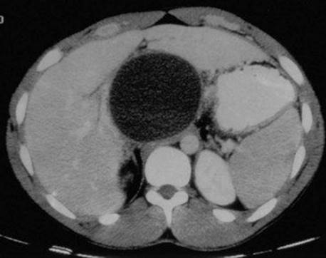
37 A patient with chronic pancreatitis is unable to eat because of persistent postprandial pain. CT is performed (shown below). What is the recommended treatment?
A Nothing by mouth and total parenteral nutrition for 4 to 6 weeks
B Percutaneous catheter drainage
C Endoscopic drainage
D Operative internal drainage
E Operative external drainage
Ref.: 1, 4, 12
Comments:
Pseudocysts that develop in patients with chronic pancreatitis can be considered mature when they are discovered unless there has also been a recent episode of acute pancreatitis. The indications for treatment of a pancreatic pseudocyst are (1) persistent symptoms (pain, inability to eat, or biliary or gastrointestinal obstruction), (2) enlargement, or (3) the onset of a pseudocyst-related complication (infection, hemorrhage, or rupture). Operative internal pseudocyst drainage into the stomach, jejunum, or duodenum is generally the preferred treatment, depending on the location of the pseudocyst. For patients with chronic pancreatitis, it is critical to evaluate the pancreatic duct to determine whether a concomitant duct drainage procedure is necessary. Pseudocyst drainage can be accomplished laparoscopically in some situations. Pseudocysts in the tail of the gland are sometimes best treated by distal pancreatectomy. Percutaneous or endoscopic drainage of established pseudocysts is still being debated. These techniques can successfully treat pseudocysts in some circumstances but have definite limitations and potential complications.
Answer
D
38 Which of the following risk factors is most strongly associated with ductal adenocarcinoma of the pancreas?
A Chronic pancreatitis
B Diabetes mellitus
C Cigarette smoking
D Coffee consumption
E Alcohol consumption
Ref.: 1, 3, 4
Comments
Epidemiologic studies have identified various demographic, medical, environmental, and dietary factors that have some relationship to pancreatic cancer. The most firmly established risk factor is cigarette smoking. Experimentally, nitrosamines have been found to be carcinogenic. In addition, the carcinogens in cigarettes have been related to K-ras oncogene mutations, which are frequent in pancreatic cancer. Alcohol has not been demonstrated conclusively to be a risk factor independent of cigarettes. The previously reported association of pancreatic cancer with coffee consumption is questionable. Diets high in fats and meat may be associated with pancreatic cancer, whereas diets high in fruits and vegetables may be protective. Certain occupational and industrial exposures have an increased risk. There may be some association with diabetes mellitus and certain forms of chronic pancreatitis, but the relationship is not considered causal. Previous gastrectomy has been associated with increased risk, whereas tonsillectomy has been observed to be protective.
Answer
C
39 A jaundiced, otherwise healthy patient is noted to have a 3-cm mass in the head of the pancreas on CT. EUS-guided fine-needle aspiration shows cancer. The mass abuts the portal vein, but there is no clear evidence of vessel involvement or metastatic disease. Which of the following is the most appropriate next step?
A MRCP to better assess vascular involvement
B Direct angiography to better assess vascular involvement
C Operative exploration and potential resection
D Endoscopic placement of a biliary stent
E Chemotherapy and radiation therapy
Ref.: 1, 2, 4
Comments
When the clinical situation suggests a resectable pancreatic neoplasm in a good-risk patient with biliary obstruction, surgery for potential resection is generally indicated without additional tests. Routine preoperative biliary decompression is not advantageous in this setting because it does not improve operative outcomes and may increase the morbidity associated with resection. Endoscopic biliary decompression is invaluable, of course, for palliation of obstruction in patients deemed inoperable or if operation is to be delayed. Angiography was formerly popular for the preoperative staging of pancreatic cancer, but its accuracy in determining resectability has limitations, and it generally adds little to good-quality Computed tomography or magnetic resonance imaging. EUS is extremely useful for identifying small tumors that are inapparent on CT, for obtaining cytologic material, and somewhat for assessing vascular invasion. Neoadjuvant chemotherapy with radiation therapy is increasingly being used before surgery for patients whose imaging studies suggest “borderline” resectability. Imaging criteria for what constitutes a borderline case vary but would commonly include tumors that abut a substantial (half or greater) circumference of adjacent vessels (hepatic artery, superior mesenteric artery, portal vein) or that narrow the portal-splenic vein confluence.
Answer
C
40 In which of the following situations is resection of a ductal carcinoma of the pancreas contraindicated?
A Age older than 80 years
B Tumor located in the body of the pancreas
C Inability to verify malignancy histologically before resection
D Presence of small peritoneal metastases
E Tumor invading the portal vein
Ref.: 1-4
Comments
Resection of a pancreatic malignancy offers the only chance for cure. Most commonly, resection of ductal carcinomas involves pancreaticoduodenectomy, because most potentially resectable tumors are located in the head or uncinate process of the gland. Tumors originating in the body or tail of the pancreas are often not diagnosed until they are beyond the confines of surgical resection. However, location alone does not contraindicate resection because, stage for stage, tumors in the body have the same survival as tumors in the head of the pancreas. Resection is indicated for physiologically fit patients (age alone is not a contraindication) who do not have metastases beyond the field of resection. Histologic or cytologic confirmation of malignancy can often be obtained intraoperatively but is not necessary before resection if the clinical circumstances suggest cancer and the surgeon is appropriately experienced. For some tumors with local vascular invasion, en bloc resection with reconstruction of the involved vessels is appropriate if a tumor-free resection can be accomplished. Positive lymph nodes outside the resection field, peritoneal metastases, and liver metastases generally contraindicate resection for adenocarcinoma of the exocrine pancreas. However, tumor “debulking” and resection of liver metastases can be beneficial in patients with functioning tumors of the endocrine pancreas.
Answer
D
41 Which of the following operations could be appropriate for a 2-cm ductal adenocarcinoma in the head of the pancreas?
A Pancreaticoduodenectomy (Whipple procedure) with preservation of the stomach and pylorus
B Duodenum-sparing pancreatectomy
C Total pancreaticoduodenectomy
D Laparoscopic enucleation
E All are potentially appropriate
Ref.: 1-4
Comments
A standard Whipple-type resection with partial gastrectomy or with preservation of the stomach and pylorus is indicated for a resectable ductal cancer in the head of the pancreas. Each of these operations yields a 5-year survival rate of approximately 15% to 20%. There is a higher incidence of initially delayed gastric emptying with pyloric-preserving operations. However, long-term studies demonstrate normal emptying and good nutritional outcomes. Likewise, the postprandial gastrin and acid responses are normal despite the loss of duodenal inhibitory factors, and marginal ulcer has not been a prohibitive problem. Additional advantages of the pyloric-preserving technique are shorter operative time and lower operative blood loss. Of course, preservation of the stomach and most proximal portion of the duodenum is not appropriate for patients with tumors in close proximity if the margins would be compromised. Duodenum-sparing resection of the pancreatic head has been used in some centers for patients with chronic pancreatitis and has been reported to better maintain enteropancreatic hormonal relationships and glucose homeostasis. This operation is not indicated for cancer.
Total pancreaticoduodenectomy is not advocated for pancreatic cancers that can otherwise be resected. It has been used based on the grounds that it produces better clearance of the lymph nodes and possible multicentric disease and avoids a pancreatic anastomosis. However, long-term survival is not improved, and total pancreatectomy is associated with a higher rate of both early and late complications. Extended pancreaticoduodenectomy involves removal of more retroperitoneal soft tissue and regional lymph nodes. U.S. studies have not demonstrated improved survival with this approach, and the rate of operative complications may be higher. Pancreatic resections can be accomplished laparoscopically, but enucleation is never an appropriate method for removing a pancreatic ductal cancer.
Answer
A
42 At laparotomy, a jaundiced patient is found to have an unresectable pancreatic cancer obstructing the bile duct. Which of the following statements regarding biliary decompression is correct?
A The preferred management is to close the patient and place an endoscopic stent postoperatively.
B Cholecystectomy plus T-tube placement is the preferred management.
C Choledochoduodenostomy is contraindicated.
D Cholecystojejunostomy should not be performed if the patient has cholelithiasis.
E Roux-en-Y choledochojejunostomy is not appropriate because of limited life expectancy.
Ref.: 1-4
Comments
Most patients with pancreatic cancer do not have resectable disease. Palliative treatment is directed to relieve obstruction of the bile duct and duodenum and to alleviate pain. For lesions demonstrated to be unresectable before laparotomy, nonoperative relief of biliary obstruction can be achieved by the endoscopic (preferred) or transhepatic route. Surgical bypass with some form of biliary enteric anastomosis generally provides more durable relief with less need for further intervention. It is preferred for patients when unresectability is determined at the time of laparotomy. Cholecystojejunostomy, choledocho- or hepatico jejunostomy, and choledochoduodenostomy are each appropriate for the management of distal bile duct obstruction.
Choledochojejunostomy usually provides the most durable relief. A Roux-en-Y configuration is preferred by many surgeons, although a simple loop (with or without distal enteroenterostomy) also suffices. Cholecystojejunostomy is relatively simple but should be avoided if the gallbladder is diseased or when cystic duct patency cannot be demonstrated or may be jeopardized by tumor proximity. It is sometimes taught that choledochoduodenostomy should be avoided with malignant obstruction because of possible tumor growth and eventual reobstruction. In reality, choledochoduodenostomy can be an effective solution provided that the common bile duct is sufficiently dilated and the duodenum is pliable and unobstructed.
Answer
D
43 When should gastrojejunostomy be performed at the time of biliary bypass in a patient with unresectable pancreatic cancer?
A Always
B Never
C If the tumor is locally unresectable and there are no peritoneal metastases
D Only if symptomatic duodenal obstruction is present at the time of surgery
E Only if endoscopic stent placement is not available
Ref.: 1-4
Comments
In addition to biliary obstruction, pancreatic cancers can obstruct the duodenum or the proximal jejunum near the ligament of Treitz. Traditionally, many surgeons have favored routine “double bypass” (biliary and duodenal) for operated patients because the rate of duodenal obstruction that develops later in patients treated by biliary bypass alone has been cited to be 5% to 30%. However, duodenal obstruction does not develop in most patients, and gastrojejunostomy is sometimes associated with problems such as bleeding or delayed gastric emptying. The selective approach is therefore appropriate. Patients with obstructive symptoms or impending obstruction as a result of tumor location should undergo gastrojejunostomy. Gastrojejunostomy is also advisable for patients with an anticipated longer survival, such as those whose lesions are not resected because of local tumor invasion rather than because of hepatic or peritoneal metastases. Endoscopic placement of a duodenal stent is another option, but the results are not always satisfactory.
Answer
C
44 A 45-year-old woman who is not an alcoholic has a septated 10-cm cystic mass in the head of the pancreas. Which of the following statements constitutes appropriate advice?
A The lesion is benign and requires no intervention.
B The lesion is malignant and probably incurable.
C Pancreaticoduodenectomy is indicated.
D Percutaneous needle biopsy is indicated.
E Drainage by Roux-en-Y cyst jejunostomy is indicated.
Ref.: 1-4
Comments
Cystadenoma and cystadenocarcinoma are cystic neoplasms of the pancreas that are most commonly manifested as mass lesions in middle-aged women. Serous and mucinous types are recognized, and the risk for malignancy is significant with the mucinous variety. Cystadenoma is more common than its malignant counterpart, but malignant transformation may occur. EUS with sampling for cytology, mucin, and carcinoembryonic antigen (CEA) can be useful for gauging the likelihood of cancer. Without resection, however, exclusion of malignancy can be difficult. Internal drainage of cystic neoplasms is not appropriate therapy. Complete excision should be carried out whenever possible. The 5-year survival rate after resection of cystadenocarcinoma is approximately 50%. Occasionally, islet cell tumors, ductal adenocarcinomas, or other unusual tumors (e.g., papillary and cystic pancreatic neoplasms) have cystic components.
Answer
C
45 A 64-year-old man is evaluated for abdominal pain. CT shows segmental dilation of the main pancreatic duct to greater than 10 mm in the head of the gland with mural nodules. Which of the following is the next most appropriate recommendation?
A EUS
B ERCP
C Serum CA 19-9
D Total pancreatectomy
E Abstinence from alcohol and CT repeated in 3 months
Ref.: 13
Comments
An intraductal papillary mucinous neoplasm (IPMN) of the pancreas is a premalignant condition characterized by papillary projections of mucin-secreting epithelial cells, excessive mucin production, and cystic dilation of the pancreatic duct. The patient may already have cancer when initially seen or may be at some other stage along the process of malignant transformation, which occurs relatively slowly. IPMNs are divided into a main duct type and a branch duct type, depending on the areas of the pancreatic ducts that are involved. The goals of evaluation are to identify factors associated with a higher risk for malignancy and to determine the anatomic extent of disease. EUS is usually the next step in evaluation when IPMN is suspected. EUS can identify diffuse or segmental dilation of the pancreatic duct and the size of cystic lesions or mural nodules and can guide fine-needle aspiration to assess cytology and molecular tumor markers. ERCP shows duct dilation without strictures, filling defects from mucus or nodules, and commonly a patulous papillary orifice with mucus. ERCP also permits sampling of mucus and therapeutic clearance if mucus obstruction is a problem. If available, direct pancreatoscopy and intraductal ultrasound can be adjuncts to ERCP for determining the extent of an IPMN.
Answer
A
46 Which of the following features of IPMN of the pancreas is associated with the lowest risk for cancer?
A Branch duct type with mural nodularity
B Branch duct type smaller than 3 cm
C Main duct type with diffuse dilation to greater than 10 mm
D Main duct type with segmental dilation
E Multifocal IPMN
Ref.: 14, 15
Comments
Progression of intraductal papillary mucinous neoplasm through the adenoma cancer sequence is considered a slow process that requires perhaps 10 to 20 years. Main duct–type tumors have a greater risk for malignancy than the branch duct type. The Sendai Consensus Guidelines identified the following features as risk factors for cancer and as general indicators for resection: main pancreatic duct dilation to greater than 10 mm, cyst size larger than 3 cm, presence of mural nodules, and atypical cytology. Additional risk factors include high-grade dysplasia, multifocal or synchronous tumors, and increasing cyst size during follow-up. In branch duct–type tumors, mural nodularity or atypical cytology may be more important determinants than size.
Answer
B
47 Which of the following would you recommend to a 60-year-old woman with an established diagnosis of IPMN?
A Mammography
B Transvaginal ultrasound
C Colonoscopy
D Genetic testing
E Screening CT for first-degree relatives
Ref.: 16
Comments
Extrapancreatic malignancies have been observed more commonly in patients with intraductal papillary mucinous neoplasm than in those with ductal cancer of the pancreas or other cystic pancreatic neoplasms. The reason for this is not known. No particular genetic predisposition for IPMN has been identified. Extrapancreatic cancer has been reported in approximately one third of patients with IPMN. Gastric cancer and colon cancer have been the most frequent. No specific guidelines exist, but based on these findings, screening upper gastrointestinal endoscopy and colonoscopy would be appropriate recommendations.
Answer
C
48 Which of the following is true regarding the diagnosis of insulinoma?
A The Whipple triad is pathognomonic.
B The serum insulin-to-glucose ratio is less than 0.3.
C An oral glucose tolerance test permits differentiation from reactive hypoglycemia.
D The tolbutamide test is useful for excluding factitious hyperinsulinemia.
E CT is the most accurate preoperative method for tumor localization.
Ref.: 1-3
Comments
The Whipple triad (fasting hypoglycemia, symptoms of hypoglycemia, relief of symptoms following the administration of glucose) clinically establishes hypoglycemia, the differential diagnosis of which requires further evaluation. The biochemical diagnosis of insulinoma is based on the findings of fasting hypoglycemia (<50 mg/dL) and hyperinsulinemia (>20 µU/mL) that yield an insulin-to-glucose ratio of greater than 0.3. The use of tolbutamide or leucine as a provocative test to release insulin may be dangerous and is not required. C peptide is cleaved from insulin before its release, and determination of C-peptide levels may be useful for excluding factitious hyperinsulinemia. In the case of organic hyperinsulinism, serial blood sampling results following oral glucose administration and subsequent fasting will demonstrate persistent hypoglycemia and hyperinsulinemia. When reactive hypoglycemia is present, insulin levels initially rise and glucose levels fall, but the levels become normal after several hours. Most insulinomas are small. Arteriography or selective venous sampling may provide useful preoperative localization. EUS and intraoperative ultrasonography can also aid in identification.
Answer
C
49 Which of the following statements is true regarding the treatment of insulinoma?
A Diazoxide is the preferred initial method of management.
B Enucleation is acceptable for localized pancreatic lesions.
C Because most lesions are multiple or diffuse, total or nearly total pancreatectomy is generally necessary.
D Because most lesions are malignant, adjuvant streptozocin is usually indicated.
E Parathyroid adenoma should be excluded or treated before pancreatic resection.
Ref.: 1-3
Comments
Insulinomas are usually single and benign and are rarely ectopic. Localization of an insulinoma can be difficult, and preoperative imaging along with thorough mobilization and exploration of the pancreas are mandatory. Intraoperative ultrasonography is indispensable. For localized lesions, simple enucleation is the preferred treatment, but the integrity of the pancreatic duct must be ascertained. If the lesion cannot be identified and the biochemical basis of the diagnosis is firm, blind distal pancreatic resection with careful histologic examination of the specimen may be necessary. Intraoperative monitoring of serum glucose levels has also been used. Diazoxide inhibits insulin release from beta cells and is occasionally used for preoperative control or for patients with recurrent postoperative hypoglycemia. For patients with metastatic malignant insulinoma, tumor debulking may be beneficial, as is the use of streptozocin and 5-fluorouracil. Gastrinoma, not insulinoma, is the most common pancreatic adenoma associated with multiple endocrine adenomatosis type I syndrome. Parathyroid disease should be excluded or treated before surgical intervention for gastrinoma.
Answer
B
50 Which of the following features is characteristic of Zollinger-Ellison syndrome but not of Verner-Morrison syndrome?
A Diarrhea
B Hypercalcemia
C Hypocalcemia
D Increased gastric acid secretion
E Malignancy
Ref.: 1-3
Comments
Zollinger-Ellison syndrome is caused by a gastrin-producing islet cell tumor. Verner-Morrison syndrome is caused by an islet cell tumor that produces vasoactive intestinal peptide. Zollinger-Ellison syndrome is associated with a marked increase in gastric acid secretion and with diarrhea. Hypercalcemia may occur because of associated parathyroid abnormalities. Verner-Morrison syndrome is characterized by watery diarrhea, hypokalemia, and achlorhydria. Hypercalcemia may occur, but the parathyroids are usually normal. Both syndromes are frequently the result of malignant islet cell tumors.
Answer
D
51 Which of the following is not a feature of the clinical syndrome associated with a glucagon-producing islet cell tumor?
A Rash
B Diabetes
C Seizures
D Glossitis
E Anemia
Ref.: 1-3
Comments
Patients with glucagon-secreting tumors have diabetes, anemia, weight loss, venous thrombosis, glossitis, and a characteristic cutaneous lesion known as necrolytic migratory erythema. The lesion is rare and often metastatic at the time of diagnosis. Treatment is directed at achieving as complete a resection as possible. Postoperatively, chemotherapy with dacarbazine or streptozocin may be useful for residual or recurrent disease.
Answer
C
52 Which of the following statements is true about pancreatic trauma?
A Blunt trauma is the most common mechanism of injury.
B Trauma is the most common cause of pancreatic pseudocyst.
C Hyperamylasemia following penetrating abdominal injury is pathognomonic.
D Negative peritoneal lavage findings following blunt trauma usually exclude pancreatic injury.
E Central retroperitoneal hematomas should be explored to exclude pancreatic injury.
Ref.: 1-3
Comments
Most pancreatic injuries are the result of penetrating trauma, although the gland is vulnerable to blunt trauma because of its fixed position anteriorly over the vertebral column. The presence of significant pancreatic injury following blunt trauma is often not initially apparent. Hyperamylasemia in serum or peritoneal fluid suggests the diagnosis, but a negative peritoneal tap or lavage does not exclude retroperitoneal injury. Retroperitoneal hematomas in the upper part of the abdomen should be explored to exclude pancreatic ductal injury. Pancreatitis is the most common cause of pseudocyst, although about 25% occur as a result of trauma.
Answer
E
53 At operative exploration following blunt abdominal trauma, complete transection of the pancreatic neck is identified. There are no associated organ injuries. Which of the following treatments is most appropriate?
A Placement of drains and closure of the abdomen
B Distal pancreatectomy with ligation of the proximal duct
C Roux-en-Y pancreaticojejunostomy to the distal pancreas with ligation of the proximal duct
D Roux-en-Y pancreaticojejunostomy to both the proximal and distal segments of the pancreas
E Pancreaticoduodenectomy with Roux-en-Y pancreaticojejunostomy to the distal duct
Ref.: 1-3
Comments
Pancreatic contusions or lacerations without ductal disruption are managed by drainage alone. The pancreatic neck is a frequent site of pancreatic injury when it occurs with blunt trauma. Distal pancreatectomy with identification and closure of the proximal duct and drainage is safe, and resections involving up to 80% of an otherwise normal gland can be accomplished without subsequent endocrine insufficiency. In theory, Roux-en-Y pancreaticojejunostomy may be desirable to preserve pancreatic tissue, but it is not recommended for the management of acute injuries because of the risk associated with a pancreatic anastomosis and the need to open the gut. Pancreaticoduodenectomy is indicated for patients with severe combined duodenal, pancreatic, and bile duct injuries.
Answer
B
References
1 Steer ML. Exocrine pancreas. In Townsend CM, Beauchamp RD, Evers BM, et al, editors: Sabiston textbook of surgery: the biological basis of modern surgical practice, ed 18, Philadelphia: WB Saunders, 2008.
2 Fisher WE, Andersen DK, Bell RH, et al. Pancreas. In Brunicardi FC, Andersen DK, Billar TR, et al, editors: Schwartz’s principles of surgery, ed 9, New York: McGraw-Hill, 2010.
3 Mulholland MW, Lilleomoe KD, Doherty GM, et al, editors. Greenfield’s surgery: scientific principles and practice, ed 4, Philadelphia: JB Lippincott, 2006.
4 Howard J, Idezuki Y, Ihse I, et al, editors. Surgical diseases of the pancreas, ed 3, Baltimore: Williams & Wilkins, 1998.
5 Mulvihill SJ. Liver, biliary tract, and pancreas. In O’Leary JP, Tabuenca A, editors: The physiologic basis of surgery, ed 4, Philadelphia: JB Lippincott, 2008.
6 Deziel DJ, Prinz RA. Bacteriology of necrotizing pancreatitis. Probl Gen Surg. 1996;13:22-28.
7 Villatoro E, Bassi C, Larvin M: Antibiotic therapy for prophylaxis against infection of pancreatic necrosis in acute pancreatitis, Cochrane Database Syst Rev 4:CD002941, 2006.
8 Buchler M, Malfertheiner P, Freiss H, et al. Human pancreatic tissue concentration of bactericidal antibiotics. Gastroenterology. 1992;103:1902-1908.
9 Sharma VK, Howden CW. Meta-analysis of randomized controlled trials of endoscopic retrograde cholangiography and endoscopic sphincterotomy for the treatment of acute biliary pancreatitis. Am J Gastroenterol. 1999;94:3211-3214.
10 McClave SA, Chang WK, Dhaliwal R, et al. Nutrition support in acute pancreatitis: a systematic review of the literature. J Parenter Enteral Nutr. 2006;30:143-156.
11 Prinz RA, Deziel DJ. Chronic pancreatitis. Probl Gen Surg. 15, 1998.
12 Deziel DJ, Prinz RA. Drainage of pancreatic pseudocysts: indications and long term results. Dig Surg. 1996;13:101-108.
13 Pais SA, Attasaranya S, Leblanc JK, et al. Role of endoscopic ultrasound in the diagnosis of intraductal papillary mucinous neoplasms: correlation with surgical histopathology. Clin Gastroenterol Hepatol. 2007;5:489-495.
14 Tanaka M, Chari S, Adsay V, et al. International consensus guidelines for management of intraductal papillary mucinous neoplasms and mucinous cystic neoplasms of the pancreas. Pancreatology. 2006;6:17-32.
15 Schmidt CM, White PB, Waters JA, et al. Intraductal papillary mucinous neoplasms: predictors of malignant and invasive pathology. Ann Surg. 2007;246:644-651.
16 Yoon WJ, Ryu JK, Lee JK, et al. Extrapancreatic malignancies in patients with intraductal papillary mucinous neoplasm of the pancreas: prevalence, associated factors, and comparison with patients with other pancreatic cystic neoplasms. Ann Surg Oncol. 2008;15:3193-3198.