
Randy R. Bindra and Jeff W. Johnson
DEFINITION
 Complete transection of a peripheral nerve is defined as interruption of all of the axons within the nerve.
Primary nerve repair is the tension-free reapproximation of severed nerve ends performed within a week of injury.
Delayed primary repair is performed up to 3 weeks from injury when local soft tissue injuries do not permit primary wound closure.
The healing of an injured peripheral nerve is different from the healing of other tissue types.
Injury is followed by an immediate degeneration, followed by incomplete recovery.
Irreversible changes in the motor and sensory end-organs make timing of repair critical to achieve useful recovery.
ANATOMY
The anatomy of the peripheral nerve can be simplified by examining its component parts (FIG 1).
Axon. The basic unit of a nerve is composed of a cell body, dendrites, and longer axons.
 All axons are surrounded by Schwann cells, which produce the myelin sheath surrounding the axon.
Interruptions in the myelin sheath are referred to as nodes of Ranvier. Impulse propagation is faster in myelinated axons, because the depolarization potential “jumps” between nodes.
Myelinated fibers are between 2 and 22 µm in diameter. The larger the fiber, the faster the conduction speeds.
Axonal transport of cytoskeletal elements and neuronal factors is oxygen-dependent. Antegrade transport along the axon occurs at roughly 1 to 4 mm per day. The transport is the rate-limiting step in nerve regeneration.

FIG 1 • Schematic of ultrastructure of the nerve. The smallest nerve unit visible to the naked eye is the nerve fascicle.
Endoneurium. Delicate connective tissue that supports and surrounds each axonal fiber and associated Schwann cells
Consists of longitudinally arranged collagen fibrils and intrinsic blood vessels
Perineurium. The connective tissue that surrounds groups of axons, creating bundles referred to as fascicles. The fascicle is the smallest visible unit of the nerve at surgery.
The fascicle is several layers thick and acts as a protective membrane and a barrier to diffusion.
Epineurium. Surrounds groups of fascicles to form the superstructure of a peripheral nerve
Forms a sheath about the entire nerve and also supports the fascicular structure by passing between all the fascicles
Forms 60% to 85% of the cross-sectional area of a peripheral nerve
Composed on longitudinally oriented collagen fibers, fibroblasts, and intrinsic vessels
Paraneurium or mesoneurium. Loose areolar tissue surrounding the epineurium
Limited to the outer surface of the nerve
Location for the extrinsic vascular supply of the nerve
Makes up the gliding apparatus of a peripheral nerve
Fascicles have a definite topographic arrangement within a peripheral nerve.
Fascicular segregation into motor and sensory components is important when aligning a sectioned nerve before repair.
This concept of functional segregation allows for use of part of a donor healthy nerve for nerve transfer with minimal functional deficit.
PATHOGENESIS
Injuries involving peripheral nerves can be simply classified as tidy or untidy.
Tidy wounds involve sharp transections with minimal to no tissue loss:
Sharp lacerations from glass or knife wounds
Most iatrogenic nerve injuries
Untidy wounds involve maceration of all tissues in the area:
Bony injury may be present.
Surrounding soft tissue may have been lost or rendered nonviable and is expected to heal with significant scarring.
NATURAL HISTORY
Complete transection of a nerve results in retraction of the nerve ends. The nerve will not heal without surgical intervention to approximate the nerve ends.
Wallerian degeneration occurs in the nerve segment distal to the level of transection.
The axon distal to the injury degenerates and does not directly contribute to repair. The axonal and myelin debris are cleared by macrophages. Schwann cells proliferate, releasing nerve growth factors or neurotrophic factors. The distal stump does produce a complex protein, neurotropic factor, that attracts regenerating axons from the proximal stump.

FIG 2 • Comparison of a normal neuron cell body (A) with that of a nerve after transection (B). Note cellular swelling, dissolution of Nissl granules in the cytoplasm, and retraction of the dendritic processes.
The cell body swells, Nissl granules in the cytoplasm diminish, and its dendritic processes retract. Several cells rupture and die, especially with more proximal nerve injuries (FIG 2).
Regenerating axons sprout from the surviving axons and migrate toward the empty tubules in the degenerate distal stump at a rate of 1 to 3 mm/day.
Proliferating Schwann cells myelinate the newly regenerated axons.
In an unrepaired nerve, the random proliferation of axons from the proximal stump forms a tender mass or neuroma.
PATIENT HISTORY AND PHYSICAL FINDINGS
History of trauma
Penetrating, ballistic, burn, stretch, blunt, fracture, or previous surgery
Timing of onset of symptoms: at initial presentation; after procedure, eg, manipulation and casting or internal fixation of a fracture
Depth and location of the injury
Severity of bleeding-associated blood vessel injury
Patient reports
Paresthesias (pins and needles) or absent sensation (numbness) in fingers
Weakness
Paralysis due to nerve or associated tendon injury
Pain: neurogenic type; can be constant and severe
Rarely, a sensation of warmth or anhydrosis
Physical examination
Note the distribution of sensory loss. The area of sensory loss varies with the nerve that is injured (FIG 3).
Examine the skin for trophic changes or dry skin. Dry, warm skin implies sympathetic interruption.
Perform thumb abduction test to check for paralysis of the abductor pollicis brevis from median nerve injury.
Perform the Froment's sign test. The test is positive if paper is held by flexing the thumb interphalangeal joint (IP), indicating recruitment of the flexor pollicis longus, which implies paralysis of the adductor pollicis from ulnar nerve injury.
The thumb IP hyperextension test may indicate paralysis of the extensor pollicis longus due to posterior interosseous palsy.
Perform the Tinel sign test. The test is positive if the patient notes a tingling sensation in the sensory distribution of the nerve. Serial progression of Tinel sign distally is useful to monitor axon progression after repair.
When performing physical examinations, it is helpful to use motor function grading according to the Medical Research Council system. This grading allows for quantitative measurement of function and allows the clinician to chart recovery objectively:
M0: no contraction
M1: palpable contraction with only a flicker of motion
M2: movement of the part with gravity eliminated
M3: muscle contraction against gravity
M4: ability to contract against moderate resistance
M5: normal function
Sensory grading is also useful in evaluation. Sensory function is evaluated within the anatomic distribution of the nerve in question. Sensation is quantified using two complementary tests—(1) Semmes-Weinstein monofilaments, which measure innervation threshold, and (2) two-point discrimination, which measures innervation density. Vibratory, pain, and temperature sensation should also be evaluated. Semmes-Weinstein filaments demonstrate subtle and early sensory loss and are more useful in evaluation of compressive neuropathy. Two-point discrimination measurements help gauge the severity of nerve injury, with two-point discrimination of less than 12 mm indicating neurapraxic injury and readings greater than 15 mm suggesting complete disruption. Used together, the various sensory tests allow for quantitative measurement of function and allow for the clinician to objectively chart recovery:
S0: lack of sensation
S1: recovery of deep cutaneous pain sensibility within the autonomous area of the nerve
S2: return of some degree of superficial cutaneous pain and tactile sensibility
S3: Return of function (S2) without evidence of hypersensibility
S3 plus: return of function (S3) with some return of twopoint discrimination
S4: normal function
Sensory recovery classification on two-point discrimination alone:
Normal: < 6 mm
Fair: 6–10 mm
Poor: 11–15 mm

FIG 3 • Distribution of sensory loss with nerve injury. Yellow, median nerve; blue, ulnar nerve; pink, radial nerve.
IMAGING AND OTHER DIAGNOSTIC STUDIES
Diagnosis in acute injuries is usually based on history and clinical examination alone without need for additional investigations.
Plain radiographs are of little use in evaluation of the nerves themselves, but may be helpful in cases of injury from fracture or projectiles.
CT myelography is useful for evaluation of injuries to the brachial plexus. The formation of a pseudomeningocele is indicative of root avulsion.
MRI is useful for evaluation of peripheral injury but is not routinely indicated for peripheral nerve injuries.
Short tau inversion recovery (STIR) MRI may show enhancement of the nerve near the site of injury or interruption of the nerve trunk on T1-and T2-weighted images.
MRI provides visualization of pseudomeningoceles at the spinal cord levels in root avulsion injuries.
Electrodiagnostic testing
Nerve conduction velocity (NCV) and electromyography (EMG) are useful in evaluation of closed nerve injuries, eg, after fracture or multiple nerve injuries such as brachial plexus injury.
If stimulation distal to the suspected injury elicits a motor response about 3 days after injury, then the lesion is likely a conduction block. However, muscle action may be present in the case of complete transection for up to 9 days.
Fibrillation potentials on EMG appear after 2 to 3 weeks and indicate muscle denervation and a severe grade nerve injury.
Recovery is best evaluated with serial examination of compound muscle action potentials. Early recovery of only a few motor units may indicate reinnervation from adjacent intact nerves and should not be used as an indicator of recovery of the repaired nerve.
DIFFERENTIAL DIAGNOSIS
Muscle or tendon injury in open lacerations
Parsonage-Turner syndrome (brachial plexus neuritis)
Peripheral nerve entrapment
NONOPERATIVE MANAGEMENT
Nonoperative management of a completely transected nerve after an open injury is doomed to failure, because cut ends retract and scar tissue forms in the gap.
Pending recovery of the nerve, splinting of the paralyzed joint maintains functional position and prevents contractures.
Serial clinical examination and electrodiagnostic testing are helpful to evaluate recovery.
SURGICAL MANAGEMENT
Nerves that have been completely interrupted require surgical measures to restore continuity.
All open injuries with neurologic impairment must be explored expeditiously.
With closed injuries or delayed presentation, consider the overall functional capacity of the injured limb.
In a largely motor nerve, eg, the radial nerve, tendon transfers may restore function more reliably than nerve repair.
Preoperative Planning
The cause of the peripheral nerve injury must be identified. The repaired nerve must have a favorable local environment if the repair is to be successful.
Underlying fractures must be stabilized.
Adequate soft tissue coverage of the nerve repair must be planned.
Repair should be delayed when multiple débridements are necessary until the bed for the repaired nerve is optimal and wound can be primarily closed.
If segmental loss is suspected, as with a crushing injury, the patient must give informed consent for additional options such as conduit repair or nerve grafting.
Injuries that present late should be evaluated with electrophysiologic studies to look for signs of recovery.
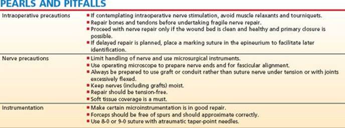
If intraoperative nerve stimulation is to be used, muscle relaxants should be avoided at induction of general anesthesia.
If associated muscle or tendon lacerations are present, muscle relaxation facilitates their repair.
Regional anesthetics such as supraclavicular block provide excellent muscle relaxation, and a supraclavicular catheter will help in administering postoperative analgesia.
Positioning
The patient is positioned supine, with the arm positioned on a hand table.
Use of a tourniquet facilitates dissection but will interfere with intraoperative nerve stimulation, because it results in ischemic conduction blocks after 15 minutes.
Use of intraoperative magnification (eg, loupes) for the dissection and a surgical microscope during nerve repair is essential.
Microinstrumentation is needed for nerve handling and repair.
Alcoholic solutions should be avoided for the preparation of open wounds to avoid chemical damage to nerve tissue.
TECHNIQUS
APPROACH
 The area of injury should be exposed both proximally and distally to allow for visualization of the proximal and distal nerve stumps using extensile approaches.
After all injured structures have been assessed, repair fractures and tendons first to take tension off the nerve repair.
Mobilize nerve ends for about 1 to 2 cm at either end, avoiding unnecessary stripping of the mesoneurium over long distances.
Preserve the common sheath of neurovascular bundles to maintain nerve vascularity and minimize tension on nerve repair.
Nerve end preparation is critical. Crushing is present even after sharp lacerations.
Under an operating microscope, the nerve end is stabilized over a sterile wooden spatula and a fresh no. 11 blade is used to progressively cut back 2-mm segments of the nerve end until sprouting fascicles are seen (TECH FIG 1).
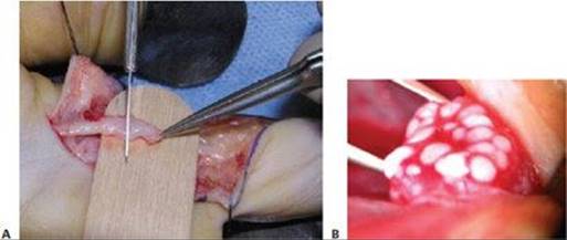
TECH FIG 1 • A. Freshening of a lacerated nerve end. The nerve is stretched over a sterile, moistened tongue depressor and cut using a sharp no. 11 scalpel. B. Sprouting fascicles must be seen at the cut surface of each nerve end before the repair.
The distal and proximal joints are placed in minimal flexion. Excessive flexion is to be avoided, because it will result in flexion contractures or tension on healing nerve.
Additional length can be gained by transposition of the proximal nerve, eg, ulnar nerve at the elbow, or bone shortening, eg, during replantation surgery.
End-to-end repair should be attempted as long as this can be accomplished with minimal tension on the repair and with minimal mobilization of the nerve.
If a single epineurial stitch of 8-0 suture fails to maintain nerve approximation, tension is excessive. Additional mobilization or alternative options such as conduit repair or nerve grafting must be considered.
EPINEURIAL REPAIR
Epineurial repair, the most common type of nerve repair, consists of alignment and approximation of the nerve ends using sutures placed in the epineurium.
After clean, pouting bundles of fascicles are exposed, the epineurium is identified circumferentially by resection or pushing back of the mesoneurium.
Correct alignment of the nerve ends is critical. Line up blood vessels and other external markings in the epineurium, and match fascicular bundles in the two ends.
Suture must be monofilament (eg, nylon) on an atraumatic needle to minimize trauma to the nerve ends. Suture size varies with the size of the nerve. Usually an 8-0 suture is used in the arm and 9-0 in the fingers. Repair with larger suture dimensions does not add strength to the repair: sutures fail by pull-out of the neural tissue.
Two simple sutures are placed 180 degrees from one another. Care must be taken to avoid penetrating fascicles with the needle (TECH FIG 2A).
One tail of each suture is left long to stabilize the nerve during repair.
Three or four additional sutures are placed on the anterior face of the repair as necessary to approximate the epineurium and prevent fascicular extrusion.
By flexing the limb further to relax the nerve, the nerve is turned over, using the suture tails, to expose the posterior wall. Each suture tail can be weighted down with a small vascular clip.
Posterior wall repair is completed with three or four simple epineurial sutures, as needed.
The long tails are then cut short, and the nerve is examined carefully to ensure that a complete epineurial seal is achieved (TECH FIG 2B).
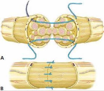
TECH FIG 2 • Steps of epineurial repair. A. The nerve ends are aligned, and two sutures are placed 180 degrees from each other. Tension across the repair is tested with two sutures. B. Additional sutures are placed in the epineurium.
GROUP FASCICULAR REPAIR
Groups of nerve fascicles are approximated by perineurial sutures (TECH FIG 3).
Group fascicular repair is indicated for partial nerve injury involving a few groups of fascicles or in a mixed nerve with distinct motor and sensory components, such as the median nerve proximal to the wrist.
The advantage of improved fascicular alignment may be counteracted by increased intraneural scarring from the increased surgical dissection and manipulation of the group fascicular repair.
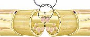
TECH FIG 3 • Technique of group fascicular repair. The epineurium is pulled back, and sutures are placed in the perineurium after fascicular groups have been aligned.
After exposing clean, pouting bundles of fascicles, the epineurium is resected back for 5 mm to clearly exposed groups of fascicles surrounded by perineurium.
The internal arrangement of the fascicles is noted, and similarly sized fascicular groups are aligned.
For partial nerve injury with a few injured fascicles, repair individual fascicles with 10-0 nylon simple sutures placed in the perineurium. Usually, two sutures per fascicle are adequate.
In the case of a complete nerve injury involving a larger nerve, a group fascicular repair that approximates groups of fascicles is faster and less traumatic.
When approximating larger groups of fascicles, four to six sutures are placed per group in a circumferential pattern. For additional stability, sutures at the external surface of the fascicular group should be passed through both the epineurium and perineurium. (These sutures are known as epi-perineurial sutures.)
After completion of fascicular repair, four additional sutures of 9-0 nylon are placed in the epineurium to take tension off the repair.
CABLE GRAFT REPAIR
Nerve grafting is indicated when end-to-end approximation is not possible, eg, after crushing of the nerve ends, retraction of nerve ends after delay in surgical intervention, or after neuroma resection.
After the nerve ends are prepared back to pouting bundles of fascicles, the epineurium is identified circumferentially.
The internal arrangement of the fascicles is noted, and a quick sketch of the fascicular arrangement helps to plan graft alignment (TECH FIG 4).
Epineurium is resected to expose the perineurium of the fascicles.
The gap between the prepared nerve ends is measured.
When grafting a larger-diameter nerve such as the median nerve using a smaller diameter nerve such as the sural nerve, several strands of donor nerve are interposed in the gap as a cable graft.
The length of nerve graft needed is calculated as follows: gap + 15% × estimated number of strands.
Donor nerves include the sural (located midway between the lateral border of the tendo achilles and the lateral malleolus), posterior interosseous (located in the floor of the fourth extensor compartment), and the medial antebrachial cutaneous nerve (located in the anteromedial forearm along branches of the basilic vein).
Each segment of graft is reversed and attached to a similar-sized group of fascicles at the proximal stump using two sutures of 9-0 nylon, 180 degrees from one another.
Although the donor nerve allows growth of regenerating axons in either direction, reversing the nerve graft helps to minimize the possibility of regenerating axons growing out along branches of the donor nerve.
Place the limb in a neutral position; then lay the graft in the defect and align it with a similar fascicular group in the distal stump. The graft is cut and sutured to the distal stump fascicles.
Follow the same sequence, laying segments of graft across the gap until the gap is filled.
The repair can be reinforced with fibrin glue placed at the anastomosis and between segments of the graft.

TECH FIG 4 • Nerve grafting using “cables” of nerve graft. After aligning the nerve ends, similar fascicular groups are bridged with segments of nerve graft.
VASCULARIZED NERVE GRAFT REPAIR
Vascularized nerve graft repair may be indicated in cases when the gap is 6 cm or more, or in a scarred tissue bed in large proximal nerve reconstruction after brachial plexus injuries.
The most common vascularized nerve graft donor is the ulnar nerve (following C8 and T1 root avulsion), along with its mesoneurium, containing the superior ulnar collateral vessels.
For local nerve defects, the ulnar nerve segment is divided, preserving the vascular pedicle. The segment is transposed with its intact pedicle, and epineurial repair is performed.
If a more remote defect is to be grafted, the vascular pedicle and nerve segment are divided. The nerve is reversed and placed in the defect. Following epineurial repair, microvascular anastomosis is performed between the artery and vein in the vascular leash to a local arterial and venous recipient vessel.
CONDUIT REPAIR
Conduit repair is indicated for clinical use in nerve gaps up to 2 cm.
Advantages over conventional repair are that it is tension free, less traumatic, permits no axonal escape, and allows spontaneous axonal orientation.
Two types are in clinical use: reversed autogenous vein or artificial conduits.
 Artificial conduits may be either manufactured using absorbable materials such as polyglycolic acid or made of collagen engineered from natural xenograft sources such as bovine tendon. Artificial conduits have obvious advantages over a vein conduit in regard to shelf availability, size variation, no additional dissection for harvesting, and resilience and elasticity. Collagen tubes degrade over time with natural processes and without any inflammatory reaction.
After nerve end preparation, the nerve diameter is measured. A conduit that is oversized by 1 mm is chosen to avoid constriction of the regenerating nerve.
The conduit is rehydrated in saline for 5 minutes.
The aim of repair is to invaginate each end of the nerve into the tube for a distance of 5 to 8 mm using a mattress suture followed by a single anchoring suture for stability (TECH FIG 5).
The suture is first passed through the tube from the outside in and about 5 mm from the tube edge. The suture is then passed transversely across the epineurium 3 mm from the edge of the nerve stump and then back through the tube in an inside-to-outside direction.
Gently ease the nerve into the tube as the knot is tightened.
Place a simple suture between the epineurium and the edge of the tube at a diametrically opposite point to anchor the tube and prevent rotation.
Repeat the same steps for the distal stump, and fill the tube with saline using a fine cannula.
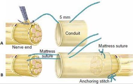
TECH FIG 5 • Technique of conduit repair. A. A horizontal mattress suture is placed between the conduit and the epineurium of the nerve. B. As the suture is tightened, the nerve is drawn into the conduit. A simple stitch is placed anchoring the epineurium to the tube opposite the location of the mattress suture.
POSTOPERATIVE CARE
Consider use of a local anesthesia infusion pump for postoperative pain control.
Immobilization is very important to prevent tension across the repair:
The elbow should be held at 90 degrees of flexion.
Wrist flexion greater than 20 degrees should be avoided.
The metacarpophalangeal joints should be held at 70 degrees of flexion.
Mobilization varies with associated tendon repair. After isolated nerve repair, gentle finger flexion and shoulder range of motion are started soon after surgery to promote nerve gliding and prevent finger stiffness.
Remove skin sutures after 2 weeks and replace the splint.
For nerve repairs around the elbow, allow motion in an extension blocking splint. Full extension is permitted after 6 weeks.
For repairs in the distal forearm and wrist level, immobilize the wrist at 20 degrees flexion and block metacarpophalangeal hyperextension for 4 weeks. Allow active finger motion within the splint. Bring the wrist to neutral at 4 weeks, and then allow mobilization out of the splint at 6 weeks.
Nerve regeneration is followed at regular intervals with clinical examination of motor and sensory recovery and Tinel sign.
The distal-most point at which the Tinel sign is observed is recorded at each visit and its distance from the suture line noted.
Expect distal progression of Tinel sign at the rate of about 1 mm per day, with a delay of 1 month after the date of repair.
Failure of Tinel progression over serial visits may indicate repair failure: consider re-exploration and grafting.
Sensory re-education is initiated early in the postoperative phase with the goal of teaching recognition of new input in a useful manner.
Three stages to this process are introduced sequentially in the recovery period:
Desensitization: the patient is presented with graded stimuli to decrease unpleasant sensations.
Early-phase discrimination and localization: the patient works with static and moving touch, using visual reinforcement.
Late-phase discrimination and tactile gnosis: the patient works with varying shaped objects.
OUTCOMES
The outcome after nerve repair is generally less favorable than that of repair of other tissues, such as bone or tendon injury.
It is difficult to predict the outcome because of several variables, including type of nerve (pure sensory versus mixed), age of patient, type of injury—clean or crushed, associated soft tissue injuries.
The single most important factor that correlates with outcome is patient age. The best results are seen in children younger than 10 years of age.
Pure motor or pure sensory nerves fare better than mixed nerves.
The outcome also correlates with level of injury. Injuries closer to the end organs fare better, because there is less distance for the regenerating axons to cover.
Peripheral factors that are determined by the injury and cannot be modified by the treating surgeon include axonal cell death, end-organ atrophy, and extensive scarring from surrounding crush injury.
The surgeon can control, to a limited extent, the scarring in and around the nerve repair.
Central factors that account for poor results include cortical remapping and reorganization, with reduced and disorganized cortical representation of denervated areas.
Children recover greater function than their adult counterparts with primarily repaired lesions at similar levels due to a combination of better axonal regeneration and cortical plasticity.
Delayed repairs fare worse than those repaired acutely, with an estimated 1% decrease in performance for every 6 days of delay in the repair.
The nature of the injury often determines the likelihood of recovery. Massive soft tissue injury or burns involving a peripheral nerve are less likely to regain function than injuries involving sharp or limited transection of a nerve.
Median nerve outcome after high injuries usually is poor because of hand intrinsic atrophy. After low injuries, useful motor function is regained in 40% to 90% of repairs, and useful sensation is restored in 53% to 100% of patients.
Ulnar nerve injuries show similarly poor results for motor recovery, with functional restoration in 35% of cases and functional sensory recovery in 30% to 68% of cases.
Because the ulnar nerve is largely a motor nerve, better results can be expected after acute radial nerve repairs, with functional return in 60% to 75% of patients. Poor results are noted with high injuries, however.
After repair of digital nerves, about 50% of patients regain static two-point discrimination of less than 10 mm. Younger children demonstrate near-normal sensory recovery due to their cortical adaptability.
Lingering symptoms of hypersensitivity and cold intolerance are common with sensory nerve injury in the upper extremity, resolving in most patients after 2 to 3 years. The cause is unclear.
Complex regional pain syndrome is more likely to be present after untreated nerve injuries. If it does occur, significant joint contractures and atrophic changes can result, and the patient generally has a prolonged recovery period and a poor outcome.
COMPLICATIONS
Causes for failure of repair include:
Tension on the initial repair
An unfavorable local tissue environment with excessive scarring
Noncompliance with protective measures or therapy and consequent joint contractures
Painful neuromas usually form in unrepaired or poorly repaired nerves close to the surface. These usually are treated with desensitization, local padding, etc. because surgical results are often disappointing.
Altered sensation is a result of axonal misdirection and cortical misrepresentation and can present as loss of temperature sensation or cold intolerance, hyperesthesia, or neuropathic pain.
Some amount of altered function is inevitable after all complete nerve injuries in the upper extremity except in young children. It is due to a combination of altered sensation and proprioception along with loss of motor strength.
Complex regional pain syndrome type II can occur after nerve injury especially in untreated cases or after delayed treatment or failure to control pain. Typical features include dramatic changes in the color and temperature of the skin accompanied by intense burning pain, skin sensitivity, sweating, and swelling. Early recognition is the key with referral to a pain management specialist for stellate blocks along with steroids, antiepileptic drugs, and therapy.
SUGGESTED READING
Al-Ghazal SK, McKiernan M, Khan K, et al. Results of clinical assessment after primary digital nerve repair. J Hand Surg Br 1994;19:255–257.
Birch R. Nerve repair. In: Green D, Hotchkiss R, Pederson W, et al, eds.
Green's Operative Hand Surgery. Philadelphia: Elsevier Churchill Livingstone, 2005.
Birch R, Bonney C, Wynn Parry CB. Surgical Disorders of the Peripheral
Nerves. Edinburgh: Churchill Livingstone, 1998.
Birch R, Raji AR. Repair of median and ulnar nerves: Primary suture is best. J Bone Joint Surg Br 1991;73B:154–157.
Chaise F, Friol JP, Gaisne E. Results of emergency repair of wounds of palmar collateral nerves of the fingers. Rev Chir Orthop Reparatrice Appar Mot 1993;79:393–397.
Clark WL, Trumble TE, Swiontkowski MF, et al. Nerve tension and blood flow in a rat model of immediate and delayed repairs. J Hand Surg Am 1992;17:677–687.
de Medinaceli L, Prayon M, Merle M. Percentage of nerve injuries in which primary repair can be achieved by end-to-end approximation: Review of 2,181 nerve lesions. Microsurgery 1993;14:244–246.
Giddins GE, Wade PJ, Amis AA. Primary nerve repair: Strength of repair with different gauges of nylon suture material. J Hand Surg Am 1989;14:301–302.
Goldberg SH, Jobin CM, Hayes AG, et al. Biomechanics and histology of intact and repaired digital nerves: an in vitro study. J Hand Surg Am 2007;32:474–482.
Goldie BS, Coates CJ, Birch R. The long term result of digital nerve repair in no-man's land. J Hand Surg Br 1992;17:75–77.
Hudson DA, de Jager LT. The spaghetti wrist: Simultaneous laceration of the median and ulnar nerves with flexor tendons at the wrist. J Hand Surg Br 1993;18:171–173.
McAllister RM, Gilbert SE, Calder JS, et al. The epidemiology and management of upper limb peripheral nerve injuries in modern practice. J Hand Surg Br 1996;21:4–13.
Puckett CL, Meyer VH. Results of treatment of extensive volar wrist lacerations: The spaghetti wrist. Plast Reconstr Surg 1985;75:714–721.
Shergill G, Bonney G, Munshi P, et al. The radial and posterior interosseous nerves. Results of 260 repairs. J Bone Joint Surg Br 2001; 83:646–649.
Sullivan DJ. Results of digital neurorrhaphy in adults. J Hand Surg Br 1985;10:41–44.
Wynn Parry CB, Salter M. Sensory re-education after median nerve lesions. Hand 1976;8:250–257.