
Randy R. Bindra and Jeff W. Johnson
DEFINITION
 A nerve injury in continuity occurs when there is loss of axonal function with preserved structure of the supportive connective tissue.
 By definition, the epineurium is preserved in a nerve injury in continuity.
Because varying degrees of axonal interruption may occur, the extent of functional loss in terms of numbness and paralysis is variable.
The severity of injury varies with degree of preservation of the endoneurium and the perineurium.
ANATOMY
The cross-sectional anatomy of the peripheral nerve is discussed in detail in Chapter HA-72.
Endoneurial tubes form the basic conduit for the Schwann cell–encased axon.
PATHOGENESIS
Several mechanisms may cause a nerve injury in continuity, but the most common is nerve stretch.
When a nerve is subject to blunt injury or stretch, axonal disruption can occur without externally visible injury to the nerve.
Stromal elements are more resilient to stretch and remain preserved to a variable extent (FIG 1).
The type of recovery seen after an injury depends on preservation of the endoneurial tube.
In the mildest forms of injury, with preserved endoneurial tubes, regenerating axons follow their original path. The destination is reached with good outcome. There is no axonal mismatch, and the recovery is termed uncomplicated regeneration.
When the endoneurial tube is disrupted, axonal regeneration is disorganized. Axons sprout and grow in a different direction, and mismatch occurs. This form of repair, termed complex regeneration, is associated with a clinically less satisfactory outcome.
With more severe forms of stretch injury, additional disruption of the perineurium occurs, resulting in a greater fibrotic response and resultant scarring of the nerve.
The nerve trunk, which externally appears uninterrupted due to the intact epineurium, demonstrates an injured segment that is enlarged due to intraneural fibrosis surrounding a mass of disorganized axons. This is referred to as a neuroma in continuity (FIG 2).
NATURAL HISTORY
Pathoanatomy associated with the injury, pathologic changes resulting from this altered anatomy, and functional recovery are closely related.
More anatomic disruption results in a stronger pathologic response and worse outcome.
Sunderland's classification of injury severity is useful to categorize injury and plan treatment.
Type I
 The mildest form of injury involves loss of axonal function without actual structural interruption: neurapraxia (FIG 3A).
Type I injury is seen after mild stretch injuries, tourniquet palsy, and external compression of a nerve, as in radial nerve compression in “Saturday night palsy.”
Although structurally intact, axons fail to conduct impulses, secondary to malfunction of ion channels along the injured segment.
No visible change in the microscopic or macroscopic appearance of the nerve is present, and there is no wallerian degeneration of the distal segment.
Electrophysiologic testing does not reveal a conduction block or denervation potentials.
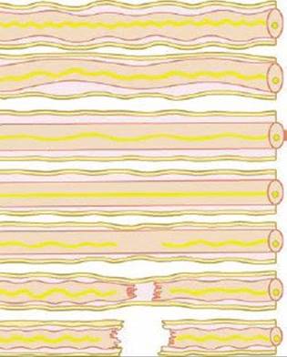
FIG 1 • Pathogenesis of a nerve injury in continuity. The effect of increasing stretch is seen, from normal nerve at the top to complete rupture at the bottom. Neural elements fail first in response to stretch; epineurium fails last.

FIG 2 • Neuroma in continuity. The enlarged part of the nerve consists of a mixture of intact and damaged axons surrounded by scar tissue and regenerating axons.

FIG 3 • Sunderland classification of nerve injury. A. Sunderland type I, neurapraxia. Nerve injury demonstrating preserved nerve structure with functional loss B. Sunderland type II, axonotmesis. Preservation of the endoneurial tube with wallerian degeneration of the distal axon. C. Sunderland type III. The fascicular structure is preserved due to intact perineurium. As the endoneurium is disrupted, regenerating axons wander within the fascicle, resulting in a less optimal recovery. D. Sunderland type IV. A severe disruption of the nerve. Although the epineurium is intact, loss of fascicular organization makes recovery unlikely without surgical intervention. E. Sunderland type V, neurotmesis. Complete structural disruption with loss of continuity.
Recovery starts within a few weeks and can be expected to be complete.
Because axons recover conductivity in a variable pattern, clinical recovery follows a random pattern.
Type II
There is structural disruption of the axon, but the endoneurium is preserved (FIG 3B).
Type II injury is seen after more severe stretch injuries, such as radial nerve palsy resulting from a closed humerus fracture.
Wallerian degeneration results and electrophysiologic tests reveal distal conduction block and denervation.
As regenerating axons progress distally, proximal muscles are reinnervated first. Clinically, recovery occurs in a proximal-to-distal direction.
Because there is no axonal mismatch, recovery usually is complete but takes longer, usually several months.
Type III
The axon, myelin sheath, and endoneurium are interrupted (FIG 3C).
Recovery is less predictable, because regenerating axons may not follow previous pathways (complicated regeneration).
With the perineurium preserved, recovery can take place without surgical intervention but usually is incomplete due to axonal misdirection.
Injury to small vessels within the endoneurium leads to an inflammatory response. Fibroblast activation results in a variable degree of interfascicular scarring that may impede nerve regeneration.
Type IV
In more severe stretch injuries, the internal nerve structure is completely disrupted, leaving only an intact epineurium (FIG 3D).
Retraction of fascicles and scarring within the nerve are present. Even though the nerve is in continuity, no clinically significant recovery can be expected without surgical intervention.
Type V
Complete rupture or laceration of the nerve with retraction of the nerve ends (see Chap. HA-93) (FIG 3E)
PATIENT HISTORY AND PHYSICAL FINDINGS
Stretch injuries that result in nerve injury in continuity usually are proximal. These injuries often take place as the nerve root exits the spinal cord or involve the brachial plexus in the neck or upper arm.
At more distal levels, nerve stretch injuries usually are the result of displaced fractures or dislocations.
There usually is a history of significant trauma, and patients complain of pain and paresthesias with a variable amount of functional loss distal to the site of injury.
Incomplete loss of function often indicates an incomplete nerve injury.
Severe pain or paresthesias after any closed fracture should alert the clinician to the possibility of an associated nerve injury.
Complete loss of function does not necessarily imply complete disruption of the nerve.
Documented lack of recovery on serial clinical examinations is essential to determine the severity of the injury and the need for surgical intervention.
Muscle strength is charted against a timeline at every visit. Progressive muscle recovery in a proximal-to-distal direction indicates spontaneous axonal regeneration.
Tinel sign and its gradual progression is also a useful measure of nerve recovery.
Recovery within a few weeks of injury and with a random pattern usually suggests a Type I injury or neurapraxia.
After incomplete injury to a peripheral nerve, function is lost in a predictable order: motor, proprioception, touch, temperature, pain, and sympathetic function.
Recovery usually occurs in the reverse order.
In a closed injury without any obvious fractures, the site of nerve injury is not always obvious.
Careful mapping of the motor and sensory deficit will help to distinguish the level of injury.
The pattern of sensory loss is a reliable way to determine the level of injury. A more proximal injury usually follows a dermatomal pattern, whereas a distal injury follows the distribution of the nerve.
IMAGING AND OTHER DIAGNOSTIC STUDIES
MRI done several weeks after injury may reveal an enlarged nerve segment, suggesting a neuroma in continuity.
Neurophysiologic studies are useful in evaluating and monitoring an injured nerve when there is no external injury.
Conduction blocks usually reverse within 10 to 14 days; therefore, tests should be delayed until this time.
Complete loss of muscle action potentials does not necessarily indicate a complete interruption of all axons.
Electromyograms (EMGs) will show variable denervation of muscle groups innervated by the nerve in question.
Fibrillation potentials on EMG usually appear within 10 to 40 days, indicating complete denervation of a muscle group.
Electromyographic evidence of reinnervation may precede voluntary muscle contraction by several weeks and may be of use in tracking the progress of nerve regeneration.
Return of a muscle action potential requires not only regeneration of the nerve to the level of the end organ but also re-establishment of a physiologic connection between the nerve and the target tissue. Re-establishment of the motor endplate is required before EMG provides evidence of functional return.
Nerve conduction studies (NCSs) also are useful in the evaluation of a closed nerve injury.
In a closed injury, lesions may be localized using NCSs.
Continuity of the nerve also may be assessed, but should be undertaken at about 10 days after the injury to prevent erroneous results, because the axons distal to a complete transection may continue to conduct during this initial period after injury.
Parameters evaluated include amplitude and latency.
DIFFERENTIAL DIAGNOSIS
Complete transection
Conduction block
Partial axonal injury
Compressive injury
NONOPERATIVE MANAGEMENT
Lesions in continuity may improve spontaneously, especially in types I and II, in which recovery is complete without any surgical intervention.
Type I through III injuries can be watched closely with serial mapping of the sensory and motor recovery.
Type IV and V injuries usually require surgical repair of the nerve to restore axonal continuity.
Preservation of some function distal to the suspected level of the injury within the distribution of the injured nerve suggests a partial injury, and observation is appropriate.
If there is a complete palsy of a nerve after a closed injury, an initial period of observation may be best until signs of denervation appear in end organs.
If signs of reinnervation appear, such as a Tinel sign distal to the level of injury, continued observation is prudent.
If no signs of innervation appear, one should strongly consider electrodiagnostic studies to evaluate the continuity of the axonal fibers.
Physical therapy is very important to maintain mobility during the period of observation.
SURGICAL MANAGEMENT
If no signs of recovery are present at 2 to 4 months, then surgical exploration may be indicated.
Electrodiagnostic testing should be used in this instance to define the level of injury.
Longer delays may compromise the efficacy of surgical repair, secondary to end organ degenerative changes.
Focal injuries are usually observed for shorter periods, because the extent of the injured nerve segment usually is smaller.
Blunt or blast injuries may be observed for up to 6 months given the often large segments of injured nerve undergoing repair.
Preoperative Planning
Intraoperative nerve action potentials (NAPs) may provide information about lesions in continuity, including the degree and extent of interruption.
If NAPs are not recordable across a lesion, then resection and direct repair rather than grafting will likely be required.
Resection is performed from the point at which NAPs are lost to the point where they return.
If NAPs are present, external neurolysis or nerve decompression may be adequate treatment.
Positioning
The patient is positioned supine, with a hand table.
If nerve grafts may be required, the opposite leg is prepared to allow access to the sural nerve. Rarely, if bilateral sural nerves are to be harvested, the patient initially is placed prone.
Use of a tourniquet may result in ischemic conduction blocks, which will render intraoperative nerve stimulation ineffective.
It generally is preferable to use a tourniquet for only the first 20 minutes of surgery, to facilitate initial dissection.
The use of an operating microscope and fine soft tissue sets or microinstrumentation is necessary for nerve handling and repair.
Approach
Surgical exposure should provide adequate access to the section of damaged nerve as well as proximal and distal to this site.
Mobilization should be minimized to prevent additional vascular insult to the nerve.
Sources of external compression should be identified and alleviated.
The bed for repair should be free of scar tissue. Nerve transposition may be required.
TECHNIQUES
EXTERNAL NEUROLYSIS
 External neurolysis is defined as the circumferential freeing of a peripheral nerve from surrounding scar tissue (TECH FIG 1A).
Dissection proceeds from normal nerve (both proximal and distal) to the area of scarring (TECH FIG 1B).
The nerve should be mobilized away from the scar tissue bed to prevent recurrence.
Use of a xenograft nerve wrap or fat graft may be considered to prevent recurrence of scarring (TECH FIG 1C).
External neurolysis may relieve neuropathic pain associated with compression, but results for sensory and motor recovery are variable.

TECH FIG 1 • External neurolysis and xenograft nerve wrap. A. The median nerve at the wrist developed painful scarring after carpal tunnel release. B. External neurolysis has been performed by excision of all scar tissue and thickened epineurium. C. A xenograft collagen nerve wrap has been placed around the nerve to minimize scar tissue formation around the nerve.
INTERNAL NEUROLYSIS
Intraoperative recording of NAPs will indicate functioning fascicular groups and help guide the surgeon during this procedure.
Internal neurolysis is performed along the fascicular segment that has lost NAPs (TECH FIG 2).
Internal neurolysis is defined as the resection of fibrotic tissue from within the structure of the nerve itself.
This procedure is indicated for late management of incomplete injuries such as stretch injuries when the nerve has regained partial function that is clinically inadequate.


TECH FIG 2 • A–C. Intraoperative microscope images of internal neurolysis of the ulnar nerve at the wrist. A. The ulnar nerve is surrounded by dense scar tissue. B. After external neurolysis, there is a persistent area of narrowing of the nerve (arrows) requiring internal neurolysis. C. Appearance after internal neurolysis—the constricting epineurium and scar between fascicles has been excised. D.Illustration of a neuroma in continuity treated by internal neurolysis. The segment of scarred epineurium is excised, and all scar tissue between fascicles also is excised.
This procedure is performed in cases of incomplete functional loss distal to the site of injury.
Some loss of intact axons can be expected as a result of the dissection, so the patient should be advised that additional loss of function could be possible with this procedure.
SPLIT REPAIR
Split repair is defined as a procedure in which intraoperative NAP recordings are used to guide the resection of individual nonconducting fascicles.
First, external neurolysis is performed to expose the injured segment of the nerve.
The epineurium is excised circumferentially to expose the injured fascicles (TECH FIG 3A).
Intraoperative NAP recordings are made to identify the injured fascicular segments (TECH FIG 3B).
Resection of the nonconducting segments is performed using either a blade or sharp microvascular scissors (TECH FIG 3C).
Repair is performed either directly or with autogenous grafting.
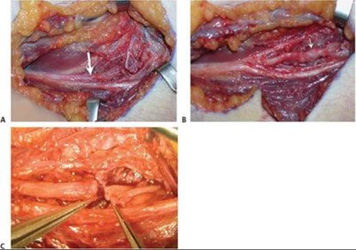

TECH FIG 3 • Exploration and split repair of a partial injury of the posterior interosseous nerve 4 weeks after palsy following a dog bite at the elbow. A. The posterior interosseous nerve demonstrates a neuroma in continuity (white arrow). B. Internal neurolysis has been performed, isolating intact peripheral fascicles with a central neuroma (white arrow). C. A gap remains in the injured fascicular group after neuroma resection. Mobilization of the intact fascicles is limited because of their proximity to the motor branches, making end-end repair of the injured fascicles difficult. D. Conduit repair of the injured fascicles has been performed. E, F. Illustrations of split repair of a partially injured nerve. Nonconducting fascicular segments are excised and either repaired by end-to-end group fascicular repair (E) or by interposing nerve grafts (F).
 Length = gap + 15% × estimated number of grafts
Grafts are attached to a group of fascicles using two sutures of 9-0 or 10-0 nylon, 180 degrees from one another.
Each graft is sutured to the proximal and distal stumps before moving on to the next graft, thus allowing for more accurate fascicular matching.
Check the repair to ensure that no stitches have pulled out. The repair may be reinforced with fibrin glue
Handling of the grafts should be minimized.
 Grafts should be kept moist from harvest to repair.
Cable grafting is the more common technique, using donor nerve from either the sural or antebrachial cutaneous nerve.
Grouped fascicular repair is then performed (TECH FIG 3D–F).
The internal arrangement of the fascicles is noted, and a quick sketch of the fascicular arrangement is made to allow alignment of the nerve ends.
Nerve grafts will not match the exact fascicular pattern—the aim is to place graft “cables” between groups of fascicles.
The gap between nerve ends is measured, and the length of graft needed is calculated.
RESECTION OF THE NERVE LESION IN CONTINUITY
If no conduction of NAPs is noted across a lesion after internal neurolysis is performed, then the entire lesion should be resected.
The proximal and distal portions of the nerve flanking the lesion should be mobilized to prevent undue tension on the repair. During mobilization, longitudinal blood vessels within the epineurium must be preserved.
The lesion is sharply excised using a fresh, sharp blade against a block (usually a moistened tongue depressor).
Epineurial Repair
If the extent of the lesion is short, then direct end-to-end epineurial repair without tension often is possible.
Direct epineurial repair is then performed as described in Chapter HA 93.
Cable Graft Repair
Cable graft repair is useful when the extent of the lesion precludes direct repair because of either tension or a large gap (TECH FIG 4A).
Cable graft repair is then performed as described in Chapter HA-93.
Sural nerve graft can be harvested through a single longitudinal or multiple transverse incisions (TECH FIG 4B).
The nerve is easily identified by careful spreading dissection in the subcutaneous tissue midway between the lateral malleolus and the tendo-achilles (TECH FIG 4C).
Use of a tendon stripper to harvest the nerve is not recommended.
This technique can result in stretch or laceration of the sural nerve.
Additionally, the posterior tibial nerve may be inadvertently injured.


TECH FIG 4 • Cable grafting for reconstruction of a sciatic nerve laceration in the thigh. A. The ends of the sciatic nerve lie 6 cm apart. B. The ipsilateral sural nerve is harvested by multiple transverse incisions in the leg. Yellow rubber slings have been placed around the nerve at each incision for identification and gentle traction to facilitate dissection. C. Multiple segments of the sural nerve have been aligned and inserted in the nerve gap and fixed with group fascicular sutures.

POSTOPERATIVE CARE
General guidelines for splinting and postoperative care are detailed in Chapter HA-72.
Serial examination is important to follow the progress after surgical repair.
OUTCOMES
Neurolysis
If NAPs are recorded through a nerve segment, recovery is thought to be about 90%.
NAP recording and subsequent neurolysis without resection have been found to consistently result in better outcomes than direct or graft repair.
Split repair
Outcomes are superior to complete repair when NAPs are recorded through some portion of the nerve.
Direct and graft repair of the injured fascicles yield similar results.
Complete resection with direct repair or graft repair
The outcome of direct repairs appears to be superior to those requiring the use of a graft; however, injuries requiring a nerve graft often are more substantial and require regeneration along a greater distance.
In general, radial nerve repairs are more successful than median nerve repairs, and both are better than ulnar nerve repairs.
Children generally have better overall outcomes than adults.
Internal neurolysis or resection of any lesion in continuity may be related to a decrease in preoperative function as some intact axons may be transected.
COMPLICATIONS
Infection
Scarring
Loss of function
Increased neuropathic pain
Either distal to the lesion or in the form of a painful neuroma
Failure of recovery of function
SUGGESTED READINGS
Birch R, Bonney C, Wynn Parry CB. Surgical Disorders of the Peripheral Nerves. Edinburgh: Churchill Livingstone, 1998.
Kline DG. Surgical repair of peripheral nerve injury. Muscle Nerve 1990;13:843–852.
Lundborg G, Rosén B, Dahlin L, et al. Tubular versus conventional repair of median and ulnar nerves in the human forearm: Early results from a prospective, randomized, clinical study. J Hand Surg Am 1997;22: 99–106.
Mackinnon SE, Novak CB. Nerve transfers: new options for reconstruction following nerve injury. Hand Clin 1999;15:643–666.
Mujadzic M, Ozyurekoglu T, Gupta A, et al. Intraoperative nerve recordings as a useful aid in the management of neuroma-in-continuity. J Reconstruct Microsurg 2005;21:341.
Seddon HJ. Nerve grafting. J Bone Joint Surg Br 1963;43B:447–461. Sunderland S. A classification of peripheral nerve injuries producing loss of function. Brain 1951;74:491–516.