
Xiaochuan Li1, Lan Zhu1 , Qing Dai2 and Jingjing Lu3
(1)
Department of Obstetrics and Gynecology, Peking Union Medical College Hospital, No. 1 Shuaifuyuan, Beiijing, 100730, P. R. China
(2)
Department of Ultrasound, Peking Union Medical College Hospital, No. 1 Shuaifuyuan, Beijing, 100730, P. R. China
(3)
Department of Radiology, Peking Union Medical College Hospital, No. 1 Shuaifuyuan, Beiijing, 100730, P. R. China
Lan Zhu
Email: zhu_julie@sina.com
2.1 Introduction
Xiaochuan Li4 and Lan Zhu4
(4)
Department of Obstetrics and Gynecology, Peking Union Medical College Hospital, No. 1 Shuaifuyuan, Beiijing, 100730, P. R. China
Lan Zhu
Email: zhu_julie@sina.com
Female genital anomalies are mainly referred to the female reproductive tract abnormalities or abnormal gonad development with or without other organ malformations. It is a large class of abnormal developmental diseases involving the vulva, vagina, uterus, fallopian tubes, and ovaries.
Female genital anomalies are based mainly on their clinical symptoms, physical examinations, and basic imaging studies to make a preliminary diagnosis, further assisted by chromosomal analysis, hormone assessment, diagnostic imaging, and even diagnostic surgical procedure to obtain an accurate diagnosis. The common clinical manifestations of these diseases are as follows:
Primary amenorrhea: It is the clinical symptoms ion women over 14 years of age who do not have any menstruation and secondary sexual characteristics or over 16 years of age when they have secondary sexual characteristics but without menstruation. About 50 % of the patients with primary amenorrhea are due to gonadal anomalies and 20 % due for reproductive tract abnormalities such as congenital absence of vagina, imperforate hymen, etc.
Symptoms related to reproductive tract obstruction: the most common symptoms are dysmenorrhea and periodic lower abdominal pain. These symptoms are often due to the presence of functional endometrium, and the menstrual outflow is obstructed. Because of retrograde menstruation, some patients present with endometriosis-related disorders of the ovaries, uterus, and pelvic endometriosis. When the menstrual outflow is incompletely obstructed, there will be irregular menstrual spotting with poor menstrual flow. When the obstruction is associated with infection, pus can form in the vagina or uterine cavity presenting as acute pelvic inflammatory disease. Obstruction at the vagina will have typical cyclical abdominal pain with increasing severity; symptoms due to imperforate hymen, transverse vaginal septum, and vaginal oblique septum may vary depending whether there are holes in the septum or not, causing either complete or incomplete obstruction symptoms.
The impact on pregnancy and delivery outcome: Different types of uterine anomalies may be related to miscarriage, premature birth, fetal malposition, fetal growth retardation, abnormal progress of labor, and postpartum hemorrhage. Rudimentary uterine horn pregnancy can lead to life-threatening uterine rupture. Among female patients with infertility, 10 % are associated with anomalies of the vulva, vagina, cervix, or uterus.
Sexual difficulties: It is common for patients with genital anomalies to complain of sexual difficulties, but rarely as the only symptom. There are usually complaints together with symptoms related to reproductive tract obstruction.
Ambiguous sexual genitalia or external genital abnormalities: Abnormal gonadal differentiation (true hermaphroditism, gonadal dysgenesis, etc.), androgen excess (congenital adrenal hyperplasia, excessive exogenous androgen exposure in early pregnancy, etc.), androgen deficiency (partial androgen insensitivity syndrome, testicular regression syndrome, etc.), and genitourinary tubercle anomalies (persistent cloaca, labia minora fusion, etc.) can cause this clinical manifestation. Ambiguous sexual genitalia can be combined with the internal genital anomalies, urinary tract anomalies, and/or anorectal malformation. Serious ambiguous sexual genitalia are more commonly found and corrected in the neonatal or infantile period, but improper corrective treatment or atypical symptoms will make it difficult for the diagnosis and subsequent treatment in the future.
The symptoms of urinary tract anomalies: 30–50 % of patients with genital anomalies are associated with urinary tract anomalies. The majority of the urinary tract anomalies are asymptomatic, yet some may present with symptoms of urinary tract infection, hematuria, urinary frequency, urgency, dysuria, and so on. Patients with urogenital fistula like vesicovaginal fistula may present with menstrual hematuria. Some patients with urogenital sinus malformation may have abnormally slack urethral opening that may be mistaken as the vaginal opening. They will present with painful sexual intercourse, sexual difficulties, stress urinary incontinence, and recurrent urinary tract infections.
Related symptoms of other organ malformations: Female genital anomalies are often associated with other organ malformations. Attention should be paid to the height, skeletal abnormalities, deafness, and other deformities of these patients.
Asymptomatic: A study of female genital anomalies among patients who had had tubal ligation showed that 3.2 % of those asymptomatic females had Müllerian duct anomalies. Therefore, some scholars estimated that the incidence of the female genital anomalies could be as high as 7 % in the general population and the majority of them were asymptomatic.
When patients presented with the above clinical manifestations, they should be included in the “high-risk” group for female genital anomalies. Progressive investigations start from different levels, from simple to complex, and from noninvasive to invasive; careful inspection and evaluation of complaints will help to raise the clinical awareness and lead to the diagnosis of these disorders. Other assessments should include detailed medical history and physical examination, sex chromosome analysis, gonadal hormone levels, and imaging assessment and finally with diagnostic surgery if it is necessary.
2.1.1 Medical History and Physical Examination
Medical history includes mother’s pregnancy history and family history: whether there was any drug exposure that occurred during pregnancy, whether there were chemicals from the environment which interfered endocrine secretions, whether there had been diethylstilbestrol (DES) exposure in early pregnancy, and so on. Family history includes other family members (especially sisters) who might have similar clinical symptoms or known genital anomalies and have suffered early neonatal death and inbreeding childbearing and other family members (especially aunts) with infertility, amenorrhea, premature menopause, and so on. Despite the causes for the majority of female genital anomalies need to be further elucidated, but at least there is familial aggregation of these diseases or there are certainly genetic characteristics, it clearly has made them genetic-related diseases.
In physical examination, the keys to make a diagnosis are the number of gonads and their positions whether they are abnormal and the certainty of the anatomy of the reproductive tract. Other examinations are to identify the other characteristic features of malformation syndromes, such as Turner syndrome (congenital ovarian hypoplasia). The typical characteristics of Turner syndrome are short stature (usually less than 150 cm), epicanthus, webbed neck, and elbow valgus. It is therefore important to familiarize with these phenotypic characteristics that will facilitate the diagnosis of genetic-related genital anomalies.
2.1.2 Sex Chromosome Analysis
Reproductive tract development and sexual differentiation are closely related to the sex chromosomes which are the basis for the gender differentiation which is one of the most critical steps of sex differentiation. Sex chromosome analysis helps to contribute to the diagnosis and differential diagnosis of female genital anomalies. Common chromosomal abnormalities causing genital anomalies are as follows: (1) Turner syndrome (congenital ovarian hypoplasia) chromosome 45, XO, expressed as ovarian dysgenesis and juvenile external and internal genitalia, accompanied with multiple organs malformations and (2) triple X chromosome 47, XXX, expressed as female phenotype but poorly developed breasts and genitalia with ovarian atrophy. Some patients may have menstrual cycles, secondary amenorrhea, and premature menopause. There is often mentally retardation. Chromosome analysis also helps to distinguish true hermaphroditism, XO/XY gonadal dysgenesis, and the like. However, in many patients with female genital anomalies, their chromosome analysis usually shows normal female phenotype.
2.1.3 Gonadal and Sex Hormone Assessment
After the sex chromosomes determine the gender, gonadal differentiation and development would lead to the differentiation and development of internal and external genitalia. The final phenotypic sex will finally develop under the influence of sex hormones. When there is gonadal dysgenesis or abnormal sex hormone synthesis or dysfunction, it could lead to genital anomalies. By measuring the serum sex hormones and gonadotropin, it helps to make the diagnosis. For example, in patients with 46, XX pure gonadal dysgenesis, their chromosome analysis and external appearance are all phenotypic female, yet they cannot develop secondary sexual characteristics. The laboratory tests will show a low serum estrogen and increased serum gonadotropin. The clinical presentation will be primary amenorrhea. Another example is the congenital adrenal hyperplasia. The chromosome karyotype is 46, XX, but it is the abnormal synthesis of cortisol that caused by the deficiency of an adrenal enzyme, leading to increased adrenal steroid precursors to generate excessive androgens. High serum 17α-hydroxyprogesterone, high testosterone, and positive dexamethasone suppression test can confirm the diagnosis.
2.1.4 Imaging Examination
Commonly used imaging examinations include ultrasound imaging, X-ray, MRI, CT scan, hysterosalpingography (HSG), and so on. The main aims of imaging are (1) to indicate the site of anomaly, anatomical features, and any related complication and (2) to exclude any morphological abnormalities in other organs. Ultrasound imaging is most widely used as a preliminary investigation because it is noninvasive, simple, and economical. MRI has an advantage in the examinations of cervix, uterine, and complicated pelvic abnormalities. It is often used as a confirmative diagnostic test. X-ray and CT scan are valuable methods for investigating skeletal malformations, but they are inferior to MRI and ultrasound for the diagnosis of female genital anomalies. HSG is useful to determine the size and shape of the uterine cavity, but because it is invasive with ionizing radiation, it is gradually replaced by ultrasound and MRI examination.
2.1.5 Diagnostic Surgical Procedures
If a certain diagnosis cannot be made despite all of the above investigations and examinations, or if there are indications for surgical treatment for the patients, diagnostic (treatment) surgery may be used as the last resort. Hysteroscopy, laparoscopy, or sometimes a combination of both is used. During a diagnostic procedure, pathological examination may be helpful for the final diagnosis. Sometimes a therapeutic surgical treatment if indicated can be completed simultaneously.
2.2 Ultrasound Diagnosis of Female Genital Anomalies
Qing Dai5
(5)
Department of Ultrasound, Peking Union Medical College Hospital, No. 1 Shuaifuyuan, Beijing, 100730, P. R. China
Qing Dai
Email: qingdai_2000@yahoo.com
During the embryonic development of female genital organs, there are internal or external influences which can lead to genital anomalies. The common reproductive tract anomalies are (1) vulvar anomalies, e.g., the imperforate hymen; (2) vaginal anomalies, e.g., congenital absence of vagina, vaginal atresia, transverse vaginal septum, longitudinal vaginal septum, and oblique vaginal septum; and (3) uterine anomalies, e.g., congenital absence of uterus, primordial uterus, infantile uterus, uterus didelphys, bicornuate uterus, rudimentary horn, arcuate uterus, septate uterus, and so on.
Imaging examination plays a very important role in the diagnosis of female reproductive tract anomalies. The commonly used imaging methods include ultrasound, X-ray imaging, MRI, CT scan, etc., in which ultrasound is the most commonly chosen method.
2.2.1 Ultrasound Imaging
Ultrasound imaging has been used in clinical obstetrics and gynecology for nearly half a century. With the ongoing development of ultrasound equipment, the image quality of ultrasound scan has greatly improved. It now plays an increasingly important role in the diagnosis and management of reproductive tract anomalies. Many uterine anomalies can also be discovered by ultrasound imaging examination alone.
Ultrasound examination techniques are as follows.
2.2.1.1 Abdominal Ultrasonography
Abdominal ultrasound examination involves placing an ultrasonic transducer on the abdominal wall for scanning. It is the most common imaging examination, suitable for all women requiring pelvic ultrasound examination.
1.
2.
3.
4.
5.
2.2.1.2 Transvaginal Ultrasonography
Transvaginal ultrasound examination involves placing the vaginal ultrasound probe for the examination. As the probe is closer to the pelvic organs at this time, it can display clearly any mass associated with the uterus, ovaries, and pelvis.
1.
2.
3.
4.
5.
2.2.1.3 Transrectal Ultrasonography
Transrectal ultrasound examination involves placing a rectal or vaginal ultrasound probe (now the majority of ultrasound equipment use the same probe) into the rectum for scanning. This scan is more often used for ultrasound examination of the male prostate. Uses in gynecology are mainly for those when abdominal scans are not clear and transvaginal ultrasound cannot be used in children and unmarried women, women with severe vaginal atrophy, etc. Before the examination, patient must empty their stool and urine, generally on the previous night by taking laxatives. During the scan, patient has to take a left lateral position or lie supine in a lithotomy position. A latex condom is used to cover the probe tip, and after the application of coupling ultrasound jelly over the head of the probe, it is then slowly inserted into the rectum. The scanning method is similar to that for a transvaginal scanning.
2.2.1.4 Perineal Ultrasonography
Perineal ultrasound examination involves placing the probe on the perineal area above the anus for scanning. Patients have to take a lithotomy position, with the probe head covered by a latex condom. The probe is placed in the area between the upper edge of the anus and the labia or on the surface of the labia majora at the vulva, for longitudinal and oblique coronal section scanning. When observing uterine adnexa, a convex array probe at frequency 3.5–5.0 MHz is used; when observing the vagina, a high-frequency linear transducer probe at frequency 5.0–12.0 MHz is used.
2.2.1.5 Ultrasound Saline Hysterography
Ultrasound saline hysterography is an ultrasound imaging method to examine the uterine cavity and other structures after the infusion of saline into the uterine cavity. An ultrasound imaging catheter (alternatives include urinary catheter, pediatric gastric tube, etc.) is inserted into the uterine cavity through the vagina and the cervix, and an appropriate amount of sterile saline is infused and fills up uterine cavity before conducting an abdominal or transvaginal ultrasound examination. For transvaginal examination, the patient has to lie supine in a lithotomy position with the probe placed in the vagina. Whether or not to use a three-way catheter with a saline distended catheter balloon at the catheter tip will depend on the tightness of the opening of the cervix and the ability to retain the saline in the uterine cavity. For ultrasound imaging, the non-echogenic saline in the uterine cavity creates good contrast with the medium to high echo signals of the endometrium. It permits an accurate evaluation of the endometrium and the uterine cavity. This method can also be used to observe the structure of the vagina. At this point, patient can be asked to raise up her buttocks. The catheter can be relocated directly in the upper vagina. The imaging can now display opened the closed upper vagina wall and reveal any incomplete oblique or transverse vaginal septum.
2.2.1.6 Three-Dimensional (3D) Ultrasonography
Three-dimensional ultrasound examination involves using a three-dimensional volume probe to collect numerous images from a targeted organ and reconstruct these images to obtain a 3D image. The examination methods and approaches are the same as the two-dimensional ultrasound. The difference is that three-dimensional ultrasonography must use a three-dimensional volume probe. When scanning, a three-dimensional volume probe is placed on the abdominal wall or inside the vagina (use an intravaginal volume probe), and scan the region required for examination. When the probe position is fixed, the 3D function can be started. Any adjustments to the three-dimensional sampling volume are made before the automatic volume scanning proceeds to obtain the 3D volume images. These images will be stored in the hard drive for analysis. Three-dimensional ultrasound imaging can be used to observe the coronal sections of reproductive organs, with the greater advantages in the diagnosis of congenital uterine abnormalities, such as septate uterus, uterus didelphys, bicornuate uterus, arcuate uterus, and so on.
Congenital uterine anomalies are the most common developmental abnormalities of the genital tract; hence it is of great clinical significance to learn these imaging features.
Congenital Absence of the Uterus
When the uterus is absent, it is because bilateral paramesonephric ducts stop growing medially to meet up at the midline. Its clinical manifestation is primary amenorrhea, but the secondary sexual characteristics are normal.
Ultrasound features: Both longitudinal scanning and transverse scanning of the lower abdomen do not detect the uterus behind the bladder.
Primordial Uterus
The bilateral paramesonephric ducts grow medially and meet up at the midline but then soon stopped growing. The resulting uterus is small and does not have uterine cavity or may have uterine cavity without endometrium. The clinical manifestation is primary amenorrhea.
Ultrasound features: The uterus is a small cord-like muscular structure with a diameter <2.0 cm, but it has no echo line to suggest a uterine cavity or no endometrial echo to suggest any endometrium (Fig. 2.1).

Fig. 2.1
A 23-year-old patient with primary amenorrhea. Transrectal ultrasonography showing a hypoechoic structure behind bladder UT and BL, with the size of 2.0 × 0.8 cm
Infantile Uterus
In late pregnancy or at any time from after birth to puberty, the uterus stops growing. After puberty, the ratio of the uterine body and the cervix is still the same at 1:2 as in infants and young children.
Ultrasound features: All uterine diameters are significantly smaller than a normal uterus; the anterior-posterior diameter (i.e., the thickness) is <2.0 cm; the cervix is relatively longer (Fig. 2.2).
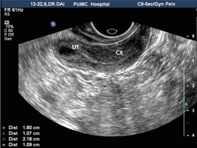
Fig. 2.2
Transvaginal ultrasound showing an infantile uterus. A 24-year-old patient presenting primary amenorrhea. All the uterine diameters are significantly smaller than normal with uterine body size: 1.8 × 1.5 × 1.1 cm, cervical length 2.2 cm, and body/cervix ratio <1
Uterus Didelphys
Both bilateral paramesonephric ducts develop normally but do not join together at midline, so each side has a set of fallopian tube and uterus.
Ultrasound Features
Uterus didelphys under ultrasonic examination would show two completely separate uteri, which are particularly clear on coronal observation. The two completely separate uterine bodies have a deep depression in between them. Both show uterine endometrial echoes (Fig. 2.3); they may be associated with double cervix; then the cervical diameter can be seen widened with two cervical echoes next to each other but completely separated (Fig. 2.4a, b).

Fig. 2.3
Abdominal ultrasound showing double uterine abnormalities. It shows both the left and right uteri, with uterine body completely separated and two separate endometrial echoes
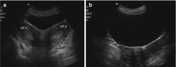
Fig. 2.4
Double uterine abnormalities (double uterus, double cervix): abdominal ultrasound showing cross-sectional images of two uteri and cervices. (a) Double uterus. (b) Double cervix
Bicornuate Uterus
The ends of the bilateral paramesonephric ducts joined, but not completely at the fundus of the uterus, resulting in a prominent horn on each side of the uterus.
Ultrasound Features
1.
2.
Rudimentary Horn Uterus
The paramesonephric duct on one side developed normally, but the middle and lower segments of the contralateral paramesonephric duct have stopped growing, thus forming different degrees of rudimentary horn. According to presence or absence of endometrial cavity, it can be divided into either rudimentary horn with or without endometrium. The latter is further divided into either connected or not connected type of rudimentary horn according to whether its endometrial cavity is connected to the contralateral uterine cavity or not.
The symptoms of rudimentary horn uterus pregnancy and interstitial tubal pregnancy are similar. They are asymptomatic in early pregnancy, but due to the poor muscular wall of the rudimentary horn, it cannot accommodate the growing fetus by becoming hypertrophied. The wall will break and rupture, usually from three to 4 months of pregnancy, thus causing severe bleeding and shock, threatening the life of the pregnant woman.
Ultrasound Features
1.
2.
Septate Uterus
After the fusion of both paramesonephric ducts, the intervening septum has not been absorbed; this divides the uterine cavity into two halves within a normal uterus; if the septum is only partly absorbed, it is an incomplete septate uterus.
Ultrasound Features
1.
2.
The two endometria of the incomplete septate uterus merge at the lower uterine segment at an acute angle <90° as a “Y”-shaped image (Fig. 2.8); for a complete septate uterus, the septum reaches the cervix, and the uterine endometria image present as a “V”-shaped image (Fig. 2.9a, b).

Fig. 2.8
Incomplete septate uterus: transvaginal 3D ultrasound showing endometrial echo as a “Y”-shaped image. The uterine cavity is separated in its upper half and shows a midline echo from its middle downward
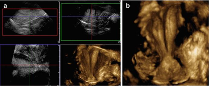
Fig. 2.9
Complete septate uterus: transvaginal 3D ultrasound showing endometrial echo as a “V”-shaped image; the uterine cavity with a hypoechoic middle septum. (a) 3D multiplanar images for 3D reconstruction 1,2,3. (b) Displaying a 3D image
Three-dimensional ultrasound scan can clearly show the septum in the uterine cavity of a septate uterus, its length, and its identification as a complete or incomplete septate uterus. Furthermore, it can also show the shape of the septum (Fig. 2.10a, b), its thickness at different levels of the uterus, etc. (Fig. 2.11a, b).

Fig. 2.10
3D transvaginal ultrasound of an incomplete septate uterus showing different shapes. (a) shows a wider septum. (b) shows a narrower and longer septum than the septum in (a)

Fig. 2.11
3D transvaginal ultrasound showing a different morphology of septate uterus. (a) shows a septum extending from the fundus to the cervix with gradually narrowing septum. (b) shows a wider upper 1/3 septum and narrower lower 2/3 septum
The clinical application of 3D ultrasonography gives a more accurate diagnosis of septate uterus and it is a direct visualization method. It also provides reliable information for the subsequent surgical treatment. The combination use of 3D ultrasound and saline hysterography (3D-HSG) further enhances the 3D ultrasound diagnosis. This will be easier to observe the morphology of the uterine septum and its relationship with the cervix and to determine the type of septate uterus. At the same time, it permits a timely diagnosis of any other associated uterine diseases such as endometrial polyps.
Arcuate Uterus
It is a minor uterine anomaly in which the intervening septum at the uterine fundus is not fully absorbed after fusion. It has a localized muscular hypertrophy in the central part of the fundus and slightly protrudes into the uterine cavity from the fundus.
Ultrasound features: The cross section showed muscular thickening at the central part of the uterine fundus, slightly protruding into the uterine cavity. This feature is shown more clearly on the coronal plane in the 3D image (Fig. 2.12). The endometrium at the uterine fundus is shown as a curved concave, the angle between the two endometria > 90°; the shape and contour of the uterus are normal.
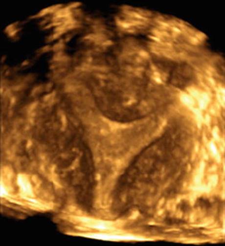
Fig. 2.12
Arcuate uterus: transvaginal 3D ultrasound image showing muscular thickening at the central part of the uterine fundus, slightly protruding into the uterine cavity, with a concave curved endometrium.
Other Ultrasound Features of Uterine Anomalies
1.
2.
3.
4.
5.
2.2.2 Ultrasound Features of Congenital Oblique Vaginal Septum Syndrome
Oblique vaginal septum syndrome is a congenital genital anomaly including uterus didelphys, double cervix, and a septum attaching to the vaginal wall extending obliquely from one side of the cervix; this often affects the patency of the ipsilateral cervix. An oblique vaginal septum is often associated with ipsilateral renal agenesis.
The clinical manifestations include painful menarche, lower abdominal pain or discomfort, excessive vaginal discharge with smell, or prolonged menstrual periods. In recent years, ultrasound examination has become the diagnostic method of choice, because of its advantages of being accurate, fast, real time, and noninvasive. Ultrasound examination shows not only the uterus and the cervix but also their number and shape and retention of blood in the vagina and the adnexa. It also gives an accurate diagnosis of renal agenesis.
Ultrasound Features
1.
2.
3.
4.
These ultrasound findings in patients with clinically dysmenorrhea and menstrual spotting can easily lead to a diagnosis of oblique vaginal septum syndrome.
2.2.2.1 The Differential Diagnoses of the Ultrasound Examination for Oblique Vaginal Septum
Oblique vaginal septum syndrome needs to be differentiated from imperforate hymen and ovarian cysts. ① Imperforate hymen may also manifest as cystic mass below the cervix, but the oblique vaginal septum syndrome mostly has uterus didelphys or septate uterus and can be associated with retention of fluid in one of the uterine cavities and renal agenesis on one side. Perineal ultrasound will help to diagnose the oblique vaginal septum. ② To differentiate it from an ovarian cyst, it is important to pay attention to the uterus. If there is a uterus didelphys anomaly, with a cystic mass below one of the uteri with sparse to dense dot-like echoes (hemorrhagic manifestation), together with renal agenesis on one side, it can easily differentiate it from an ovarian cyst.
2.2.3 The Role of 3D Ultrasound in the Diagnosis of Uterine Anomalies
2D ultrasound, particularly transvaginal 2D ultrasound, can provide clear images of the uterus, the cervix, the adnexal region, and part of the vagina. Its value in the diagnosis of the female reproductive tract anomalies is unquestionable, but because 2D ultrasound cannot display the coronal sections of the uterus, to a certain extent, its ability to diagnose uterine anomaly can be limited. 3D ultrasound imaging is a good complement to the 2D ultrasound.
3D ultrasound can produce coronal section images of the uterus, showing the shape and contour of the entire uterus, uterine endometrial echo, and the shape of the uterine cavity. The high-resolution coronal imaging is easily reproducible and can display all the anatomical relationships from the uterine fundus, the uterine horns, and the cervix. It can provide accurate classification and differential diagnosis of the uterine anomalies. As reported in national and international literatures, 3D ultrasound diagnosis of uterine anomalies has a high diagnostic sensitivity and specificity of 92 and 100 %, respectively. Hence it can provide better information for the diagnosis and the plan for surgery. In particular for septate uterus, bicornuate uterus, and arcuate uterus, all of them are uterine anomalies that cannot be identified easily by 2D ultrasound. 3D ultrasound therefore has a higher ability for the diagnosis and differential diagnosis of reproductive tract anomalies. It is at present one of the best imaging methods for the diagnosis of uterine anomalies and is recommended for wider application.
2.2.4 Other Imaging Investigations
2.2.4.1 CT Examinations
CT scans have high spatial resolution and can therefore provide clear, constant anatomical images showing the cervix and the uterine body. In CT scanning, the normal uterus shows a higher density soft tissue images, which is of either circular, triangular, or spindle in shape, with a small low-density uterine cavity.
But CT scans can only provide cross-sectional imaging of the uterus, and the resolution on the soft tissue is low. It is therefore not ideal for showing the shapes of the uterus and has limited role in the diagnosis of some uterine anomalies; in addition, CT scans are not as simple as ultrasound examination; its higher ionizing radiation will be a hazard for women of childbearing age. For these reasons, CT scans are less often used for the diagnosis of uterine anomalies.
2.2.4.2 Magnetic Resonance Imaging (MRI)
Magnetic resonance imaging (MRI) has the advantages of high resolution on tissues, good contrast for soft tissues, more image parameters, non-ionizing radiation, etc. MRI can accurately distinguish between endometrial and myometrial signals and can have multi-directional scanning at any level for imaging. Its three-dimensional images can show the shape of the uterus and directly show the signals of septum and some other complications such as endometriosis, retrograde menstruation, and so on. As reported in the literatures, the sensitivity and specificity of MRI diagnosis of bicornuate uterus and septate uterus were 100 %. Therefore, MRI is the best imaging method in the diagnosis of uterine anomalies. It is the next important complementary method after 3D ultrasound examination. However, because of its high costs, it is rarely a routine application or the first-choice method.
MRI Manifestation of Uterine Anomalies
Rudimentary horn: It is a typical MRI image showing a banana-shaped body next to a developed uterus. The endometrial cavity of the uterus is reduced in size with reduced thickness. The best display image is at axial T2WI.
Uterus didelphys: A typical feature is double uterine cavities and double cervix. Each uterus has perfect anatomical signals.
Bicornuate uterus: A sagging uterine fundus with separated uterine horns. The best plane to show the contour at the uterine fundus is the coronal axial T1WI at the middle of the uterine body, while the best image to show the separate tissue signals is at axial T2WI.
Septate uterus: The center of the uterus shows some low-echo signals for the intervening fibrous tissue.
Arcuate uterus: At the central part of uterine fundus, there is thickened myometrium slightly protruding into the uterine cavity.
2.3 MRI Diagnosis of Female Genital Anomalies
Jingjing Lu6
(6)
Department of Radiology, Peking Union Medical College Hospital, No. 1 Shuaifuyuan, Beiijing, 100730, P. R. China
The incidence of female reproductive tract anomalies in the general population is about 7 %. For patients with recurrent miscarriages, it can reach up to 13–25 % [1]. Accurate preoperative diagnosis reaching a correct anatomical and morphological classification is important for the proper planning of surgery. It can effectively improve the management and avoid unnecessary invasive investigations. Appropriate imaging methods can directly and accurately demonstrate the reproductive tract anomalies. They have irreplaceable diagnostic value for the type of genital anomalies.
At present, the common imaging methods include hysterosalpingography (HSG), ultrasonography (US), and magnetic resonance imaging (MRI). HSG only shows the morphology of uterine cavity; the operation is relatively cumbersome and has low patient’s compliance. Therefore its clinical application is very restricted. Ultrasonography is simple and low cost and has no radiation risk. It is the most popular imaging method currently used to diagnose female genital anomalies. However, it is highly operator dependent, and the image is not intuitive. In some anomalies such as rudimentary horn, the imaging display has great limitations.
With the increasing popularity of equipments and related technologies, magnetic resonance imaging has unique advantages in the female reproductive tract imaging. MRI has the advantages of high soft tissue resolution, multiplanar imaging, no radiation, and objective display of the reproductive tract anomalies. It has become the best imaging method for detecting genital anomalies. This chapter describes the clinical applications of MRI in the female genital anomalies.
2.3.1 The Recommended Imaging Criteria for MRI of the Female Reproductive System Anomalies
MRI uses the principle that different tissues possess relatively fixed and different relaxation times as T1 and T2, respectively, and they can be reflected in the MRI as the different intensities and signals in a black and white gray scale.
Before pelvic MRI scans, the general preparations include: The bladder must be at least half full to separate the uterus from the abdominal muscles, to push away the small intestines in the pelvic cavity so as to reduce peristaltic artifacts and optimize the image contrast. Before scanning, any metallic objects are to be removed from the body, including intrauterine devices. The lower abdomen should best be fixed by an abdominal belt to immobilize it. Whenever appropriate, a gel-like substance (such as the ultrasound coupling gel) should be instilled into patient’s vagina. After the vagina is filled with gel, it will be much easier to get the image and observe the structures.
MRI examination of the female reproductive tract generally includes T1-weighted images (T1WI) and T2-weighted images (T2WI) collected from transverse, sagittal, and coronal planes. T2-weighted imaging is the main sequence of the female reproductive tract. T2-weighted images clearly demonstrate the shape of the uterus and its two adnexa and any other lesions when coupled with fat-suppression technology. Sagittal images show particularly ideal uterine diseases, and transverse and coronal images show more clearly the ovaries. T1-weighted images help to show the components of any lesion, such as bleeding, adipose tissue, and so on. T2W1 can distinguish the endometrium, junctional zone, and myometrium. These images can clearly show the different structures of uterine cavity, contour of the uterine fundus, various vaginal anomalies, and other pelvic lesions. Although T1W1 may not identify junctional zone in the uterus, but any retained blood within the uterine cavity and other pelvic complications which will be displayed by TIWI may provide useful diagnostic information.
The current recommendation of scanning sequence is shown in Table 2.1 [2, 3]. During the MRI scanning of female reproductive tract anomalies, the main MRI scanning position should be taken in accordance to the axis of the uterus, which is parallel to the long axis of the uterus and as well perpendicular to the long axis as short axis of the uterus. Sagittal views of the long axis of uterus, cervix, and vagina can show a better continuity display and allow an easy assessment of the ratio of the uterine body and cervix, as well as any obstructive vaginal anomaly. The T2WI images of the long axis of uterus are most valuable to make a diagnosis and classification of genital anomalies. The routine clinical application of sagittal T2W1 images shows obstructive vaginal anomalies. Large field-of-view coronal images have the advantages to show any associated kidney anomalies. About 29 % patients with Müllerian duct anomalies are associated with kidney anomalies, such as kidney agenesis, kidney hypoplasia, ectopic kidney, horseshoe kidney, and double renal pelvis or calyces [4]. Recently, the development of 3D T2-weighted sequences can capture thin slices T2 images, and after post-processing, it can display three-dimensional pictures of anomalies. This new 3D MRI techniques have been used by some medical institutions to replace the traditional two-dimensional MRI imaging [2].
Table 2.1
MRI sequences of female genital anomalies
|
Sequence |
SSFSE |
2D T2WI FRFSE (fat saturated) |
2D T2WI FRFSE |
3D T2WI FRFSE |
TWI FSE (fat saturated) |
|
Imaging plane |
Coronal |
Axial, sagittal |
Oblique coronal |
Axial |
Axial |
|
TR (ms) |
1000–2000 |
2000–4000 |
2000–4000 |
2000–4000 |
600–700 |
|
TE (ms) |
90 |
90 |
90 |
100 |
30 |
|
Echo train length |
not applicable |
16–20 |
16–20 |
126 |
4 |
|
Total imaging matrix |
288 × 192 |
320 × 256 |
320 × 256 |
320 × 256 × 128 |
256 × 92 |
|
Field of FOV (cm) |
40 |
26 |
26 |
26 |
26 |
|
Section thickness (mm) |
6 |
5 |
5 |
1.4 |
5 |
|
Section space (mm) |
0 |
1.5 |
1.5 |
0 |
1.5 |
|
Rationale |
Give an overview display and detect the presence or absence of kidney |
Be parallel to the long axis of the uterus to show the fundal contour |
To obtain multiplanar reconstruction and curved planar reconstruction |
To detect hemorrhage within the uterus or fallopian tube |
In conclusion, MRI has high soft tissue resolution and multiplanar imaging capacity, and it produces no radiation. It is the best imaging method for reproductive tract anomalies after ultrasound imaging. Yet it is expensive, and it also has limitations to distinguish vaginal abnormalities from postoperative vaginal scar tissues.
2.3.2 Classification of Female Genital Anomalies and Their MRI Features
Müllerian ducts are the predecessors of the female genital system, which later develops into the uterus, cervix, and upper vagina. The embryo in its embryonic stage has a pair of longitudinal grooves in its posterior wall. This groove later transforms into a tube on each side as the Müllerian duct. Bilateral Müllerian ducts grow inward and downward and then fuse medially in the middle forming the uterus. If the proximal ends of the Müllerian ducts do not fuse properly or develop abnormally after fusion, these will result in female reproductive tract anomalies (or Müllerian duct anomalies).
2.3.2.1 Uterine Agenesis or Hypoplasia
Abnormal development of Müllerian ducts can lead to genital anomalies of the upper 2/3 of the vagina, the cervix, and the uterus, leading to their agenesis or hypoplasia. One type of this malformation is Mayer-Rokitansky-Kuster-Hauser syndrome (MRKH syndrome) (Fig. 2.14), which also represents the extreme form of Müllerian duct anomalies involving the proximal vagina, cervix, and uterus.

Fig. 2.14
Congenital absence of the uterus and vagina syndrome (Mayer-Rokitansky-Kuster-Hauser syndrome, MRKH syndrome). The left is fat-saturated sagittal T2-weighted image showed the complete absence of the cervix and uterus. Between the rectum (r) and bladder (b), normal vagina is invisible. The right is an axial T2-weighted image showing bilateral normal ovaries (black arrows)
For this type of genital anomalies, the sagittal T2-weighted images can be most intuitive and clear. Through the bladder and urethra in front and rectum behind, one can infer the location of the uterus, cervix, and vagina. In patients with complete agenesis of the uterus, the MRI images will show the absence of the uterus. A hypoplastic uterus will be displayed as a soft tissue mass in the pelvis, signal of which is consistent with a normal myometrium (the signal on T2-weighted images is slightly higher).
2.3.2.2 Unicornuate Uterus and Rudimentary Horn
Unicornuate uterus originates from one side of the Müllerian duct which is fully developed, while the other side is completely or nearly completely undeveloped. Approximately 40 % of these patients with unicornuate uterus have ipsilateral kidney anomaly. The most common anomaly is agenesis of the ipsilateral kidney (67 %) [4]. There are four subtypes of anomalies: (1) no contralateral rudimentary uterine horn, (2) rudimentary uterine horn with no endometrium or cavity, (3) rudimentary uterine horn with interconnected uterine cavities, and (4) rudimentary horn with no interconnected uterine cavities.
The MRI image of rudimentary horn will show a single “banana-like”-shaped horn on one side of the pelvis. The muscular wall signals and anatomy are normal. The ratio of endometrium to myometrium thickness is also normal. The clinical presentation of the rudimentary uterine horn varies with its subtype. If there is no endometrium, the entire rudimentary horn appears as diffuse low MRI signals. If endometrium exists in its cavity, the junctional zone of the rudimentary horn often presents well. If there is no communication between its endometrial cavity and the uterine cavity of the contralateral uterus, the rudimentary horn cavity will be gradually filled and distended with blood products after menarche (Fig. 2.15). The patient will suffer from abdominal pain and pelvic endometriosis will develop in some patients because of retrograde blood flow into the peritoneal cavity. From the MRI images, it is important to distinguish the endometrial lining of the rudimentary horn, because the presence of functional intrauterine endometrium in this rudimentary horn is a high-risk factor for miscarriage, ectopic pregnancy, preterm delivery, and even uterine rupture [5].

Fig. 2.15
(a) is a T2-weighted oblique coronary image and (b) is a T1-weighted axial image of the same person, in which a right unicornuate uterus and a left rudimentary uterus clearly appeared. The unicornuate uterus on the right is fully developed, and the distinct signal represents the three layers of the uterine body that can be easily detected; the rudimentary uterine on the left does not connect with the contralateral; hemorrhage within the cavity has a hyperintense signal upon T1 (white arrow)
2.3.2.3 Uterus Didelphys
Uterus didelphys arises when the uterine body and the cervical parts of both Müllerian ducts do not merge together. This results in double uterine bodies, double cervix, double vaginas, and/or single vagina. The MRI images will show two separate uterine bodies separated with a large angle toward both sides of the pelvis, associated with two cervices. The ratio of endometrium to myometrium and the junctional zone are normal. At the lower part of the uterus, there is an obvious cleft on the external contour (Fig. 2.16) probably suggestive of incomplete fusion (such as in a double uterus or bicornuate uterus), rather than from incomplete resorption after fusion (such as septate uterus). This is an important distinguishing feature of these two types of uterine anomalies.
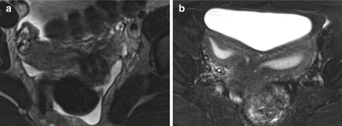
Fig. 2.16
(a) is an axial T2-weighted image showing a typical uterus didelphys, two widely splayed uterine horns with bilateral, non-fused, fully developed cervices, which is critical for distinction from bicornuate uterus. (b) is a uterus didelphys which has two relatively separate cavities
Three-quarters of these patients have repetition of the proximal end of the vagina. If there is an oblique vaginal septum on one side, it can cause blood-filled uterine cavity on the same side of the septum. Blood products often show high MRI signal on T1-weighted images. Congenital oblique vaginal septum syndrome (i.e., the Herlyn-Werner-Wunderlich syndrome, HWW syndrome) is a rare congenital disorder of the genitourinary system, involving the Müllerian and Wolffian ducts. The clinical triad includes (1) double uterus, (2) vagina hemi-obstruction, and (3) ipsilateral renal agenesis (Fig. 2.17). MRI is a noninvasive imaging method which is very sensitive to demonstrate this syndrome as well as show its anatomical anomalies and concurrent pathological changes [6].

Fig. 2.17
Schematic oblique vaginal septum syndrome and MRI images. (a) A schematic view showing a double uterus with unilateral renal agenesis. As the cavity of the uterus and cavity of oblique vaginal septum communicate through the cervix, outflow obstruction of the uterine cavity due to the oblique septum will cause blood retention behind the septum and the uterine cavity. The contralateral uterus, cervix, and kidney appear normal. (b) An axial T1-weighted MRI image showing hemorrhage is visible in the right uterine cavity and in the vagina above the oblique vaginal septum. (c) A coronal T2-weighted image showing the right kidney is absent
2.3.2.4 Bicornuate Uterus
Bicornuate uterus is also due to incomplete fusion of the bilateral Müllerian ducts. It accounts for 10 % of all Müllerian duct anomalies. The MRI image of bicornuate uterus also shows a characteristic cleft over the external contour at the lower part of the uterus.
Normal double horns can be seen on the MRI image combined with normal anatomical structure (Fig. 2.18). Whether the mid-lower horn or uterine body is fused or not is the pinpoint in differentiation between uterus didelphys and bicornuate uterus. Between two symmetrical intrauterine cavities, there is a visible soft tissue separating the horns, which is also the difference between bicornuate uterus and a double uterus. While it’s relatively difficult to accurately distinguish a bicornuate bicollis uterus from a uterus didelphys upon MRI, fortunately, the treatment strategy is not too much different between this two anomalies, in view of a surgical intervention that is not usually indicated for patients with fusion anomalies, but with reabsorption anomalies like septate uterus.

Fig. 2.18
(a, b) are two cases of bicornuate uterus, characterized by the presence of a fundal clef more than 1 cm (indicated by the white line ab and black line cd). The outer contour of the uterine is roughly outlined by the curved line. Two separate uterine horns with fusion of the mid-lower uterine body (white arrow) are distinctly manifested. The accompanying round-like leiomyoma (*) upon the left horn in (b) can be easily identified
2.3.2.5 Septate Uterus
Septate uterus is the most common, accounting for 55 % of the duct anomalies [7]. It is important to distinguish between it and the bicornuate uterus. A midline longitudinal septum arises from the fundus of the uterus, which is due to incomplete resorption of the fused septum in the uterus.
On MRI image, the uterus is of normal size. The external contour of the fundus of the uterus is similar to that of a normal uterus (Fig. 2.19); this is the most important point to distinguish a septate uterus from a bicornuate uterus. The features in ultrasound examination can be used as a standard reference (Fig. 2.20). The longitudinal septum may be incomplete or complete septum. A complete longitudinal uterine septum extends from the fundus to the external os of the cervix. In 25 % of patients, it can extend to the vagina. Uterine septum can have different lengths and tissue components, with various proportions of fibrous tissues and muscle tissues.

Fig. 2.19
A complete septate uterus: the external contour of the fundus is normal. The septum extends from the uterine fundus to the external cervical os

Fig. 2.20
Classification criteria for US differentiation of septate from bicornuate uteri. (a) When apex (3) of the fundal external contour occurs below a straight line between the tubal ostia (1, 2) or (b) 5 mm (arrow) above it, the uterus is bicornuate. (c) When apex is more than 5 mm (arrow) above the line, uterus is septate
2.3.2.6 Arcuate Uterus
Arcuate uterus occurs when the septa of the uterus and vagina have almost completely absorbed. In the MRI image, the fundus of the uterus shows slightly convex internal contour toward the uterine cavity. Arcuate uterus is a mild Müllerian duct anomaly. It usually does not cause problems in pregnancy.
On a MRI image, the uterine size is normal. The uterus has a wider uterine cavity, with a saddle-shaped bulge (Fig. 2.21), protruding into the endometrial cavity. The MRI signals of the protruding tissue are similar to the uterine myometrium. The contour of the fundus is also normal.

Fig. 2.21
The T2-weighted image of an arcuate uterus with a slightly convexed fundus. The broad-based myometrium bulge toward the endometrial cavity (white arrow)
The above anomalies are classified according to the genital anomaly classification of the American Fertility Society's definition in 1988. The typical MRI anomalies are briefly described. However, there are limitations of this classification, because there are no definition and classification of vaginal anomalies. Some complex anomalies have also been defined across different anomaly groups with the classification. As such, we cannot have an absolute pathological changes defined into any classification. It is then acceptable to have a clear and comprehensive description of the anomalies. MRI imaging for female reproductive tract anomalies is intuitive and reliable. Following our advancing MRI technology and our increased awareness of anomalies, MRI will play an increasingly important role in the diagnosis, preoperative planning, and postoperative follow-up of the female reproductive system anomalies.
References
1.
Chan YY, et al. The prevalence of congenital uterine anomalies in unselected and high-risk populations: a systematic review. Hum Reprod Update. 2011;17(6):761–71.PubMedCentralCrossRefPubMed
2.
Behr SC, Courtier JL, Qayyum A. Imaging of Mullerian duct anomalies. Radiographics. 2012;32(6):E233–50.CrossRefPubMed
3.
Robbins JB, et al. MRI of pregnancy-related issues: Mullerian duct anomalies. AJR Am J Roentgenol. 2012;198(2):302–10.CrossRefPubMed
4.
Li S, et al. Association of renal agenesis and Mullerian duct anomalies. J Comput Assist Tomogr. 2000;24(6):829–34.CrossRefPubMed
5.
Jayasinghe Y, et al. The presentation and early diagnosis of the rudimentary uterine horn. Obstet Gynecol. 2005;105(6):1456–67.CrossRefPubMed
6.
Sen KK, Balasubramaniam D, Kanagaraj V. Magnetic resonance imaging in obstructive Mullerian anomalies. J Hum Reprod Sci. 2013;6(2):162–4.PubMedCentralCrossRefPubMed
7.
Homer HA, Li TC, Cooke ID. The septate uterus: a review of management and reproductive outcome. Fertil Steril. 2000;73(1):1–14.CrossRefPubMed
Suggested Reading
Dykes TM, Siegel C, Dodson W. Imaging of congenital uterine anomalies: review and self-assessment module. AJR Am J Roentgenol. 2007;189:S1–10.CrossRefPubMed
Grimbizis GF, Gordts S, Di Spiezio Sardo A, et al. The ESHRE/ESGE consensus on the classification of female genital tract congenital anomalies. Hum Reprod. 2013;28(8):2032–44.PubMedCentralCrossRefPubMed
Jinghe Lang. Adolescent gynecology. Beijing: People’s Military Medical Press; 2011. p. 19–44.
Qinsheng Ge. Practical female reproductive endocrinology. Beijing: Scientific and Technological Literature Publishing House; 2001. p. 275–319.
Troiano RN, McCarthy SM. Müllerian duct anomalies: imaging and clinical issues1. Radiology. 2004;233:19–34.CrossRefPubMed