
Fang Jiang1, Qinjie Tian1 , Shu Wang1 and Lan Zhu1
(1)
Department of Obstetrics and Gynecology, Peking Union Medical College Hospital, No. 1 Shuaifuyuan, Beijing, 100730, P. R. China
Qinjie Tian
Email: qinjietn@sohu.com
Lan Zhu (Corresponding author)
Email: zhu_julie@sina.com
3.1 Abnormal Hymen
Fang Jiang2 and Lan Zhu2
(2)
Department of Obstetrics and Gynecology, Peking Union Medical College Hospital, No.1 Shuaifuyuan, Beijing, 100730, P. R. China
Lan Zhu
Email: zhu_julie@sina.com
The hymen is a layer of mucosal membrane located at the vaginal opening, with squamous epithelium covering its outer and inner surfaces. In between these surfaces, it contains connective tissue, blood vessels, and nerve endings. During its development, abnormal cavity formation at the urogenital sinus will lead to various anomalies which include imperforate hymen, microperforate hymen, septate hymen, and cribriform hymen (Fig. 3.1)
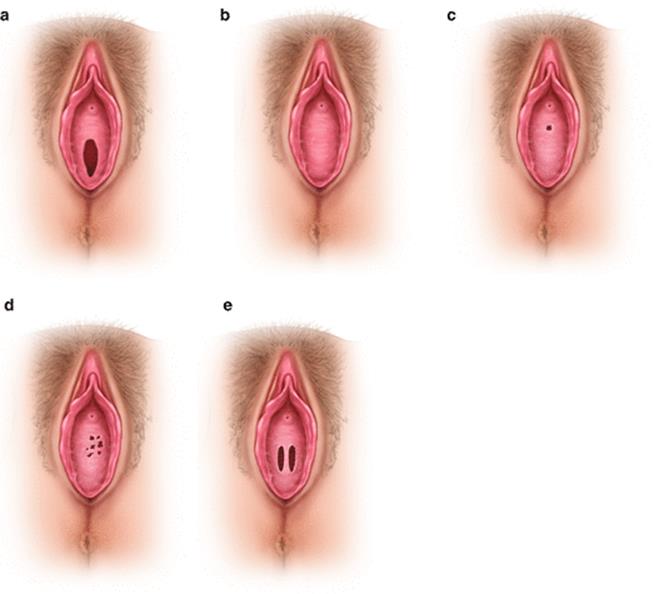
Fig. 3.1
Congenital anomalies of the hymen. (a) Normal hymen; (b) incomplete perforated hymen; (c) microperforate hymen; (d) cribriform hymen; (e) septate hymen
3.1.1 Diagnostic Criteria
3.1.1.1 Imperforate Hymen
Imperforate hymen is also known as nonporous hymen. The incidence of imperforate hymen is about 0.015 %. Imperforate hymen mainly obstructs the discharge of vaginal secretions. It may be asymptomatic at childhood due to minimal vaginal secretions, but at puberty, as both the vaginal and cervical secretions gradually increase, they accumulate in the vagina and lead to a sense of heaviness in the lower abdomen. After menarche, as the menstrual blood cannot drain out, it accumulates in the vagina forming a vaginal hematoma after several menstruations. Subsequently, it may lead to uterine and tubal hematomas, and eventually retrograde flow will enter into the pelvic cavity forming pelvic hematomas. Clinical symptoms are obvious with a cyclical lower abdominal pain, which progressively increases in intensity.
On gynecological examination, a bulging hymen can be seen, with a purple blue surface; on rectal examination, a vaginal mass bulging into the rectum is felt as a palpable pelvic mass. With a finger pressing on the vaginal mass, the bulging hymen can be made more obvious. Ultrasound scan may show accumulated fluid in the vagina and even in the uterine cavity.
3.1.1.2 Microperforate Hymen
It is a rare anomaly. There is a film covering the vaginal opening with just a pin hole opening as first described by Capraro in 1968 [1]. Since the pin hole is often difficult to detect, it is often misdiagnosed as an imperforate hymen. According to the sizes of the pin hole, the presenting symptoms will be different. Some patients have periodic menstruations with moderate amount of menstrual blood outflow, but a lot of blood still accumulates in the vagina; sometimes the periods are irregular; some patients presented with delayed menarche, with their chief complaints that include periodic abdominal pain and painful pelvic mass due to hematoma formation [2]. Microperforate or imperforate hymen can occur as an isolated anomaly, but it can also be associated with other reproductive tract anomalies, such as bicornuate uterus, vestibular anomalies, and anal atresia [3].
Similar to imperforate hymen, microperforate hymen can cause reflux of vaginal secretions and blood into the peritoneal cavity forming a pelvic mass. Unlike imperforate hymen, it will also cause recurrent urinary tract infections and sometimes a pelvic abscess. It is because the pinhole opening can have communication to the outside, through which bacteria will enter and multiply in the accumulated effusion or blood in the vagina or the pelvis leading a pelvic abscess. Bacteria can also migrate out of the vaginal opening, enter into the urethra, and cause recurrent urinary tract infections. On the contrary, patients with imperforate hymen do not have these presentations because their vaginas do not communicate with the outside [4]. The microperforate hymen is mainly diagnosed by examination under general anesthesia, and nowadays, fiber-optic hysteroscopy can be useful in its diagnosis together with simultaneous examination of any anomalies in the vagina and cervix [5].
3.1.2 Indication and Timing of Surgery
Surgery may be performed at any age. The ideal times are at the postneonatal period, puberty, or before menarche. As the development of hematocolpos can cause blood accumulation in the vagina, uterus, and fallopian tubes, followed by secondary endometriosis or pelvic infection, therefore, once hymen anomalies is diagnosed, surgery should be performed as soon as possible.
3.1.3 Surgical Contraindications
Vaginal atresia or the congenital absence of a vagina and other congenital anomalies should be ruled out before performing the surgery to incise the hymen. Surgery must be after a proper diagnosis of other anomalies (Fig. 3.2).
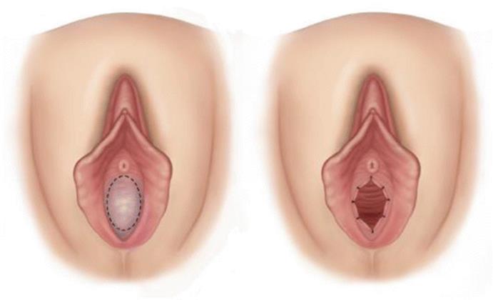
Fig. 3.2
Surgical incision of hymen
3.1.4 Preparative Preparation
Preoperative preparation is similar to other vulval surgery with routine cleansing and antiseptic preparation of the vulva.
3.1.5 Anesthesia and Positioning
The patient is placed in a lithotomy position. Local infiltration with anesthetic or intravenous anesthesia or general anesthesia can be used.
3.1.6 Surgical Procedure
Many literatures recommend the same surgical treatment for both microperforate and imperforate hymen. This includes surgical incision and removal of the accumulated blood in the vagina. After the surgery, patients will have significantly improved pregnancy rate and quality of life.
3.1.6.1 Incision
A metal urinary catheter should be used if possible to guide its position to avoid bladder injury. The surgeon should wear double gloves and insert the index finger of the left hand into the anus pressing towards the vagina for guidance, so as to avoid injury to the rectum. The incision should be at the most prominent part of the bulging hymen, though it will depend on the personal choice. The incision can be a “cross” incision, a vertical incision or a puncture incision at the center. Some surgeons think that it should begin with a small incision (especially for a thickened hymen), so as to reduce the rate of blood flow and hence to prevent vasovagal reaction due to the sudden decompression of the vaginal hematoma. Then, the outer glove of the left hand is removed to examine if the vaginal opening can accommodate one finger. Attention should be paid to avoid damaging the urethra and rectum during surgery. Finally, the vagina and cervix should be examined for other associated anomalies.
3.1.6.2 Evacuation of Hematoma
After opening the imperforate hymen, the retained menstrual blood will drain off. Any blood should be mopped up with gauze, and the cervix can now be visualized and examined. In the presence of a cervical adhesion or stenosis, a small uterine dilator is used to discharge any intrauterine blood retention. Tubal blood collection will gradually drain out following the operation. The abdomen should not be pressed or kneaded to avoid rupture of a hematoma or to force more retained blood flowing into the peritoneal cavity.
3.1.6.3 Suturing Edges of the Incisions
Any redundant hymen tissue should be cut off from the incision wounds. Absorbable sutures are used to suture the edges, while a very thin hymen wound without bleeding may not require suturing. In the literatures, absorbable sutures No. 3 “0” or No. 4 “0” are recommended to suture the wound edges or to cauterize wound edges with diathermy, so as to avoid wound adhesion with subsequent stenosis.
3.1.7 Postoperative Management
1.
2.
3.
4.
3.2 Virilization of the External Genitalia
Qinjie Tian3
(3)
Department of Obstetrics and Gynecology, Peking Union Medical College Hospital, No. 1 Shuaifuyuan, Beijing, 100730, P. R. China
Qinjie Tian
Email: qinjietn@sohu.com
3.2.1 Perineal Incision and Repair
3.2.1.1 Diagnostic Criteria
Female patients with a normal vagina but with a high perineal body or fused labia minora would have their vaginal opening completely or partially obliterated, resulting in poor menstrual blood flow or poor sex life. This vulval anomaly is mainly due to abnormal androgenic influence. Clinically it can be manifested as ambiguous external genitalia, labial fusion, and common vaginal and urethral opening. Prader classified external genital anomalies into the five types according to the varying degrees of virilization of the external genitalia: As illustrated by the following photos (Fig. 3.3), these types are described as:
· Type I: Larger clitoris, with normal vagina and urethral openings.
· Type II: Larger clitoris, with a funnel-shaped vaginal opening, but the openings for the vagina and urethra are still separated.
· Type III: Significantly enlarged clitoris, both the vagina and urethra open from a common urogenital sinus.
· Type IV: Significantly enlarged clitoris like a penis, the base of the penis is at the urogenital sinus, similar to hypospadias with fusion of the reproductive uplifted part.
· Type V: Clitoris looks like a penis with the urethral opening at the penis tip. There is a complete fusion of the reproductive uplifted part; it is often mistaken as a male penis with cryptorchidism and hypospadias.
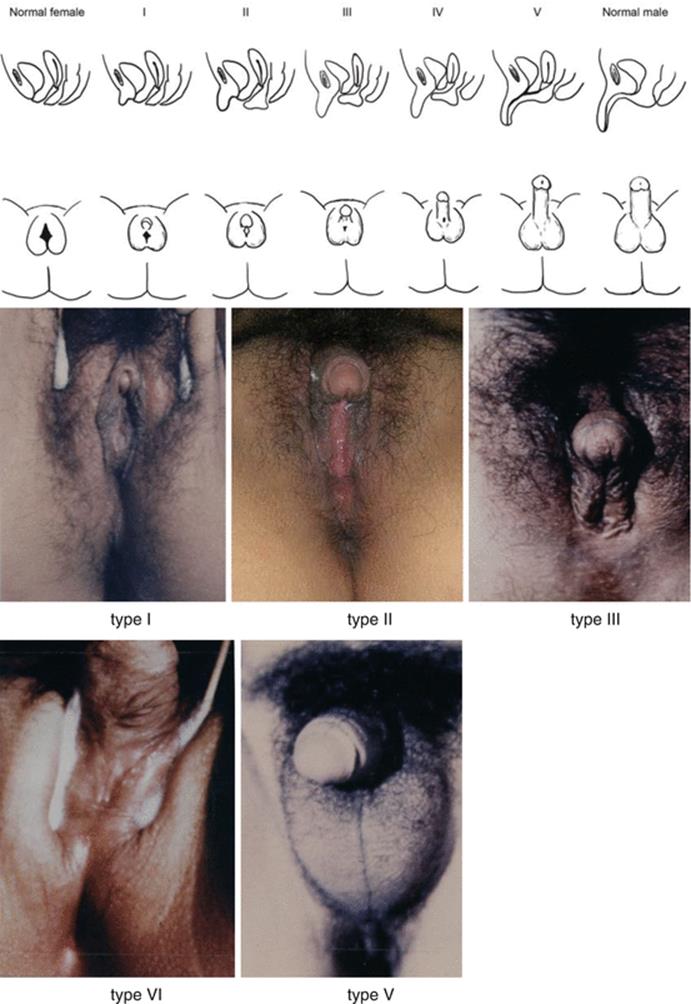
Fig. 3.3
Appearances of the external genitalia of normal female, 5 types of various degrees of virilisation and normal male
3.2.1.2 Operative Indications
Diagnosis can be easily made and accurately confirmed by examination. They are patients with a normal vagina and high vulval body or fusion of labia minora.
3.2.1.3 Timing of Surgery
1.
2.
3.
3.2.1.4 Preoperative Preparation
Routine vulval skin cleaning and antiseptic preparation are performed.
3.2.1.5 Anesthesia
Local anesthesia is used for the labial incision and repair. If there is also a simultaneous corrective surgery for the clitoris or for vaginoplasty, general anesthesia or epidural anesthesia is recommended for these procedures.
3.2.1.6 Position
Patient is placed in a lithotomy position same as other vaginal surgery.
3.2.2 Surgical Procedures
1.
2.
3.
3.2.2.1 Postoperative Management
1.
2.
3.2.2.2 Key Points of Surgical Techniques
1.
2.
3.2.3 Surgical Excision of Long Clitoris and Repair
The clitoris is located between the top ends of the labia minora, and at the meeting point of the upper ends of the labia majora. It is a small cylindrical organ about 1.5–3.5 cm long, surrounded by foreskin. The swollen tip is called the glans clitoris. Under the skin, there is a bunch of erectile tissue. The erectile tissue is composed of cavernous tissue (there are two erectile clitoris cavernosa, similar to a male penis, and it is divided into head, body, and feet), with rich venous plexus and nerve endings. Therefore, it is very sensitive and bleeds easily after injury.
The size of the clitoris head is about 3–4 mm wide and 4–5 mm long, smaller than the eraser head of a pencil. There may be ethnic differences. The clitoris is a sensitive organ which plays an important role in achieving and maintaining sexual satisfaction and orgasm. The simple clitoral excision in the past has been abandoned and changed to clitoral plastic surgery which would keep the blood vessels and nerves.
3.2.3.1 Diagnostic Criteria
The cause of an enlarged clitoris must be accurately diagnosed before surgery. In many women, the common causes for an enlarged clitoris include:
1.
2.
3.
3.2.3.2 Surgical Indication
1.
2.
3.2.3.3 Timing of Surgery
1.
2.
3.2.3.4 Preoperative Preparation
The cleaning and antiseptic preparation of the vulva before operation is performed to avoid infection.
3.2.3.5 Anesthesia
Patient will require either general anesthesia or epidural anesthesia.
3.2.3.6 Position
Patient should be positioned in a lithotomy position as in other vaginal surgery.
3.2.3.7 Surgical Procedures
1.
2.
3.
4.
5.
6.
7.
8.
9.
10.
3.2.3.8 Postoperative Management
1.
2.
3.
4.
5.
6.
3.2.3.9 Key Points of Surgical Techniques
1.
2.
3.
4.
3.3 Labial Fusion/Adhesion
Shu Wang4 and Lan Zhu4
(4)
Department of Obstetrics and Gynecology, Peking Union Medical College Hospital, No. 1 Shuaifuyuan, Beijing, 100730, P. R. China
3.3.1 Diagnosis
3.3.1.1 Symptoms
Patients with labial fusion will have normal menstruation at puberty, but menstrual blood and urine will be noticed coming out from the same opening which might be mistaken as “cyclical hematuria”; patients also complain of failure to have sexual intercourse as the penis cannot penetrate into the vagina.
In addition, due to different extents of fusion of the labia minora, the degree of covering to the vaginal opening can be different. If it is a narrow opening close to the pubic symphysis, the outflow of urine and blood is likely to be sluggish. Sometimes even before puberty, pelvic mass will form as a result of urine reflux to the vagina or uterus cavity. Worse still, the obstruction by the distended vagina and uterus can lead to hydroureters and hydronephrosis. After menarche, it may be associated with retention of blood in the vagina or uterine cavity [6–8] leading to obstruction of the lower urinary tract and recurrent urinary tract infections.
3.3.1.2 Examination
At the perineum, there are two small openings. An anterior hole-like opening is located at the normal urethral opening position; the posterior opening is the anal opening. They are in their normal positions. With a metal urinary probe, the anterior opening can be probed into and lead to some urine outflow, thus confirming it as the urethral opening. The metal probe can then be withdrawn just behind the anterior small opening and move downwards in a perpendicular direction towards the anus; it can be felt probing into the vagina. Between the anterior urethral and anal openings, there is a layer of normal perineal skin covering the hidden vaginal opening. Gynecological examination can reveal a normal uterus, normal cervix, and bilateral adnexal masses, though it is less likely to be associated with other Müllerian abnormalities. Almost all patients can be diagnosed by the above physical examination, and if necessary, perineal ultrasound and cystoscopy can assist confirming the diagnosis.
It is important to exclude other differential diagnosis such as MRKH syndrome, vaginal atresia, labial adhesion after infection, or radiotherapy. The first two disorders show normal vulvar vestibule and normal appearance of the labia majora and minora. Patients do not have normal menstrual flow, with (in vaginal atresia)/or without (in MRKH syndrome) periodic abdominal pain. The treatment of these vaginal anomalies is different from that of the labial fusion. Labial adhesion generally has a history of chronic inflammation of the vulva, vulval dysplasia, or a history of radiotherapy. Attention should be taken to have detailed history and physical examination.
3.3.2 Indication
If the condition is diagnosed, early surgical treatment is necessary.
3.3.3 Timing of Surgery
During puberty, patients with labial fusion usually present with abnormal menstrual blood flows or may also seek treatment because of difficulty at intercourse. Vulval surgery can expose the opening of the vagina, through which the menstrual blood can flow out normally after the surgery.
3.3.4 Contraindication
Before any surgery, it is important to identify the openings and positions of urethra, vagina, and rectum. If it confirms simple labia minora fusion, the following surgery can be performed. Otherwise, there can be risk of the urinary tract injury.
3.3.5 Preoperative Preparation
Preoperative pelvic ultrasound should be performed to define whether kidneys, ureters, and internal genital organs are normal, so as to exclude other congenital genital anomalies. If necessary, karyotyping and sex hormone and sex-related hormone or enzyme tests, such as 21-hydroxylase and cortisol levels, should be performed in order to confirm whether this vulval anomaly is due to clinical manifestations of other systemic endocrine disorders.
3.3.6 Surgical Procedure
Figures 3.6, 3.7, 3.8, 3.9, 3.10, and 3.11.
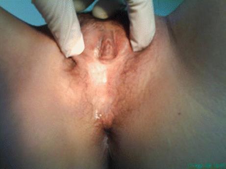
Fig. 3.6
The perineum should be adequately exposed and the vestibular structures defined. The perineum can be seen almost totally covered by perineal skin; there is a small opening below the clitoris without normal urethral and vaginal openings
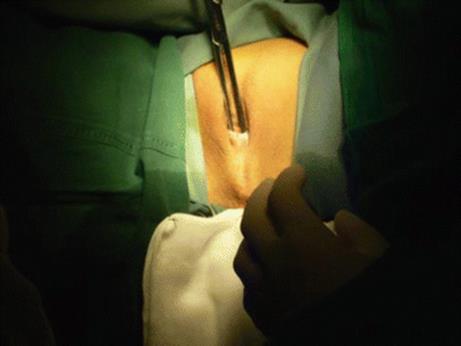
Fig. 3.7
An artery forceps is used to enlarge the above small opening. The artery forceps can then be inserted perpendicularly close to the perineal skin and run down towards the direction of the anus
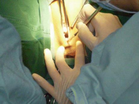
Fig. 3.8
Being guided by the artery forceps, the perineal skin can be incised down to the level of posterior vaginal wall, to expose the vaginal and urethral openings
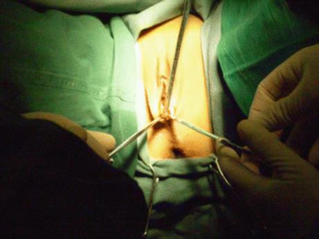
Fig. 3.9
Alice forceps are used to clamp the edges of incised perineal skins to provide hemostasis
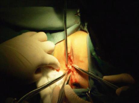
Fig. 3.10
The perineal wounds can then be sutured using 3/0 absorbable suture
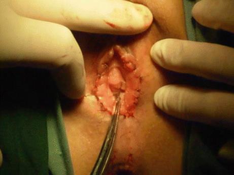
Fig. 3.11
After complete suturing, the labia minora will become the new vaginal opening, exposing the urethra and vagina
3.3.7 Key Points of Surgery
The success of this surgery is based on accurate diagnosis and better understanding of the anatomy of the vulva. It is important to adequately divide the fused perineal skin, to expose the vaginal and urethral openings.
References
1.
Winderl LM, Silverman RK. Prenatal diagnosis of congenital imperforate hymen. Obstet Gynecol. 1995;85(5 Pt 2):857–60.CrossRefPubMed
2.
Capraro VJ, Dillon WP, Gallego MB. Microperforate hymen: a distinct clinical entity. Obstet Gynecol. 1974;44(6):903–5.PubMed
3.
Sanfilippo AM, Mansuria SM. Microperforate hymen resulting in pelvic abscess. J Pediatr Adolesc Gynecol. 2006;19(2):95–8.CrossRefPubMed
4.
Stewart JF. Recurrent urinary tract infection in the female child secondary to a partial imperforate hymen. J Urol. 1970;103(3):353–6.PubMed
5.
Shukunami K, Kaneshima M, Kurokawa T, Kubo M, Kotsuji F. Microperforate hymen at 27 years of age diagnosed with withdrawal bleeding and hysterofiberscopy. Arch Gynecol Obstet. 2000;264(1):49–50.CrossRefPubMed
6.
Guldberg R, Thybo S, Andersen B. Synechia vulva—an unusual cause of urinary symptoms in a 15-year-old girl. Acta Obstet Gynecol Scand. 2007;86(2):251–2. doi:10.1007/s00404-008-0598-3.CrossRefPubMed
7.
Puhl AG, Steiner E, Krämer WW, Weltzien A, Skala C, Passuello V, et al. Fetal urogenital sinus with consecutive hydrometrocolpos because of labial fusion prenatal diagnostic difficulties and postpartal therapeutic management. Fetal Diagn Ther. 2008;23(4):287–92. doi:10.1159/000123615.CrossRefPubMed
8.
Gul A, Yıldırım G, Gedıkbası A, Gungorduk K, Ceylan Y. Prenatal ultrasonographic features of persistent urogenital sinus with hydrometrocolpos and ascites. Arch Gynecol Obstet. 2008;278:493–6.CrossRefPubMed