
Henning A. Gaissert
 INDICATIONS
The transcervical approach to thymectomy (TCT) is almost exclusively reserved for nonthymomatous myasthenia gravis (MG). TCT is an attractive surgical option because it is the least invasive approach for an operation to treat generalized weakness with the lowest reported rate of postoperative myasthenic crisis, the single most concerning disease-specific morbidity. Detractors of TCT argue that this less invasive technique limits the complete resection of thymic tissue. There are no prospective randomized comparisons between thymectomy and medical therapy, and none between the various approaches to the procedure.
The practice guideline of the American Academy of Neurology regards thymectomy as “an option to increase the probability of remission or improvement” of MG. Complete remission and clinical improvement of symptom severity have been reported in multiple observational studies: These provide the basis for selective resection of the gland. Thymectomy for purely ocular MG is not supported by some neurologists; however, about one half of these patients later progress to generalized MG; early thymectomy may reduce this proportion.
By inference, thymectomy is also an option in other immunologic disease associated with thymic hyperplasia, aplastic anemia for example.
TCT has been reported for the resection of selected, small thymomas, an indication not favored by the author.
TCT may be selected for resection of an intrathymic parathyroid. The resection of parathyroid adenoma, known or suspected to be located within the mediastinal thymus, has been recommended at the time of neck exploration, particularly when less than four glands are found.
 CONTRAINDICATIONS
Antecedent sternotomy leaves planes obliterated on which the execution of this operation depends. Prior tracheostomy or neck exploration create obstacles, but do not preclude TCT.
Thymoma, however small or favorable in location, is not an appropriate target for the cervical approach, no matter whether associated with MG.
Emergent thymectomy for severe MG with respiratory impairment is inadvisable due to the high risk of postoperative respiratory failure.
Antecedent cervical vertebral operations may lead to severe restriction in the range of neck extension and obstruct the surgeon’s view. A different approach should be selected in these patients.
 PREOPERATIVE PLANNING
The diagnosis of MG is considered in patients with muscle weakness and easy fatigability and must be secured by at least one or all three of the following tests: Presence of acetylcholine receptor antibodies in peripheral blood, a positive edrophonium chloride (Tensilon) test, or characteristic electromyographic findings in extremity muscles.
The operation has no role in the management of acute, severe MG since postoperative results as measured in remission or improvement evolve one or more years after thymectomy. Symptoms of MG should be controlled before thymectomy with cholinesterase inhibitors, with or without addition of immunosuppressive agents. In preparation for thymectomy, neurologist and surgeon should mutually consider the prevention of postoperative myasthenic crisis. The incidence of myasthenic crisis after TCT is reported as 0.7% to 1%, the lowest of all operative approaches. Intravenous immunoglobulins (IVIG) or plasmapheresis should, therefore, be administered before the operation only to selected patients at increased risk of respiratory failure.
To evaluate respiratory muscle function, forced vital capacity (FVC) is useful as a simple test suitable for bedside comparison early after operation. An FVC of less than 15 to 20 mL/kg, a value extrapolated from other conditions, predicts postoperative respiratory failure. The most sensitive test of clinical respiratory fatigability, however, is the maximal breathing capacity (MBC) measured as the product of frequency and volume of breaths during 1 minute. An impaired MBC may initiate the preoperative administration of IVIG or plasmapheresis, although the precise indication for either treatment in TCT has not been studied.
In every patient undergoing thymectomy for MG, the absence of thymoma should be confirmed either with chemical shift magnetic resonance imaging or computed tomography. In the surgeon’s office, neck mobility and maximal extension are tested to ensure exposure of the mediastinum during operation. The anesthetic team must be aware of the underlying diagnosis to plan the respiratory management of MG and avoid depolarizing muscle relaxants. Following IVIG administration and immediately before operation, a complete blood count is repeated to exclude hemolytic anemia.
 SURGERY
The description follows the technique of Cooper et al.
Positioning
Correct positioning of the patient on the operative table is critical to surgical exposure and thus to the success of the operation. The patient is placed supine and the crest of the head is flush with the top of the table. The arms are padded and tucked at the patient’s side. An inflatable bag is placed underneath the shoulders. Once anesthetized, the patient is intubated with a single-lumen endotracheal tube. The operating table is turned 90 degrees counterclockwise and the shoulder bag is inflated to achieve maximum neck flexion while the head remains supported on a head rest. Neck, chest, and upper abdomen are included in the sterile field.

Figure 27.1 The cervical incision as placed within the contours of the sternocleidomastoid muscles exposes thymus following separation of the strap muscles.
Technique
A short cervical collar incision and subplatysmal flaps extending to manubrium and cricoid expose the strap muscles; these are divided in midline. Self-retaining Gelpi retractors are inserted (Fig. 27.1). The surgeon inserts an index finger between sternum and thoracic thymus to enlarge this plane and gently separate thymus and underlying innominate vein from the bone. In the neck, the left cervical thymus is commonly observed to be longer while the right cervical thymus is often short and blunt. Both sides are bluntly mobilized using peanut gauze at the tip of a short clamp. The superior pole vessels are isolated, ligated with silk ties, and divided. The cervical thymus is lifted from its bed down to the thoracic inlet.
Intraoperative Positioning
To expose the mediastinum to the surgeon (and, unfortunately, only the surgeon), Gelpi retractors are removed. A right-angle blade, part of the Cooper thymectomy retractor (Pilling Company, Ft. Washington, PA, Fig. 27.2), is inserted behind the manubrium and suspended on a cross bar. The cross bar and its supporting poles are placed by the surgeon precisely above the manubrium. The manubrial blade is lifted and secured on the bar, with the patient’s head still supported on a rest; the head should not be floating (Fig. 27.3). The rubber bag lifting the shoulders is now deflated to open the thoracic inlet. Two Parker retractors, available from various sources, are inserted to retract the skin and kept in place by a loop fashioned of two 1-in Latex Penrose drains; non-Latex drains do not possess sufficient extensibility. The other end is hooked on the side rail of the operating table.
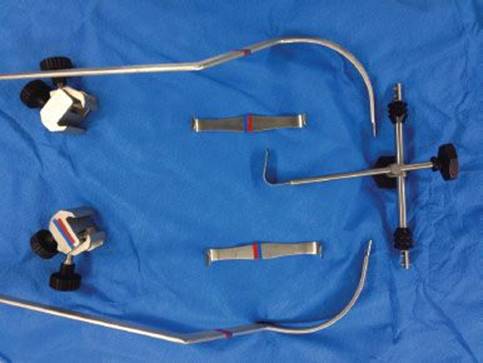
Figure 27.2 The components of the Cooper thymectomy retractor (Pilling Company, Ft. Washington, PA): Manubrial retractor on crossbar with hinges detached from both lateral posts, Parker retractors, and clamps to anchor the posts.

Figure 27.3 The inserted manubrial retractor lifts the sternum to open the thoracic inlet. Note support of the head in neck extension. The Parker blades are inserted and retracted by a latex loop.
Technique
The surgeon, seated with a headlight above the patient’s head, establishes by dissection whether both poles of the thymus are located anterior to the innominate vein; venous branches are also identified. Sometimes, the left thymus passes underneath the vein; this variant requires recognition and dissection at this time.
The accurate division of thymic veins and the complete removal of the gland are the central technical issues. With gentle traction on the ligatures, the cervical thymus is pulled forward and handed to the first assistant. The venous branches entering the top and anterior faces of the vein are sequentially identified and encircled with thin silk ties (Fig. 27.4). While clips seem desirable, they are easily dislodged during the subsequent dissection in front of the vein; all branches are, therefore, individually ligated. A tonsil sponge is used to expose these branches. Those entering the lower surface of the innominate vein are ligated last.

Figure 27.4 Surgeon’s view of the mediastinal dissection during separation of the thymus from the innominate vein. Note retraction of the superior thymic pole ties by the assistant and depression of the innominate vein with a tonsil sponge.
Returning the thymus to its bed covering the vein, the mediastinum is entered with two tonsil sponges, one to fix the gland with gentle downward pressure, the other to separate both pleurae and the pericardium from the gland. A systematic approach begins to separate the thymus from pleura and pericardium (Fig. 27.5) on one side, establishes the lower extent of the gland and ends on the other pleura. Pericardial fat is excluded, while the fatty tissues of the aortopulmonary window close to the left phrenic nerve are cautiously included. The gland is then folded off the pericardium with judicious use of electrocautery.
The gland is delivered into the wound and marked with respect to side so pathologic findings may be correlated. The mediastinum is checked for bleeding and any violation of the pleural space. The neck incision is closed in layers. The lungs are inflated just before the platysma is closed.

Figure 27.5 Below the innominate vein, the thymic gland is separated from pleura and pericardium with blunt dissection.
 POSTOPERATIVE MANAGEMENT
Following recovery from anesthesia, patients are usually discharged on the day of operation unless postanesthetic symptoms require overnight observation. A chest radiograph is obtained.
 COMPLICATIONS
Total rates of complications are below 10%. Myasthenic crisis, postoperative respiratory failure due to muscle weakness, is the most severe occurring in one series once in 100 consecutive patients and in another series once in 151 patients; both patients responded to treatment. The incidence of myasthenic crisis is thus either the lowest or among the lowest among the reported approaches.
Uncontrolled intraoperative bleeding is not reported in clinical series. The management of bleeding from thymic branches or the innominate vein consists of compression of the bleeding vessel and passage of ties around the branch. Bleeding from the innominate vein itself, not previously encountered by the author, may be controlled either with Swedish DeBakey clamps on either side of the injury or after passage of vascular tapes around the vein; sternotomy or transfusion should remain an exception.
Pneumothorax is occasionally observed and may require aspiration when symptomatic.
 RESULTS
The outcome measures after thymectomy for MG are (1) complete remission referenced to follow-up period; (2) improvement in the Osserman classification, a severity measure of MG consisting of five grades originally proposed as a pretreatment assessment (Table 27.1).
Calhoun et al. reported operative results in 78 patients who underwent TCT more than 12 months before assessment; the mean Osserman grade improved from a preoperative 2.73 to 0.94 after a mean of 5-year follow-up. The need for pyridostigmine declined from 90% to 54%, whereas the proportion of patients receiving prednisone decreased from 33% to 27% and the mean dose from 27 to 16 mg per patient. While more than half of all patients had thymic hyperplasia, 80% of preoperative prednisone use was observed in patients with involuted glands.
Khicha et al. observed 151 patients after extended TCT over a mean interval of 53 months. The Kaplan–Meier estimates of complete stable remission were 33% at 3 and 35% at 6 years. When they included asymptomatic patients on low-dose, single immunosuppressive therapy in their definition of complete remission, the rates were 43% at 3 and 45% at 6 years. These results were preserved in the patients with longer follow-up.
DeFilippi et al. reported TCT in 53 selected patients with MG with a mean follow-up of 4.3 years. Freedom from symptoms was observed in 81% and 43% (9 of 21) were in complete remission 5 or more years after operation. Thymectomy within the first year after onset of symptoms was associated with a higher rate of improvement or remission.
TABLE 27.1 Modified Osserman Classification[s3]

 CONCLUSIONS
In summary, TCT is a minimal invasive technique of thymectomy that is followed by a consistent response to treatment with improvement of symptoms in a large majority of patients and complete remission in 30% to 40% of patients. Comparisons with medical and alternative surgical therapy are pending. The technique requires most attention during the ligation of thymic branches of the innominate vein and in the clear separation of the tissues that are, and are not, part of the specimen. Maximal mediastinal dissection should not be attempted via a cervical incision.
Recommended References and Readings
1. Gronseth GS, Barohn RJ. Practice parameter: Thymectomy for autoimmune myasthenia gravis (an evidence-based review): Report of the Quality Standards Subcommittee of the American Academy of Neurology. Neurology.2000;55(1):7–15.
2. Monsul NT, Patwa HS, Knorr AM, et al. “The effect of prednisone on the progression from ocular to generalized myasthenia gravis”. J Neurol Sci. 2004;217(2):131–133.
3. Deeb ME, Brinster CJ, Kucharzuk J, et al. Expanded indications for transcervical thymectomy in the management of anterior mediastinal masses. Ann Thorac Surg. 2001;72:208–211.
4. Powell AC, Alexander HR, Chang R, et al. Reoperation for parathyroid adenoma: A contemporary experience. Surgery. 2009; 146(6):1144–1155.
5. Khicha SG, Kaiser LR, Shrager JB. Extended transcervical thymectomy in the treatment of myasthenia gravis. Ann N Y Acad Sci. 2008;1132:336–343.
6. Calhoun RF, Ritter JH, Guthrie TJ, et al. Results of transcervical thymectomy for myasthenia gravis in 100 consecutive patients. Ann Surg. 1999;230:555–561.
7. Inaoka T, Takahashi K, Mineta M, et al. Thymic hyperplasia and thymus gland tumors: Differentiation with chemical shift MR imaging. Radiology. 2007;243:869–876.
8. Tsukada H, Sunkara R, Dorcas DC, et al. Intravenous immunoglobulin-induced hemolytic anemia following thoracoscopic thymectomy for myasthenia gravis: Case report. Ann Thorac Surg. in press.
9. Cooper JD, Al-Jilaihawa AN, Pearson FG, et al. An improved technique to facilitate transcervical thymectomy for myasthenia gravis. Ann Thorac Surg. 1988;45:242–247.
10. DeFilippi VJ, Richman DP, Ferguson MK. Transcervical thymectomy for myasthenia gravis. Ann Thorac Surg. 1994;57:194–197.