Geoffrey H. Sperber
“…find out the cause of this effect, or rather say, the cause of this defect, For this effect defective comes by cause.”
--Shakespeare, Hamlet, act II, scene II
The secondary palate, so called because it forms after the appearance of the primary palate, constitutes both the floor of the nasal cavities and the roof of the mouth. The secondary palate is comprised of the anterior hard and the posterior soft palate and is an essential component of normal respiration, mastication, deglutition, and speech.
The very existence of the definitive secondary palate in humans characterizes a major mammalian evolutionary separation of the respiratory from the masticatory functions of the primitive stomodeal (oronasal) chamber of vertebrate antecedents. The transition of the single chamber of the embryonic stomodeum into the divided compartments of the two nasal cavities for respiration and the oral cavity for mastication represents a subdivision of this common chamber found in primitive crossopterygian fish, reptiles, birds, and early mammals. The separation of a continuous respiratory channel (the nostrils) from an intermittently required food-ingestion channel (the mouth) enabled the evolutionary development of leisurely mastication without respiratory interference. This separation occurs only anteriorly since the nasopharynx and oropharynx share a channel posteriorly, which accounts for momentary asphyxiation during swallowing. However, in the neonate and infant until 6 months of age, concomitant breathing and swallowing can occur due to the epiglottis and soft palate being in contact, forming a seal that maintains a continuous airway between the nasopharynx and laryngopharynx. Milk passes on either side of this continuous airway channel into the esophagus. An oropharynx develops only upon postnatal descent of the larynx, thereby separating the soft palate from the epiglottis. As a consequence, the risk of aspirating food may occur, protected by development of the cough reflex.
The existence of an intact palate has enabled sophisticated masticatory movements and epicurian enjoyment to evolve by virtue of the taste and texture senses embedded in the palate, accounting for the popular concept of the palate being the seat of taste (hence, palatable). Moreover, an intact palate is essential for the production of normal speech. Deficiencies of palatal development will result in food and salivary spillage into the nostrils and impaired articulation.
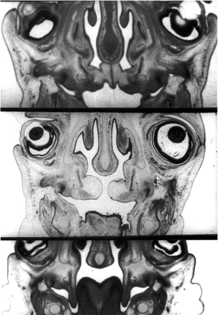
Three elements make up the secondary definitive palate: the two lateral palatal processes projecting into the stomodeum from the maxillary prominences and the primary palate derived anteriorly from the frontonasal prominence (Fig. 2.1). These three elements are initially widely separated due to the advancing edges of the palatal processes being deflected down on either side of the obtruding tongue, which initially occupies most of the stomodeal chamber (Kimes et al., 1991) (Fig. 2.2). Concomitantly, the midline cartilaginous nasal septum descends from the roof of the stomodeum as a feature of nasal capsular development (Mooney et al., 1994) (Fig. 2.3). During the 8th week postconception, a remarkable transformation of the palatal shelves occurs, when they elevate into a horizontal position as a prelude to their fusion with each other, the primary palate, and the nasal septum, thereby partitioning the oronasal chamber (Fig. 2.4).
|
|
|
FIG. 2.1. Schematic depiction of midcoronal sections of embryonic heads at 7 and 12 weeks postconception. (From Sperber, 2001, with permission.) |
The transition from vertical to horizontal is completed within hours during the 8th week postconception. There is a sex difference in the timing of palatal closure. Shelf elevation and fusion begin a few days earlier in male than in female embryos (Burdi and Faist, 1967), the slight delay possibly accounting for the higher incidence of cleft palates in females. Several mechanisms have been proposed for the rapid elevation of the palatal shelves. They include biochemical transformations in the physical consistency of the connective tissue matrix of the shelves; variations in vasculature and blood flow to these structures (Amin et al., 1994), resulting in a sudden increase in tissue fluid turgor; rapid differential mitotic growth, muscular movements, and an intrinsic shelf force (Young et al., 1997; McGonnell et al., 1998). The intrinisic shelf elevating force is generated chiefly by the synthesis, accumulation, and hydration of hyaluronic acid and glycosaminoglycans within the extracellular matrix of the shelves (Singh et al., 1997).
The alignment of mesenchymal cells and orientation of collagen within the palatal shelves may direct the elevating forces, while palatal mesenchymal cells are themselves contractile. The withdrawal of the fetus's face from against the heart prominence by uprighting the head facilitates jaw opening (Fig. 2.5). Mouth-opening reflexes and extrinsic tongue muscle activity have been implicated in the withdrawal of the tongue from between the vertical shelves (Humphrey, 1969). Depressed fetal swallowing may delay palatal shelf elevation, precluding their conjunction, leading to clefting. Prenatal ultrasonography revealing aphagia has been correlated with cleft palate formation (Laster et al., 2001). Absence of functioning of the hyoglossus muscle, which depresses the tongue, resulting from Hoxa gene mutations, prevents palatal shelf lifting and consequent clefting (Barrow and Capecchi, 1999). Tongue withdrawal is further aided by movement of the lower jaw through the functioning of pharyngeal arch muscles, which in turn requires an intact neural motor system (Wragg et al., 1972; Kjaer, 1997). In this regard, the trigeminal nerve is the first motor nerve to function in the fetus and, moreover, requires a functioning incudomalleal primary jaw joint to allow opening of the mouth. The fetus must be in a floating condition in the amniotic sac to permit jaw movements; in the event of oligohydramnios,1 deficient amniotic fluid inhibits mouth opening and tongue withdrawal, thereby precluding palatal shelf elevation, accounting for a possible source of clefting. Extracellular agents such as growth factors, hormones, and neuropeptides have been implicated in the regulation of various cellular responses during palate development (Greene and Pratt, 1976; Morris-Wiman and Brinkley, 1992; Abbott et al., 1998; Izadnegahdar et al., 1999; Machida et al., 1999). Most significantly, epidermal growth factor (EGF), the transforming growth factors (TGF-α, TGF-β1, TGF-β3), and their receptor molecules have been identified during all stages of palate formation (Citterio and Gaillard, 1994; Proetzel et al., 1995; Kaartinen et al., 1997; Cui et al., 1998; Sun et al., 1998). Docking of TGF-α with the EGF receptor results in the production of matrix metalloproteinases, a class of proteins that regulate palate closure (Miettinen et al., 1999).
|
|
|
FIG. 2.2. Coronal section scanning electron micrographs of stomodeal chambers of embryos at (A) 4, (B) 6, (C) 7, and (D) 8 weeks, revealing secondary palate development. T, tongue; np, nasal prominence (septum); mee, medial edge epithelium; double arrows, palatal shelves; single arrow, midpalatal fusion seam. (Courtesy of Dr. H. C. Slavkin. University of Southern California.) |
During palate closure, the mandible becomes more prognathic and the vertical dimension of the stomodeal chamber increases, though maxillary width remains stable, allowing shelf contact to occur. Also, forward growth of Meckel's cartilage relocates the tongue more anteriorly, concomitant with head elevation. Mandibular growth retardation causes retrognathia that enforces a high tongue position, preventing the shelves from fusing (Seegmiller and Fraser, 1977) to create the Pierre Robin sequence (Lavrin, 2000).
The epithelium overlying the edges of the palatal shelves is especially thickened, and their fusion upon mutual contact is crucial to intact palatal development. This fusion is dependent on targeted removal of the epithelium between the palatal shelves, which is anticipated by upregulation of keratin K5/6 and expression of vimentin mRNA in the medial edge epithelium (Gibbins et al., 1999). Fusion also occurs between the dorsal surfaces of the fusing palatal shelves and the lower edge of the midline nasal septum. The fusion seam initially forms anteriorly in the hard palate region, with subsequent merging of the soft palate region (Ferguson, 1988). The mechanisms of adhesive contact, fusion, and subsequent degeneration of the epithelium are not clearly understood. The combination of degenerating epithelial cells and a surface coat accumulation of glycoproteins and desmosomes facilitates epithelial adherence between contacting palatal shelves. The cell adhesion molecule syndecan is expressed as the shelves elevate, and its expression decreases during fusion (Fitchett et al., 1990). At the time of palatal shelf fusion, there is increased expression of N-cadherin. This may be instrumental in the transformation of epithelium into its different phenotypes (nasal, medial edge, and oral) and into mesenchyme. Only the medial edge epithelium of the palatal shelves (in contrast to their oral and nasal surface epithelia) undergoes cytodifferentiation, which involves a decline of EGF receptors leading to apoptotic cell death (Greene and Pratt, 1976). The oral surface of the palatal shelves differentiates into stratified squamous epithelium, while the nasal surface becomes respiratory ciliated pseudostratified columnar epithelium (Carette et al., 1991).
|
|
|
FIG. 2.3. Coronal sections of embryos at (top) 54, (middle) 57, and (lower) 63 days postconception, revealing elevation of lateral palatal shelves from vertical to horizontal and fusion with the nasal septum. (From the Carnegie Embryo Collection.) |
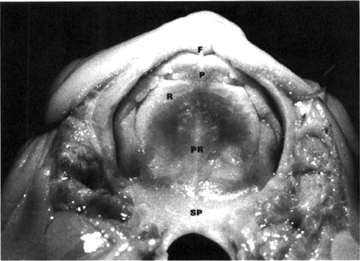
Programmed cell death of the fusing medial edge epithelia is restricted to the periderm, with basal cells remaining healthy. Epithelial-mesenchymal transformation of medial edge epithelium is essential to mesenchymal coalescence of the shelves. The surface cells lose their epithelial cell junctions and adopt a fibroblastic morphology. Cytokeratins (characteristic of epithelia), vimentin (typical of mesenchyme), and cadherins are variously expressed during this transformation (Montenegro et al., 2000). Some epithelial cells also migrate into the palatal mesenchyme, undergoing epithelial-mesenchymal transformation and contributing to seam disruption (Fitchett et al., 1990; Shuler et al., 1991, 1992; Griffith and Hay, 1992; Hay, 1995; Lavrin and Hay, 2000). The epithelium, at its leading edges, may contribute to failure of fusion by not breaking down after shelf approximation, leading to epithelial pearl formation, or by not maintaining adhesiveness beyond a critical time should palatal shelf elevation be delayed.2 Obstructed, unfused medial palatal shelf epithelium stratifies and keratinizes before birth (Goss et al., 1970). However, the capability to fuse is retained until shortly before birth (Lavrin et al., 2001). This provides the possibility that unfused palatal shelves brought into contact by in utero surgical intervention could forestall clefting. Fusion of the three palatal components initially produces a flat, unarched roof to the mouth (Fig. 2.6). The fusing lateral palatal shelves overlap the anterior primary palate, as indicated later by the sloping pathways of the junctional incisive neurovascular canals that carry the previously formed incisive nerves and blood vessels. The site of junction of the three palatal components is marked by the incisive papilla overlying the incisive canal. The line of fusion of the lateral palatal shelves is traced in the adult by the midpalatal suture3 and on the surface by the midline raphe of the hard palate (Fig. 2.6). This fusion seam is minimized in the soft palate by invasion of extraterritorial mesenchyme.
|
|
|
FIG. 2.4. Coronal section of the palate of an embryo immediately after midline fusion. PS, palatal shelves; NS, nasal septum; 1C, inferior concha; C, cartilage of nasal septum; VN, vomeronasal organ; OC, ossification centers; NF, nasal fossa; T, tongue. (From O'Rahilly, 1975, with permission.) |
Ossification of the palate proceeds during the 8th week postconception from the spread of bone into the mesenchyme of the fused lateral palatal shelves and from trabeculae appearing in the primary palate as premaxillary centers, all derived from the single primary ossification centers of the maxillae (Jacobson, 1955; Wood et al., 1967; Kjaer, 1989; Nijo and Kjaer, 1993; Silau et al., 1994; Vacher et al., 1999; Vacher et al., 2001). The existence of an incisive fissure in the ossified palate at the site of fusion of the primary and secondary palates has been demonstrated in archeological material (Sejrsen et al., 1993; Maureille and Bar, 1999; Kieser et al., 1999). However, the coincidence of the incisive fissure with alveolar palatal clefts has been disputed (Lisson and Kjaer, 1997). Posteriorly, the hard palate is ossified by trabeculae spreading from the single primary ossification center of each of the palatine bones.
|
|
|
FIG. 2.5. Schematic depiction of fetal head movements from frontal (left) and lateral (right) perspectives at (1,2) 6, (3,4) 7, and (5,6) 8 weeks. (Courtesy of Dr. V. M. Diewert. University of British Columbia.) |
|
|
|
FIG. 2.6. Palate and upper lip of a 22-week-old fetus. F, frenulum; P, papilla overlying incisive foramen; PR, palatal raphe; R, rugae; SP, soft palate. (From Sperber, 2001, with permission.) |
The midpalatal sutural structure is first evident at 10.5 weeks, when an upper layer of fiber bundles develops across the midline (Del Santo et al., 1998). In infancy, the midpalatal suture in coronal section has a Y shape, and it binds the vomer with the palatal shelves. In childhood, the junction between the three bones rises into a T shape, with the interpalatal section taking a serpentine course. In adolescence, the suture becomes so interdigitated that mechanical interlocking and interstitial islets of bone are formed. Cartilage may appear in islands in the suture in the neonatal period, but after 3 years the suture is exclusively fibrous (Persson, 1973). Application of expansional forces on the maxillae to widen the palate in orthodontic practice induces osteogenesis in the midpalatal suture (distraction osteogenesis) (Latham, 1971; Kobayashi et al., 1999). The palatine bone elements remain separated from the maxillary elements by the transverse palatomaxillary sutures into adulthood (Nijo and Kjaer, 1993).
Ossification does not occur in the most posterior part of the palate, giving rise to the soft palate. Myogenic mesenchymal tissue of the first and fourth pharyngeal arches migrates into this faucial region, supplying the musculature of the soft palate and fauces (Cohen et al., 1993, 1994). The tensor veli palatini is derived from somitomeres associated with the first pharyngeal arch; the levator palatini and uvular and faucial pillar muscles are derived from somitomeres associated with the fourth pharyngeal arch, accounting for the innervation by the first arch trigeminal nerve of the tensor veli palatini muscle and by the fourth arch pharyngeal plexus and vagus nerves for all of the other muscles.
The tensor veli palatini is the earliest of the five palatal muscles to develop, forming myoblasts at 40 days postconception. It is followed by the palatopharyngeus (45 days), the levator veli palatini (8th week), the palatoglossus (9th week), and the uvular muscles (llth week). The palatoglossus, derived from the tongue musculature, attaches to the soft palate during the llth week postconception. The hard palate grows in length, breadth, and height, becoming an arched roof for the mouth (Fig. 2.7). The fetal palate increases in length more rapidly than in width between 7 and 18 weeks postconception, after which the width increases faster than the length (Lee et al., 1992). In early prenatal life, the palate is relatively long, but from the 4th month postconception it widens as a result of midpalatal sutural growth and appositional growth along the lateral alveolar margins. At birth, the length and breadth of the hard palate are almost equal. The postnatal increase in palatal length is due to appositional growth hi the maxillary tuberosity region and, to some extent, at the transverse maxillopalatine suture (Sejrsen et al., 1996).
Growth at the midpalatal suture ceases between 1 and 2 years of age, but no synostosis occurs to signify its cessation.4 Growth in width of the midpalatal suture is larger in its posterior than in its anterior part.
Obliteration of the midpalatal suture may start in adolescence, but complete fusion is rarely found before 30 years of age. The timing and degree of fusion of this suture vary greatly (Wehrbein and Yildizhan, 2001).
|
|
|
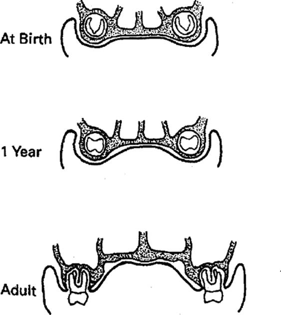
FIG. 2.7. Schematic cross-sectional views of the palate at various ages. Note the increasing depth of the palatal arch concomitant with tooth eruption. (From Sperber, 2001, with permission.) |
Lateral appositional growth continues until 7 years of age, by which time the palate achieves its ultimate anterior width. Posterior appositional growth continues after lateral growth has ceased so that the palate becomes longer, rather than wider, during late childhood. During infancy and childhood, bone apposition also occurs on the entire inferior surface of the palate, accompanied by concomitant resorption from its superior (nasal) surface. This bone remodeling results in descent of the palate and enlargement of the nasal cavity. Nasal capacity must increase, to keep pace with the increasing respiratory requirements engendered by general body growth. A fundamental drive in facial growth is provision of an adequate nasal capacity; if this need is not met, the space capacity requirement is diverted to the mouth for maintenance of respiration.
The appositional growth of the alveolar processes contributes to deepening, as well as widening, of the vault of the bony palate, at the same time adding to the height and breadth of the maxillae. The lateral alveolar processes help to form an anteroposterior palatal furrow, which together with a concave floor produced by a tongue curled from side to side results in a palatal tunnel ideally suited to receive a nipple. A variable number of transverse palatal rugae develop in the mucosa covering the hard palate (Harris et al., 1990; Thomas and Rossouw, 1991). They appear even before palatal fusion, which occurs at 56 days postconception. The rugae, which are most prominent in the infant, hold the nipple while it is being milked by the tongue. Palatal rugae are utilized as landmarks in cephalometry and orthodontics because of their stability (Hoggan and Sadowsky, 2001). The anterior palatal furrow is well marked during the first year of life (i.e., the active suckling period) and normally flattens out into the palatal arch after 3 to 4 years of age, when suckling has been discontinued. Persistence of thumb or finger sucking may retain the accentuated palatal furrow into childhood.
Anomalies of the palate occur as a consequence of disturbances of the above-mentioned developmental processes. Successful fusion of the three embryonic components of the palate involves complicated synchronization of shelf movements with growth and withdrawal of the tongue and growth of the mandible and head. Mistiming of any of these critical events, because of environmental agents or genetic predisposition, results in failure of fusion, leading to clefts of the palate. Palatal clefting is multifactorial in its etiology, and no single genetic locus has been identified as a source of its cause, despite its hereditary familial associations (Hibbert and Field, 1996). Postnatal surgical repair of cleft palate results in cicatricial formation, the scarring compromising the growth potential and final result. However, in utero repair of fetal cleft palate offers the possibility of scarless healing and is a potential future treatment (Christ, 1990; Weinzweig et al., 1999).
The entrapment of epithelial rests or pearls in the line of fusion of the palatal shelves, particularly the midline raphe of the hard palate, may give rise later to median palatal rest cysts (Arnold et al., 1998). A common superficial expression of these epithelial entrapments is development of epithelial cysts or nodules, known as Epstein's pearls, along the median raphe of the hard palate and at the junction of the hard and soft palates. Small mucosal gland retention cysts (Bohn's nodules) may occur on the buccal and lingual aspects of the alveolar ridges, and dental lamina cysts composed of epithelial remnants of this lamina may develop on the crests of the alveolar ridges. All of these superficial cysts of the palate in the newborn usually disappear by the third postnatal month. An anterior midline maxillary cyst developing in the region of the primary palate cannot be of fissural origin, but is a nasopalatine duct cyst encroaching anteriorly into the palate. Cysts are rare in the soft palate because of the mesenchymal merging of the shelves in this region, although submucous clefts may occur.
Delay in elevation of the palatal shelves from the vertical to the horizontal while the head is growing continuously results in a widening gap between the shelves so that they cannot meet and, therefore, cannot fuse. When eventually they do become horizontal, this leads to clefting of the palate (palatoschisis). Other causes of cleft palate are defective shelf fusion, failure of medial edge epithelial cell death, possible postfusion rupture (Kitamura, 1991), and failure of mesenchymal consolidation and differentiation (Lavrin and Hay, 2000).
|
|
|
FIG. 2.8. Schematic depiction of cleft palate variations. (From Sperber, 2001, with permission.) |
The least severe form of cleft palate is the bifid uvula, of relatively common occurrence. Increasingly severe clefts always incur posterior involvement, the cleft advancing anteriorly in contradistinction to the direction of normal fusion (Fig. 2.8). The lines of fusion of the lateral palatal shelves with the primary palate dictate the diversion from the midline of a severe palatal cleft anteriorly to either the right or left or, in rare instances, to both. If the cleft involves the alveolar arch, it usually passes between the lateral incisor and canine teeth (Lisson and Kjaer, 1997). Such severe clefting of the palate may or may not be associated with unilateral or bilateral cleft upper lip; the two conditions are determined independently. The vertical nasal septum may fuse with the left or right palatal shelf or neither in cases of severe cleft palate (Figs. 2.9,2.10).
Clefts of the soft palate alone incur varying degrees of speech difficulty and swallowing problems because of the inability to close off the oropharynx completely from the nasopharynx during these pharyngeal functions. Clefts of the hard palate, which almost invariably include soft palate clefts, give rise to feeding problems, particularly in infants in whom the vacuum-producing sucking processes demand an intact hard palate. Spillage of food into the nasal cavity is symptomatic of feeding difficulties. The efficacy of suckling is compromised, with the necessary negative oral pressure for ingestion shifted toward the nasal cavity and pharynx. The consequent altered physiological pressures result in altered morphology of adjacent structures (Ihan-Hren et al., 2001). Such infants require early reparative surgery and/or obturator fitting to maintain good nutrition and to aid development of correct enunciation.
|
|
|
FIG. 2.9. Schematic coronal section through a unilateral cleft palate with an oral opening into one nasal cavity only. (From Sperber, 2001, with permission.) |
|
|
|
FIG. 2.10. Schematic coronal section through a bilaterally cleft palate with an oral opening into both nasal cavities. (From Sperber, 2001, with permission.) |
Cleft palate is a feature of a number of congenital defect syndromes, among which are mandibulofacial dysostosis (Treacher Collins syndrome), micrognathia (Pierre Robin sequence), van der Woude syndrome, and orodigitofacial dysostosis syndrome. The palate is narrower, shorter, and lower than normal in Down syndrome (trisomy 21), although it is often described as having a high midline elevation but being horizontally flattened laterally along the alveolar ridges, creating a steeple palate (Panchon-Ruiz et al., 2000). A highly arched palate is also characteristic of Marfan syndrome, an inherited disorder manifesting skeletal and cardiovascular anomalies. Cleidocranial dysostosis, a congenital defect of intramembranous bones, also manifests a highly arched palate, with or without a cleft. Other congenital conditions displaying a highly arched palate are craniofacial dysostosis (Crouzon syndrome), acrocephalosyndactyly (Apert syndrome), progeria, Turner syndrome (XO sex chromosome complement), and oculodentodigital dysplasia. A great variety of other congenital anomalies may be associated with clefts of the lip and palate (Milerad et al., 1997; Natsume et al., 2001).
A fairly common genetic anomaly of the palate is a localized midpalatal overgrowth of bone, of varying size, known as torus palatinus. This may enlarge in adulthood, and although it does not directly influence dental occlusion, if prominent, it may interfere with the seating of a removable orthodontic appliance or upper denture.
Acknowledgements
I thank Anne-Marie McLean for meticulous word processing of the manuscript.
References
Abbott, BD, Probst, MR, Perdew, GH, Buckalew, AR (1998). AH receptor, ARNT, glucocorticoid receptor, EGF receptor, EGF, TGFα, TGFβ1, TGFβ2 and TGFβ3 expression in human embryonic palate and effects of 2, 3, 7, 8 tetrachlorodibenzo-p-dioxin (TCDD). Teratology 58: 30–43.
Amin, N, Ohashi, Y, Chiba, J, et al. (1994). Alterations in vascular pattern of the developing palate in normal and spontaneous cleft palate mouse embryos. Cleft Palate Craniofac J 31: 332–344.
Arnold, WH, Rezwani, T, Baric, I (1998). Location and distribution of epithelial pearls and tooth buds in human fetuses with cleft lip and palate. Cleft Palate Craniofac J 35: 359–365.
Barrow, JR, Capecchi, MR (1999). Compensatory defects associated with mutations in Hoxa 1 restore normal palatogenesis to Hoxa 2 mutants. Development 226: 5011–5026.
Burdi, AR, Faist, K (1967). Morphogenesis of the palate in normal human embryos with special emphasis on the mechanisms involved. Am J Anat 120: 149.
Carette, MJ, Lane, EB, Ferguson, MW (1991). Differentiation of mouse embryonic palatal epithelium in culture: selective cytokeratin expression distinguishes between oral, medial edge and nasal epithelial cells. Differentiation 47: 149–161.
Christ, JE (1990). Plastic surgery for the fetus. Plast Reconstr Surg 86: 1238.
Citterio, HL, Gaillard, DA (1994). Expression of TGF2, EGF-R and cell proliferation during human palatogenesis: an immunohistochemical study. Int J Dev Biol 38: 499–505.
Cohen, SR, Chen, L, Burdi, AR, et al. (1994). Patterns of abnormal myogenesis in human cleft palates. Cleft Palate Craniofac 31: 345.
Cohen, SR, Chen, L, Trotman, CA, et al. (1993). Soft palate myogenesis: a developmental field paradigm. Cleft Palate Craniofac J 30; 441–446.
Cui, X-M, Warburton, D, Zhao, J, et al. (1998). Immunohistochemical localization of TGF-β type II cell receptor and TGFβ3 during palatogenesis in vivo and in vitro. Int J Dev Biol 42: 817–820.
Del Santo, M, Jr, Minarelli, AM, Liberti, EA (1998). Morphological aspects of the mid-palatal suture in the human foetus: a light and scanning electron microscope study. Eur J Orthod 20: 93–99.
Ferguson, MWJ (1988). Palate development. Development 103(SuppL): 41–60.
Fitchett, JE, McAlmon, KR, Hay, ED, Bernfield, M (1990). Epithelial cells lose syndecans prior to epithelial-mesenchymal transformation in the developing rat palate. J Cell Biol 111: 1459.
Gibbins, JR, Manthey, A, Tazawa, YM, et al. (1999). Midline fusion in the formation of the secondary palate anticipated by upregulation of keratin K5/6 and localized expression of vimentin mRNA in medial edge epithelium. Int J Dev Biol 43: 237–244.
Goss, AN, Bodner, JW, Avery, JK (1970). In vitro fusion of cleft palate shelves. Cleft Palate J 7: 737–747.
Greene, RM, Pratt, RM (1976). Developmental aspects of secondary palate formation. J Embryol Exp Morphol 36: 225–245.
Griffith, CM, Hay, ED (1992). Epithelial-mesenchymal transformation during palatal fusion: carboxyfluorescein traces cells at light and electron microscopic levels. Development 116: 1087–1099.
Harris, MJ, Juriloff, DM, Peters, CE (1990). Disruption of pattern formation in palatal rugae in fetal mice heterozygous for First Arch (FAR). J Craniofac Genet Dev Biol 10: 363–371.
Hay, ED (1995). An overview of epithelio-mesenchymal transformation. Acta Anat (Basel) 154: 8–20.
Hibbert, SA, Field, JK (1996). Molecular basis of familial cleft lip and palate. Oral Dis 2: 238–241.
Hoggan, BR, Sadowsky, C (2001). The use of palatal rugae for the assessment of anteroposterior tooth movements. Am J Orthod Dentofac Orthoped 119: 482–488.
Humphrey, T (1969). The relation between human fetal mouth opening reflexes and closure of the palate. Am J Anat 125: 317–344.
Ihan-Hren, N, Oblak, P, Kozelj, V (2001). Characteristic forms of the upper part of the oral cavity in newborns with isolated cleft palate. Cleft Palate Craniofac J 38: 164–169.
Izadnegahdar, MF, Rathanaswami, P, Shah, RM (1999). Effects of EGF and TGFβ1 on c-myc gene expression and DNA synthesis in embryonic hamster palate mesenchymal cells. Anat Rec 254: 453–464.
Jacobson, A (1955). Embryological evidence for the non-existence of the premaxilla in man. J Dent Assoc S Afr 10: 189–210.
Kaartinen, V, Cui, XM, Heisterkamp, N, et al. (1997). TGFβ3 regulates transdifferentiation of medial edge epithelium during palatal fusion and associated degradation of the basement membrane. Dev Dyn 209: 255–260.
Kieser, JA, Dennison, KJ, Dias, GR (1999). Premaxillary suture in early Polynesians. Int J Osteoarchaeol 9: 244–247.
Kimes, KR, Mooney, MP, Siegel, MI, Todhunter, JS (1991). Size and growth rate of the tongue in normal and cleft lip and palate human fetal specimens. Cleft Palate Craniofac J 28: 212–216.
Kitamura, H (1991). Evidence for cleft palate as a postfusion phenomenon. Cleft Palate Craniofac J 28: 195–210.
Kjaer, I (1989). Prenatal skeletal maturation of the human maxilla. J Craniofac Genet Dev Biol 9: 257–264.
Kjaer, I (1997). Mandibular movements during elevation and fusion of palatal shelves evaluated from the course of Meckel's cartilage. J Craniofac Genet Dev Biol 17: 80–85.
Kobayashi, ET, Hashimoto, F, Kobayashi, Y, et al. (1999). Forceinduced rapid changes in cell fate at midpalatal suture cartilage of growing rats. J Dent Res 78: 1495–1504.
Laster, Z, Temkin, D, Zarfin, Y, Kushnir, A (2001). Complete bony fusion of the mandible to the zygomatic complex and maxillary tuberosity: case report and review. Int J Oral Maxillofac Surg 30: 75–79.
Latham, RA (1971). The development, structure and growth pattern of the human midpalatal suture. J Anat 108: 31–41.
Lavrin, IG (2000). Pierre Robin sequence—a review and an animal model. In: Davidovitch, Z, Mah, J (Eds.) The Biological Mechanisms of Tooth Movement and Craniofacial Adaptation. Harvard Society for the Advancement of Orthodontics. Boston, pp. 299–303.
Lavrin, IG, Hay, ED (2000). Epithelial-mesenchymal transformation, palatogenesis and cleft palate. Angle Orthod 70: 181–182.
Lavrin, IG, McLean, W, Seegmiller, RE, Olsen, BR, Hay, ED (2001). The mechanism of palatal clefting in the Colllal mutant mouse. Arch Oral Biol 46: 865–869.
Lee, SK, Kim, YS, Lim, CY, Chi, JG (1992). Prenatal growth pattern of the human maxilla. Acta Anat (Basel) 145: 1–10.
Lisson, JA, Kjaer, I (1997). Location of alveolar clefts relative to the incisive fissure. Cleft Palate Craniofac J 34: 292–296.
Machida, J, Yoshiura, K, Funkhauser, CD, et al. (1999). Transforming growth factor a: genomic structure, boundary sequences and mutation analysis in non-syndromic cleft lip/palate and cleft palate only. Genomics 61: 237–242.
Mann, SE, Nijland, MJ, Ross, MG (1996). Mathematical modeling of human amniotic fluid dynamics. Am J Obstet Gynecol 175: 937–944.
Maurielle, B, Bar, D (1999). The premaxilla in Neanderthal and early modern children: ontogeny and morphology. J Hum Evol 37: 137–152.
McGonnell, IM, Clarke, JD, Tickle, C (1998). Fate map of the developing chick face: an analysis of expansion of facial primordia and establishment of the primary palate. Dev Dyn 212: 102–118.
Miettinen, PJ, Chin, JR, Shum, L, et al. (1999). Epidermal growth factor receptor function is necessary for normal craniofacial development and palate closure. Nat Genet 22: 69–73.
Milerad, J, Larson, O, Hagberg, C, Ideberg, M (1997). Associated malformations in infants with cleft lip and palate: a prospective, population-based study. Pediatrics 100: 180–186.
Montenegro, MA, Rojas, M, Dominguez, S, Vergara, A (2000). Cytokeratin, vimentin and E-cadherin immunodetection in the embryonic palate in two strains of mice with different susceptibility to glucocorticoid-induced clefting. J Craniofac Genet Dev Biol 20: 137–143.
Mooney, MP, Siegel, MI, Kimes, KR, et al. (1994). Anterior paraseptal cartilage development in normal and cleft lip and palate human fetal specimens. Cleft Palate Craniofac J 31: 239–245.
Morris-Wiman, J, Brinkley, L (1992). An extracellular matrix infrastructure provides support for murine secondary palatal shelf remodelling. Anat Rec 234: 575–586.
Natsume, N, Niimi, T, Furukawa, H, et al. (2001). Survey of congenital anomalies associated with cleft lip and/or palate in. 701,181 Japanese people. Oral Surg Oral Med Oral Pathol Oral Radiol Endod 91: 157–161.
Nijo, BJ, Kjaer, I (1993). The development and morphology of the incisive fissure and the transverse palatine suture in the human fetal palate. J Craniofac Genet Dev Biol 13: 24–34.
O'Rahilly, R (1975). A Color Atlas of Human Embryology. Philadelphia: Saunders.
Panchon-Ruiz, A, Jornet-Carrillo, V, Sanchez del Campo, F (2000). Palate vault morphology in Down syndrome. J Craniofac Genet Dev Biol 20: 198–200.
Persson, M (1973). Structure and growth of facial sutures. Odont Rev 24(Suppl 26): 1–146.
Proetzel, G, Pawlowski, SA, Wiles, MV, et al. (1995). Transforming growth factor fa is required for secondary palate fusion. Nat Genet 11: 409–14.
Seegmiller, RE, Fraser, FC (1977). Mandibular growth retardation as a cause of cleft palate in mice homozygous for chondrodysplasia gene. J Embryol Exp Morphol 38: 227–238.
Sejrsen, B, Kjaer, I, Jakobsen, J (1993). The human incisal suture and premaxillary area studied on archeological material. Acta Odont Scand 51: 143–151.
Sejrsen, B, Kjaer, I, Jakobsen, J (1996). Human palatal growth evaluated on medieval crania using nerve canal openings as references. Am J Phys Anthropol 99: 603–611.
Shuler, CF, Guo, Y, Majumder, A, Luo, R (1991). Molecular and morphological changes during the epithelial-mesenchymal transformation of palatal shelf medial edge epithelium in vitro. Int J Dev Biol 35: 463–472.
Shuler, CF, Halpern, DE, Guo, Y, Sank, AC (1992). Medial edge epithelium fate traced by lineage analysis during epithelialmesenchymal transformation. Dev Biol 154: 318–330.
Silau, AM, Njio, B, Solow, B, Kjaer, I (1994). Prenatal sagittal growth of the osseous components of the human palate. J Craniofac Genet Dev Biol 14: 252–256.
Singh, GD, Moxham, BJ, Langley, MS, Embery, G (1997). Glycosaminoglycan biosynthesis during 5-fluro-2-deoxyuridine-induced palatal clefts in the rat. Arch Oral Biol 42: 355–363.
Sun, D, Vanderburg, CR, Odierna, GS, Hay, ED (1998). TGFβ3 promotes transformation of chicken palatal medial edge epithelium to mesenchyme in vitro. Development 125: 95–105.
Thomas, CJ, Rossouw, RJ (1991). The early development of palatal rugae in the rat. Aust Dent J 36: 342–348.
Vacher, C, Copin, H, Sakka, M (1999). Maxillary ossification in a series of six human embryos and fetuses aged from 9 to 12 weeks of amenorrhea: clinical implications. Surg Radiol Anat 21: 261–266.
Vacher, C, Sakka, M, Dauge, M-C (2001). Incisive suture (fissure) in the human fetus: radiographic and histologic study. Cleft Palate Craniofac J 38: 330–336.
Wehrbein, H, Yildizhan, F (2001). The mid-palatal suture in young adults. A radiological-histological investigation. Eur J Orthod 23: 105–114.
Weinzweig, J, Panter, KE, Pantaloni, M, et al. (1999). The fetal cleft palate: II scarless healing after in utero repair of a congenital model. Plast Reconstr Surg 104: 1356–1364.
Wood, NK, Wragge, LE, Stuteville, OH (1967). The premaxilla: embryological evidence that it does not exist in man. Anat Rec 158: 485–489.
Wragg, LE, Smith, JA, Borden, CS (1972). Myoneural maturation of the foetal rat tongue at the time of secondary palate closure. Arch Oral Biol 27: 673–682.
Young, AV, Hehn, BM, Sanghera, JS, et al. (1997). The activation of MAP kinase during vertical palatal shelf development in hamster. Growth Dev Aging 61: 27–38.
Appendix: World Wide Web Site Addresses for CL/P
· http://www.nuds.nwu.edu/clphome.htm
· http://www.healthanswers.com/database/ami/converted/001051.html
· http://www.scottishiritechildrens.org/services/cleftwhatis/shtml
· http://www.healthgate.com/hic/cleft-lip_palate/
· http://www.clapa.mcmail.com/
· http://www.widesmiles.org/index.html
· http://www.cleft.org/index.html
· http://www.cleft.com/cpf.htm
The American Cleft Palate-Craniofacial Association Web Site address is http://www.cleft.com, their Cleftline is http://www.cleftline.org, and the Cleft Palate-Craniofacial Journal is available online at http://www.cpcj.allenpress.com.
For genetic counseling, consult the following Web Sites:
· American College of Medical Genetics
o http://www.faseb.org/genetics/acmg/acmgmenu.htm
· National Society of Genetic Counselors
o http://www.nsgc.org
· Genetic Alliance
o http://www.geneticalliance.org
· Office of Rare Disorders
o http://www.rarediseases.info.nih.gov/ord/patient-support.html
· Gene Tests
o http://www.genetests.org/servlet/access