Samuel S. Chong
Felicia S. H. Cheah
Ethylin Wang Jabs
Normal development of facial structures such as the lip and palate is a dynamic, highly regulated, and complex process (Francis-West et al., 1998; Schutte and Murray, 1999). Signaling interactions control the normal outgrowth of facial primordia from undifferentiated mesenchymal cells, as well as the subsequent fusion of the frontal nasal mass and the left and right maxillary primordia to form intricate facial structures such as the lip and palate. From mouse mutant models, human syndromes, and both association and expression studies, a spectrum of gene products, such as transcription factors, growth factors, and signaling molecules, are postulated to be involved in these interactions (Table 3.1). These factors interact in a series of intra- and intercellular events that culminate in a developmentally significant pattern of gene expression. When the structure or expression of these genes is modified, a cleft of some type [cleft lip only (CL), cleft palate only (CP), or cleft lip and palate (CLP)] may occur.
Transcription Regulators
Expression of every gene is uniquely controlled, whether in its spatial or temporal pattern or in its response to extracellular signals. Transcription regulators control gene expression via activating or repressing signal-transduction pathways. These transcription regulators possess specific DNA binding domains, such as homeodomain, basic helix-loop-helix, and basic leucine zipper. Protein-protein interactions are essential for specifying the actions of transcription regulators. Such interaction permits these regulators to distinguish relevant target sequences from many nonspecific binding sites in the genome and confers highly precise transcriptionregulatory properties.
Since their discovery in 1984, homeobox genes have attracted widespread interest among molecular biologists, biochemists, geneticists, embryologists, and evolutionary biologists. These genes are defined by the presence of a homeobox, which is a characteristic 180-base pair DNA sequence coding for a relatively conserved block of 60 amino acids called the homeodomain. In mammals, there are four highly conserved homeobox-containing gene clusters (Hox clusters) that are closely related to the Drosophila Antennapedia and Bithorax complexes.
Extensive analysis of the gene expression pattern for the Hox clusters has shown that they can mediate patterning in craniofacial development (Krumlauf, 1993). Many of these homeobox-containing genes are expressed in specific regions of neural crest-derived mesenchyme of the developing facial primordia. Much interest has been focused on these genes as they may control patterning of the developing facial primordia. Expression of Hox-cluster genes during craniofacial development is highly conserved between mice and humans (Vieille-Grosjean et al., 1997). This supports the validity of employing the mouse as a model for understanding the role of HOX and other homeoboxcontaining genes during human craniofacial development.
MSX Genes
MSX genes are homeobox-containing genes homologous to the Drosophila msh (muscle segment homeobox) gene (Hill et al., 1989). The murine Msx family consists of three members, Msxl-3. Msxl and Msx2 have been well characterized with respect to their DNA binding and transcriptional properties (Catron et al., 1995,1996; Woloshin et al., 1995; Zhang et al., 1996), while Msx3 has been less studied (Holland, 1991; Shimeld et al., 1996). They share a highly conserved homeobox motif that encodes a DNA binding domain (the homeodomain). In addition, Msx proteins share conserved regions such as the extended homeodomain and Msx homology regions, which flank the homeodomain (Zhang et al., 1997).
Both Msxl and Msx2 proteins function as transcriptional repressers in cellular differentiation (Catron et al., 1995, 1996; Woloshin et al., 1995; Zhang et al., 1996). Interestingly, this represser activity occurs independently of homeodomain DNA binding sites in target genes, even though the Msx proteins exhibit sequence-specific DNA binding activity. Rather, Msx proteins interact with other protein factors to modulate differentiation and/or proliferation. For example, the Msxl homeodomain interacts directly with TATAbinding protein (Zhang et al., 1996). Additionally, Msx proteins dimerize in vitro and in vivo with other homeoproteins, such as the Dlx proteins, to modulate their own transcriptional activities (Zhang et al., 1997). This heterodimer formation results in a functional antagonism that counteracts the represser and activator actions of Msx and Dlx, respectively, and has been proposed as a mechanism by which Msx and Dlx proteins mutually regulate their transcriptional activities in vivo.
Embryonic expression patterns of Msx genes are consistent with a role for Msx proteins in epithelialmesenchymal tissue interactions during craniofacial development (Hill et al., 1989; MacKenzie et al., 1991a,b; Mina et al., 1995). Msx genes are differentially expressed in both migratory and postmigratory neural crest cells. The role for Msx proteins in active morphogenesis is further suggested by the lack of Msxl expression in cells undergoing terminal differentiation (Woloshin et al., 1995) and by restricted cellular expression of Msxl transcript during periods of rapid cellular proliferation in tissues that are maintained in a developmentally plastic state during regeneration (Pavlova et al., 1994; Reginelli et al., 1995; Simon et al., 1995).
|
TABLE 3.1. Genes Implicated in Lip/Palate Development and Disease |
||||||||||||||||||||||||||||||||||||||||||||||||||||||||||||
|
||||||||||||||||||||||||||||||||||||||||||||||||||||||||||||
In mice, targeted disruption of Msxl leads to significant craniofacial abnormalities ranging from the loss of structures such as palatal shelves and maxillary bones to a slight shortening of the maxilla and mandible and absence of tooth development beyond the bud stage (Satokata and Maas, 1994) (Color Fig. 3.1a-c). That MSX1 is a candidate gene for human orofacial clefting is further supported by the findings of van den Boogaard et al. (2000), who identified a Dutch family with tooth agenesis and various combinations of CP and CLP, a phenotype similar to that of Msxl gene knockout mice. Mutational analysis of the MSX1 gene in affected individuals revealed heterozygosity for a nonsense mutation (S104X) in exon 1. Further evidence comes from an Iowa case-control study, which reported positive association between MSX1 and CP (Lidral et al., 1998; Romitti et al., 1999), although a previous study in a Philippine population had shown no association (Lidral et al., 1997).
Msx2 is also involved in craniofacial development, although its role in lip and palate development is less well defined. Msx2-deficient mice show defects of skull ossification and persistent calvarial foramen (Satokata et al., 2000). These features closely resemble the defective cranial osteogenesis and enlarged parietal foraminae observed in humans with functional haploinsufficiency of MSX2 (Wilkie et al., 2000). In contrast, a gain-of-function mutation in MSX2 has been shown to be the cause of autosomal dominant craniosynostosis in a family with one member having a CP (Jabs et al., 1993; Ma et al., 1996). Furthermore, transgenie mice harboring either a wild-type or a mutant (P148H) human MSX2 gene show perinatal lethality and various degrees of craniofacial malformation, including mandibular hypoplasia, cleft secondary palate, exencephaly, and median facial cleft (Winograd et al., 1997). In addition, aplasia of the interparietal bone and reduced ossification of the hyoid have been observed in these transgenic mice. These data allude to the importance of MSX2 dosage effects during skull development.
DLX Genes
DLX genes are homeobox-containing genes homologous to the Drosophila Distal-less (Dll) gene (Price et al., 1991). They include at least six members in mice (Dlxl, Dlx2, Dlx3, DlxS, Dlx6, and Dlx7) and encode transcription factors that are involved in the patterning of the orofacial skeleton derived from cephalic neural crest cells (Porteus et al., 1991; Price et al., 1991; Robinson et al., 1991; Robinson and Mahon, 1994; Simeone et al., 1994; Nakamura et al., 1996; Davideau et al., 1999). In mice, only Dlxl and Dlx2 are expressed in the developing maxillary arch, while Dlxl, -2, -3, -5, and -6 are expressed in overlapping domains in the developing mandibular primordia (Dolle et al., 1992; Qiu et al., 1995, 1997).
Targeted disruption of either Dlxl or Dlx2 in mice affects the development of the palatine and pterygoid bones of the palate, aside from features unique to each knockout (Qiu et al., 1995,1997). In Dlxl -'-/Dlx2-'- double gene knockout mice, no additional skeletal structures are affected or replaced except for the absence of maxillary molars.
LHX Genes
The LHX (LIM homeobox) genes encode a class of transcription regulators that possess two tandemly repeated cysteine-rich double-zinc finger motifs called LIM (lin-11 isl-1 mec-3) domains, followed by a homeodomain (Freyd et al., 1990). Each LIM domain is composed of 50 to 60 amino acids with a conserved pattern of cysteine and histidine residues forming a pair of zinc fingers and separated by a linker of two amino acids.
In contrast to the DNA binding activity of the homeodomain, the LIM domains regulate the activity of the LIM protein molecules by protein-protein interaction (Sanchez-Garcia et al., 1993; Xue et al., 1993; Feuerstein et al., 1994; Taira et al., 1994; Dawid et al., 1995; Agulnick et al., 1996; Jurata et al., 1996; Bach et al., 1997; Morcillo et al., 1997; Breen et al., 1998).
Genetic studies conducted in various organisms such as Drosophila and mice suggest that members of the Lhx gene family are required for tissue patterning or specification and differentiation of different cell types during embryonic development (Cohen et al., 1992; Taira et al., 1994; Shawlot and Behringer, 1995; Sharma et al., 1998; Zhao et al., 1999b). Two of these genes, Lhx6 and Lhx7, are expressed prior to initiation of tooth formation in presumptive oral and odontogenic mesenchyme of the maxillary and mandibular processes during mouse embryogenesis (Grigoriou et al., 1998). Another LIM homeobox gene, Lhx8 (also called L3), is differentially expressed in the maxillary primordia, mandibular primordia, and ventral forebrain during mouse embryogenesis (Matsumoto et al., 1996). Lhx8 has been mapped to mouse chromosome 3 syntenic segment H3-4 (Kitanaka et al., 1998), which is homologous to a human chromosomal region (4q25-q31) that has been associated with craniofacial clefting (Beiraghi et al., 1994; Mitchell et al., 1995). During different stages of palatogenesis, continuous expression of Lhx8 is observed in the mesenchyme (Zhao et al., 1999a). In mice homozygous for deletion of the Lhx8 gene, the palatal shelves are not able to contact and fuse normally, resulting in clefting of the secondary palate (Zhao et al., 1999a) (Color Fig. 3.1d-f). The CP phenotype observed in Lhx8-/- mutant mice thus makes Lhx8 a candidate gene for the isolated nonsyndromic form of CP in humans.
PRRX Genes
The PRRX (paired-related homeobox) genes encode a class of transcription regulators that have a homeodomain homologous to the Drosophila paired and gooseberry genes and to the mouse Pax3, Pax6, and Pax7 genes (Nohno et al., 1993; Norris et al., 2000). The Prrx proteins have three conserved domains: the homeodomain, the aristaless domain, and the Prrx-specific domain (ten Berge et al., 1998). These proteins do not contain a paired box and have a glutamine at amino acid position 50 of the homeodomain in place of the paired-specific serine. This amino acid difference confers the DNA binding specificity of the Prrx proteins (Treisman et al., 1989).
The two best studied Prrx genes, Prrxl (also known as Mhox, Prxl, Phoxl, Pmxl, and K2) and Prrx2 (also known as Prxl and S8), are expressed in a variety of tissues, especially in the mesenchyme during embryonic development (Leussink et al., 1995, and references therein). The expression patterns of Prrxl and Prrx2 in the developing mouse embryo overlap but differ considerably. The Prrxl and Prrx2 genes have similar expression patterns in the cranium, branchial arches, body wall, and limbs; but major differences have been observed in the brain and heart. For example, Prrxl expression is detected in most of the ectoderm, including the precursors of the brain, whereas Prrx2 expression is not detected in the developing brain.
Inactivation (loss of function) of the Prrxl gene in mice results in homozygous mutants with craniofacial defects such as microcephaly, low-set ears, pointed snout, clefting of the secondary palate, and mild mandibular hypoplasia (Martin et al., 1995; ten Berge et al., 1998). In contrast, inactivation of PrrxZ does not result in any morphological abnormalities, suggesting that Prrxl functionally compensates for the loss of Prrx2 in Prrx2-/- mice (ten Berge et al., 1998; Lu et al., 1999). Prrxl-/-/Prrx2-/- double-mutant mice exhibit abnormalities similar to but more severe than Prrxl-/- mice. For example, double-mutant mice exhibit absence of external ears, and 8% of them have clefting of the mandible and tongue (ten Berge et al., 1998). Prrxl-/-/Prrx2-/- mice die shortly after birth, while Prrxl-/- mice survive up to 24 h after birth. These results indicate that Prrx may be essential for normal craniofacial development, including palate and mandibular formation (ten Berge et al., 1998; Lu et al., 1999).
Goosecoid
Goosecoid (GSC) is a homeodomain-containing protein that was originally isolated from a Xenopus dorsal blastopore lip cDNA library (Blumberg et al., 1991). Its name is derived from the fact that parts of the homeodomain are similar to the Drosophila genes gooseberry and bicoid (Cho et al., 1991). Highly conserved Goosecoid homologs have been identified in chick, zebrafish, and mouse. In addition to the homeodomain, they share a highly conserved seven-amino acid stretch known as the Goosecoid engrailed homology domain (Goriely et al., 1996; Mailhos et al., 1998).
Goosecoid acts as a transcription factor and is expressed in two distinct phases of mouse embryonic development: initially during gastrulation [embryonic day 6.4 (E6.4) to E6.7] in regions of the embryo with axial patterning activity and subsequently during organogenesis (El0.5 onward) in craniofacial regions, the ventral body wall, and limbs (Blum et al., 1992; Gaunt et al., 1993). Generally, Goosecoid appears to be involved in orchestrating the final stages of the formation of craniofacial structures such as the ear, nose, and mouth.
Homozygous disruption of the Gsc gene in mice results in numerous developmental defects affecting structures in which Goosecoid is expressed during the organogenesis phase of embryonic development (Rivera-Perez et al., 1995; Yamada et al., 1995). The defects predominantly involve the lower mandible and its associated musculature, including the tongue, nasal cavity, nasal pits, malleus, and external auditory meatus (Yamada et al., 1995, 1997). Rivera-Perez et al. (1995) demonstrated numerous craniofacial and rib cage abnormalities in Gsc-/- mutant mice, including reduction or absence of maxillary and frontal bones and structures of the middle ear, as well as malformation of various bones at the base of the skull, e.g., palatine. Mice chimeric for the Gsc-/- mutation display defects similar to those observed in constitutive Gsc-/- mutant mice (Rivera-Perez et al., 1999), suggesting that Goosecoid acts in a cell-autonomous manner in mesenchyme-derived tissues during craniofacial development; i.e., Goosecoid is required for the emergence, proliferation, or survival of Goosecoid-expressing cells.
Growth Factors
Growth factors such as fibroblast growth factor (FGF), transforming growth factor-a (TGF-α), the TGF-β superfamily, platelet-derived growth factor-a (PDGF-α), and epidermal growth factor (EGF) together with some structural extracellular matrix molecules control epithelial-mesenchymal interactions during normal palatogenesis (Ferguson, 1988; Fitzpatrick et al., 1990; Crossley and Martin, 1995; Wall and Hogan, 1995). Some of these growth factors are expressed at several stages of facial development, making it unclear which stage(s) of development is affected by mutations in any of these genes.
Fibroblast Growth Factors and Receptors
To date, at least 23 structurally similar members of the FGF family have been identified. They are associated with a wide spectrum of functions, such as angiogenesis, wound healing, embryonic development, and malignant transformation (reviewed in Basilico and Moscatelli, 1992; Yamaguchi and Rossant, 1995). They regulate cell proliferation, differentiation, and migration in many different tissues through complex signaltransduction pathways. Upon binding of FGF to its receptor, FGFR, receptor dimerization occurs, followed by kinase activation in one of the receptors. This results in the transphosphorylation of the other receptor in the dimer. The autophosphorylated FGFR can then bind and phosphorylate intracellular gene products to activate downstream signal-transduction pathways.
Although the expression patterns of many members of the Fgf gene family have not been fully characterized, at least seven members (Fgfl, Fgf2, Fgf4, Fgf5, Fgf8, Fgf9, and Fgf 12) are expressed in the developing facial primordia (reviewed in Francis-West et al., 1998; Colvin et al., 1999). Some members of the Fgf family may control outgrowth of the developing facial primordia, analogous to their roles in limb bud development (Richman and Crosby, 1990; Niswander and Martin, 1992; Drucker and Goldfarb, 1993; Ohuchi et al., 1994; Crossley and Martin, 1995; Wall and Hogan, 1995; Richman et al., 1997).
The Fgfrs (Fgfrl, Fgfr2, and Fgfr3) are also expressed in the facial primordia and later associate with some regions of chondrogenesis. In a study by Celli et al. (1998), transgenic expression of a kinase-deficient mutant, Fgfr2b (a dominant-negative mutant receptor), resulted in embryonic lethality and limb and craniofacial abnormalities such as CP and reduced maxillary bone. These features are reminiscent of several human craniofacial syndromes associated with activating mutations in FGFR genes, e.g., Apert, Crouzon, Pfeiffer, Jackson-Weiss and Beare-Stevenson syndromes (reviewed in Yamaguchi and Rossant, 1995; Passos-Bueno et al., 1999). Each of these syndromes can be associated with CP, especially Apert syndrome.
Transforming Growth Factors
The TGFs are extracellular signaling molecules that play widespread roles in regulating development in both invertebrates and vertebrates. TGF-α and TGF-β contribute to facial development, especially palate formation.
TGFA Gene
The TGFA gene encodes a 160-amino acid transmembrane glycoprotein called TGF-α (Derynck et al., 1984). It interacts with the EGF receptor (EGFR) and elicits downstream responses during cell-to-cell interaction (Brachmann et al., 1989). Biological relevance for TGFA as a candidate gene for oral clefting is supported by its expression pattern in palatal tissues, especially in the midline seam and subjacent mesenchyme of the palatal shelves at the time of shelf fusion (Dixon et al., 1991). Interestingly, however, Tgfa gene knockout mice have failed to demonstrate a cleft phenotype (Luetteke et al., 1993).
Some human population-based studies have yielded a positive association between TGFA and CL with or without CP (CL/P). Ardinger et al. (1989) first reported evidence that specific alleles of TGFA are associated with nonsyndromic CL/P. Subsequent results supported this association in independent Caucasian populations: Australian (Chenevix-Trench et al., 1992), British (Holder et al., 1992), French (Stoll et al., 1992), and another U.S. population, in Pennsylvania (Sassani et al., 1993). Another study, on a Chilean Caucasoid-Mongoloid population, demonstrated an association between TGFA and nonsyndromic CL/P (Jara et al., 1995). Conversely, two studies involving a population from the Philippines and a U.S. Caucasian population failed to replicate the earlier findings (Lidral et al., 1997, 1998).
Rather than being a necessary and sufficient determinant on its own, it has been postulated that TGFA acts as a modifier gene (Stoll et al., 1992; Jara et al., 1995; Murray, 1995). Nevertheless, meta-analyses of TGFA continue to provide strong support for a possible role in cleft etiology. Machida et al. (1999) utilized single-strand conformational polymorphism analysis to search for causal TGFA mutations in the coding sequence, splice junctions, and a portion of the 3′ untranslated region in 250 individuals with nonsyndromic CL/P or nonsyndromic CP. Several novel sequence substitutions were identified. Five variants were found in conserved regions of the gene and may represent rare causes of clefting in individuals.
TGF-β Superfamily
The TGF-β superfamily of polypeptides performs a wide range of regulatory functions. Members include the TGF-β family (TGF-β1, TGF-β2, TGF-β3, TGF-β4, and TGF-β5) and the more distantly related bone morphogenetic proteins, growth and differentiation factors, and the activin/inhibin family.
TGFB genes
Members of the TGF-β family, which are encoded by the TGFB genes, display a remarkable spectrum of effects on patterning, epithelial-mesenchymal interactions, cellular proliferation, apoptosis, and chondrogenesis during development (Kingsley, 1994). The mature and active form of TGF-β contains 110 to 140 amino acids, derived from the C-terminal region of the TGF-β precursor protein through a proteolytic process. This precursor contains an N-terminal signal peptide followed by a prodomain containing 50 to 375 amino acids and a C-terminal region. The mature domain contains six cysteine residues, which form three intrachain disulfide bonds. Each monomer has an additional N-terminal cysteine, which can form an interchain disulfide bond to generate a population of either homodimers or heterodimers, thus indirectly increasing the functional diversity of these proteins.
Tgf-β1, Tgf-β2, and Tgf-β3 are expressed in early embryogenesis and are associated later with some regions of skeletal development (Pelton et al., 1989, 1991; Millan et al., 1991). Expression of each Tgf-β is temporally and spatially regulated in the developing palate, suggesting an important role for these isoforms in this process (Fitzpatrick et al., 1990). Brunet et al. (1995) showed that depletion of Tgf-β3 using either antisense oligonucleotides or neutralizing antibodies prevents in vitro palatal fusion. In contrast, inhibition of Tgf-β1 and Tgf-β2 activities by either strategy failed to affect palate development and fusion. Studies done on Tgfb2-/- and Tgfb3-/- mutant mice have shown that both play important roles in palatogenesis (Kaartinen et al., 1995, 1997; Proetzel et al., 1995; Sanford et al., 1997). Sanford et al. (1997) demonstrated that 23% of their Tgfb2-/- mutant mice had complete anteroposterior clefting of the secondary palate extending to the soft palate, leaving the nasal septae exposed. Histological analysis of these mutants at El8.5 revealed failure of the palatal shelves to elevate into the horizontal orientation. Kaartinen et al. (1997) also demonstrated impaired basement degradation and medial edge epithelium (MEE) transdifferentiation in all Tgfb3-/- mutant mice, resulting in CP (Fig. 3.1g-i). These studies provide strong evidence for a critical role of Tgf-β2 and Tgf-β3 in the molecular control of palatal fusion.
That the TGFB3 gene is involved in human oral clefting is supported by the findings in an Iowa casecontrol study, showing an association between polymorphic markers in TGFB3 and CP (Lidral et al., 1998).
Activins, follistatin, and receptors
Activins are dimeric glycoproteins that are widely expressed during rodent development (Feijen et al., 1994; Roberts and Earth, 1994; Phillips, 2000). The first of these proteins was originally purified from ovarian follicular fluid and shown to stimulate release of follicle-stimulating hormone from the pituitary. Various other nonreproductive functions have since been attributed to the activins, such as in mesodermal and neuronal induction and in erythroid differentiation (reviewed in Phillips, 2000). The two best studied activin subunits, (3A and βB, are encoded by the INHBA and INHBB genes, respectively. Activin A and activin B consist of homodimers of βA and βB, respectively, while activin AB is a heterodimer of βA and βB. Activins interact with binding proteins such as follistatin and initiate intracellular signaling by binding to serine/threonine kinase receptors, types I and II (reviewed in Phillips, 2000). The type II activin receptors (Acvr2 and Acvr2b) are constitutively active kinases. Upon binding activin, they associate with and phosphorylate a type I activin receptor (Acvrl or Acvrlb), which then initiates intracellular signaling.
The expression patterns of the activin βA and βB subunits in the developing mouse embryo overlap but differ considerably (Feijen et al., 1994). At E10.5 to 12.5, EβA (but not 0B) mRNA is abundant in mesenchymal tissues such as the developing face, body wall, heart, and precartilaginous limb condensations. The sites of type II activin receptor expression also overlap with or are adjacent to the sites of activin ft subunit expression, but there are some differences between Acvr2 and Acvr2b. For example, only Acvr2 is expressed in the whisker follicles and the mandibular component of the first branchial arch.
Targeted disruption of the Inhbb gene, which encodes the activin βB subunit, causes defective (open) eyelids in otherwise viable homozygous mutant mice; however, females are unable to rear offspring normally (Vassalli et al., 1994). This is in contrast to mice homozygous for targeted disruption of the Inhba gene, which encodes the activin βA subunit. Inhba-/- mutant mice lack whiskers and incisors; have a secondary defect in the alveolar ridge of the mandible, the site of formation of the lower molars; and die within 24 h (Matzuk et al., 1995a). Significantly, approximately one-third of these mice had clefting of the secondary palate. Of the mice that did not show clefting, detailed examination revealed that one-third lacked a hard palate, while the remainder had incomplete hard palate development.
Acvr2 gene knockout mice also exhibit variable craniofacial defects (Matzuk et al., 1995b). Approximately one-fifth of Acvr2-/- mutant mice had variable mandibular hypoplasia (micrognathia) and secondary defects including CP, absence of incisors, defects in Meckel's cartilage, and open eyelids.
Matzuk et al. (1995c) also created follistatin (Fst)-deficient mice and reported that Fst-/- mutant mice were growth-retarded, had shiny skin, and died within hours of delivery. A majority of the mice either had delayed incisor development or lacked incisors altogether. Clefting of the secondary palate was observed in 6% of the null mice, while 21% (hybrid background) and 55% (inbred background) lacked a hard palate.
The incomplete penetrance of the CP phenotype in Inhba and Fst gene knockout mice is reminiscent of the complex nature of human nonsyndromic oral clefting. The malformations seen in Acvrl gene knockout mice, however, appear to mimic the human condition Pierre Robin syndrome, which involves defects in the development of the first branchial arch.
Plcrfelef-Derived Growth Factors and Receptors
The PDGFs are a family of homo- and heterodimers of disulfide-bonded α- and β-polypeptide chains. The mature forms of α and β chains of PDGF are ~100 amino acids long and show ~60% amino acid sequence identity (reviewed in Heldin and Westermark, 1999). The PDGF isoforms bind to two receptors, PDGFR-α and PDGFR-β. The first has high affinity for both α and α chains of PDGF, whereas the second binds only to the β chain with high affinity. Hence, Pdgf-αα can induce PDGFR-αα homodimers only, PDGF-ββ can induce all 3 dimeric combinations of α and β receptors, and PDGF-αβ can induce both PDGFR-αα homodimers and PDGFR-αβ heterodimers. Aside from their regulatory roles in central nervous and vascular system development, in the maintenance of tissue homeostasis, and in wound healing (reviewed in Heldin and Westermark, 1999), PDGFs and PDGFRs may play an important role in palate development, particularly in regulating palatal midline epithelial seam formation and degradation.
The Pdgfs and Pdgfrs are differentially expressed during development of the embryonic mouse secondary palate. For example, PDGFR-αα is normally present during palate formation, while Pdgfr-ββ is sparse or absent (Qiu and Ferguson, 1995). Furthermore, Pdgf-α is expressed in the mandibular arch of El0.5 embryos, while Pdgfr-αα is expressed in the surface ectoderm underlying branchial arch mesenchyme (Orr-Urtreger and Lonai, 1992). The expression patterns of these genes change temporally during palatal shelf elevation and midline epithelial seam formation and degradation. At El3, before elevation of the palatal shelf, PDGFR-αα is expressed in the palatal mesenchyme and MEE, while only low-level expression of Pdgf-αα is detected in the palatal epithelia. During palatal midline epithelial seam formation and degradation throughout E14, intensive co-localization of Pdgf-αα and its receptor in the nasal and midline seam epithelia is observed (Qiu and Ferguson, 1995).
Homozygous knockout of the Pdgfra gene causes some midline defects and under-development of the face, with absence of some facial bones (Soriano, 1997). The phenotype was similar to that observed in the recessive mouse mutant Patch (Ph), which involves a deletion encompassing the Pdgfra gene (Morrison-Graham et al., 1992). In both mutants, neuronal derivatives of the neural crest were unaffected, suggesting that Pdgfα affects survival of the subset of neural crest cells giving rise to nonneuronal derivatives.
Epidermal Growth Factors and Receptor
The EGF family consists of several different members, including EGF, heparin-binding EGF-like factor, amphiregulin, epiregulin, betacellulin, the neuregulins, and the neuregulin-2s (reviewed in Riese and Stern, 1998). Most of the precursor proteins are membrane-bound and can be proteolytically cleaved into soluble forms or remain as membrane-anchored hormones in signaling. They share a domain of homology of approximately 50 amino acids and together with the EGFR regulate proliferation and differentiation of various tissue types, including the palate.
Egfr gene knockout mice exhibit facial mediolateral defects such as narrow and elongated snouts, underdeveloped lower jaws, and CP (Miettinen et al., 1999) (Fig. 3.1J-1). Interestingly, palatal shelf tissues extracted from Egfr-/- mutant mice are capable of fusing but with residual epithelium in the midline frequently observed. Cultured mandibular cells from these mice exhibit diminished morphogenesis of Meckel's cartilage. Secretion of matrix metalloproteinases (MMPs) was also diminished in palatal shelf tissues of Egfr-/- mutant mice. Usually, Egf is able to increase MMP secretion; absence of Egfr prevents Egf ligand binding and downstream signal transduction, leading to decreased MMP expression. Hence, Egf/Egfr signaling is necessary for normal craniofacial development, and downstream effectors such as the MMPs mediate this role indirectly.
To further understand the respective roles of these growth factors during facial development, especially in the area of palatogenesis, functional studies are needed to determine how members of these growth factor families control the outgrowth and patterning via the different signal-transduction pathways.
Signaling Molecules
Signaling molecules are specific substances synthesized and released by signaling cells. They produce a specific response only in the target cells that have receptors for them. Endothelins, jagged proteins, and thrombospondins are some of the signaling molecules involved in normal facial development.
Endothelins and Receptors
Endothelins (ETs) were initially identified as vasoconstrictor proteins secreted by vascular endothelial cells (Yanagisawa et al., 1988). They consist of three closely related 21-amino acid polypeptides (ET1, ET2, ETS) that are capable of binding one or both ET receptors, ETA and ETB (Inoue et al., 1989; Arai et al., 1990; Sakurai et al., 1990). Both receptors belong to the seven-transmembrane, G protein-coupled receptor family. At physiological concentrations, only ET1 and ET2 can bind to ETA but all three ET isoforms are able to bind to ETB.
The ETs are first synthesized as preproendothelins of approximately 200 amino acid residues, which are then processed by a furin-like protease into biologically inactive intermediates called big ETs (Yanagisawa et al., 1998). A further cleavage at the common Trp21 residue by highly specific ET-converting enzymes (ECEs) yields the mature ET. Two isozymes of ECE (ECE1 and ECE2) are known, both belonging to the type II membrane-bound metalloprotease family (Xu et al., 1994; Emoto and Yanagisawa, 1995).
Recent gene knockout studies have revealed unexpected and interesting roles for Et isopeptides and their receptors. For example, mice homozygous for a null mutation of the EtA gene demonstrate craniofacial and cardiovascular abnormalities similar to those associated with the CATCH-22 (cardiac defects, abnormal facies, thymic hypoplasia, cleft palate, hypocalcemia, associated with chromosome 22 microdeletion) syndrome (Clouthier et al., 1998). In contrast, mutant mice deficient in Et3 or Ets gene product show developmental abnormalities of other neural crest derivatives, including melanocytes and enteric neurons, giving rise to coat-color spotting and aganglionic megacolon reminiscent of Hirschsprung disease in humans (Baynash et al., 1994; Hosoda et al., 1994). These observations suggest that two distinct Et signaling pathways likely contribute to the development of different neural crest lineages and that only some of the Et isopeptides and their receptors are essential for craniofacial development.
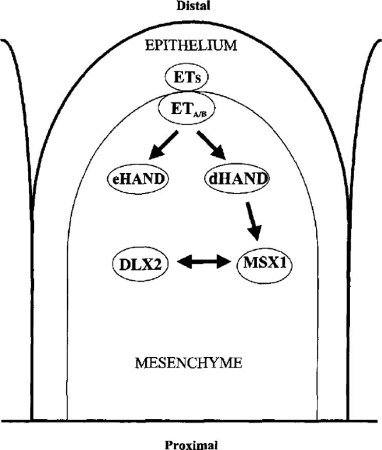
Endothelin 1 is expressed mainly in the epithelium of the pharyngeal arches, endothelium of the aortic arch artery, and cardiac outflow tracts; homozygous disruption of this gene results in malformations of pharyngeal arch-derived craniofacial structures, including a nonfused mandible (Kurihara et al., 1994,1995). The zebrafish craniofacial mutant sucker was also shown to be caused by a mutation in the etl gene (Miller et al., 2000). The exact role of Etl in neural crest development is unclear. However, one novel basic helixloop-helix transcriptional factor, dHAND, has been shown to interact with Etl in controlling neural crest branchial arch formation in mice (Thomas et al., 1998). Expression of dHAND is limited to the mesenchyme of the distal branchial arches, just beneath the Etlexpressing epithelium. In dHAND null mutant mice, lack of expression of dHAND in branchial and aortic arches results in the branchial arches becoming hypoplastic at s early as E9.5, apparently secondary to the programmed cell death of the mesenchyme. Furthermore, Msxl expression is undetectable in dHAND-/- mutant mice.
|
|
|
FIG. 3.2. Endothelin-HAND-MSX pathway: proposed model for regulation of branchial arch outgrowth. Endothelins (ETs) are secreted from the epithelial layer of the branchial arch into the mesenchyme. They bind to receptors (ETA/B), which are expressed in mesenchymal and surrounding endothelial cells. This binding upregulates the expression of two basic helix-loop-helix transcription factors, dHAND and eHAND, in the distal mesenchyme. In turn dHAND upregulates expression of the homeobox protein MSXI also in the distal mesenchyme. MSXI interacts with another homeobox protein, DLX, and they regulate each other in an antagonistic manner. An appropriate balance between MSXI and DLX is probably crucial in maintaining normal cell proliferation, differentiation, and death in the branchial arch, which is essential for formation of the secondary palate. |
These data suggest a potential molecular model for the regulation of branchial arch outgrowth. In this model, Etl is secreted from the branchial arch epithelium into the mesenchyme, while its receptor, EtA, is expressed in mesenchymal cells and surrounding endothelial cells (Fig. 3.2). Intracellular signaling is initiated by the binding of secreted Etl to activated EtA receptor, which enhances the expression of dHAND in the mesenchyme. In turn, dHAND upregulates expression of Msxl in the distal branchial arch (Thomas et al., 1998). An appropriate balance between Msxl and its modulator Dlx2 in the distal branchial arch may be crucial for normal proliferation, differentiation, and cell death in the branchial arch, processes necessary for formation of a continuous mesenchyme in the secondary palate.
Jagged Molecules and Notch Receptors
The Notch family of receptors are important signaling molecules regulating cell fate during development. They are membrane-bound and have a large extracellular domain containing multiple tandemly arranged EGF-like repeats (Rebay et al., 1991). Jaggedl and Jagged2 (also known as Serrate 1 and Serrate2) are transmembrane ligands for the Notch receptors (reviewed in Artavanis-Tsakonas et al., 1995; Robey, 1997; Francis-West et al., 1998). Their extracellular domains also contain multiple EGF-like motifs as well as a conserved motif called the DSL (delta, serrate, lag-2) domain. The DSL domain is required for interaction with their receptors.
Binding of Jaggedl and Jagged2 to the Notch receptors elicits a series of intracellular processes involving receptor proteolysis and interactions with several novel cytoplasmic and nuclear proteins (Artavanis-Tsakonas et al., 1995; Kopan et al., 1996). The Notch intercellular signaling pathway is evolutionarily conserved. It is essential for proper cell-fate specification and embryonic development in a diverse group of organisms (reviewed in Artavanis-Tsakonas et al., 1995; Gridley, 1997; Robey, 1997). In humans, mutations in the Notch genes have been implicated in cancer and an inherited condition causing stroke and dementia (Ellisen et al., 1991; Joutel et al., 1996).
In contrast, Jaggedl and Jagged2 proteins play a role in craniofacial and limb development. In mice, Jaggedl is expressed in both developing maxillary and mandibular primordia (Mitsiadis et al., 1997), while Jagged2 is expressed in the first to third branchial arches and the developing limb (Valsecchi et al., 1997). Haploinsufficiency of the JAGl gene in humans causes Alagille syndrome (Alagille et al., 1987; Li et al., 1997; Oda et al., 1997). This syndrome is characterized by a broad forehead, deep-set eyes, malformed ears, long straight nose, and pointed mandible. Some cases of Alagille syndrome result from truncation of the Jaggedl protein, which acts in a dominant-negative fashion during craniofacial development. A single-amino acid change in the Jagged2 protein is thought to be responsible for the mouse syndactylism (sra) mutant, in which the digits of the feet are fused (Sidow et al., 1997). In contrast, targeted deletion of the Jag2 exons encoding the DSL domain results in craniofacial defects and perinatal lethality in mice (Jiang et al., 1998). Jag2-/- mutant mice have CP mainly due to failure of the palatal shelves to elevate and fuse. In addition, these mutant mice exhibit syndactyly of the fore- and hindlimbs. The limb defects are more severe than those seen in sm mutants. These studies suggest that Notch signaling mediated by Jagged2 plays an essential role during limb and craniofacial development in mice and that sm is a hypomorphic allele of the Jag2 gene.
Thrombospondins
Thrombospondins (TSPs) are a family of multifunctional, extracellular matrix glycoproteins secreted by a variety of cells and consisting of five members (TSP1, TSP2, TSP3, TSP4, and COMP/TSP5) encoded by distinct genes (reviewed in Bornstein, 1992; Vacca et al., 1999). The first, TSP1, is a homotrimeric multidomain glycoprotein. Each monomer contains an aminoterminal heparin binding domain, followed by a procollagen homology domain, three TSP (properidin-like) repeats, three EGF-like repeats, seven Ca2+-binding repeats, and a carboxy-terminal cell attachment domain (Bornstein and Sage, 1994). The TSPs have an affinity for cell surfaces and extracellular matrix macromolecules such as fibronectin, laminin, heparan sulfate proteoglycans, plasminogen, collagens, and histidine-rich glycoproteins (Lawler, 1986; Frazier, 1987). For example, TSP1 is involved in platelet aggregation, inflammation, and inhibition of angiogenesis, as well as cell adhesion, migration, growth, and differentiation (Bornstein and Sage, 1994).
As discussed in an earlier section, the TGF-β family is one of the numerous growth factor families involved in modulating mesenchymal proliferation of the palatal shelves. There are several pathways available for activating the TGF-βs, one of which utilizes TSPs. Expression of TSPs is prominent in head mesenchyme, including the palate. Tspl and Tsp2 are expressed in different but overlapping regions of cartilage and bone mesenchyme (Tooney et al., 1998; Melnick et al., 2000).
Tspl activates latent Tgf-β1, while Tsp2 functions as an antagonist to the function of Tspl by competitively binding latent TGF-β and not activating it (Crawford et al., 1998; Melnick et al., 2000).
Although the precise roles of Tspl and Tsp2 in embryogenesis are poorly understood, studies based on their transcript expression and protein localization shed some light on their functions during palatogenesis. High expression of Tspl, Tsp2, and Tgf-β mRNAs and high protein quantities are localized throughout the extracellular matrix of the palatal mesenchyme (Melnick et al., 2000). At the vertical palatal shelf stage of palatogenesis, Tsp2 protein is found throughout the extracellular matrix of the shelf mesenchyme. By the horizontal palatal shelf stage, Tsp2 protein is far less evident in the palatal shelf proper. It is principally localized in the ossification centers of the maxilla, including the palate. This is consistent with its previously purported role in craniofacial morphogenesis (Tooney et al., 1998). However, in Thbsl (Tsp2) null mutant mice, CP or other craniofacial anomalies are absent (Kyriakides et al., 1998), suggesting that Tsp2 may not be an essential participant in palatogenesis.
γ-Aminobutyric Acid and Receptors
γ-Aminobutyric acid (GABA) is a major inhibitory neurotransmitter with many critical functions as an intercellular signaling molecule in the nervous system and in a number of nonneuronal cell types (reviewed in Erdo and Wolff, 1990; Chebib and Johnston, 1999). In the GABA-utilizing neurons of the central nervous system, it is synthesized from glutamic acid by glutamic acid decarboxylase (GAD). Outside of the central nervous system, GABA is synthesized by a number of cell types, including pancreatic β cells. It binds to GABA receptors, namely GABAA, GABAB, and GABAC. Both GABAA and GABAcbelong to a superfamily of transmitter-gated ion channels that includes nicotinic acetylcholine, strychnine-sensitive glycine, and 5-hydroxytryptamine 3 receptors. The GABAA receptors are heterooligomeric Cl- channels that can be selectively blocked by the alkaloid bicuculline and modulated by steroids, barbiturates, and benzodiazepines. In contrast, GABAc receptors represent a relatively simple form of Cl-channel made up of a single type of protein subunit. The GABAB receptors are heterooligomeric receptors coupled to G proteins and can activate second-messenger systems and Ca2+ and K+ ion channels upon binding GABA (Chebib and Johnston, 1999).
Cell culture studies have demonstrated that GABA is capable of promoting the survival, differentiation, and migration of embryonic neurons (Barbin et al., 1993; Liu et al., 1997). These observations suggest a role for GABA and its receptors in normal embryonic and fetal development. In addition, both genetic and teratological studies have suggested that GABA signaling may be involved in normal craniofacial development, including palate formation (Zimmerman and Wee, 1984; Culiat et al., 1993, 1995; Homanics et al., 1997). Teratological studies have shown that drugs such as diazepam, which can potentiate GABA action, are capable of inducing CP formation during a critical phase of mouse palate development (Miller and Becker, 1975; Wee and Zimmerman, 1983). However, the CP phenotype is observed only at high drug doses, suggesting that the effect may be nonspecific. Nonetheless, mice lacking the GABAA receptor β3 subunit (Gabrβ3) show clefting of the secondary palate (Culiat et al., 1993, 1995; Homanics et al., 1997). Furthermore, mice deficient in Gad67, one of two GAD enzymes in mice, show a similar phenotype (Condie et al., 1997), clearly demonstrating a role for GABA signaling in normal palatogenesis.
RYK
The RYK (related to tyrosine kinases) gene encodes a catalytically inactive member of the receptor protein tyrosine kinase (RTK) family (Hovens et al., 1992; Halford et al., 1999; Katso et al., 1999). The RTKs typically are transmembrane signal-transduction glycoproteins with an extracellular N-terminal ligand binding domain and an intracellular C-terminal tyrosine kinase domain (reviewed in van der Geer et al., 1994). They regulate diverse cellular functions, among them mitogenesis, differentiation, and morphogenesis. Because of its inability to undergo autophosphorylation or to phosphorylate substrates, RYK is unique within the RTK family. Despite this, it is able to activate downstream signaling pathways (Katso et al., 1999, and references therein).
In a study by Halford et al. (2000), most mice homozygous for targeted disruption of the Ryk gene failed to survive more than 24 h. In addition to some limb abnormalities, these mice have a slightly smaller and more rounded cranial vault, reflecting minor changes in size and shape of individual calvarial elements. They have a shorter snout due to shortened nasal bones, a flattened midface due to premaxillary/maxillary hypoplasia, and a reduced mandible. In addition, over 88% of the mice presented with a completely cleft secondary palate. These results indicate that Ryk is essential for normal development of craniofacial structures, including the secondary palate. These authors also provide biochemical evidence to suggest that Ryk activates downstream signaling via interaction with other members of different RTK subfamilies.
Future Perspectives
The preceding list of candidate genes for lip and palate development is by no means exhaustive and underscores the concept that CL and CP are a complex, multifactorial group of disorders. The interactions of multiple genes and their polymorphisms might explain the observed variations in normal and abnormal lip and palate development. Several of these genes have been extensively characterized with respect to their roles in lip and palate development. However, evidence for the role of the other genes in lip and palate pattern formation is less conclusive, and our current understanding of the genetic factors controlling these processes remains rudimentary.
Completion of the sequencing phase of the Human Genome Project and transition to the functional genomics phase holds the promise of unraveling the complex inter- and intracellular signaling pathways controlling this developmental process. Continued utilization of targeted mutagenesis in the mouse and application of random mutagenesis methods in the mouse and other vertebrate model systems, such as the zebrafish, should accelerate the pace of gene discovery and functional analyses.
Acknowledgements
Work on this chapter was sponsored in part by National Institutes of Health grants DE 13078, DE 13939 (E. W. J.), and DE 13707 (S. S. C. and E. W. J.) and by Johns Hopkins Singapore (S. S. C., F. S. H. C., and E. W. J.).
References
Agulnick, AD, Taira, M, Breen, JJ, et al. (1996). Interactions of the LIM-domain-binding factor Ldbl with LIM homeodomain proteins. Nature 384: 270–272.
Alagille, D, Estrada, A, Hadchouel, M, et al. (1987). Syndromic paucity of interlobular bile ducts (Alagille syndrome or arteriohepatic dysplasia): review of 80 cases. J Pediatr 110: 195–200.
Arai, H, Hori, S, Aramori, I, et al. (1990). Cloning and expression of a cDNA encoding an endothelin receptor. Nature 348: 730–732.
Ardinger, HH, Buetow, KH, Bell, GI, et al. (1989). Association of genetic variation of the transforming growth factor-alpha gene with cleft lip and palate. Am J Hum Genet 45: 348–353.
Artavanis-Tsakonas, S, Matsuno, K, Fortini, ME (1995). Notch signaling. Science 268: 225–232.
Bach, I, Carriere, C, Ostendorff, HP, et al. (1997). A family of LIM domain-associated cofactors confer transcriptional synergism between LIM and Otx homeodomain proteins. Genes Dev 11: 1370–1380.
Barbin, G, Pollard, H, Gaiarsa, JL, Ben-Ari, Y (1993). Involvement of GABAA receptors in the outgrowth of cultured hippocampal neurons. Neurosci Lett 252: 150–154.
Basilico, C, Moscatelli, D (1992). The FGF family of growth factors and oncogenes. Adv Cancer Res 59: 115–165.
Baynash, AG, Hosoda, K, Giaid, A, et al. (1994). Interaction of endothelin-3 with endothelin-B receptor is essential for development of epidermal melanocytes and enteric neurons. Cell 79: 1277–1285.
Beiraghi, S, Foroud, T, Diouhy, S, et al. (1994). Possible localization of a major gene for cleft lip and palate to 4q. Clin Genet 46: 255–256.
Blum, M, Gaunt, SJ, Cho, KW, et al. (1992). Gastrulation in the mouse: the role of the homeobox gene goosecoid. Cell 69: 1097–1106.
Blumberg, B, Wright, CV, De Robertis, EM, Cho, KW (1991). Organizer-specific homeobox genes in Xenopus laevis embryos. Science 253: 194–196.
Bornstein, P (1992). Thrombospondins: structure and regulation of expression [published erratum appears in FASEB J (1993) 7: 237]. FASEB J 6: 3290–3299.
Bornstein, P, Sage, EH (1994). Thrombospondins. Methods Enzymol 245: 62–85.
Brachmann, R, Lindquist, PB, Nagashima, M, et al. (1989). Transmembrane TGF-α precursors activate EGF/TGF-α receptors. Cell 56: 691–700.
Breen, JJ, Agulnick, AD, Westphal, H, Dawid, IB (1998). Interactions between LIM domains and the LIM domain-binding protein Ldbl. J Biol Chem 273: 4712–4717.
Brunei, CL, Sharpe, PM, Ferguson, MW (1995). Inhibition of TGF- β3 (but not TGF-β1 or TGF-β2) activity prevents normal mouse embryonic palate fusion. Int J Dev Biol 39: 345–355.
Catron, KM, Wang, H, Hu, G, et al. (1996). Comparison of MSX-1 and MSX-2 suggests a molecular basis for functional redundancy [published erratum appears in Mech Dev (1996) 56: 223]. Mech Dev 55: 185–199.
Catron, KM, Zhang, H, Marshall, SC, et al. (1995). Transcriptional repression by Msx-1 does not require homeodomain DNA-binding sites. Mol Cell Biol 15: 861–871.
Celli, G, LaRochelle, WJ, Mackem, S, et al. (1998). Soluble dominantnegative receptor uncovers essential roles for fibroblast growth factors in multi-organ induction and patterning. EMBO J 17: 1642–1655.
Chebib, M, Johnston, GA (1999). The “ABC” of GABA receptors: a brief review. Clin Exp Pharmacol Physiol 26: 937–940.
Chenevix-Trench, G, Jones, K, Green, AC, et al. (1992). Cleft lip with or without cleft palate: associations with transforming growth factor alpha and retinoic acid receptor loci. Am J Hum Genet 51: 1377–1385.
Cho, KW, Blumberg, B, Steinbeisser, H, De Robertis, EM (1991). Molecular nature of Spemann's organizer: the role of the Xenopus homeobox gene goosecoid. Cell 67: 1111–1120.
Clouthier, DE, Hosoda, K, Richardson, JA, et al. (1998). Cranial and cardiac neural crest defects in endothelin-A receptor-deficient mice. Development 125: 813–824.
Cohen, B, McGuffin, ME, Pfeifle, C, et al. (1992). apterous, a gene required for imaginal disc development in Drosophila encodes a member of the LIM family of developmental regulatory proteins. Genes Dev 6: 715–729.
Colvin, JS, Feldman, B, Nadeau, JH, et al. (1999). Genomic organization and embryonic expression of the mouse fibroblast growth factor 9 gene. Dev Dyn 216: 72–88.
Condie, BG, Bain, G, Gottlieb, DI, Capecchi, MR (1997). Cleft palate in mice with a targeted mutation in the γ-Aminobutyric acidproducing enzyme glutamic acid decarboxylase 67. Proc Natl Acad Sci USA 94: 11451–11455.
Crawford, SE, Stellmach, V, Murphy-Ullrich, JE, et al. (1998). Thrombospondin-1 is a major activator of TGF-β1 in vivo. Cell 93: 1159–1170.
Crossley, PH, Martin, GR (1995). The mouse Fgf8 gene encodes a family of polypeptides and is expressed in regions that direct outgrowth and patterning in the developing embryo. Development 121: 439–451.
Culiat, CT, Stubbs, L, Nicholls, RD, et al. (1993). Concordance between isolated cleft palate in mice and alterations within a region including the gene encoding the β3 subunit of the type A yaminobutyric acid receptor. Proc Natl Acad Sci USA 90: 5105–5109.
Culiat, CT, Stubbs, LJ, Woychik, RP, et al. (1995). Deficiency of the β3 subunit of the type A γ-Aminobutyric acid receptor causes cleft palate in mice. Nat Genet 11: 344–346.
Davideau, JL, Demri, P, Gu, TT, et al. (1999). Expression of DLX5 during human embryonic craniofacial development. Mech Dev 81: 183–186.
Dawid, IB, Toyama, R, Taira, M (1995). LIM domain proteins. C R Acad Sci III 318: 295–306.
Derynck, R, Roberts, AB, Winkler, ME, et al. (1984). Human transforming growth factor-alpha: precursor structure and expression in E. coli. Cell 38: 287–297.
Dixon, MJ, Garner, J, Ferguson, MW (1991). Immunolocalization of epidermal growth factor (EGF), EGF receptor and transforming growth factor alpha (TGF-α) during murine palatogenesis in vivo and in vitro. Anat Embryol 184: 83–91.
Dolle, P, Price, M, Duboule, D (1992). Expression of the murine Dlx-1 homeobox gene during facial, ocular and limb development. Differentiation 49: 93–99.
Drucker, BJ, Goldfarb, M (1993). Murine FGF-4 gene expression is spatially restricted within embryonic skeletal muscle and other tissues. Mech Dev 40: 155–163.
Ellisen, LW, Bird, J, West, DC, et al. (1991). TAN-1, the human homolog of the Drosophila notch gene, is broken by chromosomal translocations in T lymphoblastic neoplasms. Cell 66: 649–661.
Emoto, N, Yanagisawa, M (1995). Endothelin-converting enzyme-2 is a membrane-bound, phosphoramidon-sensitive metalloprotease with acidic pH optimum. J Biol Chem 270: 15262–15268.
Erdo, SL, Wolff, JR (1990). γ-Aminobutyric acid outside the mammalian brain. J Neurochem 54: 363–372.
Feijen, A, Goumans, MJ, van den Eijnden-van Raaij, AJ (1994). Expression of activin subunits, activin receptors and follistatin in postimplantation mouse embryos suggests specific developmental functions for different activins. Development 120: 3621–3637.
Ferguson, MW (1988). Palate development. Development 203: 41–60.
Feuerstein, R, Wang, X, Song, D, et al. (1994). The LIM/double zincfinger motif functions as a protein dimerization domain. Proc Natl Acad Sci USA 91: 10655–10659.
Fitzpatrick, DR, Denhez, F, Kondaiah, P, Akhurst, RJ (1990). Differential expression of TGF-β isoforms in murine palatogenesis. Development 109: 585–595.
Francis-West, P, Ladher, R, Barlow, A, Graveson, A (1998). Signalling interactions during facial development. Mech Dev 75: 3–28.
Frazier, WA (1987). Thrombospondin: a modular adhesive glycoprotein of platelets and nucleated cells. J Cell Biol 205: 625–632.
Freyd, G, Kim, SK, Horvitz, HR (1990). Novel cysteine-rich motif and homeodomain in the product of the Caenorhabditis elegans cell lineage gene lin-11. Nature 344: 876–879.
Gaunt, SJ, Blum, M, De Robertis, EM (1993). Expression of the mouse goosecoid gene during mid-embryogenesis may mark mesenchymal cell lineages in the developing head, limbs and body wall. Development 117: 769–778.
Goriely, A, Stella, M, Coffinier, C, et al. (1996). A functional homologue of goosecoid in Drosophila. Development 222: 1641–1650.
Gridley, T (1997). Notch signaling in vertebrate development and disease. Mol Cell Neurosci 9: 103–108.
Grigoriou, M, Tucker, AS, Sharpe, PT, Pachnis, V (1998). Expression and regulation of Lhx6 and Lhx7, a novel subfamily of LIM homeodomain encoding genes, suggests a role in mammalian head development. Development 225: 2063–2074.
Halford, MM, Armes, J, Buchert, M, et al. (2000). Ryk-deficient mice exhibit craniofacial defects associated with perturbed Eph receptor crosstalk. Nat Genet 25: 414–18.
Halford, MM, Gates, AC, Hibbs, ML, et al. (1999). Genomic structure and expression of the mouse growth factor receptor related to tyrosine kinases (Ryk). J Biol Chem 274: 7379–7390.
Heldin, CH, Westermark, B (1999). Mechanism of action and in vivo role of platelet-derived growth factor. Physiol Rev 79: 1283–1316.
Hill, RE, Jones, PF, Rees, AR, et al. (1989). A new family of mouse homeo box-containing genes: molecular structure, chromosomal location, and developmental expression of Hox-7.1. Genes Dev 3: 26–37.
Holder, SE, Vintiner, GM, Farren, B, et al. (1992). Confirmation of an association between RFLPs at the transforming growth factor-alpha locus and non-syndromic cleft lip and palate. J Med Genet 29: 390–392.
Holland, PW (1991). Cloning and evolutionary analysis of msh-iike homeobox genes from mouse, zebrafish and ascidian. Gene 98: 253–257.
Homanics, GE, DeLorey, TM, Firestone, LL, et al. (1997). Mice devoid of y-aminobutyrate type A receptor β3 subunit have epilepsy, cleft palate, and hypersensitive behavior. Proc Natl Acad Sci USA 94: 4143–148.
Hosoda, K, Hammer, RE, Richardson, JA, et al. (1994). Targeted and natural (piebald-lethal) mutations of endothelin-B receptor gene produce megacolon associated with spotted coat color in mice. Cell 79: 1267–1276.
Hovens, CM, Stacker, SA, Andres, AC, et al. (1992). RYK, a receptor tyrosine kinase-related molecule with unusual kinase domain motifs. Proc Natl Acad Sci USA 89: 11818–11822.
Inoue, A, Yanagisawa, M, Kimura, S, et al. (1989). The human endothelin family: three structurally and pharmacologically distinct isopeptides predicted by three separate genes. Proc Natl Acad Sci USA 86: 2863–2867.
Jabs, EW, Muller, U, Li, X, et al. (1993). A mutation in the homeodomain of the human MSX2 gene in a family affected with autosomal dominant craniosynostosis. Cell 75: 443–450.
Jara, L, Blanco, R, Chiffelle, I, et al. (1995). Evidence for an association between RFLPs at the transforming growth factor alpha (locus) and nonsyndromic cleft lip/palate in a South American population. Am J Hum Genet 56: 339–341.
Jiang, R, Lan, Y, Chapman, HD, et al. (1998). Defects in limb, craniofacial, and thymic development inJaggedZ mutant mice. Genes Dev 22: 1046–1057.
Joutel, A, Corpechot, C, Ducros, A, et al. (1996). Notch3 mutations in CADASIL, a hereditary adult-onset condition causing stroke and dementia. Nature 383: 707–710.
Jurata, LW, Kenny, DA, Gill, GN (1996). Nuclear LIM interactor, a rhombotin and LIM homeodomain interacting protein, is expressed early in neuronal development. Proc Natl Acad Sci USA 93: 11693–11698.
Kaartinen, V, Cui, XM, Heisterkamp, N, et al. (1997). Transforming growth factor-β3 regulates transdifferentiation of medial edge epithelium during palatal fusion and associated degradation of the basement membrane. Dev Dyn 209: 255–260.
Kaartinen, V, Voncken, JW, Shuler, C, et al. (1995). Abnormal lung development and cleft palate in mice lacking TGF-β3 indicates defects of epithelial-mesenchymal interaction. Nat Genet 22: 415–21.
Katso, RM, Russell, RB, Ganesan, TS (1999). Functional analysis of H-Ryk, an atypical member of the receptor tyrosine kinase family. Mol Cell Biol 29: 6427–6440.
Kingsley, DM (1994). The TGF-β superfamily: new members, new receptors, and new genetic tests of function in different organisms. Genes Dev 8: 133–146.
Kitanaka, J, Takemura, M, Matsumoto, K, et al. (1998). Structure and chromosomal localization of a murine LIM/homeobox gene, Lhx8. Genomics 49: 307–309.
Kopan, R, Schroeter, EH, Weintraub, H, Nye, JS (1996). Signal transduction by activated mNotch: importance of proteolytic processing and its regulation by the extracellular domain. Proc Natl Acad Sci USA 93: 1683–1688.
Krumlauf, R (1993). Hox genes and pattern formation in the branchial region of the vertebrate head. Trends Genet 9: 106–112.
Kurihara, Y, Kurihara, H, Oda, H, et al. (1995). Aortic arch malformations and ventricular septal defect in mice deficient in endothelin- 1. J Clin Invest 96: 293–300.
Kurihara, Y, Kurihara, H, Suzuki, H, et al. (1994). Elevated blood pressure and craniofacial abnormalities in mice deficient in endothelin- 1. Nature 368: 703–710.
Kyriakides, TR, Zhu, YH, Smith, LT, et al. (1998). Mice that lack thrombospondin 2 display connective tissue abnormalities that are associated with disordered collagen fibrillogenesis, an increased vascular density, and a bleeding diathesis. J Cell Biol 140: 419–430.
Lawler, J (1986). The structural and functional properties of thrombospondin. Blood 67: 1197–1209.
Leussink, B, Brouwer, A, Khattabi, ME, et al. (1995). Expression patterns of the paired-related homeobox genes MHox/Prxl and S8/Prx2 suggest roles in development of the heart and the forebrain. Mech Dev 52: 51–64.
Li, L, Krantz, ID, Deng, Y, et al. (1997). Alagille syndrome is caused by mutations in human Jaggedl, which encodes a ligand for Notchl. Nat Genet 16: 243–251.
Lidral, AC, Murray, JC, Buetow, KH, et al. (1997). Studies of the candidate genes TGFB2, MSX1, TGFA, and TGFB3 in the etiology of cleft lip and palate in the Philippines. Cleft Palate Craniofac J 34: 1–6.
Lidral, AC, Romitti, PA, Basart, AM, et al. (1998). Association of MSX1 and TGFB3 with nonsyndromic clefting in humans. Am J Hum Genet 63: 557–568.
Liu, J, Morrow, AL, Devaud, L, et al. (1997). GABAA receptors mediate trophic effects of GABA on embryonic brainstem monoamine neurons in vitro. J Neurosci 17: 2420–2428.
Lu, MF, Cheng, HT, Kern, MJ, et al. (1999). prx-1 functions cooperatively with another paired-related homeobox gene, prx-2, to maintain cell fates within the craniofacial mesenchyme. Development 126: 495–504.
Luetteke, NC, Qiu, TH, Peiffer, RL, et al. (1993). TGF-α deficiency results in hair follicle and eye abnormalities in targeted and waved-1 mice. Cell 73: 263–278.
Ma, L, Golden, S, Wu, L, Maxson, R (1996). The molecular basis of Boston-type craniosynostosis: the Prol48 → His mutation in the N-terminal arm of the MSX2 homeodomain stabilizes DNA binding without altering nucleotide sequence preferences. Hum Mol Genet 5: 1915–1920.
Machida, J, Yoshiura, K, Funkhauser, CD, et al. (1999). Transforming growth factor-alpha (TGFA): genomic structure, boundary sequences, and mutation analysis in nonsyndromic cleft lip/palate and cleft palate only. Genomics 61: 237–242.
MacKenzie, A, Ferguson, MW, Sharpe, PT (1991a). Hox-7 expression during murine craniofacial development. Development 113: 601–611.
Mackenzie, A, Leeming, GL, Jowett, AK, et al. (1991b). The homeobox gene Hox 7.1 has specific regional and temporal expression patterns during early murine craniofacial embryogenesis, especially tooth development in vivo and in vitro. Development 111: 269–285.
Mailhos, C, Andre, S, Mollereau, B, et al. (1998). Drosophila Goosecoid requires a conserved heptapeptide for repression of paired-class homeoprotein activators. Development 125: 937–947.
Martin, JF, Bradley, A, Olson, EN (1995). The paired-like homeobox gene MHox is required for early events of skeletogenesis in multiple lineages. Genes Dev 9: 1237–1249.
Matsumoto, K, Tanaka, T, Furuyama, T, et al. (1996). L3, a novel murine LIM-homeodomain transcription factor expressed in the ventral telencephalon and the mesenchyme surrounding the oral cavity. Neurosci Lett 204: 113–116.
Matzuk, MM, Kumar, TR, Vassalli, A, et al. (1995a). Functional analysis of activins during mammalian development. Nature 3 74: 354–356.
Matzuk, MM, Kumar, TR, Bradley, A (1995b). Different phenotypes for mice deficient in either activins or activin receptor type II. Nature 374: 356–360.
Matzuk, MM, Lu, N, Vogel, H, et al. (1995c). Multiple defects and perinatal death in mice deficient in follistatin. Nature 374: 360–363.
Melnick, M, Chen, H, Zhou, Y, Jaskoll, T (2000). Thrombospondin- 2 gene expression and protein localization during embryonic mouse palate development. Arch Oral Biol 45: 19–25.
Miettinen, PJ, Chin, JR, Shum, L, et al. (1999). Epidermal growth factor receptor function is necessary for normal craniofacial development and palate closure. Nat Genet 22: 69–73.
Millan, FA, Denhez, F, Kondaiah, P, Akhurst, RJ (1991). Embryonic gene expression patterns of TGF /SI, jS2 and β3 suggest different developmental functions in vivo. Development 111: 131–143.
Miller, CT, Schilling, TF, Lee, K, et al. (2000). sucker encodes a zebrafish endothelin-1 required for ventral pharyngeal arch development. Development 127: 3815–3828.
Miller, RP, Becker, BA (1975). Teratogenicity of oral diazepam and diphenylhydantoin in mice. Toxicol Appl Pharmacol 32: 53–61.
Mina, M, Gluhak, J, Upholt, WB, et al. (1995). Experimental analysis of Msx-1 and Msx-2 gene expression during chick mandibular morphogenesis. Dev Dyn 202: 195–214.
Mitchell, LE, Healey, SC, Chenevix-Trench, G (1995). Evidence for an association between nonsyndromic cleft lip with or without cleft palate and a gene located on the long arm of chromosome 4. Am J Hum Genet 57: 1130–1136.
Mitsiadis, TA, Henrique, D, Thesleff, I, Lendahl, U (1997). Mouse Serrate-1 (Jagged-1): expression in the developing tooth is regulated by epithelial-mesenchymal interactions and fibroblast growth factor-4. Development 124: 1473–1483.
Morcillo, P, Rosen, C, Baylies, MK, Dorsett, D (1997). Chip, a widely expressed chromosomal protein required for segmentation and activity of a remote wing margin enhancer in Drosophila. Genes Dev 11: 2729–2740.
Morrison-Graham, K, Schatteman, GC, Bork, T, et al. (1992). A PDGF receptor mutation in the mouse (Patch) perturbs the development of a non-neuronal subset of neural crest-derived cells. Development 115: 133–142.
Murray, JC (1995). Face facts: genes, environment, and clefts. Am J Hum Genet 57: 227–232.
Nakamura, S, Stock, DW, Wydner, KL, et al. (1996). Genomic analysis of a new mammalian distal-less gene: Dlx7. Genomics 38: 314–324.
Niswander, L, Martin, GR (1992). Fgf-4 expression during gastrulation, myogenesis, limb and tooth development in the mouse. Development 114: 755–768.
Nohno, T, Koyama, E, Myokai, F, et al. (1993). A chicken homeobox gene related to Drosophila paired is predominantly expressed in the developing limb. Dev Biol 158: 254–264.
Norris, RA, Scott, KK, Moore, CS, et al. (2000). Human PRRX1 and PRRX2 genes: cloning, expression, genomic localization, and exclusion as disease genes for Nager syndrome. Mamm Genome 11: 1000–1005.
Oda, T, Elkahloun, AG, Pike, BL, et al. (1997). Mutations in the human Jaggedl gene are responsible for Alagille syndrome. Nat Genet 16: 235–242.
Ohuchi, H, Yoshioka, H, Tanaka, A, et al. (1994). Involvement of androgen-induced growth factor (FGF-8) gene in mouse embryogenesis and morphogenesis. Biochem Biophys Res Commun 204: 882–888.
Orr-Urtreger, A, Lonai, P (1992). Platelet-derived growth factor-A and its receptor are expressed in separate, but adjacent cell layers of the mouse embryo. Development 115: 1045–1058.
Passos-Bueno, MR, Wilcox, WR, Jabs, EW, et al. (1999). Clinical spectrum of fibroblast growth factor receptor mutations. Hum Mutat 14: 115–125.
Pavlova, A, Boutin, E, Cunha, G, Sassoon, D (1994). Msxl (Hox- 7.1) in the adult mouse uterus: cellular interactions underlying regulation of expression. Development 120: 335–345.
Pelton, RW, Nomura, S, Moses, HL, Hogan, BL (1989). Expression of transforming growth factor j32 RNA during murine embryogenesis. Development 106: 759–767.
Pelton, RW, Saxena, B, Jones, M, et al. (1991). Immunohistochemical localization of TGF)81, TGF β2, and TGF β3 in the mouse embryo: expression patterns suggest multiple roles during embryonic development. J Cell Biol 115: 1091–1105.
Phillips, DJ (2000). Regulation of activin's access to the cell: why is mother nature such a control freak? Bioessays 22: 689–696.
Porteus, MH, Bulfone, A, Ciaranello, RD, Rubenstein, JL (1991). Isolation and characterization of a novel cDNA clone encoding a homeodomain that is developmentally regulated in the ventral forebrain [published erratum appears in Neuron (1992) 9: 187]. Neuron 7: 221–229.
Price, M, Lemaistre, M, Pischetola, M, et al. (1991). A mouse gene related to Distal-less shows a restricted expression in the developing forebrain. Nature 352: 748–751.
Proetzel, G, Pawlowski, SA, Wiles, MV, et al. (1995). Transforming growth factor-β3 is required for secondary palate fusion. Nat Genet 22: 409–414.
Qiu, CX, Ferguson, MW (1995). The distribution of PDGFs and PDGF-receptors during murine secondary palate development. J Anat 186: 17–29.
Qiu, M, Bulfone, A, Ghattas, I, et al. (1997). Role of the Dlx homeobox genes in proximodistal patterning of the branchial arches: mutations of Dlx-1, Dlx-2, and Dlx-1 and -2 alter morphogenesis of proximal skeletal and soft tissue structures derived from the first and second arches. Dev Biol 185: 165–184.
Qiu, M, Bulfone, A, Martinez, S, et al. (1995). Null mutation of Dlx- 2 results in abnormal morphogenesis of proximal first and second branchial arch derivatives and abnormal differentiation in the forebrain. Genes Dev 9: 2523–2538.
Rebay, I, Fleming, RJ, Fehon, RG, et al. (1991). Specific EGF repeats of Notch mediate interactions with Delta and Serrate: implications for Notch as a multifunctional receptor. Cell 67: 687–699.
Reginelli, AD, Wang, YQ, Sassoon, D, Muneoka, K (1995). Digit tip regeneration correlates with regions of Msxl (Hox 7) expression in fetal and newborn mice. Development 222: 1065–1076.
Richman, JM, Crosby, Z (1990). Differential growth of facial primordia in chick embryos: responses of facial mesenchyme to basic fibroblast growth factor (bFGF) and serum in micromass culture. Development 209: 341–348.
Richman, JM, Herbert, M, Matovinovic, E, Walin, J (1997). Effect of fibroblast growth factors on outgrowth of facial mesenchyme. Dev Biol 189: 135–147.
Riese, DJ II, Stern, DF (1998). Specificity within the EGF family/ErbB receptor family signaling network. Bioessays 20: 41–48.
Rivera-Perez, JA, Mallo, M, Gendron-Maguire, M, et al. (1995). Goosecoid is not an essential component of the mouse gastrula organizer but is required for craniofacial and rib development. Development 222: 3005–3012.
Rivera-Perez, JA, Wakamiya, M, Behringer, RR (1999). Goosecoid acts cell autonomously in mesenchyme-derived tissues during craniofacial development. Development 226: 3811–3821.
Roberts, VJ, Earth, SL (1994). Expression of messenger ribonucleic acids encoding the inhibin/activin system during mid- and lategestation rat embryogenesis. Endocrinology 234: 914–923.
Robey, E (1997). Notch in vertebrates. Curr Opin Genet Dev 7: 551–557.
Robinson, GW, Mahon, KA (1994). Differential and overlapping expression domains of Dlx-2 and Dlx-3 suggest distinct roles for Distal-less homeobox genes in craniofacial development. Mech Dev 48: 199–215.
Robinson, GW, Wray, S, Mahon, KA (1991). Spatially restricted expression of a member of a new family of murine Distal-less homeobox genes in the developing forebrain. New Biol 3: 1183–1194.
Romitti, PA, Lidral, AC, Munger, RG, et al. (1999). Candidate genes for non-syndromic cleft lip and palate and maternal cigarette smoking and alcohol consumption: evaluation of genotypeenvironment interactions from a population based case-control study of orofacial clefts. Teratology 59: 39–50.
Sakurai, T, Yanagisawa, M, Takuwa, Y, et al. (1990). Cloning of a cDNA encoding a non-isopeptide-selective subtype of the endothelin receptor. Nature 348: 732–735.
Sanchez-Garcia, I, Osada, H, Forster, A, Rabbitts, TH (1993). The cysteine-rich LIM domains inhibit DNA binding by the associated homeodomain in Isl-1. EMBO J 22: 4243–4250.
Sanford, LP, Ormsby, I, Gittenberger-de Groot, AC, et al. (1997). TGFβ2 knockout mice have multiple developmental defects that are non-overlapping with other TGFβ knockout phenotypes. Development 224: 2659–2670.
Sassani, R, Bartlett, SP, Feng, H, et al. (1993). Association between alleles of the transforming growth factor-alpha locus and the occurrence of cleft lip. Am J Med Genet 45: 565–569.
Satokata, I, Ma, L, Ohshima, H, et al. (2000). Msx2 deficiency in mice causes pleiotropic defects in bone growth and ectodermal organ formation. Nat Genet 24: 391–395.
Satokata, I, Maas, R (1994). Msxl deficient mice exhibit cleft palate and abnormalities of craniofacial and tooth development. Nat Genet 6: 348–356.
Schutte, BC, Murray, JC (1999). The many faces and factors of orofacial clefts. Hum Mol Genet 8: 1853–1859.
Sharma, K, Sheng, HZ, Lettieri, K, et al. (1998). LIM homeodomain factors Lhx3 and Lhx4 assign subtype identities for motor neurons. Cell 95: 817–828.
Shawlot, W, Behringer, RR (1995). Requirement for Liml in headorganizer function. Nature 374: 425–430.
Shimeld, SM, McKay, IJ, Sharpe, PT (1996). The murine homeobox gene Msx-3 shows highly restricted expression in the developing neural tube. Mech Dev 55: 201–210.
Sidow, A, Bulotsky, MS, Kerrebrock, AW, et al. (1997). Serrate2 is disrupted in the mouse limb-development mutant syndactylism. Nature 3S9: 722–725.
Simeone, A, Acampora, D, Pannese, M, et al. (1994). Cloning and characterization of two members of the vertebrate Dlx gene family. Proc Natl Acad Sci USA 92: 2250–2254.
Simon, HG, Nelson, C, Goff, D, et al. (1995). Differential expression of myogenic regulatory genes and Msx-1 during dedifferentiation and redifferentiation of regenerating amphibian limbs. Dev Dyn 202: 1–12.
Soriano, P (1997). The PDGF alpha receptor is required for neural crest cell development and for normal patterning of the somites. Development 124: 2691–2700.
Stoll, C, Qian, JF, Feingold, J, et al. (1992). Genetic variation in transforming growth factor alpha: possible association of BamHI polymorphism with bilateral sporadic cleft lip and palate. Am J Hum Genet 50: 870–871.
Taira, M, Otani, H, Saint-Jeannet, JP, Dawid, IB (1994). Role of the LIM class homeodomain protein Xlim-1 in neural and muscle induction by the Spemann organizer in Xenopus [published erratum appears in Nature (1995) 373: 451]. Nature 372: 677–679.
ten Berge, D, Brouwer, A, Korving, J, et al. (1998). Prxl and Prx2 in skeletogenesis: roles in the craniofacial region, inner ear and limbs. Development 125: 3831–3842.
Thomas, T, Kurihara, H, Yamagishi, H, et al. (1998). A signaling cascade involving endothelin-1, dHAND and msxl regulates development of neural-crest-derived branchial arch mesenchyme. Development 125: 3005–3014.
Tooney, PA, Sakai, T, Sakai, K, et al. (1998). Restricted localization of thrombospondin-2 protein during mouse embryogenesis: a comparison to thrombospondin-1. Matrix Biol 17: 131–143.
Treisman, J, Gonczy, P, Vashishtha, M, et al. (1989). A single amino acid can determine the DNA binding specificity of homeodomain proteins. Cell 59: 553–562.
Vacca, A, Di Marcotullio, L, Giannini, G, et al. (1999). Thrombospondin- 1 is a mediator of the neurotypic differentiation induced by EGF in thymic epithelial cells. Exp Cell Res 248: 79–86.
Valsecchi, C, Ghezzi, C, Ballabio, A, Rugarli, El (1997). JAGGED2: a putative Notch ligand expressed in the apical ectodermal ridge and in sites of epithelial-mesenchymal interactions. Mech Dev 69: 203–207.
van den Boogaard, MJ, Dorland, M, Beemer, FA, van Amstel, HK (2000). MSX1 mutation is associated with orofacial clefting and tooth agenesis in humans [published erratum appears in Nat Genet (2000) 25: 125]. Nat Genet 24: 342–343.
van der Geer, P, Hunter, T, Lindberg, RA (1994). Receptor proteintyrosine kinases and their signal transduction pathways. Annu Rev Cell Biol 10: 251–337.
Vassalli, A, Matzuk, MM, Gardner, HA, et al. (1994). Activin/inhibin βB subunit gene disruption leads to defects in eyelid development and female reproduction. Genes Dev 8: 414–427.
Vieille-Grosjean, I, Hunt, P, Gulisano, M, et al. (1997). Branchial HOX gene expression and human craniofacial development. Dev Biol 183: 49–60.
Wall, NA, Hogan, BL (1995). Expression of bone morphogenetic protein-4 (BMP-4), bone morphogenetic protein-7 (BMP-7), fibroblast growth factor-8 (FGF-8) and sonic hedgehog (SHH) during branchial arch development in the chick. Mech Dev 53: 383–392.
Wee, EL, Zimmerman, EF (1983). Involvement of GABA in palate morphogenesis and its relation to diazepam teratogenesis in two mouse strains. Teratology 28: 15–22.
Wilkie, AO, Tang, Z, Elanko, N, et al. (2000). Functional haploinsufficiency of the human homeobox gene MSX2 causes defects in skull ossification. Nat Genet 24: 387–390.
Winograd, J, Reilly, MP, Roe, R, et al. (1997). Perinatal lethality and multiple craniofacial malformations in MSX2 transgenic mice. Hum Mol Genet 6: 369–379.
Woloshin, P, Song, K, Degnin, C, et al. (1995). MSX1 inhibits myoD expression in fibroblast X 10T1/2 cell hybrids. Cell 82: 611–620.
Xu, D, Emoto, N, Giaid, A, et al. (1994). ECE-1: a membrane-bound metalloprotease that catalyzes the proteolytic activation of big endothelin- 1. Cell 78: 473–485.
Xue, D, Tu, Y, Chalfie, M (1993). Cooperative interactions between the Caenorhabditis elegans homeoproteins UNC-86 and MEC-3. Science 261: 1324–1328.
Yamada, G, Mansouri, A, Torres, M, et al. (1995). Targeted mutation of the murine goosecoid gene results in craniofacial defects and neonatal death. Development 121: 2917–2922.
Yamada, G, Ueno, K, Nakamura, S, et al. (1997). Nasal and pharyngeal abnormalities caused by the mouse goosecoid gene mutation. Biochem Biophys Res Commun 233: 161–165.
Yamaguchi, TP, Rossant, J (1995). Fibroblast growth factors in mammalian development. Curr Opin Genet Dev 5: 485–491.
Yanagisawa, H, Yanagisawa, M, Kapur, RP, et al. (1998). Dual genetic pathways of endothelin-mediated intercellular signaling revealed by targeted disruption of endothelin converting enzyme-1 gene. Development 125: 825–836.
Yanagisawa, M, Kurihara, H, Kimura, S, et al. (1988). A novel potent vasoconstrictor peptide produced by vascular endothelial cells. Nature 332: 411–415.
Zhang, H, Catron, KM, Abate-Shen, C (1996). A role for the Msx- 1 homeodomain in transcriptional regulation: residues in the Nterminal arm mediate TATA binding protein interaction and transcriptional repression. Proc Natl Acad Sci USA 93: 1764–1769.
Zhang, H, Hu, G, Wang, H, et al. (1997). Heterodimerization of Msx and Dlx homeoproteins results in functional antagonism. Mol Cell Biol 17: 2920–2932.
Zhao, Y, Guo, YJ, Tomac, AC, et al. (1999a). Isolated cleft palate in mice with a targeted mutation of the LIM homeobox gene Lhx8. Proc Natl Acad Sci USA 96: 15002–15006.
Zhao, Y, Sheng, HZ, Amini, R, et al. (1999b). Control of hippocampal morphogenesis and neuronal differentiation by the LIM homeobox gene LhxS. Science 284: 1155–1158.
Zimmerman, EF, Wee, EL (1984). Role of neurotransmitters in palate development. Curr Top Dev Biol 19: 37–63.