Michael M. Cohen Jr.
Cleft lip with or without cleft palate is etiologically distinct from isolated cleft palate. In a patient with cleft lip with or without cleft palate, if another family member is affected, he or she will have either isolated cleft lip or cleft lip together with cleft palate but not isolated cleft palate alone. Similarly, if a patient has isolated cleft palate, another affected family member can have only isolated cleft palate. There are three exceptions. First, in genetic isolates that are inbred, both types may concur by chance (Cohen, 1978, 2000). Second, cleft lip and palate and cleft palate alone but not cleft lip alone have been associated with MSX1 (J. C. Murray, personal communication, 2000). Third, in many genetic syndromes with orofacial clefting, both types may be found. For example, in the autosomal dominantly inherited Van der Woude syndrome, in which orofacial clefting is found together with lip pits, patients may have cleft lip, cleft lip and palate, or isolated cleft palate (Cohen, 1978, 2000).
When cleft data are broken down by subtype, isolated cleft palate (13%-50%) is associated more frequently with congenital malformations than cleft lip (7%-13%) or cleft lip and palate (2%-ll%). Some studies have suggested that associated anomalies occur with a frequency of 44% to 64% in patients with clefts. In general, the more malformations that occur together with orofacial clefting, the lower the birth weight (Cohen, 1978, 2000; Cohen and Bankier, 1991; Hagberg et al., 1997; Kallen et al, 1996).
Syndromes, Sequences, and Associations
Syndromology is a broad and diverse field of endeavor spanning almost all areas of medicine. Approximately 1 % of all newborns have multiple anomalies, or syndromes. Of these, about 40% can be diagnosed as having specific, recognized syndromes. The other 60% have unknown entities that need to be further delineated. Although many syndromes are individually rare, in the aggregate they constitute a significant portion of medicine (Cohen, 1997b). Many syndromes with orofacial clefting are known (Cohen, 1978; Cohen and Bankier, 1991).
Syndromes are composed of multiple malformations. A particular malformation can be minimally or maximally expressed. For example, bifid uvula is a minimal expression of cleft palate. More complex malformations also can be minimally or maximally expressed, as exemplified by holoprosencephaly and its attendant facial dysmorphism (Fig. 6.1). A syndrome can be defined as a pattern of multiple anomalies thought to be pathogenetically related and not representing a sequence. In contrast, sequence can be defined as a pattern of multiple anomalies derived from a single known or presumed prior anomaly or mechanical factor. In a syndrome, the level of understanding of a pathogenetically related set of anomalies is usually lower than in a sequence, in which the initiating event and the cascading of secondary events are frequently known. A syndrome commonly, but not always, implies a unitary etiology, e.g., del(4p) syndrome; a sequence commonly has multiple causes, e.g., oligohydramnios sequence (Cohen, 1997b; Spranger et al., 1982).
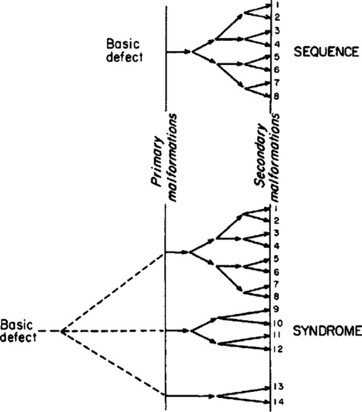
A true malformation syndrome is characterized by embryonic pleiotropy in which a pattern of developmentally unrelated malformation sequences occurs, i.e., the malformations that make up the syndrome occur in embryonically noncontiguous areas. They are not related to one another at the descriptive embryonic level; at a more basic level, the malformations have, or are presumed to have, a common cause and are thus pathogenetically related. The difference between a malformation sequence and a malformation syndrome is diagrammed in Figure 6.2. When holoprosencephaly occurs alone, it is a malformation sequence, but when it occurs with multiple noncontiguous anomalies, such as in trisomy 13 syndrome or with multiple noncontiguous anomalies in the autosomal recessively inherited Meckel syndrome, it is a malformation syndrome composed of several malformation sequences (Cohen, 1997b).
|
|
|
FIG. 6.1. Spectrum of dysmorphic faces associated with variable degrees of holoprosencephaly. A: Cyclopia without proboscis formation. Note single central eye. B: Cyclopia with proboscis. C: Ethmocephaly. D: Cebocephaly. Ocular hypotelorism with single-nostril nose. E: Median cleft lip, flat nose, and ocular hypotelorism. F: Ocular hypotelorism and surgically repaired cleft lip. (A-D, F from Cohen et al., 1971. E from DeMyer and Zeman, 1963. Montage from Cohen 1997b, with permission.) |
Because malformations can be relatively simple or complex, the later a defect is initiated, the simpler the malformation, and the earlier during organogenesis that a defect is initiated, the more far-reaching the consequences. The primary defect sets off a chain of secondary and tertiary events, resulting in what appear to be multiple anomalies. In holoprosencephaly, the embryonic forebrain fails to cleave sagittally into cerebral hemispheres, transversely into telencephalon and diencephalon, and horizontally into olfactory and optic bulbs. Holoprosencephaly varies in severity. At the mild end of the spectrum is simple absence of the olfactory tracts and bulbs. Holoprosencephaly is associated with facial dysmorphism, which also varies from mild to severe expression (Fig. 6.1). A single eye or closely set eyes, proboscis formation, single-nostril nose, flattened nose, median cleft lip, or lateral cleft lip may be observed variably or in combination. All malformations encountered trace their origin developmentally to a single primary defect in morphogenesis thought to be an abnormality in the prechordal mesoderm (Cohen, 1997b).
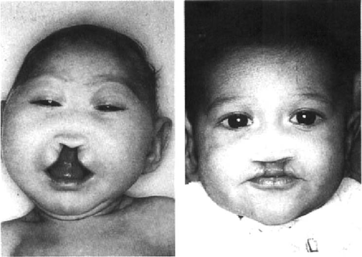
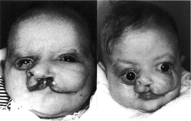
Although median cleft lip may occur with holoprosencephaly, cases occur without holoprosencephaly (Fig. 6.3). In holoprosencephaly with median cleft lip, the head circumference is dramatically reduced,abut when median cleft lip occurs without holoprosencephaly, the head circumference is within two standard deviations of the mean and normotelorism is the rule. Median cleft lip, reasonable head circumference, and normotelorism predict lack of holoprosencephaly. The distinction is important clinically and surgically because survival is the rule, necessitating surgical cleft lip repair. However, some degree of mental deficiency can accompany such cases (Cohen, 2000).
An association can be defined as a nonrandom occurrence of several anomalies in two or more individuals. An association cannot be reduced to a sequence or syndrome. For example, orofacial clefting is associated with congenital heart defects more commonly than expected by chance. Cleft lip-palate is also associated with neural tube defects more commonly than expected by chance. However, when cleft palate occurs with a basilar encephalocele that herniates through the sphenoid bone, the cleft palate occurs on a mechanical basis.
|
|
|
FIG. 6.2. Comparison of a malformation sequence (top) with a true malformation syndrome (bottom). Isolated holoprosencephaly is an example of a malformation sequence. The combination of holoprosencephaly, ventricular septal defect, and polydactyly caused by trisomy 13 is a malformation syndrome. (From Cohen, 1997b, with permission.) |
Syndrome Delineation
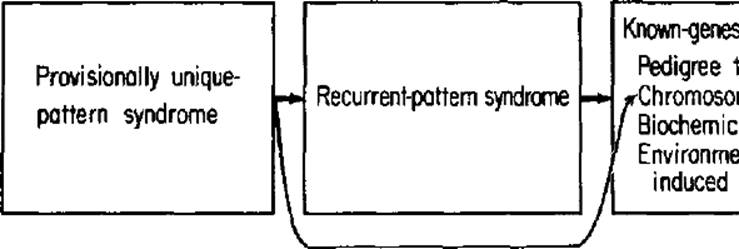
The process of syndrome delineation can be divided into the following stages: (1) unknown-genesis syndromes, including provisionally unique-pattern syndromes and recurrent-pattern syndromes, and (2)known-genesis syndromes, including pedigree syndromes, chromosomal syndromes, biochemical-defect syndromes, and environmentally induced syndromes (Cohen, 1997b).
In an unknown-genesis syndrome, the cause is simply not known. In a provisionally unique-pattern syndrome, several anomalies are observed in the same patient such that the clinician does not recognize the overall pattern of defects from his or her own experience, from searching the literature, or from consultation with the most learned colleagues in the field (Cohen, 1997b).
|
|
|
FIG. 6.3. Median cleft lip with holoprosencephaly (left), note microcephaly, without holoprosencephaly (right), note normal head circumference. (From Cohen, 2001, with permission.) |
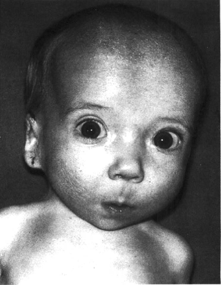
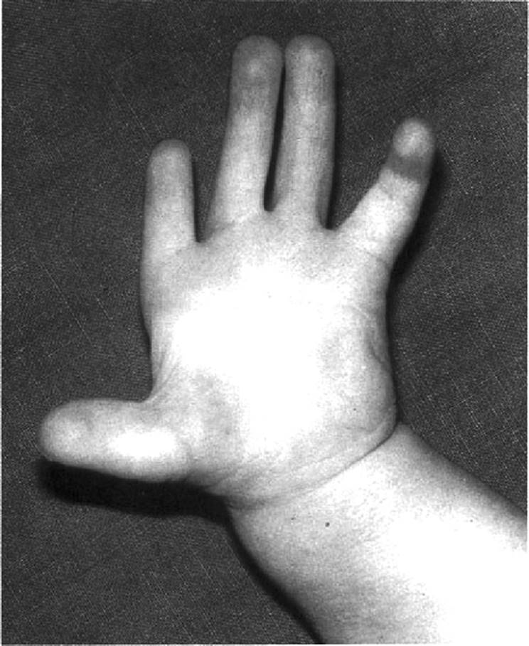
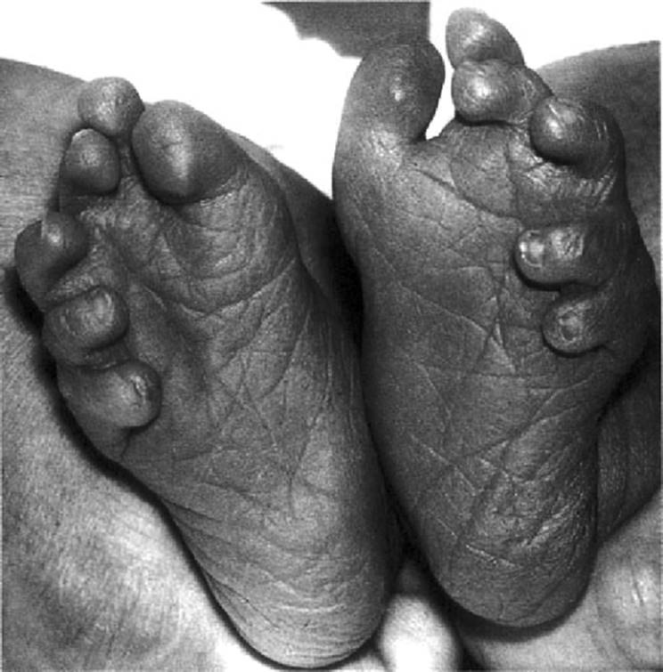
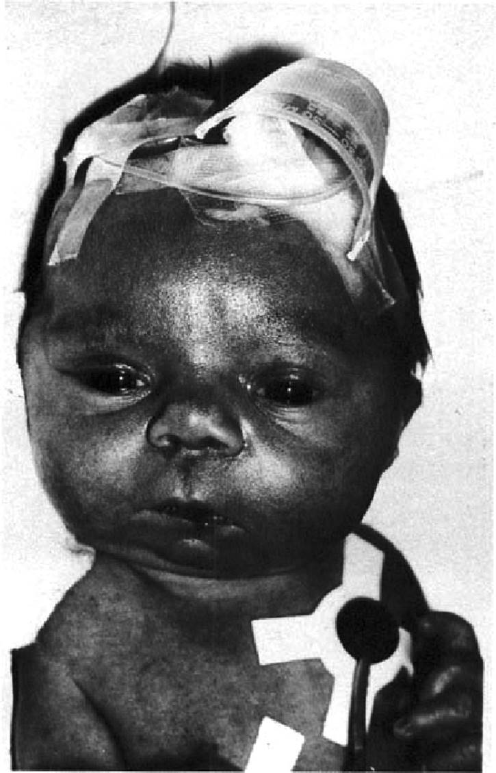
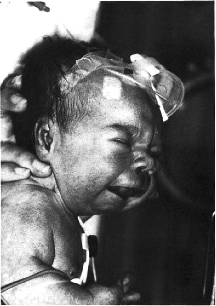
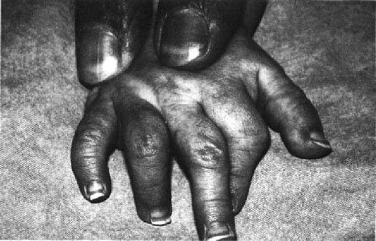
An example of a provisionally unique-pattern syndrome (Fig. 6.4,6.5 and 6.6) consists of a dysmorphic face with a prominent square forehead and Robin sequence, broad proximally placed thumbs, hypoplasia of the third and fifth middle phalanges, absent middle phalanges in the second fingers, broad halluces, postaxial polydactyly, rhizomelic short stature, and radiographic abnormalities of the spine and pelvis (Martsolf et al., 1997).
|
|
|
FIG. 6.4. Provisionally unique-pattern syndrome. Dysmorphic face with Robin sequence. (From Martsolf et al., 1977, with permission.) |
|
|
|
FIG. 6.5. Provisionally unique-pattern syndrome. Broad proximally placed thumb, hypoplasia of third and fifth middle phalanges, and absence of middle phalanx of index finger. (From Martsolf et al., 1977, with permission.) |
|
|
|
FIG. 6.6. Provisionally unique-pattern syndrome. Postaxial hexadactyly and abnormal toes. (From Martsolf et al., 1977, with permission.) |
Most likely, the anomalies in this provisionally unique-pattern syndrome have a common cause, though unknown, rather than different causes acting independently. The probability that such anomalies occur in the same patient by chance becomes less likely the more anomalies the patient has and the rarer these anomalies occur individually in the general population (Cohen, 1997b).
Obviously, if a second example of the syndrome comes to light, the condition is no longer unique. A provisionally unique-pattern syndrome is a one-of-a-kind syndrome to a particular observer at a particular point in time. There may be a nineteenth century description of a similar instance that escapes his or her attention. There may also be some instances of the syndrome in different parts of the world that remain as yet unrecognized. Thus, many syndromes appear to be unique at the time the initial patient is discovered but are no longer unique when two or more examples become known (Cohen, 1997b).
|
|
|
FIG. 6.7. Recurrent-pattern syndrome. Catel-Manzke syndrome with micrognathia (Robin sequence), ventricular septal defect, and talipes equinovarus. |
A recurrent-pattern syndrome can be defined as a similar or identical set of anomalies in two or more unrelated patients (Cohen, 1997b). An example is the Catel-Manzke syndrome, consisting of a globular cranium, Robin sequence, clinodactyly of the index finger with a supernumerary proximal phalanx in the index finger, congenital heart defects (50%), particularly ventricular septal defect, and talipes equinovarus (Gorlin et al., 1990) (Fig. 6.7,6.8,6.9,6.10 and 6.11).
The same abnormalities in two or more patients suggest, but do not prove, that the pathogenesis may be the same. At the recurrent-pattern stage of syndrome delineation, the etiology is still not known. In general, the validity of a recurrent-pattern syndrome increases with the more abnormalities found in the condition and the more patients recognized as having the syndrome (Cohen, 1997b). About 25 cases of Catel-Manzke syndrome have been described, mostly in males but several females have also been noted (Fig. 6.7,Fig. 6.8,6.9,6.10,6.11). Although a few examples of familial aggregation have been reported, no pattern of inheritance has been discerned. Most cases are sporadic, and the syndrome is still of the recurrentpattern type.
|
|
|
FIG. 6.8. Recurrent-pattern syndrome. Catel-Manzke syndrome. Micrognathia (Robin sequence). |
|
|
|
FIG. 6.9. Recurrent-pattern syndrome. Catel-Manzke syndrome. Micrognathia (Robin sequence). |
At the recurrent-pattern stage of syndrome delineation, the number of findings is usually expanded as the number of patients increases. However, because the etiology remains unknown at this time, other examples of the syndrome tend to be selected because they most closely resemble the first case. This results in an artificial homogeneity of cases that emphasizes the most severe aspects of the syndrome. Thus, we should be wary estimated frequencies given in review articles and textbooks for various anomalies that occur in a recurrentpattern syndrome; they tend to be overestimates that can affect the prognostic risk counseling for possibly developing some features of the syndrome such as mental retardation (Cohen, 1997b).
|
|
|
FIG. 6.10. Recurrent-pattern syndrome. Catel-Manzke syndrome. Marked clinodactyly of right index finger. |
|
|
|
FIG. 6.11. Recurrent-pattern syndrome. Catel-Manzke syndrome. Radiograph of left hand showing extra bone at base of proximal phalanx of index finger. |
A known-genesis syndrome can be defined as several anomalies causally related on the basis of (1) occurrence in the same family or, less conclusively, the same mode of inheritance in different families; (2) a chromosomal defect; (3) a specific defect in an enzyme or structural protein; or (4) an environmental factor. The term pedigree syndrome refers to known genesis on the basis of pedigree evidence alone; the basic defect itself remains undefined, although the condition is known to represent a monogenic disorder. Several examples with orofacial clefts include the autosomal recessively inherited Meckel syndrome and oral-facial-digital syndrome I, which has X-linked dominant inheritance, lethal in the male. Chromosomal syndromes, such as trisomy 13 syndrome, are cytogenetically determined. In a biochemical-defect syndrome, specific enzymatic defects are known in recessive syndromes. An example is the autosomal recessive Smith-Lemli-Opitz syndrome. An environmentally induced syndrome is defined in terms of the environmental factor or causative teratogen, such as diphenylhydantoin syndrome (Cohen, 1997b).
Comments on the Process of Syndrome Delineation
The process of syndrome delineation is summarized in Figure 6.12. Generally, a syndrome can be placed into one of the categories previously discussed. Occasionally, a syndrome may be delineated in one step, thus bypassing several of the stages mentioned earlier. For example, if a new chromosomal abnormality is discovered during the laboratory investigation of a patient clinically defined as having a provisionally unique-pattern syndrome, the patient represents a known-genesis syndrome of the chromosomal type in a one-step delineation. However, the variability of the clinical expression must await the discovery of more patients. In other instances, such as a large dominant pedigree with many affected individuals, a known-genesis syndrome of the pedigree type and much of its phenotypic variability can be determined in one step (Cohen, 1997b).
Provisionally unique-pattern syndromes occur with some frequency. Further delineation will often occur, given sufficient time. A truly unique-pattern syndrome may occur with a chromosomal anomaly involving two or more breaks. The condition may be sporadic or segregate within a family. Since the chance of an identical duplication-deficiency syndrome occurring in another family is very slight, the syndrome may be considered unique to an affected individual or an affected family (Cohen, 1997b).
The multiple anomalies that make up any syndrome are thought to be pathogenetically related. However, anomalies may concur either by chance or as a statistically related association in the same patient. Some associations are well-stocked pools for future syndrome delineation (Cohen, 1997b).
Syndrome delineation and the use of the delineation terms proposed should be thought of as a dynamic, flexible, and continually changing framework in which to view various syndromes. These categories should never be thought of as static or immutable, even at the higher stages of syndrome delineation. Etiologic and clinical heterogeneity is common should be expected to occur even when not readily apparent. Moreover, we should not confuse syndrome delineation with understanding of the syndrome's pathogenesis, even at the higher stages of delineation. In a pedigree syndrome such as the autosomal recessively inherited Meckel syndrome, we know nothing about how the homozygous state of the Meckel gene produces such diverse features as encephalocele, orofacial clefting, polydactyly, and polycystic kidneys (Cohen, 1997b).
|
|
|
FIG. 6.12. Summary of the process of syndrome delineation. See text. (From Cohen, 1997b, with permission.) |
Significance of Syndrome Delineation
The significance of syndrome delineation cannot be overestimated. As an unknown-genesis syndrome becomes delineated, its phenotypic spectrum, its natural history, and its inheritance pattern or risk of recurrence become known, allowing for better patient care and family counseling. If the phenotypic spectrum is known, the clinician can search for suspected defects that may not be immediately apparent but that may produce clinical problems at a later date, such as a hemivertebra in Goldenhar syndrome. If a certain complication can occur in a given syndrome, such as Wilms tumor in Beckwith-Wiedemann syndrome, the clinician is forewarned to monitor the patient for possible development of neoplasia. Finally, if the recurrence risk is known, the parents can be counseled properly about future pregnancies. This is particularly important if the risk is high and the disorder is severely disabling or disfiguring, has mental deficiency as one component, or has a dramatically shortened life span. For example, Stickler syndrome is an autosomal dominant disorder with a 50% recurrence risk when one parent is affected. Retinal detachment occurs in 20% of reported cases, and blindness affects 15%. Genetic counseling is important because the risk of developing serious ocular problems is high. This relatively common condition also illustrates the importance of syndrome delineation because the entity was unrecognized before 1965, although clearly it existed before then. Thus, syndrome delineation fosters good patient care. The overall treatment program gains rationality. In contrast, with a provisionally unique-pattern syndrome, the treatment program and overall management frequently leave something to be desired (Cohen, 1997b).
Syndrome Nomenclature
In general, a newly recognized syndrome can be denoted by (1) an eponym, (2) one or more striking features, (3) an acronym, (4) a numeral, (5) a geographic term, or (6) some combination of the above. None of these systems of nomenclature is without fault. Each has advantages and disadvantages. In general, nomenclature usage evolves with time. Sometimes international working groups are very helpful in standardizing nomenclature in various subfields (Cohen, 1976).
Mythical terms such as elfin fades syndrome and animal nomenclature such as bird-headed dwarf are pejorative and should be discouraged. One commonly recognized principle in the field is never to use apostrophe 5 in eponymic designations. Thus, we use Apert syndrome, not Apert's syndrome, because Apert neither had nor owned the syndrome he described (Cohen, 1976, 1997a,b).
Pace of Syndrome Delineation
Syndrome delineation is proceeding at a very rapid pace. Table 6.1 illustrates syndrome delineation through the years with respect to orofacial clefting. Toriello (1988) has estimated that newly recognized syndromes in general are being described at the rate of one or more per week and, although some represent variable expression of previously recorded conditions, many actually represent newly recognized syndromes.
Orofacial clefting is estimated for the years 1971, 1978, and 1990 in Table 6.1 (Cohen and Bankier, 1991). The 1990 estimate is based on the POSSUM computer based system. POSSUM (1990) is a computer program that provides an extensive catalogue of multiple anomaly syndromes, including photographs (on video disk) and references. The POSSUM estimate is compared with those of Cohen (1978) and Gorlin et al. (1971). The category “unknown cause” in Table 6.1 includes distinctive syndromes of unknown genesis and various simple associations.
|
TABLE 6.1. Syndrome Delineation Involving Orofacial Clefting |
||||||||||||||||||||||||||||||||||||||||
|
||||||||||||||||||||||||||||||||||||||||
|
TABLE 6.2. Genetic Aspects of Some Syndromes with Clefting |
||||||||||||||||||||||||||||||||||||||||
|
||||||||||||||||||||||||||||||||||||||||
Syndromes with orofacial clefting have been reviewed by Gorlin et al. (2001). Genetic aspects of a few syndromes are summarized in Table 6.2, and some teratogenic syndromes with occasional clefting are shown in Table 6.3.
Robin Sequence and Robin Complexes
In a classic article titled “Not All Dwarfed Mandibles Are Alike,” the late Samuel Pruzansky (1969) observed that “Pierre Robin syndrome” (as he and others called it in those days) was not only causally heterogeneous but also pathogenetically and phenotypically variable.
Shprintzen (1988) showed that by using different definitions of Robin sequence proposed by various authors, he could calculate different frequencies of syndromic and nonsyndromic Robin sequence in his own hospital series. Different criteria for Robin sequence are summarized in Table 6.4. Definitions I through IV are clearly Robin sequence, but definition V is non-Robin in type. The cleft palate is V-shaped, and by polysomnography, some respiratory compromise is evident. The mandible is normal but may be slightly small subjectively, though it is impossible to be certain. Thus, at the interface, it is not always possible to distinguish between Robin sequence and ordinary cleft palate (Cohen, 1997b, 2000).
|
TABLE 6.3. Some Human Teratogenic Syndromes* |
||||||||||||||||||||||||||||||||
|
||||||||||||||||||||||||||||||||
|
TABLE 6.4. Defining Robin Sequence Using Different Criteria |
||||||||||||||||||||||||||||||||
|
||||||||||||||||||||||||||||||||
Shprintzen (1988) demonstrated that different mechanisms of obstruction can occur within the same syndrome and noted that glossoptosis is frequently not the cause of upper airway obstruction in some cases. Types of obstruction based on Sher (1992) are summarized in Table 6.5. Kreiborg and Cohen (1996) documented the growth pattern in Robin patients and found that catch-up growth was partial, never complete.
Because of etiologic, pathogenetic, and phenotypic differences, Robin sequence or Robin complexes of various types can occur. Table 6.6 summarizes several conditions with mandibular differences, cranial base angle changes, and different types of respiratory compromise. These changes are shown in Figure 6.13. Double lines indicate general causes: malformation, deformation, or connective tissue dysplasia. Thick arrows indicate Robin sequence. Thin lines show possible ways that Robin complexes can occur (Cohen, 1999).
A distinction is made between micrognathia and retrognathia (Fig. 6.13). Micrognathia refers to size, retrognathia to position. In Treacher Collins syndrome, the mandible is short. In deletion 22qll.2 syndrome, the mandible is essentially normal in size but retrognathia in position because the cranial base angle is larger than normal (Table 6.6). Most Robin conditions are micrognathic or retrognathic but not both together (so-called “microretrognathia”) (Cohen, 1999). Different results were found by Glander and Cisneros (1992), but their results may be questioned. These authors compared del(22qll.2) with Robin in Stickler dysplasia. No controls from the general population or from first-degree relatives were provided. Furthermore, with multiple measurements on each cephalogram, confidence intervals would have produced a more meaningful interpretation than f-tests of significance.
Phenotypic variability (Table 6.6) is illustrated by comparing the mandible in Treacher Collins syndrome with that found in deformational Robin sequence (Cohen, 1999). In both conditions, the mandibles are short. Catchup growth in deformational Robin sequence is incomplete. Mandibular growth is severely affected in Treacher Collins syndrome. The two conditions differ greatly in shape; the Treacher Collins syndrome mandible is highly specific (Cohen, 1999; Kreiborg and Cohen, 1996).
Different Robin complexes are exemplified by del(22qll.2) syndrome and spondyloepiphyseal dysplasia congenita (Table 6.6). In del(22qll.2) syndrome, retrognathia, caused by an obtuse cranial base angle, and cleft palate, either submucous or true, are found. The flat cranial base angle and retrognathia do not contribute to pharyngeal obstruction, which in fact results from hypotonia (Arvystas and Shprintzen, 1984). Robin sequence does not occur. Rather, in this form of Robin complex, all of the manifestations are causally, but not sequentially, related (Cohen, 1999).
|
TABLE 6.5. Types of Obstruction in Robin Sequence/Complex (n = 52) |
|||||||||||||||||||||||||||||||||||
|
|||||||||||||||||||||||||||||||||||
|
|
|||||||||||||||||||||||||||||||||||
|
FIG. 6.13. Robin sequence and complexes. Double lines indicate general causes: malformation, deformation, or connective tissue dysplasia. Thick arrows indicate Robin sequence. Thin lines show all possible ways that Robin sequence can occur. (From Cohen, 1999, with permission.) |
|||||||||||||||||||||||||||||||||||
|
TABLE 6.6. Robin Sequences and Complexes |
||||||||||||||||||||||||||||||||||||||||||||||
|
||||||||||||||||||||||||||||||||||||||||||||||
An example of multiple mechanisms of respiratory compromise can be found in spondyloepiphyseal dysplasia congenita (Table 6.6): small mechanically abnormal chest, tracheobronchomalacia, and/or central apnea due to cervical or medullary compression caused by cervical instability (Harding et al., 1990) in addition to the possibility of upper respiratory obstruction based on Robin sequence.
Unusual Craniofacial Clefts and the Tessier Classification
Tessier (1976) outlined an anatomic and descriptive classificatory system in which the various types of bony and soft tissue defects, which he called clefts, are situated along definite axes with numbers assigned to the sites of clefting, depending on their relationships to the sagittal midline (Fig. 6.14). Clefting may involve bone and/or soft tissue but rarely to the same extent. From the sagittal midline to the infraorbital foramen, abnormalities of soft tissue predominate. From the infraorbital foramen to the temporal bone, however, osseous defects are more severe than those of soft tissue, a notable exception being the ear. Clefts through the orbit use the lower eyelid as an equator. Cleft numbered lines may be either northbound (cranial) or southbound (facial). Cranial numbered lines have facial numbered counterparts, yet these numbers are different to avoid the implication that they necessarily have the same etiopathogenesis. Thus, the Tessier classification permits description of both the location and the extent of unusual facial clefts. Several different types of Tessier clefts may occur in the same patient (Fig. 6.15).
|
|
|
FIG. 6.14. Tessier craniofacial clefting system. A: Soft tissue clefts. B: Bony clefts. Dotted lines represent uncertain localization or uncertain clefting. Note that northbound cranial line has a different number from southbound facial line. Thus, system is descriptive and anatomic and avoids etiologic or pathogenetic speculation. For example, the cause of a number 10 cleft may be different from the cause of a number 4 cleft. (From Cohen, 1999, with permission.) |
|
|
|
FIG. 6.15. Multiple clefts. Left: Cleft lip with Tessier number 7 cleft on left side and left numbers 5 and 6 on right. Right: Cleft lip and palate with Tessier number 6 cleft on left side and number 4 cleft on right. Courtesy of A. Richieri-Costa (Bauru, Brazil). |
|
|
|
FIG. 6.16. Complex clefting. Hairline indicator points to cleft area. Computed tomographic scan showed anterior cranium bifidum occultum and gap in sphenoid bone. Note ear tags. Courtesy of A. Richieri-Costa (Bauru, Brazil). |
In some instances, overlying soft tissue defects predict the possibility of underlying bony clefts. Elsewhere I have referred to such features (e.g., colobomatous notching of the upper or lower eyelids or the nostrils or the interruption of the eyelashes or the eyebrows) as Tessier signs (Cohen, 1997b). Also included in this category are hairline indicators that may point to the cleft (Moore et al., 1988) (Fig. 6.16).
The causes of most Tessier clefts are unknown. The overwhelming majority occur sporadically. One exception is Treacher Collins syndrome, which is autosomal dominantly inherited. Some Tessier clefts are malformations that can be explained by faulty embryogenesis. However, others represent disruptions, such as those associated with amniotic bands (Cohen, 1997b).
References
Arvystas, M, Shprintzen, RJ (1984). Craniofacial morphology in the velocardiofacial syndrome. J Craniofac Genet Dev Biol 4: 39–45.
Cohen, MM, Jr (1976). Syndrome designations. J Med Genet 13: 266–270.
Cohen, MM, Jr (1978). Syndromes with cleft lip and palate. Cleft Palate J 15: 306–328.
Cohen, MM, Jr (1997a). Apert syndrome, not Apert's syndrome: Apert neither had nor owned the syndrome that bears his name. Plast Reconstr Surg 100: 532–533.
Cohen, MM, Jr (1997b). The Child with Multiple Birth Defects. New York: Oxford University Press.
Cohen, MM, Jr (1999). Robin sequences and complexes: causal heterogeneity and pathogenetic variability. Am J Med Genet 84: 311–315.
Cohen, MM, Jr (2000). Etiology and pathogenesis of orofacial clefting. In: Cleft Lip and Palate: A Physiological Approach, Oral and Maxillofacial Surgery Clinics of North America vol. 12. pp. 379–397.
Cohen, MM, Jr (2001). Perspectives on craniofacial anomalies, syndromes, and other disorders. In: Craniofacial Surgery: A Multi-Disciplinary Approach to Craniofacial Anomalies, edited by K. Y. Lin, R. C. Ogle, and J. A. Jane, 2002, Chapter 1, pp. 1–38.
Cohen, MM, Jr, Bankier, A (1991). Syndrome delineation involving orofacial clefting. Cleft Palate-Craniofac J 28: 119–120.
Cohen, MM, Jr, Jirasek, JE, Guzman, RT, Gorlin, RJ, Peterson, MQ (1971). Holoprosencephaly and facial dysmorphia: nosology, etiology and pathogenesis. Birth Defects 7(7): 125–135.
DeMyer, WE, Zeman, W (1963). Alobar holoprosencephaly (arhinencephaly) with median cleft lip and palate: clinical, electroen cephalographic and nosologic considerations. Confin Neurol 23: 1–36.
Glander, K II, Cisneros, GJ (1992). Comparison of the craniofacial characteristics of two syndromes associated with the Pierre-Robin sequence. Cleft Palate Craniofac J 29: 210–219.
Gorlin, RJ, Cervenka, J, Pruzansky, S (1971). Facial clefting and its syndromes. Birth Defects 7(7): 3–49.
Gorlin, RJ, Cohen, MM, Jr, Hennekam, RCM (2001). Syndromes of the Head and Neck, 4th ed. New York: Oxford University Press.
Gorlin, RJ, Cohen, MM, Jr, Levin, LS (1990). Syndromes of the Head and Neck, 3rd ed. New York: Oxford University Press.
Hagberg, C, Larson, O, Milerad, J (1997). Incidence of cleft lip and palate and risks of additional malformations. Cleft Palate-craniofac J 35: 40–45.
Harding, CO, Green, CG, Perloff, WH (1990). Respiratory complications in children with spondyloepiphyseal dysplasia congenita. Pediatr Pulmonol 9: 49–54.
Kallen, B, Harris, J, Robert, E (1996). The epidemiology of orofacial clefts: 2. Associated malformations. J Craniofac Genet Dev Biol 16:242-248.
Kreiborg, S, Cohen, MM, Jr (1996). Syndrome delineation and growth in orofacial clefting and craniosynostosis. In: Facial Clefts and Craniosynostosis Principles and Management, edited by TA Turvey, KWL Vig, and RJ Fonseca. Philadelphia: WB Saunders, Chapter 3, pp. 57–75.
Martsolf, JT, Reed, MH, Hunter, AGW (1977). Case report 56. Skeletal dysplasia, Robin anomalad, and polydactyly. Syndrome Ident 5(1): 14–18.
Moore, MH, David, DJ, Cooler, RD (1988). Hairline indicators of craniofacial clefts. Plast Recontr Surg 82: 589–593.
Murray, JC (1995). Face facts: genes, environment, and clefts. Am J Hum Genet 57: 227–232.
Novelli, G, Amati, F, Dallapiccola, G (1999). UFDIL and CDC45L: a role in DiGeorge syndrome and related phenotypes? Trends Genet 15: 251–253.
POSSUM (1990). Computer software system. Melbourne, Australia: Murdoch Institute for Research into Birth Defects.
Pruzansky, S (1969). Not all dwarfed mandibles are alike. Birth Defects 5(2): 120–129.
Saitta, SC, McGrath, JM, Mensch, H, et al. (1999) A 22qll.2 deletion that excludes UFDIL and CDC45L in a patient with conotruncal and craniofacial defects. Am J Hum Genet 65: 562–566.
Schutte, BC, Basart, AM, Watanabe, Y (1999). Microdeletions at chromosome bands Iq32-q41 as a cause of Van der Woude syndrome. Am J Med Genet 84: 145–150.
Sher, AE (1992). Mechanisms of airway obstruction in Robin sequence: implications for treatment. Cleft Palate-Craniofac J 29: 224–231.
Shprintzen, RJ (1988). Pierre Robin, micrognathia, and airway obstruction: the dependency of treatment on accurate diagnosis. Int Anesthesiol Clin 26: 64–71.
Spranger, JW, Benirschke, K, Hall, JG, Lenz, W, Lowry, RB, Opitz, JM, Pinsky, L, Schwarzacher, HG, Smith, DW (1982). Errors of morphogenesis: concepts and terms. J Pediatr 100: 160–165.
Srivastava, D, Yamagishi, H (1999). Role of the dHAND-UFDIL pathway. Trends Genet 25: 253–254.
Tessier, P (1976). Anatomical classifications of facial, craniofacial and laterofacial clefts. J Maxillofac Surg 4: 69–92.
Toriello, HV (1988). New syndromes from old: the role of heterogeneity and variability in syndrome delineation. Am J Med Genet Suppl 1: 50–70.
Wulfsberg, EA, Leana-Cox, J, Neri, C (1997). Reply to “What's in a name? The 22qll.2 deletion” (letter to the editor). Am J Med Genet 72: 248–249.
Yamagishi, H, Garg, V, Matsuoka, R (1999). A molecular pathway revealing a genetic basis for human cardiac and craniofacial defects. Science 283: 1158–1161.