
John S. Leard* & Chris L. Wells
INTRODUCTION
The purpose of this chapter is to describe common musculoskeletal and integumentary pathologies and the evidence found in the literature regarding their influence on the cardiopulmonary system. Impairments of bony structures, joints, skin, fascia, and musculature of the thorax may lead to a decrease in cardiopulmonary function. These impairments will lead to impaired circulation, aerobic capacity/endurance, ventilation, and respiration/gas exchange by restriction of movement of structures in the body. This results in decreased functional mobility of the patient and disability. This chapter will also describe the evidence in the literature as it relates to the interventions commonly associated with these pathologies.
MUSCULOSKELETAL CONDITIONS
Osteoporosis (Practice Patterns 4A, 4B, 4C, 4F, 4G; ICD-9-CM Code: 733.0)
Introduction
Osteoporosis is a general term for a decrease in the mass of normal bone per unit volume and leads to an increase risk of fracture. Many conditions and diseases may be involved in the etiology of developing osteoporosis including metabolic disorders of osteoclastic versus osteoblastic activity, endocrine disease, bone disuse, genetic factors, and postmenopausal state. Bone loss is generally associated with advancing age in both sexes but proceeds at a faster rate in women, especially following menopause.1,2
The current intervention strategy is prevention of normal bone loss per unit volume by increasing peak bone mass by the third decade through diet, weight-bearing exercise, and cessation of smoking. Inherited factors account for an estimated 60% to 80% of the variability in peak bone mass. Diet, physical activity, and hormonal status are important modifiers of bone accrual.3 Reversal of widespread osteoporosis is extremely difficult to achieve.
Classic spinal deformities associated with osteoporosis are increased kyphosis with loss of height, thoracic vertebral body fractures, and back pain.4,5 The increased kyphosis is related to thoracic wedge fractures but also has nonskeletal contributing factors.6 One of these factors is the intervertebral disc shape.7 The loss of height does not seem to be due to the patient having small vertebral bodies but instead is due to the wedging, which causes an increase in the kyphosis.8 Besides osteoporosis, there are many reasons why thoracic vertebral body fractures occur in patients older than 50 years. Metastases, multiple myeloma, and trauma9,10 are associated with these fractures as well. In Finland, fractures occur in men and women with equal frequency, and gradually increase with age. At age 65, the frequency sharply increases in women. This is attributed to the pattern of age-related osteoporosis between the sexes due to menopause. In a study of 942 women in Rochester, Minnesota, increasing age was also associated with vertebral fractures.11 The level of peak frequency for spinal fractures occurs at T7-T8 and T11–T12.8 Vertebral body fractures have radiological characteristics that are different from fractures associated with osteoarthritis. Osteoporotic fractures have a greater height difference posterior to anterior in the saggital plane than fractures in patients with osteoarthritis.12
Back pain in patients suffering from osteoporosis is associated with the number of vertebral body fractures and the severity of the kyphotic curve.13 The literature is not conclusive regarding the pathomechanics of the increased kyphotic curve. Poor posture, decreased strength of the muscles that extend the thoracic spine, and the spinal fractures themselves are all possible beginnings to this increased curve.
Impairments That Influence Cardiopulmonary Function
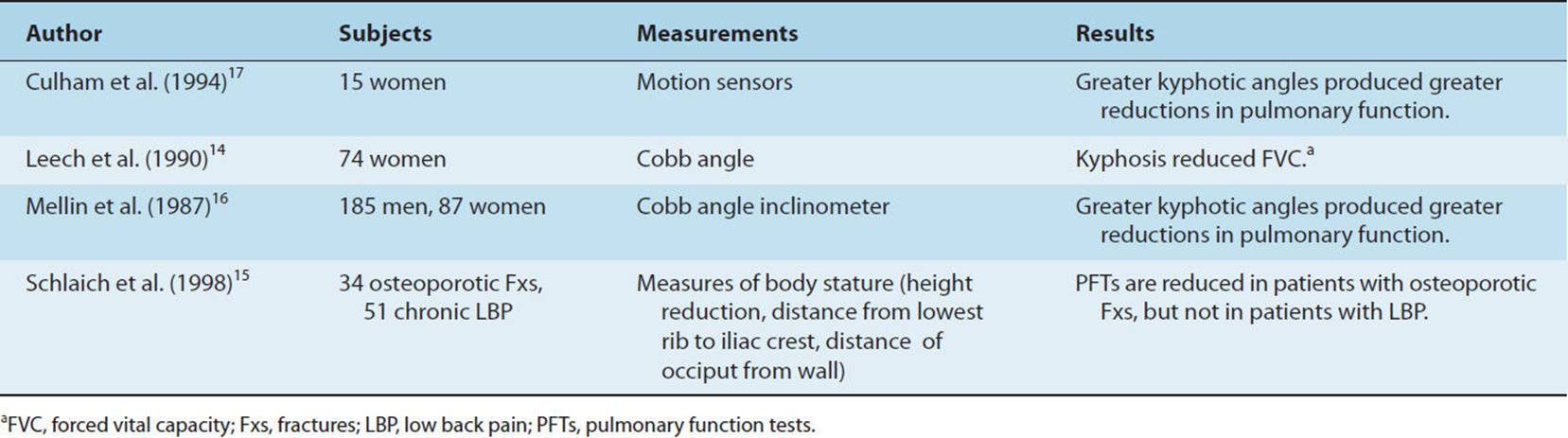
There appears to be no literature that supports the notion that back pain leads to pulmonary dysfunction. However, pulmonary function loss is associated with women suffering from osteoporosis due to their increased kyphotic curve and thoracic compression fractures. Kyphosis and thoracic compression fractures caused by osteoporosis produce some predictable declines in vital capacity in women.14 Schlaich et al. noted that pulmonary function is reduced in patients who have spinal osteoporotic fractures and not in patients suffering from chronic low back pain.15 It is questionable as to whether this decrease in pulmonary function is only related to the spinal hypomobility, causing a decrease in the ability to expand the chest, or whether it is related to osteoporosis which is progressive with age, such that older patients experience more severe impairments. In 1987, Mellin and Harjula studied 187 men and 87 women and concluded that patients with increased spinal curves had decreased vital capacities and forced expiratory volumes.16 Culham et al. studied 15 women with kyphosis and found a significant negative correlation among kyphotic angle and inspiratory capacity, vital capacity, and lateral expansion of the thorax.17 Table 13-1 summarizes the findings of these articles, which examined cardiopulmonary function and bone deformities.
TABLE 13-1 Summary of Studies Comparing Pulmonary Function and Bony Deformities
Interventions to Improve Cardiopulmonary Function
No treatment effectiveness studies have been found that have examined the benefits of physical therapy on pulmonary function with osteoporotic patients. A group of physical therapists did study the effects of physical therapy on chronic pain and performance associated with osteoporotic patients. Treatments included balance training, muscle strength training, and lumbar stabilization exercises. They concluded that the program improved balance and level of daily function, decreased pain, decreased the use of analgesic medication, and improved quality of life after the active training period.18
Associated Conditions With Osteoporosis
If the musculoskeletal impairments described previously are associated with other comorbidities, such as congestive heart failure (CHF) or cardiovascular disease, the pulmonary function of the patient and the outcomes of interventions may be negatively influenced. Vogt et al.19 studied 1,492 older white women with osteoporosis and found no relationship between the incidence and the prevalence of vertebral fractures and cardiovascular disease. However, they discovered that women with cardiovascular disease had more disabling back pain than women without cardiovascular disease. A single case report of an older woman with osteoporosis and congestive heart failure (CHF), who required mitral valve replacement, suggests that osteoporosis may have a role in the morbidity and mortality of patients. This subject had a profound collapse of the thoracic spine that resulted in pulmonary insufficiency and death.20 Although this was a single case study, such situations may occur to a greater extent in older patients with cardiovascular and pulmonary diseases and may require further investigation.
Summary
The relationship between osteoporosis and cardiopulmonary impairments is difficult to identify, because osteoporosis has many associated impairments that could affect cardiopulmonary function. Osteoporosis affects older men and women and is associated with an increased kyphotic curve, thoracic compression fractures, and decreased lateral chest expansion. It is difficult to separate these factors individually to identify whether the overall decrease in chest wall size or expansion is altering cardiopulmonary function or whether other comorbidities are influencing function. No treatment effectiveness studies have been found that have examined the effects of physical therapy on pulmonary function with patients diagnosed with osteoporosis.
Ankylosing Spondylitis (Practice Patterns 4E, 4F; ICD-9-CM Code: 720)
Introduction
Ankylosing spondylitis is a chronic inflammatory disease that primarily affects the sacroiliac joint and the spine. The shoulder and hip, as well as other lower extremity joints, may be affected. It is a form of chronic seronegative spondyloarthritis. The disease results in progressive stiffening of the joints, typically beginning with the spine and sacroiliac joints. It attacks the site of insertion of tendons, ligaments, fascia, and fibrous joint capsules.2 There is also a high risk of cardiomyopathy, arrhythmias, pericarditis, and aortic valvular disease associated with ankylosing spondylitis. Males are afflicted at a rate of 10:1, male-to-female ratio, and Caucasians are afflicted more frequently than Afro-Americans. The onset of the disease occurs between 20 and 40 years of age.21,22 The cause of ankylosing spondylitis is unknown, but its etiology may be a hyper-reactivity response of the immune system.
Initially, the patient may present with complaints of poorly localized back pain that is provoked by sudden movements. The back pain is differentiated from “mechanical low back pain” because movement relieves the symptoms, the symptoms do not diminish with rest, and the patient experiences night pain. The progression of these signs and symptoms is either continuous or intermittent, spreading cephalically along the spine. In the most severe cases, the spine and hips will become stiff, a spinal flexion deformity develops, and pathological vertebral fractures occur. Only one-third of the patients diagnosed will progress to these severe symptoms.2
Impairments That Influence Cardiopulmonary Function
Pulmonary involvement affecting the upper lobes of the lungs has been documented in 1.2% of patients with ankylosing spondylitis. Histological findings can include nonspecific fibrosis, dilated bronchi, and bulla formation and occur at a rate of 50:1, male-to-female ratio.22
Over time, the stiffening and straightening of the spine cause a decrease in chest wall compliance and result in a mechanical restrictive process. This chest wall restriction is associated with an increase in pneumothorax, atelectasis, and aspiration due to esophageal dysfunction that can lead to pulmonary infection. In the presence of fibrotic changes, the recurrence of infection can lead to progressive pulmonary fibrosis.21
Pulmonary involvement is documented by abnormal findings on standard laboratory tests. Pulmonary function tests show a decline in vital capacity and total lung volumes. Maximal expiratory and inspiratory efforts are reduced. Radiological examination demonstrates small apical nodules, infiltrates, and pleural thickening. There is an increase in B lymphocytes and a lower level of neutrophils upon bronchoalveolar lavage.21,23 As the disease progresses, the spine becomes hypomobile at the costovertebral joints, resulting in decreased chest expansion and sometimes provoking pain with deep breathing.2
Interventions to Improve Cardiopulmonary Function
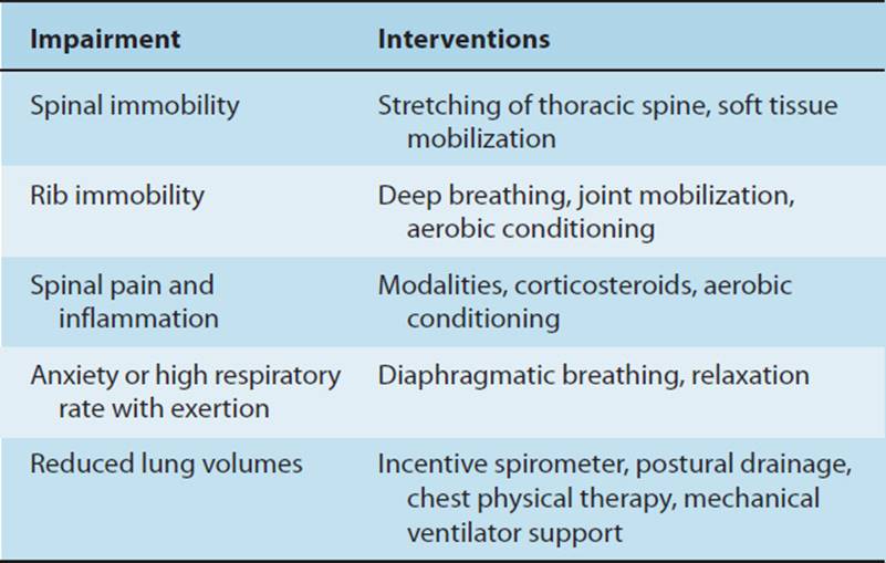
Treatment for pulmonary involvement includes general spinal and rib cage mobility intervention, such as general trunk stretching, soft tissue mobilization, and deep breathing exercises to improve mobility. Modalities and medications can be used to control pain and spinal inflammation. Corticosteroids may be utilized if intrinsic pulmonary disease impairs function.21,23 Depending on the severity of the disease, aerobic exercises are prescribed to maintain cardiopulmonary conditioning and trunk range of motion. Diaphragmatic breathing or inspiratory hold-breathing can be used as a breathing strategy when the patient has a high respiratory rate, anxiety, or signs of oxygen desaturation with exertion. An incentive spirometer may also be used as visual feedback in order to facilitate larger tidal volumes. In severe cases, the patient may not be able to generate a large enough lung volume to manage his or her own secretions; therefore, chest physical therapy and postural drainage will be indicated to address these impairments. If the patient is retaining carbon dioxide, he or she may need mechanical ventilatory support (see Table 13-2).
TABLE 13-2 Common Interventions for Ankylosing
Prognosis varies considerably. Approximately 35% of patients with ankylosing spondylitis will succumb to deadly arrhythmias. Infection and fractures of the cervical spine are also leading causes of death. These occur once the disease has progressed and has produced a brittle and rigid cervical spine that is susceptible to injury, even after minimal trauma. Death from pulmonary involvement is rare, but when it occurs, it is usually associated with infection and respiratory failure secondary to cervical spine injury.21
Summary
Pulmonary function is altered with patients suffering from ankylosing spondylitis, mainly due to restriction of chest wall movement. This results in decreased chest expansion and sometimes provokes pain with deep breathing. As the disease progresses, the restriction of chest wall movement and stiffening of the spine cause other impairments such as pneumonia, inability to remove secretions, small apical nodules, infiltrates, and pleural thickening. Initial intervention is aimed at maintaining or increasing chest wall movement.
Idiopathic Scoliosis (Practice Pattern 4B; ICD-9-CM Code: 737.3)
Introduction
Scoliosis is the development of an abnormal lateral curvature of the spine. Approximately 80% of all abnormal lateral curvatures of the spine have mechanisms that are unknown, so they are classified as idiopathic. The onset generally appears in childhood or adolescence and has a 10:1 female-to-male ratio.24 The curve may progress from mild to severe, particularly during rapid stages of growth, and can be located in the lumbar, thoracic, or both regions of the spine.2 The lateral curve usually presents with rotation and increased kyphosis. The lateral curve plus the rotation of the involved thoracic vertebrae around a vertical axis causes a decrease in lung function.25
Impairments That Influence Cardiopulmonary Function
The scoliosis results in a compression of the lung on the concave side of the abnormal thoracic curve and may decrease lung volumes.24,26 The greater the lateral curve of the spine, the more likely the patient will have pulmonary symptoms. As the curve progresses, the first indication of pulmonary limitation occurs during exercise with breathlessness.27 Upon examination, there is a decrease in diaphragmatic excursion and breath sounds on the concave side. These impairments are related to the severity of the curve.28 If the curvature becomes greater than 60 degrees, which is classified as severe, the skeletal deformity begins to affect pulmonary function and the patient will present with more respiratory complaints. It is difficult to determine how much of the dyspnea is associated with deconditioning.27 In some cases, the deformity actually causes a pulmonary restrictive breathing pattern when the PFTs are examined. There is a decrease in vital capacity; forced vital capacity; forced expiratory volume in 1 second (VC, FVC, FEV1), and an elevation of the FEV1/FVC ratio. When the curve is more than 90 degrees, there is mechanical impairment of inspiratory muscles and decreased lung compliance leading to a decrease in inspiratory muscle strength.24,29 With patients having curves between 100 and 136 degrees, the pattern of respiratory muscle activation is similar to that in patients with severe chronic obstructive pulmonary disease (COPD). This pattern does not use the sternocleidomastoid at rest but has a stronger than normal recruitment of the rib cage inspiratory muscles, the abdominal muscles, and the diaphragm with less mechanical efficiency.30 In both COPD and severe kyphoscoliosis, abnormal biomechanics are the major causes in abnormal pulmonary function. Patients with COPD have increased lung compliance and decreased chest wall compliance, whereas patients with severe kyphoscoliosis have marked reduction in lung and chest wall compliance. The acute decompensation occasionally seen in patients with severe kyphoscoliosis appears to be primarily due to the marked reduction in lung and chest wall compliance and less to airway resistance and positive end-expiratory pressure.31 Patients with severe scoliosis are more susceptible to developing atelectasis, pneumonia, and a reduction in exercise tolerance. Pulmonary complications are a contributing factor to the death of 82% of patients with clinically significant scoliosis.29
Kyphoscoliosis is most frequently associated with respiratory complications due to increased chest wall stiffness. Patients will present with low lung volumes particularly VC and expiratory reserve volume with little change in the right verticle (RV). This is consistent with a restrictive pattern. If the tidal volume is significantly impaired, hypoventilation may occur. Hypoventilation is associated with hypoxemia and ventilation–perfusion mismatch. Pulmonary vascular resistance is elevated because of the distortion and compression of vascular tissue caused by this skeletal deformity.32 Severe kyphoscoliosis can lead to bronchial torsion, which will obstruct the airway and cause moderate to severe air trapping.33 Boyer et al.34 found moderate-to-severe air trapping in 46% of 44 children with idiopathic scoliosis.
Surgical Interventions to Improve Cardiovascular Function
Surgical correction of the deformity is the intervention of choice in cases of severe scoliosis. Severe lateral curves are usually greater than 40 degrees, as measured by the Cobb method2(see Fig. 13-1). The surgery may be done through either an anterior and/or a posterior approach. Both approaches involve bone grafts from the ribs or the iliac crest and utilization of spinal instrumentation. Surgical intervention has an impact on pulmonary function. In long-term follow-up, it has been reported that the posterior approach restores pulmonary function to normal predicted levels, whereas the anterior approach does not correct the restrictive pulmonary pattern.35 In a study with long-term follow-up of patients who had undergone the anterior approach, there was a small decrease in forced vital capacity. The authors concluded that improvement in pulmonary function could not be achieved with this surgery.36 The anterior approach is also associated with an increased incidence of pulmonary complication in the postoperative period because one lung must be deflated in order to achieve surgical access. Harvesting bone grafts from the ribs also has a longer-term negative effect in restoration of PFTs than grafts taken from the pelvis. Patients who have undergone the placement of spinal instrumentation (Harrington rod) demonstrate little to no improvement in the restrictive pulmonary process.35
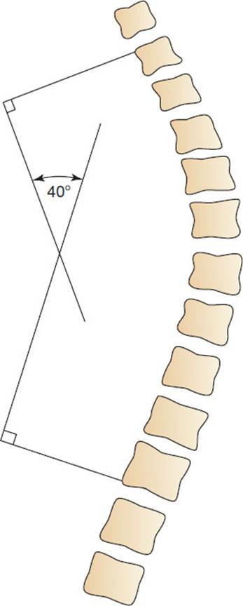
FIGURE 13-1 The Cobb method is used to measure this 40-degree lateral thoracic spinal curvature of the spine seen on an AP radiograph.
Nonsurgical Interventions to Improve Cardiopulmonary Function
Sixty-eight percent of patients with idiopathic scoliosis do not have pulmonary symptoms because the curve is not great enough either to cause symptoms or to have surgery.27 These less severe curves are often treated with bracingto decrease progression of the curve. A meta-analysis of the efficacy of nonoperative treatment found brace wear for 23 hours a day was significantly more effective than brace wear, or paravertebral electrical surface stimulation, for shorter periods of time.37
Some bracing may be a concern in the nonsurgical treatment of these patients.24,29,38 The Boston brace, a common thoracolumbosacral (TLSO) brace, was shown to have reduced lung volume and pulmonary compliance in patients wearing this brace for treatment of idiopathic scoliosis.39 Bracing increased respiratory effort, NVD, and dyspnea scores during progressive-cycle ergometry exercise.40 However, long-term effects of wearing a TLSO brace for 2 years showed no harm to lung function, in contrast to the short-term effects of bracing which showed impaired pulmonary function.41
Physical therapy interventions do not appear to reverse the lateral curvature but are aimed at slowing the progression of the curve. Treatments include stretching of soft tissue, strengthening on the convex side of the curve, lower extremity flexibility, and postural instruction.
Patients with severe kyphoscoliosis with ventilatory insufficiency requiring nocturnal nasal ventilation delivered by volume-cycled or pressure-cycled ventilators have been observed to improve symptoms, pulmonary function, and arterial blood gasses.42 Long-term noninvasive ventilation for patients with thoracic cage abnormalities appears to improve hypoventilation and functional abilities and decreases the need for tracheostomy.43
Treatment of mild idopathic scoliosis has included aerobic training and has been shown to be effective in maintaining and improving pulmonary function. Athanasopoulos et al.44 observed improved forced vital capacity, FEV1/FVC, and exercise tolerance after 2 months of cycling exercise performed 4 times per week for 30 minutes. A control group was observed to have reductions in the FVC, VC, respiratory rate, and exercise tolerance.
Summary
The greater the lateral curve of the spine, the more likely the patient will have pulmonary symptoms. Interventions for idiopathic scoliosis can be classified as nonsurgical for the less severe curves (less than 40 degrees) and as surgical for the more severe curves. The posterior surgical approach to decrease the lateral curve has many advantages over the anterior approach and demonstrates better return of pulmonary function postoperatively. Sixty-eight percent of the patients with idiopathic scoliosis with less than a 40-degree curve do not have pulmonary symptoms. These less severe curves are often treated with bracing to decrease progression of the curve. Some bracing has been shown to cause pulmonary impairments and may be of concern during the nonsurgical treatment of these patients. Aerobic conditioning has improved the cardiopulmonary function of patients with less severe scoliosis.
Pectus Deformities (Practice Patterns 4B, 4C, 4D, 4F, 4I; ICD-9-CM Code: 733)
Introduction
Pectus excavatum, which is commonly referred to as funnel chest, is the most common of all the chest wall deformities. It results from an overgrowth of costal cartilage with depression of the sternum, usually the lower portion.
Pectus carinatum or pigeon breast is a less common deformity that is caused by abnormal growth of the costal cartilage or abnormal fusion of the sternal body and manubrium that causes an anterior protrusion of the sternum. This deformity is not associated with respiratory difficulty, but dysplasia and cardiac defects may present as pulmonary symptoms.24
Impairments That Influence Cardiopulmonary Function
With pectus excavatum, there is often right sternal rotation as well as depression. This results in more depression on the right side and causes rotation and displacement of the heart to the left side. Heart murmur from altered blood flow is common due to the shift of the mediastinum. Occasionally patients may report dyspnea, chest pain, and palpitations. If pectus excavatum is severe, there is a decrease in stroke volume and respiratory reserve.
Surgical Interventions to Improve Cardiopulmonary Function
Surgical intervention is performed primarily for cosmetic reasons. Surgery is also warranted in the severe case where the deformity is interfering with cardiac output and causing significant pulmonary restrictions.24
A surgically produced deformity, a median sternotomy, is a common surgical approach for open heart procedures, such as coronary artery bypass graft and valvular repair or replacement. When compared to nonsternotomy procedures, patients who undergo sternotomy demonstrate a greater decrease in FEV1, FVC, PaO2, and PaCO2 in the early postoperative phase. The decline in lung volumes is correlated with pain level. Patients who undergo surgical intervention involving the chest wall generally have a transient decrease in chest wall compliance and increase their risk of pulmonary complications, including atelectasis and pneumonia.45
Sternal Precautions
Postoperatively, a patient is provided with sternal precautions in order to protect the incision site, enhance healing, and decrease pain. Many sternal precaution protocols exist and appear to be institution specific. In general, they last from postoperative day 1 until weeks 6 to 8. The goals of these protocols are to prevent shear and distraction forces across the surgical incision site. See Box 13-1 for one example of a sternal precaution protocol.
BOX 13-1
Sternal Precaution Protocol
For the first 6 to 8 weeks after surgery, patient may perform
•Bilateral shoulder exercises in forward flexion and extension, but below shoulder level
•Gentle upper-extremity isometric exercises
Patient must avoid
•Bilateral horizontal shoulder abduction, external rotation, and scapular retraction
•Unilateral arm exercises
•Lifting weights greater than 10 lb
•Pushing and pulling heavy objects
•Heavy housework
•Driving a car
Summary
Pectus deformities are usually treated surgically for cosmetic reasons or because of impairments in cardiopulmonary function. Postoperatively, cardiopulmonary function is decreased because of pain or decrease in chest wall compliance. Specific postoperative guidelines are given for the first 21 days, and general guidelines, for 8 weeks.
The Relationship of Shoulder Motion to Pulmonary Function (Practice Patterns 4D, 4E, 4F, 4G, 4H, 4I; ICD-9-CM Codes: 726, 810, 811, 812)
Introduction
Shoulder hypomobility is an impairment that is theorized to influence pulmonary function. The premise of this theory is that a relationship exists between shoulder motion and rib cage motion. This theory posits that shoulder motion influences rib cage and spinal movement and eventually influences pulmonary function. Therefore, treatment techniques have been developed using breathing to facilitate shoulder movement and shoulder movement to facilitate breathing.
Literature Supporting Theoretical Relationship
There appears to be no literature that directly associates shoulder immobilization or dysfunction with a decrease in pulmonary function, although many of the muscles of the shoulder share a respiratory function as well as an arm positional function. Patients suffering from lung disease and athletes recovering from workouts will often use their arms for support in order to facilitate accessory muscle breathing and to assist in ventilation. Studies show that there is an increase in metabolic and ventilatory requirements in patients who breathe at tidal volume with arms elevated and unsupported. It appears that the diaphragm contributes more to ventilatory pressure changes when the arms are unsupported and flexed to shoulder level.46 Mackey et al. agreed with this study and felt that the intercostals and accessory respiratory muscles act to stabilize the arms and torso, impeding chest wall movement and placing more emphasis on the diaphragm.47 These studies seem to indicate that there is a direct relationship between the position/support of the arms and pulmonary function. Hodges et al. found that predictable, anticipatory postural trunk motions occur during unilateral upper arm movement.48 The trunk movements are synergistic to the direction of the shoulder motion. The authors indicated that the trunk did not simply provide stabilization but worked with the shoulder muscles to facilitate movement. This also provides evidence to suggest that there is a direct relationship between shoulder motion and trunk/rib cage motion that is predictable and synergistic with movement of the glenohumeral joint. With movement of the glenohumeral joint above 160 degrees of flexion, Kapanji noted that thoracic spine extension is necessary in order to allow the scapula to fully depress and full shoulder range of motion to be attained.49 With thoracic spine extension, there is an increase in chest expansion and a subsequent increase in thoracic volume. This also provides evidence directly connecting the motion of the glenohumeral joint to thoracic cavity expansion.
Intervention to Improve Pulmonary Function
Massery has obtained positive results when utilizing different shoulder positions in order to facilitate breathing. This approach uses breathing, the motions of the trunk, and the motions of the shoulder in a coordinated fashion to facilitate synergistic trunk/shoulder motion and restore shoulder or pulmonary function. Such a treatment technique combines shoulder flexion and trunk extension with inhalation and shoulder extension and trunk flexion with expiration. Improved inspiratory and expiratory capacities have been obtained with this technique.50 This technique is currently being investigated but enjoys widespread use. More research is needed to determine its effectiveness in treating pulmonary and glenohumeral dysfunction.
INTEGUMENTARY CONDITIONS
Sarcoidosis (Practice Pattern 6H; ICD-9-CM Code: 457)
Introduction
Sarcoidosis is a systemic granulomatous disease that primarily affects the lungs and the lymphatic system.51 Noncaseating tumor-like epitheloids or granulomas are the hallmark of sarcoidosis. There is difficulty in collecting epidemiological information on this disease because of the inconsistency in the definition, variable methods for diagnosis, and variable presentation of disease.52 There appear to be two peaks for onset of sarcoidosis. One peak is in the third decade of life, followed by another peak in the sixth decade of life. The incidence is estimated at 6 per 100,000. There is a three times higher incidence in Afro-Americans than in Caucasians.52,53
Etiology of Pulmonary Involvement
It has been suggested that the granulomas are formed in response to a persistent and poorly degradable immune response that stimulates a T-cell–mediated response to some unidentified stimulus. This chronic immune activity results in fibrosis. There are other theories that have been proposed about the etiology of sarcoidosis. There appears to be a genetic predisposition underlying this disease with an abnormal regulation of antigen recognition. Patients who have been exposed to certain infections, talc, or clay, have a higher incidence of sarcoidosis than does the general population. Approximately 60% of patients who are diagnosed with sarcoidosis will have complete resolution of the granulomas. In the remaining patients, the disease will cause some degree of pulmonary dysfunction. At the site of the granulomas there is an accumulation of T-helper cells that chronically stimulates ongoing inflammation. There is also the detrimental effect of chronic inflammation with the release of enzymes that degrades the delicate tissue of the lungs to create more granulomas and fibrosis.51–53
Other Systems Affected
Other systems besides the lungs can be affected from the infiltration of granulomas and fibrosis. Twenty percent of the patients will have kidney involvement, which can extend from interstitial nephritis, and fibrosis that interferes with filtration, kidney stones, and urinary obstruction. Twenty to thirty percent of patients will have cardiac involvement, but most are asymptomatic. There may be fibrosis of the myocardium that both impairs contractility and causes arrhythmias and sudden death. There may be granuloma formation that predominantly affects the meninges, particularly the posterior fossa of the central nervous system (CNS). Sarcoidosis of the CNS can mimic tumors with progressive hydrocephalus and cranial nerve impairment. The gastrointestinal system may be impaired, as well as the liver and the eyes, and the skin, which typically clears.54
Diagnosis
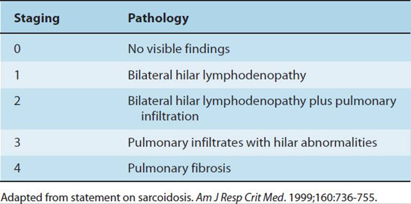
The disease is usually diagnosed by both clinical presentation and radiological findings. There is typically bilateral lymphadenopathy in 90% of the cases. This disease can be placed into a staging classification (Table 13-3)51 that has become helpful in advancing the understanding of sarcoidosis and medical management.
TABLE 13-3 Staging Classification of Sarcoidosis
Open lung biopsy is used to confirm the diagnosis by identifying the presence of granulomas.55 Pulmonary function tests typically demonstrate a restrictive pattern but may reveal an obstructive pattern if the granulomas are obstructing the airways. As in many fibrotic diseases, the PFTs in the presence of sarcoidosis show a decrease in total lung capacity, vital capacity and a decrease in diffusion capacity.55
Patients typically present with nonspecific constitutional symptoms that include fever, fatigue, and malaise; and weight loss, which usually occurs before any other symptom. If there is pulmonary involvement, 95% of the patients’ primary complaint will be dyspnea and decreased exercise tolerance. There may be a nonproductive cough and chest pain with exercise that is typically associated with efforts to increase tidal volume. Approximately half of the patients will have clubbing and one-third will have palpable lymph nodes. Upon auscultation there will be basilar crackles associated with fibrosis and arrhythmias. Thirty to sixty percent of patients will be asymptomatic if there is no significant pulmonary involvement.52,55,56
Intervention
It is recommended that patients be medically monitored closely for at least two years for signs of remission or progression so that medications can be adjusted as necessary. As with many of the other restrictive diseases that have been discussed in this chapter, corticosteroids are the most common medications administered initially. It has been documented that corticosteroids can actually shrink the size of the granulomas and contribute to remission. Cytotoxic drugs such as methotrexate and azathioprine may suppress the progression of disease but do not cause remission.
Two-thirds of those with the diagnosis will have complete remission without any pulmonary dysfunction. The remaining patients will have some degree of pulmonary dysfunction, and approximately 10% will develop progressive pulmonary fibrosis. Prognosis depends on the stage of the disease at the time of diagnosis. Patients in stage 1 or stage 2 typically have a 70% remission rate. Only 10% to 20% of patients in stage 3 will go into remission, and it is rare to have any recovery when diagnosis is made in stage 4.52 Almost 90% of patients die from sarcoidosis because of pulmonary failure. A patient who presents with acute symptoms has a higher incidence of recovery than a patient who has an insidious onset with multiple-organ involvement.51
Systemic Lupus Erythematosus (Practice Patterns 4D, 7C, 7D, 7E; ICD-9-CM Codes: 695.4, 710.0)
Etiology
The etiology of systemic lupus erythematosus (SLE) is unknown, but it has been suggested that there are genetic, environmental, and hormonal influences. Autoantibodies are activated against various nuclear antigens. These autoantibodies and the stimulated immune response are believed to mediate many of the manifestations of SLE. Lupus tends to affect women predominately in their childbearing years. Patients who develop interstitial pulmonary disease tend to be 45 to 50 years of age. Drug-induced SLE typically presents in the older patient, and there appears to be no gender differences. In general the prevalence is 50 per 100,000.57
Pulmonary Involvement and Clinical Presentation
Lupus affects the pulmonary system more frequently than any other collagen vascular disease. Pulmonary involvement may present as infectious pneumonia, pleuritis, pleural effusion, alveolar hemorrhage, pulmonary fibrosis, pulmonary hypertension, and obstructive airway disease. It can also be associated with muscular weakness that impairs ventilation. In approximately 5% to 10% of patients, it is suggested that the onset of SLE may be due to exposure to the following drugs: procainamide, penicillamine, quinidine, and hydralazine.58
The pleura is the most commonly affected tissue within the pulmonary system. The incidence of pleuritis is more common in men and in African Americans. The pleura becomes infiltrated with lymphocytes and plasma cells. A fibrotic process can develop from the inflammatory responses that cause pleural adhesions. This may result in a reduction in diaphragmatic excursion and chest wall expansion. These patients complain of chest pain with dyspnea, cough, and fever. Pleural effusion is usually present but will vary in size; therefore, signs and symptoms will also vary.56–58 Pleural effusion may present alone or may also be associated with infection, pulmonary embolism, congestive heart failure, or renal dysfunction.58
Pneumonitis that is associated with SLE will be associated with acute onset of dyspnea, hypoxia, fever, and hemoptysis. There will be patchy, diffuse infiltrates on chest X-ray, with a predilection for lower-lobe involvement. It often mimics the clinical presentation of adult respiratory distress syndrome. Acute lupus pneumonitis is associated with a 50% mortality rate.57,58
Pulmonary interstitial fibrosis primarily affects the lower lobes. It has been suggested that it may be a sequela of pneumonitis with a chronic insidious onset. These patients will present with a restrictive pattern on pulmonary function tests, low diffusion capacity, moderate to severe desaturation with exertion, and pleuritic chest pain with increased tidal volumes.57,58
Pulmonary hypertension may be associated with pulmonary fibrosis or may be the primary presentation of the pulmonary disease. It is associated with muscular hypertrophy, intimal proliferation, and capillary fibrosis and thrombus. These patients present with progressive dyspnea, exercise intolerance, and signs of right heart failure.57,58
Lupus may be associated with shrinking lung disorder. Upon radiographic examination there is a progressive loss of lung volume with basilar atelectasis. Shrinking lung disease is associated with diaphragmatic dysfunction that is not related to peripheral muscle weakness. This results in a decrease in lung compliance and a decrease in expiratory pressure that exceeds inspiratory weakness. Dyspnea is the most pronounced symptom, with many patients also reporting orthopnea.57,58
Finally, patients with SLE may present with alveolar hemorrhage that results in dyspnea, cough, and a decline in hemoglobin/hematocrit levels. A low-grade fever may also be present. Alveolar hemorrhage is uncommon but is highly life threatening with a 70% mortality rate. It resembles pneumonitis with infiltrates, but also encompasses alveolar necrosis, edema, and microvascular thrombus. There is a higher prevalence in young women.57,58
Diagnosis
Diagnosis of SLE depends on the clinical presentation. Analysis of pleural effusion will show a high concentration of glucose, and the antinuclear antibody test will be positive. The majority of diagnoses will be made on the basis of open lung biopsy.57,58
Intervention and Prognosis
Thalidomide and cyclosporine are the leading medications for the management of lupus. Ninety percent of patients will report an improvement in their symptoms, and 60% will go into remission. Ultraviolet A (UV-A) phototherapy is also used where it is suggested to promote DNA repair and enhance cell-mediated immunity. Methylprednisone, plasmaphoresis, azathioprine, and methotrexate can also be utilized to suppress the chronic immune and inflammatory processes that are progressively destroying pulmonary function.56
When there is pulmonary involvement with SLE, there is an approximate 50% mortality rate due to pneumonitis, fibrosis, and hemorrhage. Infection is the most common, accounting for the high mortality rate.58
Summary
Systemic lupus erythematosus, more frequently than any other collagen vascular disease, affects the pulmonary system. Pulmonary involvement may present as infectious pneumonia, pleuritis, pleural effusion, alveolar hemorrhage, pulmonary fibrosis, pulmonary hypertension, and obstructive airway disease. The majority of diagnoses will be made on the basis of open lung biopsy. Intervention is determined by clinical presentation along with medications for the management of lupus.
Scleroderma and Crest Syndrome (Practice Pattern 7E; ICD-9-CM Code: 710.1)
Introduction
Scleroderma or systemic sclerosis is a rare disease that affects multisystems and is characterized by intense fibrosis. It affects 2 to 10 people per 1 million. There is a higher incidence in women with a 7:1 female-to-male ratio, which decreases after the fifth decade of life. There is a slightly higher prevalence in Afro-Americans, 1.6% in males and 4.3% in females. Patients with scleroderma have twice the incidence of breast and bronchoalveolar cancer than the general population.23,59
Clinical Presentation and Etiology
Scleroderma is characterized by tightness and thickening of the skin, digital pitting and loss of pulp of the fingertips, and sclerodactyly and pulmonary fibrosis. The etiology of this disease is unknown, but several theories exist. It has been suggested that there is a hormonal influence, because there is an increased onset during pregnancy. It is possible there is a genetic link, but this theory has little evidence. There appears to be an immunological basis for both scleroderma and CREST. It has been documented that there is an increased incidence of scleroderma in males who have been exposed to silica, coal dust toxic oil, and organic solvents.59,60
CREST syndrome stands for a cluster of clinical presentations: calcinosis, Raynaud phenomenon, esophageal dysfunction, sclerodactyly, and telangiectasia. The incidence is 4 to 5 per million. Patients with CREST or limited systemic sclerosis typically suffer from Raynaud phenomenon a long time before any other symptoms appear. Raynaud phenomenon involves the vasoconstriction of the digits that leads to local hypoxia and cyanosis, pallor skin, and cold skin temperature. Distal ulcers of fingers and toes are common and frequently result in infections and autoamputation of distal digital segment.23,59,60 Scleroderma typically has a more acute onset that is associated with fatigue, arthralgias, carpal tunnel syndrome, swelling of hands and feet, and skin thickening.60
Pulmonary Involvement
Interstitial pulmonary fibrosis occurs in 74% of the cases of scleroderma, but the pathogenesis is not clearly defined. The incidence of pulmonary fibrosis is lower in CREST, but the pathology is believed to be similar. It may be mediated by growth factors and cytokines that are chemoattractants for fibroblasts, which enhance the synthesis and deposition of collagen within the alveolar walls. Through the inflammatory process, alveolar macrophages also stimulate various chemicals that stimulate fibroblastic activity and collagen formation. There is also a disruption of the capillary permeability that allows protein from the blood to leak into the interstitium that also contributes to collagen production. Typically the bases of the lungs are predominantly affected with alveolar space and capillary obliteration. The alveolar walls are infiltrated with edema and collagen. There is also a risk of further pulmonary fibrosis caused by aspiration of gastric acid from gastroesophageal reflux disease.23
With the presence of capillary destruction, there is a risk for the development of pulmonary hypertension. Up to 65% of the diagnosed cases of scleroderma will have pulmonary hypertension. There is a decrease in lumen size in the pulmonary vessels and obliteration due to hypertrophy of the medial layer of the blood vessels with collagen and plaque deposition within the intimal layer and vascular spasm.23 Patients may also be diagnosed with pleural thickening and effusion, which are usually found incidentally, but the presence of a pneumothorax may cause symptoms.60
Other Systems Involved
CREST syndrome and scleroderma can also affect other organs. The skin thickens and tightens, and the patient also suffers from Raynaud phenomenon. Patients may also have difficulty with secretory glands that reduce saliva and tears as well as cause sexual dysfunction. Anorexia, malnutrition, diarrhea, reflux, and osteoporosis are associated with involvement of the gastrointestinal system. Renal dysfunction and failure are associated with vascular insufficiency due to fibrosis. Finally, a small percentage of patients may present with cardiac involvement, pericarditis, pericardial effusion, arrhythmias, and on rare occasions left ventricular heart failure. Right heart failure is more frequently seen in this patient population and is associated with moderate-to-severe pulmonary hypertension.60
Diagnosis
The majority of patients will present for medical intervention due to skin changes and Raynaud syndrome. Sixty percent of the patients present with dyspnea. If the chest X-ray is normal in the presence of dyspnea, there must be a concern for pulmonary hypertension. Approximately 20% of patients will complain about a persistent nonproductive cough that usually worsens with exertion. If the cough is productive or occurs with ingestion, aspiration pneumonia must be considered in the differential diagnosis. Pulmonary fibrosis is associated with crackles and desaturation with exertion. Pleural disease may present with pleuritic chest pain.
Chest X-ray will depict interstitial opacities particularly within the lower lobes, which will progress to other lung segments as the disease progresses, decreasing lung volumes. Chest X-ray and a high-resolution computed tomographic (CT) scan will illustrate honeycombing in end-stage disease. The pulmonary function tests will be consistent with a restrictive process. There will be a decrease in total lung volume, FVC, and residual volume if interstitial fibrosis exists. If the primary pulmonary dysfunction is associated with hypertension, PFTs may be normal, but the patient may present with signs and symptoms of right heart failure. Disruption in gas exchange is common and associated with a decrease in diffusion capacity and may be the earliest sign of pulmonary involvement.23
Intervention and Prognosis
Many patients will go untreated until the lung disease has caused significant functional impairments. The standard medical intervention includes corticosteroids and cytotoxic drugs such as cyclophosphamide, D-penicillamine, and cyclosporine to decrease the immune response. Lung transplantation is becoming an accepted medical intervention in certain cases.23
The prognosis of patients with pulmonary involvement secondary to scleroderma or CREST is very dismal. If the diffusion capacity is less than 40% there is a 95% mortality rate at 5 years as opposed to 75% if diffusion capacity is greater than 40%. With progressive pulmonary hypertension, survival rates are even lower.23,61,62
Mixed Tissue Connective Disease (Practice Patterns 4D, 7E; ICD-9-CM Codes: 710, 714)
Etiology
The etiology of mixed tissue connective disease (MTCD) is unknown but is classified as a rheumatological disease. The clinical features are a combination of lupus, scleroderma, and polymyositis. There is a female prevalence to this disease, but there appears to be no ethnic or racial preponderance. The onset is typically in the mid-thirties.63
Eighty-five percent of patients diagnosed with MTCD have pulmonary involvement; but nearly 70% are asymptomatic, even though many of them will have a low diffusion capacity. Pulmonary involvement can involve pneumonitis, pneumonia, interstitial fibrosis, pleural effusion, pulmonary hypertension, and diaphragmatic dysfunction.64
Pulmonary Involvement and Clinical Presentation
The patient usually presents with insidious complications that have been present for an average of 4.5 years prior to seeking medical intervention. The most common signs and symptoms are sclerodactyly, and a nonproductive cough, Raynaud phenomenon, polyarthritis, and dyspnea on exertion. Occasionally, the patient may present acutely with severe hypoxemia, hemoptysis, or acute heart failure.63
The clinical presentation will vary based on the pulmonary involvement. If the underlying pulmonary disease is interstitial fibrosis, the peripheral parenchymal layer in the lower lobes is the first affected, and the fibrosis extends superiorly in an asymmetrical pattern as the disease worsens. Clinically the patient will suffer from severe hypoxemia. Pulmonary hypertension may develop secondary to hypoxemia from significant interstitial fibrosis. Pulmonary hypertension may also be caused by thromboembolic disease or pulmonary arteriopathy that leads to the reduction of arteriole lumen size from thrombosis, vasoconstriction, and hypertrophy of the arterial walls.64
Dyspnea on exertion, chest discomfort, and light-headedness are common signs of hypertension. As the hypertension increases the patient may present with signs and symptoms of right-heart failure. PFTs will demonstrate a decline in total lung capacity (TLC) and VC and a reduced diffusion capacity associated with hypoxemia. Examination of cardiac function will demonstrate elevations in pulmonary arterial pressure and reduced right ventricular function, which is consistent with pulmonary hypertension. Closer inspection of bronchoalveolar fluid shows an elevation of neutrophils that can lead to further fibrotic changes.22
Intervention and Prognosis
As with other diseases that lead to pulmonary fibrosis, the mainstay of treatment involves corticosteroids and the use of immunosuppressive medications. These drugs also increase the risk of infections. Research has begun to analyze the effectiveness of prostacyclin medications such as Flolan and UT15, which are potent vasodilatators that are delivered to the pulmonary arterial system to reduce the pulmonary arterial pressure.
Prognosis depends on the progression of fibrosis or hypertension, the response to treatment, the minimizing adverse effects of medications, and the extent of other tissue involvement such as the kidneys. Death from pulmonary involvement is the result of end-stage pulmonary fibrosis, infection, heart failure, or severe hemoptysis due to pulmonary hypertension.22
Sjögren Syndrome (Practice Patterns 4A, 4C, 4D, 4H, 5B, 6B; ICD-9-CM Code: 714)
Etiology
Sjögren syndrome is a chronic autoimmune inflammatory disease that causes exocrinopathy and epithelitis. Sjögren syndrome is one of the most common systemic rheumatic diseases and affects 1% of the general population and 3% of the elderly.65,66 Exocrinopathy involves the infiltration of T- and B-cell lymphocytes that progressively destroy salivary and lacrimal glands. Similar infiltrates may also invade visceral organs including the lungs. Sjögren syndrome is characterized by the infiltration of lymphocytes in the lungs; pancreas; gastrointestinal, hepatobiliary, and renal systems; and bone marrow. There have been several theories to explain the pathology of this syndrome. There appears to be a genetic predisposition, which is supported by the expression of certain antigens in the salivary epithelial cells. It also appears that herpes viruses and retrovirus may contribute to the pathology of this syndrome.65
The onset of Sjögren syndrome peaks in the fourth and fifth decades of life. Nine percent of the patient’s diagnosed with Sjögren Syndrome will suffer from pulmonary involvement. This syndrome is more pronounced in females with a 9:1 female-to-male ratio. Sjögren syndrome is classified as primary if it occurs without other connective tissue disease and secondary if Sjögren syndrome is associated with another connective tissue disease. It commonly occurs with rheumatoid arthritis.65
Pulmonary Involvement and Clinical Presentation
The pulmonary involvement includes various etiologies such as interstitial pneumonitis and fibrosis. Pleural disease—thickening, effusion, or pleuritis—and lymphoproliferative disorders are also associated with Sjögren Syndrome. Sjögren syndrome is also associated with shrinking lung syndrome that may be linked to phrenic nerve neuropathy, intrinsic diaphragm muscle weakness, or another unknown cause.66
Dryness in the upper airways leads to impairment of smell and taste, epistaxis, and perforation of the nasal septum. Dysfunction of the small peripheral airways leads to obstruction and the potential of hyperactivity of the airways. The end result may be mild obstructive pattern due to small airway disease, to subclinical alveolitis to pneumonitis, or to interstitial fibrotic disease.65
Many of the signs and symptoms are nonspecific for many other pulmonary diseases, but classic complaints for Sjögren syndrome include complaints of dry mouth and eyes, and arthritic symptoms. The signs and symptoms related to pulmonary involvement include a nonproductive cough, dyspnea on exertion and at rest, and ill-defined chest pain.65 In the presence of shrinking lung syndrome, the chest X-ray will illustrate small clear lung fields without evidence of intrinsic or pleural disease and an elevated right diaphragm. This disease is associated with a restrictive breathing pattern on pulmonary function tests.66
Diagnosis
The diagnosis of Sjögren syndrome is made based on clinical presentation of dry eyes and mouth and elevation of IgM, presence of a rheumatoid factor, or elevated antinuclear antibodies. The patient should be worked up for other connective tissue disorders, such as rheumatoid arthritis, lupus, or scleroderma.66
Intervention and Prognosis
Patients may respond to corticosteroids, but generally speaking, immunosuppressive medications are ineffective. Medications are given for palliative treatment of dryness of mucous lining, and in the presence of hyperreactive airways, bronchodilators are prescribed. Supplemental oxygen will be utilized when hypoxemia is documented.65–67
Prognosis is poor if the bronchoalveolar fluid contains excessive neutrophils. Prognosis is also dependent on the presence and progression of other connective diseases.
Summary
Diseases that affect the integumentary system do not just affect the skin. Multiple system involvement is quite common. The cardiopulmonary system is often a target of these diseases, with significant impairments to structures leading to functional limitations and disabilities for the patient.
SUMMARY
We have seen that impairments of bony structures, joints, fascia, skin, and musculature can result in decreases in cardiopulmonary function. The nature of these impairments is usually restrictive, which in turn can lead to decreased pulmonary function and poor exercise tolerance. Diminished movement of air into and out of the lungs may produce secretion retention, thus predisposing the patient to lung infection. Early physical therapy interventions should be directed toward maintaining long expansion, mobilizing secretions (if present) and improving exercise tolerence. These interventions may prevent many of these impairments and keep them from becoming functional limitations or disabilities.
REFERENCES
1.Wolf RL, Zmuda JM, Stone KL, Cauley JA. Update on epidemiology of osteoporosis. Curr Rheumatol Rep. 2000;2(1):74-86.
2.Salter RB. Textbook of Disorders and Injuries of the Musculoskeletal System. 3rd ed. Baltimore, MD: Williams & Wilkins; 1999.
3.Bachrach L. Acquisition of optimal bone mass in childhood and adolescence. Trends Endocrinol Metabol. 2001;12(1):22-28.
4.Enstrud KE, Black DM, Harris F, Ettinger B, Cummings SR. Correlates of kyphosis in older women. The fracture intervention trial research group. Am Geriatr Soc. 1997;45(6):682-687.
5.Ismail AA, Cooper C, Felsenberg D, et al. Number and type of vertebral deformities: epidemiological characteristics and relation to back pain and height loss. European vertebral osteoporosis study group. Osteoporos Int. 1999;9(3):206-213.
6.De Smet AA, Robinson RG, Johnson BE, Lukert BP. Spinal compression fractures in osteoporotic women: patterns and relationship to hyperkyphosis. Radiology. 1988;166(2):497-500.
7.Goh S, Price RI, Leedman PJ, Singer KP. The relative influence of vertebral body and intervertebral disc shape on thoracic kyphosis. Clin Biomech (Bristol, Avon). 1999;14(7):439-438.
8.Hedlund LR, Gallagher JC, Meeger C, Stoner S. Change in vertebral shape in spinal osteoporosis. Calcif Tissue Int. 1989;44(3):168-172.
9.Biyani A, Ebraheim NA, Lu J. Thoracic spine fractures in patients older than 50 years. Clin Orthop. 1996 July;(328):190-193.
10.Harma M, Heliovaara M, Aromaa A, Knekt P. Thoracic spine compression fractures in Finland. Clin Orthop. 1986;205:188-194.
11.Melton LJ, Atkinson EJ, Khosla S, O’Fallon WM, Riggs BL. Secondary osteoporosis and the risk of vertebral deformities in women. Bone. 1999;24(1):49-55.
12.Abdel-Hamid Osman A, Bassiouni H, Koutri R, Nijs J, Geusens P, Dequeker J. Aging of the thoracic spine: distinction between wedging in osteoarthritis and fracture in osteoporosis—a cross-sectional and longitudinal study. Bone. 1994;15(4):437-442.
13.Ryan PJ, Blake G, Herd R, Fogelman I. A clinical profile of back pain and disability in patients with spinal osteoporosis. Bone. 1994;15(1):27-30.
14.Leech JA, Dulberg C, Kellie S, Pattee L, Gay J. Relationship of lung function to severity of osteoporosis in women. Am Rev Respir Dis. 1990;141(1):68-71.
15.Schlaich C, Minne HW, Bruckner T, et al. Reduced pulmonary function in patients with spinal osteoporotic fractures. Osteoporos Int. 1998;8(3):261-267.
16.Mellin G, Harjula R. Lung function in relation to thoracic spinal mobility and kyphosis. Scand J Rehabil Med. 1987;19(2):89-92.
17.Culham EG, Jimenez HA, King CE. Thoracic kyphosis, rib mobility, and lung volumes in normal women and women with osteoporosis. Spine. 1994;1(11):1250-1255.
18.Malmros B, Mortensen L, Jensen MB, Charles P. Positive effects of physiotherapy on chronic pain and performance in osteoporosis. Osteoporos Int. 1998;8(3):215-221.
19.Vogt MT, Nevitt MC, Cauley JA. Back problems and atherosclerosis—the study of osteoporotic fractures. Spine. 1997;22(23):2741-2747.
20.Blansfield HN, Andrew CB. Osteoporosis—a factor in mortality following cardiac surgery. Comm Med. 2000;64(2):71-73.
21.Lee-Chiong T. Pulmonary manisfestations of ankylosis spondylitis and relapsing polychondritis. Clin Chest Med. 1998;19(4):747-757.
22.Wiedemann H, Matthay R. Pulmonary manisfestations of the collagen vascular disease. Clin Chest Med. 1989;10(4):677-715.
23.Fishman A, ed. Pulmonary Diseases and Disorders: Companion Handbook. 2nd ed. New York: McGraw-Hill; 1994.
24.Grissom L, Harcke H. Thoracic deformities and the growing lung. Semin Reontgenol. 1998;33(2):199-208.
25.Leong JC, Lu WW, Luk KD, Karlberg EM. Kinematics of the chest cage and spine during breathing in healthy individuals and in patients with adolescent idiopathic scoliosis. Spine. 1999;24(13):1310-1315.
26.Aaro S, Ohlund C. Scoliosis and pulmonary function. Spine. 1984;9(2):220-222.
27.Vedantam R, Crawford A. The role of preoperative pulmonary function tests in patients with adolescent idiopathic scoliosis undergoing posterior spinal fusion. Spine. 1997;22(23):2731-2734.
28.Giordano A, Fuso L, Galli M, Calcagni M, Aulsia L. Evaluation of pulmonary ventilation and diaphragmatic movement in idiopathic scoliosis using radioaerosol ventilation scintigraphy. Nucl Med Commun. 1997;18:105-111.
29.Widmann R, Bitan F, Laplaza J, Burke S, DiMaio M. Spinal deformity, pulmonary compromise, and quality of life in osteogenesis imperfecta. Spine. 1999;24(16):1673-1678.
30.Estenne M, Deroom E, DeTroyer A. Neck and abdominal muscle activity in patients with severe thoracic scoliosis. Am J Resp Crit Care Med. 1998;158(2):452-457.
31.Conti G, Rocco M, Antonelli M, et al. Respiratory system mechanics in the early phase of acute respiratory failure due to severe kyphoscoliosis. Intensive Care Med. 1997;23(5):539-544.
32.Krachman S, Criner G. Hypoventilation syndrome. Clin Chest Med. 1998;19(1):139-156.
33.Al-Katten K, Simonds A, Chung KF, Kaplan DK. Kyphoscoliosis and bronchial torsion. Chest. 1997;111(4):1134-1137.
34.Boyer J, Amin N, Taddonio R, Dozor AJ. Evidence of airway obstruction in children with idiopathic scoliosis. Chest. 1996;109(6):1532-1535.
35.Vedantam R, Lenke L, Bridwell K, Haas J, Linville D. A prospective evaluation of pulmonary function in patients with adolescent idiopathic scoliosis relative to the surgical approach used for spinal arthrodesis. Spine. 2000;25(1):82-90.
36.Wong CA, Cole AA, Watson L, Webb JK, Johnston ID, Kinnear WJ. Pulmonary function before and after anterior spinal surgery in adult idiopathic scoliosis. Thorax. 1996;51(5):534-536.
37.Rowe DE, Bernstein SM, Riddick MF, Adler F, Emans JB, Gardner-Bonneau D. A meta-analysis of the efficacy of non-operative treatments for idiopathic scoliosis. J Bone Joint Surg Am. 1997;79(5):664-674.
38.Cline CC, Coast JR, Arnall DA. A chest wall restrictor to study effects on pulmonary function and exercise. Development and validation. Respiration. 1999;66(2):182-187.
39.Katsaris G, Loukos A, Valavanis J, Vassiliou M, Behrakis PK. The immediate effect of a Boston brace on lung volumes and pulmonary compliance in mild adolescent idiopathic scoliosis. J Eur Spine. 1999;8(1):2-7.
40.Ferrari K, Goti P, Sanna A, et al. Short term effects of bracing on exercise performance in mild idiopathic thoracic scoliosis. Lung. 1997;175(5):299-310.
41.Korovessis P, Filos KS, Georgopoulos D. Long-term alterations of respiratory function in adolescents wearing a brace for idiopathic scoliosis. Spine. 1996;21(17):1979-1984.
42.Ferris G, Servera-Pieras E, Vergara P, et al. Kyphoventilatory insufficiency: noninvasive management outcomes. Am J Phys Med Rehab. 2000;79(1):24-29.
43.Leger P. Long-term noninvasive ventilation for patients with thoracic cage abnormalities. Resp Care Clinic North Am. 1996;2(2):241-252.
44.Athanasopoulos S, Paxinos T, Tsafantakis E, Zachariou K, Chatziconstantinou S. The effect of aerobic training in girls with idiopathic scoliosis. Scand J Med Sci Sports. 1999;9(1):36-40.
45.Aris A, Camara M, Casan P, Litvan H. Pulmonary function following aortic valve replacement: a comparison between ministernotomy and median sternotomy. J Heart Valve Dis. 1999;8: 605-608.
46.Couser JI, Martinez FJ, Celli BR. Respiratory response and ventilatory muscle recruitment during arm elevation in normal subjects. Chest. 1992;101(2):336-340.
47.Mackey M, Ellis E, Nicholls M. Breathing patterns and heart rate during simulated occupational upper limb tasks in normal subjects. Physiother Res Int. 1998;3(2):83-99.
48.Hodges PW, Cresswell AG, Daggfeldt K, Thorstensson A. Three dimensional preparatory trunk motion precedes asymmetrical upper limb movement. Gait Posture. 2000;11(2):92-101.
49.Kapandji IA. The Physiology of the Joints. Vol 1. 2nd ed. Edinburgh, UK: Churchill Livingstone; 1970.
50.Massery M. A patient with neuromuscular and musculoskeletal dysfunction. In: Frownfelter D, Dean E, eds. Principles and Practice of Cardiopulmonary Physical Therapy. 3rd ed. St Louis, MO: Mosby; 1996:679.
51.Anonymous. Statement on sarcoidosis. Am J Resp Crit Med. 1999; 160:736-755.
52.Nagai S, Shigematsu M, Hamada K, Izumi T. Clinical courses and prognoses of pulmonary sarcoidosis. Curr Opin Pulm Med. 1999;5:293-298.
53.Costabel U, Hunninghake G. ATS/ERS WASOG statement on sarcoidosis. Eur Resp J. 1999;14:735-737.
54.Belfer M, Stevens R. Sarcoidosis: a primary care review. Am Fam Physician. 1998;58(9):2041-2052.
55.Sheffield E. Pathology of sarcoidosis. Clin Chest Med. 1997;18(4):741-750.
56.Lynch J, Kazerooni E, Gay S. Pulmonary sarcoid. Clin Chest Med. 1997;18(4):755-780.
57.Godfrey T, Khamashta M, Hughes G. Therapeutic advances in systemic lupus erythematosus. Curr Opin Rheumatol. 1998;10(5):435-441.
58.Murin S, Weidemann H, Matthay RA. Pulmonary manifestations of systemic lupus erythematosus. Clin Chest Med. 1998;19(4):641-665.
59.Minai O, Dweik R, Arroliga A. Manifestations of scleroderma pulmonary disease. Clin Chest Med. 1998;19(4):713-727.
60.Silman A. Epidemiology of scleroderma. Ann Rheum Dis. 1991;50:846-853.
61.Steen V. Clinical manifestations of systemic sclerosis. Semin Cutan Med Surg. 1998;17(1):48-54.
62.Bulpitt K, Clements P, Lachenbruch P, Paulus H, Peter J. Early undifferentiated connective tissue disease: III. Outcome and prognostic indicators in early scleroderma (systemic sclerosis). Ann Int Med. 1993;118:602-609.
63.Prakash U. Respiratory complications in mixed connective tissue disease. Clin Chest Med. 1998;19(4):733-745.
64.Corley D, Winterbauer R. Collagen vascular disease. Semin Resp Infect. 1995;10(2):78-85.
65.Cain H, Noble P, Matthay R. Pulmonary manifestations of Sjögren’s Syndrome. Clin Chest Med. 1998;19(4):687-697.
66.Mialon M, Barthelemy L, Sebert P, LeHenaff C, Sarni D. A longitudinal study of lung impairment in patients with primary Sjögren’s syndrome. Clin Exp Rheumatol. 1997;15(4):49-54.
67.Tavoni A, Cirigliano C, Frigelli S, Stampacchia G, Bombardieri S. Shrinking lung in primary Sjögren’s syndrome. Arthritis Rheum. 1999;42(10):2249-2250.