Philip S. Barie
Frederic M. Pieracci
Soumitra R. Eachempati
Infection is morbid and costly, but also preventable to some degree; therefore, it behooves every practitioner to do the utmost to prevent infection. An ensemble of prevention methods is required, because no single method is universally effective. Infection control is paramount, but often underemphasized. Surgical incisions and traumatic wounds must be handled gently, inspected daily, and dressed if necessary using aseptic technique. Drains and catheters must be avoided if possible, and removed as soon as practicable. Prophylactic and therapeutic antibiotics should be used sparingly so as to minimize antibiotic selection pressure on the development of multidrug-resistant (MDR) pathogens.
Surgical patients are at particular risk of infection for many reasons. Surgery is inherently invasive, which creates portals of entry in natural epithelial barriers for pathogens to invade the host. Surgical illness is immunosuppressive (e.g., trauma, burns, malignant tumors), as is therapeutic immunosuppression following solid organ transplantation. General anesthesia almost always means a period of mechanical ventilation and a period of reduced consciousness during emergence from anesthesia that poses a risk of pulmonary aspiration of gastric contents, all of which increase the risk of pneumonia. Nosocomial pneumonia occurs more frequently among surgical patients than comparably ill medical patients. Surgical patients are also uniquely afflicted by infections of incisions. Considering that the development of a postoperative infection has a negative impact on surgical outcomes, recognizing risk, minimizing it, and taking an aggressive approach to the diagnosis and treatment of such infections is crucial to improve surgical outcomes.
Surgical infections are traditionally considered to be infections that require surgical therapy (e.g., complicated intra-abdominal and soft tissue infections). However, the recognition that surgical patients are especially vulnerable to nosocomial infection has led to an expansive definition to include any infection that affects surgical patients. Intra-abdominal and soft tissue infections are considered in detail elsewhere in this textbook. The emphasis of this chapter is on the epidemiology, prevention, and management of postoperative and posttraumatic infections.
Risk Factors for Postoperative Infection
The general principles of surgical care, critical care, and infection control cannot be overemphasized. Resuscitation must be rapid, yet precise. Pathology must be identified and treated as soon as possible. Infection control is sometimes sacrificed under the often chaotic conditions of resuscitation, but it must not be (see below). Central venous catheters inserted under suboptimal barrier precautions (i.e., lack of cap, mask, sterile gown, and sterile gloves for the operator and a full-bed drape for the patient) must be removed and replaced (if necessary) by a new puncture at a new site as soon as the patient's condition permits. Detailed evidence-based guidelines for the general prevention of infection (1,2) and the prevention of ventilator-associated pneumonia have been published (3,4). All who provide critical care must be familiar with the guidelines and adhere to them insofar as possible.
Control of Blood Sugar
Hyperglycemia is deleterious to host immune function, and may also reflect the catabolism and insulin resistance associated with the surgical stress response. Poor perioperative control of blood glucose increases the risk of infection and worsens outcome from sepsis. Diabetic patients undergoing cardiopulmonary bypass surgery have a higher risk of infection of both the sternal incision and the vein harvest incisions on the lower extremities (5). Moderate hyperglycemia (>200 mg/dL) at any time on the first postoperative day increases the risk of surgical site infection (SSI) fourfold after cardiac (6) and noncardiac surgery (7). Insulin infusion to keep blood glucose concentrations <110 mg/dL was associated with a 40% decrease in mortality among critically ill postoperative patients, and also fewer nosocomial infections and less organ dysfunction (8). Meta-analysis of 35 trials of control of blood glucose indicates that the risk of mortality is decreased significantly (risk ratio [RR] 0.85, 95% confidence interval [CI] 0.75–0.97) by tight glucose control, especially so for critically ill surgical patients (RR 0.58, 95% CI 0.22–0.62), regardless of whether the patients had diabetes mellitus (RR 0.71, 95% CI 0.54–0.93) or stress-induced hyperglycemia (RR 0.73, 95% CI 0.58–0.90) (9).
Nutritional support is crucial, considering that surgical stress causes catabolism and that restoration of anabolism requires the provision of calories and nitrogen far in excess of basal requirements of 25 to 30 kcal and 1 g nitrogen/kg/day. In the midst of the stress response, it is challenging to provide adequate calories and protein while avoiding hyperglycemia. Parenteral nutrition appears to convey no advantage over not feeding the patient at all (10), perhaps because of the inherent morbidity of central intravenous feeding (i.e., the risk of catheter-related bloodstream infection [CR-BSI] and hyperglycemia). In contrast, early enteral feeding (within the first 48 hours, perhaps immediately if the gut is functional) is clearly beneficial, with the possible exception of pneumonia prevention (see below). The risk of infection was reduced by 55% (odds ratio [OR] 0.45, 95% CI 0.30–0.66) in a meta-analysis of 15 randomized trials of early enteral feeding following surgery, trauma, or burns (11).
Blood Transfusion
Blood transfusion can be life saving after trauma or hemorrhage, but an increased risk of infection is the consequence. Transfusions exert immunosuppressive effects through presentation of leukocyte antigens and the induction of a shift to the T-helper 2 (immunosuppressive) phenotype, although the mechanism remains somewhat controversial because transfusion of leukocyte-depleted red blood cell concentrates does not reduce the risk of infection (12). Claridge et al. identified an exponential relationship between transfusion risk and infection risk among trauma patients, detectable with even 1 unit of transfusion and becoming a virtual certainty after more than 15 units of transfused blood (RR 1.084, 95% CI 1.028–1.142) (13). Hill et al. has estimated by meta-analysis the risk of infection related to blood transfusion to be increased for trauma patients by more than fivefold (OR 5.26, 95% CI 5.03–5.43), and for surgical patients by more than threefold (14). This increased risk for infection by transfusion has also been identified for critically ill patients in general (15), and for CR-BSI (16) and ventilator-associated pneumonia (VAP) (17) specifically.
Banked blood is affected by a “storage lesion” characterized by loss of membrane 2-3-diphosphoglycerate and adenosine triphosphate, leading to loss of membrane deformability (18). As a result, erythrocytes cannot deform as they must to transit the microcirculation, causing disruption of nutrient blood flow and impaired oxygen offloading. Consequently, blood transfusion does not increase oxygen consumption for critically ill patients with sepsis (19), and may actually increase the risk of organ dysfunction. The storage lesion becomes fully manifest after about 14 days of storage; transfusion of older blood is an independent risk factor for the development of infection (20). It is safe to be conservative in the administration of red blood cell concentrates to stable patients in the intensive care unit (ICU) (21).
Infection Control
Infection control is an individual and a collective responsibility of the critical care team and unit. Hand hygiene is the most effective means known to reduce the spread of infection, yet whenever studied it is invariably underutilized. Hand cleansing with soap and water requires a minimum of 30 to 45 seconds to be effective. Alcohol gel hand cleansers are equally effective (except against the spores of Clostridium difficile), compliance with use is higher, and when used, the prevalence of MDR bacteria is reduced (22). Universal precautions (i.e., cap, mask, gown, gloves, and protective eyewear) must be observed whenever there is a risk of splashing of body fluids.
The source of most bacterial pathogens is the patient's endogenous flora. Skin surfaces, artificial airways, gut lumen, wounds, catheters, and inanimate surfaces (e.g., bed rails, computer terminals) may become colonized. Any break in natural epithelial barriers (e.g., incisions, percutaneous catheters, airway or urinary catheters) creates a portal of entry for invasion of pathogens. The fecal-oral route is the most common manner by which pathogens reach the portal, but health care workers definitely facilitate the transmission of pathogens around a unit. Many organisms that cause infection following surgery are inherently avirulent (e.g., Candida, Enterococcus, Pseudomonas). Whether infection develops is determined by complex interactions among host defenses, pathogen, and therapy.
Contact isolation is an important part of infection control, and should be used selectively to prevent the spread of pathogens such as methicillin-resistant Staphylococcus aureus (MRSA) and vancomycin-resistant enterococci (VRE), or MDR Gram-negative bacilli. However, contact isolation may decrease the amount of direct patient contact. An appropriate balance must be struck, because reduced nurse staffing of ICUs has been independently associated with an increased risk of a number of nosocomial infections (23).
Catheter Care
Optimal catheter care includes avoidance of use when unnecessary, appropriate skin cleansing and barrier protection during insertion, proper catheter selection, proper dressing of indwelling catheters, and removal as soon as possible when no longer needed, or after insertion under less than ideal circumstances (e.g., trauma bay, cardiac resuscitation).
The benefit of the information gained by catheterization must always be weighed against the risk of infection. Almost all indwelling catheters carry a risk of infection, but nontunneled central venous catheters (and pulmonary artery catheters) pose the highest risk, including local site infections and CR-BSIs (see below). Other catheters that pose increased infection risk include intercostal thoracostomy catheters (if inserted as an emergency), ventriculostomy catheters for intracranial pressure monitoring, and urinary bladder catheters. Each day of endotracheal intubation and mechanical ventilation increases the risk of pneumonia by 1% to 3%; it is controversial whether tracheostomy decreases that risk.
Chlorhexidine (which is bactericidal, viricidal, and fungicidal) should be used preferentially for skin preparation for vascular catheter insertion, having been shown to be superior to povidone-iodine solution (24). If povidone-iodine solution is used (of which use is discouraged), it must be allowed to dry, as it is not bactericidal when wet. Full barrier precautions are mandatory for all bedside catheterization procedures (2) except arterial and urinary bladder catheterization, for which sterile gloves and a sterile field suffice. Whenever a central venous catheter is inserted under suboptimal conditions it must be removed (and replaced at a different site if still needed) as soon as permitted by the patient's hemodynamic status, but no more than 24 hours after insertion. A single dose of a first-generation cephalosporin (e.g., cefazolin), but no more, may prevent some infections following emergency tube thoracostomy or ventriculostomy, but is not indicated for vascular or bladder catheterizations. Topical antiseptics placed postprocedure at the insertion site are of no benefit for any type of indwelling catheter, and may actually increase the risk of infection.
Dressings must be maintained carefully (25). Maintaining the integrity of dressings is challenging if the patient is agitated or the body surface is irregular (e.g., the neck [internal jugular vein catheterization] as opposed to the chest wall [subclavian vein catheterization]), but its importance is crucial. Marking the dressing clearly with the date and time of each change is a simple and effective way to manage dressing changes. Dressing carts or similar apparatus should not be brought from patient to patient; rather, sufficient supplies should be kept in each patient's room. The possibility for inanimate objects (e.g., scissors) to be transmission vectors if not cleansed thoroughly after contact with each patient must be borne in mind. Dedicated catheter care teams reduce the risk of CR-BSI substantially (26).
The choice of catheter may play a role in decreasing the risk of infection related to endotracheal tubes, central venous catheters, and urinary catheters. Continuous aspiration of subglottic secretions (CASS), via an endotracheal tube with an extra lumen that opens to the airway just above the balloon, facilitates the removal of secretions that accumulate below the vocal cords but above the endotracheal tube balloon, an area that cannot be reached by routine suctioning. The incidence of VAP is decreased by one half by CASS (26). Silver-impregnated endotracheal tubes are effective in reducing airway colonization, but whether the incidence of VAP is reduced is not yet known (27). Antibiotic- (e.g., minocycline/rifampin) or antiseptic-coated central venous catheters (e.g., chlorhexidine/silver sulfadiazine) can reduce the incidence of CR-BSI (28), especially in high-prevalence units; minocycline/rifampin-coated catheters may be more effective. Urinary bladder catheters coated with ionic silver reduce the incidence of catheter-related bacterial cystitis by a similar amount (29).
Ventilator weaning by protocol, combined with daily sedation holidays and spontaneous breathing trials, allows earlier endotracheal extubation and decreases the risk of VAP (see below) (30). An even better strategy may be avoidance of endotracheal intubation entirely. Respiratory failure can sometimes be managed with noninvasive positive pressure ventilation delivered by mask (e.g., continuous positive airway pressure [CPAP]) (31). Improved resuscitation techniques and noninvasive monitoring techniques have decreased the utilization of pulmonary artery catheters, which pose a particularly high risk of infection (32). Most drains do not decrease the risk of infection; in fact, the risk is probably increased (33) because the catheters hold open a portal for invasion by bacteria.
Risk Factors for Surgical Site Infection
The spectrum of bacterial contamination of the surgical site is well described (34). Clean surgical procedures affect only integumentary and musculoskeletal soft tissues. Clean-contaminated procedures open a hollow viscus (e.g., alimentary, biliary, genitourinary, respiratory tract) under controlled circumstances (e.g., elective colon surgery). Contaminated procedures involve extensive introduction of bacteria into a normally sterile body cavity, but too briefly to allow infection to become established during surgery (e.g., penetrating abdominal trauma, enterotomy during adhesiolysis for mechanical bowel obstruction). Dirty procedures are performed to control established infection (e.g., colon resection for perforated diverticulitis).
Numerous factors determine whether a patient will develop an SSI, including factors related to the patient, the environment, and the therapy (Table 106.1) (33). As incorporated in the National Nosocomial Infections Surveillance System (NNIS) (34,35), the most recognized factors are the wound classification, American Society of Anesthesiologists class ≥3 (class 3: Chronic active medical illness), and prolonged operative time, where time is longer than the 75th percentile for each such procedure. According to the NNIS, the risk of SSI increases with an increasing number of risk factors present, irrespective of the type of operation (35). Laparoscopic surgery decreases the incidence of SSI under most circumstances (36). The reasons that laparoscopic surgery decreases the risk of SSI are possibly several, including decreased wound size, limited use of cautery in the abdominal wall, or a diminished stress response to tissue injury.
|
Table 106.1 Rates of health care-associated pneumonia and catheter-related bloodstream infection among various intensive care unit (ICU) types |
||||||||||||||||||||||||||||||||||||||||
|
||||||||||||||||||||||||||||||||||||||||
Host-derived factors contribute importantly to the risk of SSI, including increased age (36), obesity, malnutrition, diabetes mellitus (5,7), hypocholesterolemia (38), and numerous other factors that are not accounted for specifically by the NNIS system (Table 106.1). In one 6-year study of 5,031 patients undergoing noncardiac surgery, the overall incidence of SSI was 3.2%. Independent risk factors for the development of SSI included ascites, diabetes mellitus, postoperative anemia, and recent weight loss, but not chronic obstructive pulmonary disorder, tobacco use, or corticosteroid use (39). In another prospective study of 9,016 patients, 12.5% of patients developed an infection of some type within 28 days after surgery (40). Multivariable analysis revealed that decreased serum albumin concentration, increased age, tracheostomy, and amputations were associated with an increased probability of an early infection, whereas factors associated with readmission due to infection included a dialysis shunt, vascular repair, and an early infection. Factors associated with 28-day mortality included increased age, low serum albumin concentration, increased serum creatinine concentration, and an early infection (40).
Lapses in the modern operating room can result in increased rates of SSI. Proper sterilization, ventilation, and skin preparation techniques require continuous vigilance. The operating team must be attentive to personal hygiene (e.g., hand scrubbing, hair). Recent data indicate that a brief rinse with soap and water followed by use of an alcohol gel hand rub is equivalent to the prolonged (and ritualized) session at the scrub sink (41).
Hypothermia during surgery is common if patients are not warmed actively, owing to evaporative water losses, administration of room temperature fluids, and other factors (42). Maintenance of normal core body temperature is unequivocally important for decreasing the incidence of SSI. Mild intraoperative hypothermia is associated with an increased rate of SSI following elective colon surgery (43) and diverse operations (44).
It is intuitive that oxygen administration in the postoperative period would be beneficial for wound healing (45,46). The ischemic milieu of the fresh surgical incision is vulnerable to bacterial invasion. Moreover, oxygen has been postulated to have a direct antibacterial effect (46). However, clinical trials have had conflicting results (47,48). Supplemental oxygenation administration specifically to reduce the incidence of SSI remains plausible, and further studies are needed.
Closure of a contaminated or dirty incision is widely believed to increase the risk of SSI, but few good studies exist to help sort out the multiplicity of wound closure techniques available to surgeons. “Open abdomen” techniques of temporary abdominal closure for management of trauma or severe peritonitis are utilized increasingly. Retrospective studies indicate that antibiotics are not indicated for prophylaxis of the open abdomen (49), but infection of the abdominal wall, should it occur, is highly morbid. Inability to achieve primary abdominal closure is associated with several infectious complications (pneumonia, bloodstream infection, and SSI). Infectious complications, in turn, significantly increased costs from prolonged length of stay, but not mortality (50).
Drains placed in incisions probably cause more infections than they prevent. Epithelialization of the wound is prevented and the drain becomes a conduit, holding open a portal for invasion by pathogens colonizing the skin. Several studies of drains placed into clean or clean-contaminated incisions show that the rate of SSI is not reduced (51,52); in fact, the rate is increased (53,54,55,56). Considering that drains pose a risk and accomplish little of what is expected of them, they should be used as little as possible and removed as soon as possible (57). Under no circumstances should prolonged antibiotic prophylaxis be administered to “cover” indwelling drains.
Wound irrigation is a controversial means to reduce the risk of SSI. Routine low-pressure saline irrigation of an incision does not reduce the risk of SSI (58), but high pressure (i.e., pulse irrigation) may be beneficial (59). An increasing body of knowledge suggests that intraoperative topical antibiotics can minimize the risk of SSI (60,61,62), but the use of antiseptics rather than antibiotics might minimize the possibility of the development of resistance.
Risk Factors for Pneumonia
Surgical patients are susceptible to pneumonia, particularly if they require mechanical ventilation. Ventilator-associated pneumonia, defined as pneumonia occurring 48 to 72 hours after endotracheal intubation, is the most common ICU infection among surgical and trauma patients. Unfortunately, VAP is partially iatrogenic. Nonspecific diagnostic criteria, indiscriminate antibiotic use, and unclear therapeutic end points have all contributed to increased episodes of VAP caused by MDR pathogens. In turn, MDR pathogens increase the likelihood of inadequate initial antimicrobial therapy, which exerts further selection pressure for these pathogens, and results in higher mortality.
Distinction is sometimes made between early-onset VAP (occurring <5 days after intubation) and late-onset VAP (occurring ≥5 days after intubation). Early-onset VAP, to which trauma patients are particularly prone, is often a result of aspiration of gastric contents, and is usually caused by antibiotic-sensitive bacteria such as methicillin-sensitive S. aureus, Streptococcus pneumoniae, and Haemophilus influenzae (4,63,64). Conversely, patients with late-onset VAP are at increased risk for infection with MDR pathogens (e.g., MRSA, Pseudomonas aeruginosa, or Acinetobacter spp.).
The incidence of VAP depends upon the diagnostic criteria utilized, and thus varies in published reports. Clinical criteria alone overestimate the incidence of VAP as compared with either microbiologic or histologic data (65,66). A systematic review of 89 studies of VAP among mechanically ventilated patients (67) reported a pooled incidence of VAP of 22.8% (95% CI 18.8–26.9). The NNIS system reported recently that VAP occurred at a rate of 4.9 cases per 1,000 ventilator-days in medical ICUs and 9.3 per 1,000 ventilator-days in surgical ICUs (36) (Table 106.2). The risk for trauma patients, especially those with traumatic brain injury, is especially high. The incidence of VAP increases with the duration of mechanical ventilation at a rate of 3% per day during the first 5 days, 2% per day during days 5 to 10, and 1% per day after that (68).
Risk factors for VAP are summarized in Table 106.3 (27). Perhaps most important is airway intubation itself. The risk of VAP increases six- to 20-fold in mechanically ventilated patients (69,70); VAP is also especially common in patients with acute respiratory distress syndrome (ARDS), owing to prolonged mechanical ventilation and devastated local airway host defenses (71,72,73).
|
Table 106.2 Risk factors for the development of surgical site infections |
|||
|
Several evidence-based strategies can prevent VAP, but to be used effectively a thorough understanding of modifiable risk factors is required (Table 106.4). Prevention of VAP begins with minimization of endotracheal intubation and the duration of mechanical ventilation. Noninvasive positive pressure ventilation (NIPPV) should be considered in lieu of intubation, as management of respiratory failure with NIPPV leads to a lower incidence of VAP (74). If endotracheal intubation is required, the orotracheal route is preferred to nasotracheal intubation to decrease the risk of VAP by as much as one half (75), by decreasing the risk of nosocomial sinusitis (76), which often precedes and is caused by the same pathogen as that which caused VAP subsequently. Evidence-based strategies to decrease the duration of mechanical ventilation include daily interruption of sedation (77), standardized weaning protocols, and adequate ICU staffing (78).
|
Table 106.3 Risk factors for ventilator-associated pneumonia |
||||||||||||||||||||||||||||||
|
||||||||||||||||||||||||||||||
|
Table 106.4 Strategies to prevent ventilator-associated pneumonia |
||||||||||||||||||||||||||||||
|
After intubation, most VAP preventive measures aim to decrease the risk of aspiration. Both maintenance of endotracheal cuff pressure >20 cm H2O (51) and CASS reduce the incidence of VAP significantly (26).
Semirecumbent positioning (30- to 45-degree head-up) is also protective as compared to supine positioning, especially during enteral feeding (79,80,81). Compared to postpyloric feeding, intragastric feeding increases both gastroesophageal reflux and aspiration (82). A meta-analysis of 11 randomized trials reported a RR of 0.77 (95% CI 0.60–1.00, p = 0.05) for VAP with postpyloric as compared to gastric feedings (83). Promotility agents such as erythromycin may facilitate safe intragastric feeding, should this route be used (84). However, early enteral feedings may increase the risk of VAP. Shorr et al. reported that enteral nutrition begun ≤48 hours after the initiation of mechanical ventilation was independently associated with the development of VAP (OR 2.65, 95% CI 1.93–3.63, p <0.0001) (17).
Pharmacologic strategies to minimize the risk of aspiration include minimization of stress ulcer prophylaxis, and selective decontamination of the digestive tract (SDD) with either topical or systemic antibiotics or antiseptics. Myriad clinical trials have reported a significant decrease in the incidence of VAP, but the literature is limited by questionable study methodology (85), study in ICUs in which MDR pathogens were rare, and an increased number of infections caused by MDR bacteria observed in the SDD groups (86,87,88). For these reasons, use of SDD is currently not recommended for the routine prevention of VAP. However, meta-analysis of studies of oropharyngeal decontamination with topical chlorhexidine provides sufficient evidence to recommend the practice (89), especially for cardiac surgical patients.
Stress ulcer prophylaxis is a known risk factor for VAP (90); its use should be reserved for patients at high risk for gastrointestinal mucosal hemorrhage (e.g., mechanical ventilation >2 days, intracranial hemorrhage, coagulopathy, glucocorticoid therapy). Results of randomized trials comparing histamine type 2 antagonists, sucralfate, and antacids are conflicting for prevention of VAP (91,92).
Ample data document the relationship between blood transfusion and infection risk in surgical, trauma, and critically ill patients. Shorr et al. found red blood cell transfusion to be an independent risk factor for VAP (OR 1.89, 95% CI 1.33–2.68, p = 0.0004) (17). Earley et al. documented a 90% decreased incidence of VAP in a surgical ICU following implementation of an anemia management protocol that resulted in fewer blood transfusions (93).
Risk Factors for Catheter-related Bloodstream Infection
Critically ill patients often require reliable large-bore central venous access (e.g., femoral, internal jugular, or subclavian vein), but the catheters are highly prone to infection. Strict adherence to infection control and proper insertion technique is crucial for prevention (94), because surgical and especially trauma patients are at high risk (Table 106.1). When placed under elective (controlled) circumstances, optimal insertion technique includes chlorhexidine skin preparation (not povidone-iodine) (24), draping the entire bed into the sterile field, and donning a cap, a mask, and sterile gown and gloves (2). If technique is breached, the risk of infection increases exponentially, and the catheter should be removed and replaced (if still needed) at a different site using strict asepsis and antisepsis as soon as the patient's condition permits, but certainly within 24 hours. Infection risk for femoral vein catheters is highest, and is lowest for catheters placed via the subclavian route (49). Peripheral vein catheters, peripherally inserted central catheters (PICCs), and tunneled central venous catheters (e.g., Hickman, Broviac) pose less risk of infection than percutaneous central venous catheters (25). Information campaigns, educational initiatives (95), and strict adherence to insertion protocols are all effective to decrease the risk of CR-BSI. Antibiotic- and antiseptic-coated catheters are controversial, but may help decrease the risk of infection in units that have a high rate of infection (96).
Risk Factors for Sinusitis
In the ICU, nosocomial sinusitis is an uncommon closed-space infection that may be clinically occult but can have serious consequences (97). Whereas sinusitis is often part of the differential diagnosis of fever, the incidence is low in comparison to other nosocomial infections in the ICU, and the diagnosis can be difficult to document convincingly. The likely pathogenesis of sinusitis is anatomic obstruction of the ostia draining the facial sinuses, especially the maxillary sinuses. Transnasal endotracheal intubation is the leading risk factor, with an incidence of sinusitis estimated to be one third after 7 days of intubation. Maxillofacial trauma, with obstruction of drainage by retained blood clots, is another clear risk factor. Nasogastric intubation, nasal packing for epistaxis, and corticosteroid therapy have also been implicated, but the evidence is less convincing.
Antibiotic Prophylaxis
Prophylactic antibiotics are used most often to prevent infection of a surgical incision. Antibiotic prophylaxis of surgery does not prevent postoperative nosocomial infections, which actually occur at an increased rate after prolonged prophylaxis (98), selecting for more resistant pathogens when infection does develop (99).
Preoperative antibiotic prophylaxis is proved to reduce the risk of postoperative SSI in many circumstances. However, only the incision itself is protected, and only while it is open and thus vulnerable to inoculation. Therefore, antibiotics are not a panacea. If not administered properly, antibiotic prophylaxis is ineffective and may be harmful.
Antibiotic prophylaxis is indicated for most clean-contaminated and contaminated (or potentially contaminated) operations. An example of a clean-contaminated operation where antibiotic prophylaxis is usually not indicated is elective laparoscopic cholecystectomy (100); meta-analysis of five trials including 899 patients revealed no benefit compared with placebo for prevention of SSI (OR 0.68, 95% CI 0.24–1.91), “major infection,” or “distant infection.” Antibiotic prophylaxis is indicated for high-risk biliary surgery; high-risk is conferred by age older than 70 years, diabetes mellitus, or a recently instrumented biliary tract (e.g., biliary stent).
Elective colon surgery is a clean-contaminated procedure where preparatory practices are in evolution (101,102), although the evidence of benefit of systemic antibiotic prophylaxis is unequivocal. Antibiotic bowel preparation, standardized in the 1970s by the oral administration of nonabsorbable neomycin and erythromycin base in addition to mechanical cleansing, reduced the risk of SSI to its present rate of approximately 4% to 8% (101). However, mechanical bowel preparation and preoperative oral antibiotics are omitted increasingly out of the belief that there is no additive benefit beyond parenteral antibiotic prophylaxis. Current Surgical Care Improvement Project (SCIP) guidelines for antibiotic prophylaxis of elective colon surgery give equal weighting to oral prophylaxis alone, parenteral prophylaxis alone, or the combination (102) (Table 106.5), despite the fact that two meta-analyses (that asked different questions) are in conflict as to the efficacy of oral prophylaxis for colorectal surgery. Song and Glenny (103) examined oral antibiotics alone compared with oral/systemic antibiotic prophylaxis (five trials), and found a higher SSI rate with oral prophylaxis alone (OR 3.34, 95% CI 1.66–6.72). Lewis performed a meta-analysis of 13 randomized trials of systemic versus combined oral and systemic prophylaxis, and showed significant benefit for the combined approach (RR 0.51, 95% CI 0.24–0.78) (101).
|
Table 106.5 Surgical care improvement program: Approved antibiotic prophylactic regimens for elective surgery |
|||||||||||||
|
|||||||||||||
Antibiotic prophylaxis of clean surgery is controversial. Where bone is incised (e.g., craniotomy, sternotomy) or a prosthesis is inserted, antibiotic prophylaxis is generally indicated. Some controversy persists with clean surgery of soft tissues (e.g., breast, hernia). Meta-analysis of randomized controlled trials shows some benefit of antibiotic prophylaxis of breast cancer surgery without immediate reconstruction (104,105), but no decrease of SSI rate for groin hernia surgery (106,107), even when a nonabsorbable mesh prosthesis is implanted.
Arterial reconstruction with prosthetic graft material is an example of clean surgery where the susceptibility to infection is high, owing to the presence of ischemic tissue and the infrainguinal location of many such operations. A recent meta-analysis (108) identified 23 randomized, controlled trials of prophylactic systemic antibiotics for peripheral arterial reconstruction (Table 106.6). Prophylactic systemic antibiotics reduced the risk of SSI by approximately 75%, and early graft infection by about 69%. There was no benefit to prophylaxis for more than 24 hours, antibiotic bonding to the graft material itself, or preoperative bathing with an antiseptic agent compared with unmedicated bathing.
|
Table 106.6 Meta-analysis of measures to prevent infection following arterial reconstruction |
||||||||||||||||||||||||||||||||||||||||||||||||||||||||||||
|
||||||||||||||||||||||||||||||||||||||||||||||||||||||||||||
Four principles guide the administration of antimicrobial agents for prophylaxis: safety, an appropriate narrow spectrum of coverage of relevant pathogens, little or no reliance upon the agent for therapy of infection (owing to the possible induction of resistance with heavy usage), and administration within 1 hour before surgery and for a defined, brief period of time thereafter (no more than 24 hours [48 hours for cardiac surgery]; ideally, a single dose) (109). According to these principles, quinolones or carbapenems are undesirable agents for surgical prophylaxis, although ertapenem and quinolone prophylaxes have been endorsed by the SCIP for prophylaxis of colon surgery (the latter with metronidazole for penicillin-allergic patients) (Table 106.5).
Most SSIs are caused by Gram-positive cocci; therefore, prophylaxis should be directed primarily against staphylococci for clean cases and high-risk clean-contaminated elective biliary and gastric surgery. A first-generation cephalosporin is preferred in almost all circumstances (Table 106.7), with clindamycin used for penicillin-allergic patients (109). If Gram-negative or anaerobic coverage is required, a second-generation cephalosporin or the combination of a first-generation agent plus metronidazole is most experts' regimens of first choice. Vancomycin prophylaxis is generally appropriate only in institutions where the incidence of MRSA infection is high (>20% of all SSIs caused by MRSA).
The optimal time to give parenteral antibiotic prophylaxis is within 1 hour prior to incision (110). Antibiotics given sooner are ineffective, as are agents given after the incision is closed. A 2001 audit of prescribing practices in the United States indicated that only 56% of patients who received prophylactic antibiotics did so within 1 hour prior to the skin incision (111); timeliness was documented in only 76% of cases in a 2005 audit in U.S. Department of Veterans Affairs hospitals (112). Most inappropriately timed first doses of prophylactic antibiotic occur too early (111,112); changing institutional processes to administer the drug in the operating room can improve compliance with best practices (112). Antibiotics with short half-lives (<2 hours, e.g., cefazolin or cefoxitin) should be redosed every 3 to 4 hours during surgery if the operation is prolonged or bloody (113). Even though the SCIP specifies a 24-hour limit for prophylaxis, single-dose prophylaxis (with intraoperative redosing, if indicated) is equivalent to multiple doses for the prevention of SSI (114). Unfortunately, excessively prolonged antibiotic prophylaxis is both pervasive and potentially harmful. Recent U.S. data show that only 40% of patients who receive antibiotic prophylaxis do so for less than 24 hours (111). As a result of ischemia caused by surgical hemostasis, antibiotic penetration into the incision immediately after surgery is questionable until neovascularization occurs (24–48 hours). Antibiotics should not be given to “cover” indwelling drains or catheters, in lavage or irrigation fluid, or as a substitute for poor surgical technique. That prophylaxis is prolonged excessively is demonstrated by antibiotic utilization data for U.S. surgical ICUs (Table 106.8); high usage of first-generation cephalosporins in ICUs cannot be explained by therapeutic use.
Prolongation of antibiotic prophylaxis beyond 24 hours is not only nonbeneficial, but also may be harmful. Clostridium difficile–associated disease (CDAD) follows disruption of the normal balance of gut flora, resulting in overgrowth of the enterotoxin-producing C. difficile (115). Although virtually any antibiotic may cause CDAD (even a single dose), prolonged antibiotic prophylaxis increases the risk. Prolonged prophylaxis also increases the risk of nosocomial infections unrelated to the surgical site, and the emergence of MDR pathogens. Both pneumonia and vascular catheter–related infections have been associated with prolonged prophylaxis (116,117), as has the emergence of SSI caused by MRSA (99).
Evaluation of Possible Postoperative Infection
A new temperature elevation usually triggers an automatic evaluation that includes many costly tests of limited utility, based on suspicion of a nosocomial infection. During the evaluation, the patient may be exposed to unneeded radiation, require transport outside the controlled environment of the ICU, or experience considerable blood loss due to this testing, which is often repetitive. With utilization of resources under intensive scrutiny, it is appropriate to assess such fevers in a prudent and cost-effective manner.
However, some infected patients do not become febrile, and may even become hypothermic. A hypothermic or euthermic patient may have a life-threatening infection (118,119). Such patients include elderly patients, those with open abdominal wounds or large burns, patients on extracorporeal support (e.g., continuous renal replacement therapy) (120), patients with end-stage liver disease or chronic renal failure, and patients taking anti-inflammatory or antipyretic drugs. Absent a fever, any of hypotension, tachycardia, tachypnea, confusion, rigors, skin lesions, respiratory manifestations, oliguria, lactic acidosis, leukocytosis, leukopenia, immature neutrophils (i.e., bands >10%), or thrombocytopenia may indicate a workup for infection and immediate empiric therapy.
|
Table 106.7 Appropriate cephalosporin prophylaxis for selected operationsa |
|||||||||||||||||||||||||||||||||||||||||||||||||||||||||||||||
|
|||||||||||||||||||||||||||||||||||||||||||||||||||||||||||||||
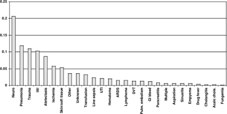
The definition of fever is arbitrary, and depends on how and when temperature was measured. In addition to host biology, a variety of environmental forces in an ICU can also alter body temperature, such as specialized mattresses, lighting, heating or air conditioning, peritoneal lavage, and renal replacement therapy (121,122,123). Thermoregulatory mechanisms can be disrupted by drugs or by injury to the central nervous systems. Thus, it is often difficult to determine if an abnormal temperature is a reflection of a physiologic process, a drug, or an environmental influence. Moreover, in surgical patients, the substantial possibility (~50%) that a fever is due to a noninfectious cause must be considered (Fig. 106.1) (124).
|
Table 106.8 Comparison of antibiotic utilization (median defined daily doses) for surgical intensive care units (SICUs) and inpatient units, 1998–2002 |
||||||||||||||||||||||||
|
||||||||||||||||||||||||
Many ICUs consider any patient with a core temperature ≥38.3°C (≥101°F) to be febrile and to warrant evaluation to determine if infection is present. However, a lower threshold may be decided upon for immunocompromised patients. However, laboratory tests or imaging studies to search for infection should be performed only after a clinical assessment (history and physical examination) indicates that infection might be present.
Blood Cultures
Blood cultures should be obtained from patients with a new fever when clinical evaluation does not strongly suggest a noninfectious cause. The site of venipuncture should be cleaned with either 2% chlorhexidine gluconate in 70% isopropyl alcohol or 1% to 2% tincture of iodine. Povidone-iodine (10%), while acceptable, is not bactericidal until dry; some false-positive blood cultures may be due to premature specimen collection (125,126). One blood culture is defined as a 20- to 30-mL sample of blood drawn at a single time from a single site, regardless of how many bottles or tubes are filled for processing. The sensitivity of blood culturing for detection of true bacteremia or fungemia is related to many factors, most importantly the volume of blood drawn and obtaining the cultures before initiation of anti-infective therapy (127,128).
Recent data suggest that the cumulative yield of pathogens is optimized when three blood cultures with adequate volume (20- to 30-mL each) are drawn (127). Each culture should ideally be drawn by separate venipuncture or through a separate intravascular device, but not through multiple ports of the same intravascular catheter (129). There is no evidence that the yield of cultures drawn from an artery or vein is different. Drawing two to three blood cultures with appropriate volume from separate sites of access at the onset of fever is the most effective way to discern whether an organism found in blood culture represents a true pathogen (multiple cultures are often positive), a contaminant (only one of multiple blood cultures is positive for an organism commonly found on skin and clinical correlation does not support infection), or a bacteremia/fungemia from an infected catheter (one culture from the source catheter is positive, often with a positive catheter tip, and other cultures are not) (130).
|
|
|
Figure 106.1. Causes of fever (%) among more than 600 consecutive surgical intensive care unit patients. IAI, intra-abdominal; UTI, urinary tract infection; ARDS, acute respiratory distress syndrome; DVT, deep venous thrombosis; pulm., pulmonary; GI, gastrointestinal; chole., cholecystitis. |
Intravascular Devices
All intravascular devices and insertion sites must be assessed daily as part of a comprehensive physical examination to determine if they are still needed and whether signs of infection are present locally (e.g., inflammation or purulence at the exit site or along the tunnel) or systemically. Contaminated catheter hubs are common portals of entry for organisms colonizing the endoluminal surface of the catheter (131). Additionally, infusate (parenteral fluid, blood products, or intravenous medications) can become contaminated and produce bacteremia or fungemia, which is especially likely to result in septic shock. Abrupt onset of signs and symptoms of sepsis or shock in patients with an indwelling vascular catheter should prompt suspicion of infection of an intravascular device. Recovery of certain micro-organisms in multiple blood cultures, such as staphylococci or Candida spp., strongly suggests infection of an intravascular device.
Removal and culture of the catheter has historically been the gold standard for the diagnosis of CR-BSI, particularly with short-term catheters. Studies have demonstrated the reliability of semiquantitative or quantitative catheter tip culture methods for the diagnosis of CR-BSI (132), which is confirmed when a colonized catheter is associated with concomitant bloodstream infection with the identical organism, with no other plausible source. Some ICU clinicians culture central venous catheters routinely on removal, regardless of whether infection is suspected. Because ~20% of central venous catheters are colonized at removal, most unassociated with local or systemic infection, this practice is expensive and can lead to unnecessary therapy. The predictive value of a positive catheter culture is very low when there is a low pretest probability of line sepsis (132), and catheters removed from ICU patients should only be cultured if there is strong clinical suspicion of CR-BSI (133).
For patients with fever alone who do not have systemic inflammatory response syndrome (SIRS) (134), there is usually no need to remove or change all indwelling catheters immediately, although such an approach would be prudent in a patient with a prosthetic heart valve or a fresh arterial graft (133). If patients have severe sepsis or septic shock, peripheral embolization, disseminated intravascular coagulation, or acute respiratory distress syndrome (ARDS), removal of all intravascular catheters is indicated, even if the catheters are cuffed or tunneled devices (135).
Infected phlebitis (suppurative phlebitis) of a central vein due to a centrally placed catheter is unusual. With suppurative phlebitis, bloodstream infection characteristically persists and originates from a peripheral vein catheter site with infected intravascular thrombus, producing a picture of overwhelming sepsis with high-grade bacteremia or fungemia. This syndrome is most often encountered in burn patients or other ICU patients who develop catheter-related infection that goes unrecognized, permitting micro-organisms to proliferate. In patients with persistent S. aureus bacteremia or fungemia, echocardiography is appropriate to assess for endocarditis and guide further therapy (133).
Intensive Care Unit–acquired Pneumonia
Pneumonia is the second most common cause of ICU-acquired infection and a ubiquitous cause of fever, with the majority of cases occurring in mechanically ventilated patients (65,136). The diagnosis of VAP is especially challenging, as patients commonly also have other, noninfectious processes producing abnormal chest radiographs and gas exchange (e.g., congestive heart failure, atelectasis, ARDS). Intubated, sedated patients cannot cough, or otherwise mobilize abnormal secretions without assistance. In addition, immunocompromised patients such as solid organ transplant recipients may have pneumonia without fever, cough, sputum production, or leukocytosis (137,138).
The diagnosis of VAP requires determination if the patient has pneumonia, and the etiologic pathogen. Poor specificity is problematic because it not only exposes patients to unnecessary risk from overtreatment with antibiotics, but also increases selection pressure and thus the emergence of MDR bacteria (139,140). Conversely, inadequate initial therapy in patients with VAP (poor sensitivity) is associated with increased mortality that cannot be reduced by subsequent changes in antibiotics (141).
Historically, the diagnosis of VAP requires one or more of the following: fever, leukocytosis or leukopenia, purulent sputum, hypoxemia, or a new or evolving chest radiographic infiltrate. However, several noninfectious processes may mimic these nonspecific signs, such as congestive heart failure, atelectasis, pulmonary thromboembolism, pulmonary hemorrhage, and ARDS, making clinical criteria alone unreliable. A new chest radiographic infiltrate, along with two of the three aforementioned criteria, was only 69% sensitive and 75% specific for VAP as compared to postmortem histology (142). Several subsequent reports have confirmed the low specificity of clinical acumen in the diagnosis of VAP (143,144); clinically diagnosed VAP is confirmed microbiologically in <50% of cases (66,145,146). Computerized tomography (CT) is particularly sensitive for demonstrating parenchymal or pleural disease in posterior-inferior lung segments (147,148), although there is only a fair correlation with the diagnosis of pneumonia in complex patients (149). Clinical judgment determines whether suspicion is high enough to warrant transporting the patient to the radiology suite for a higher-resolution study such as CT.
The Clinical Pulmonary Infection Score (CPIS) incorporates clinical, radiographic, and microbiologic criteria (i.e., temperature, leukocyte count, chest radiographic infiltrates, appearance and volume of tracheal secretions, PaO2:FiO2, culture and Gram stain of tracheal aspirate [0–2 points each]) to yield a maximum score of 12 points (150). A CPIS of >6 points indicates a high probability of VAP. However, the specificity of CPIS is no better than clinical acumen alone when compared to lower respiratory tract cultures obtained via bronchoscopic bronchoalveolar lavage (BAL) or protected specimen brush (PSB) (150,151,152). However, the negative predictive value of a Gram stain showing no organisms in a clinically stable patient approaches 100% (153).
Because of the low specificity of traditional diagnostic criteria, culture of lower respiratory tract samples is mandatory for nosocomial pneumonia prior to any manipulation of antibiotics in order to minimize false-negative results. The method of specimen collection (invasive vs. noninvasive) and the method of specimen analysis (semiquantitative vs. quantitative) are debated. Noninvasive techniques include endotracheal suction aspiration (EA), blinded plugged telescoping catheter, blinded PSB, and mini-BAL. Endotracheal aspirates are less specific due to an increased likelihood of contamination by oropharyngeal flora reflecting colonization rather than infection.
Invasive techniques (BAL or PSB) collect samples using fiberoptic bronchoscopy, and also allow direct inspection of the airways, but are more expensive and resource intensive than noninvasive techniques. Furthermore, arterial desaturation may persist for up to 24 hours postbronchoscopy, possibly due to alveolar flooding caused by residual lavage fluid. However, this desaturation has not been correlated with poorer outcomes (154).
Respiratory tract cultures may be reported either semiquantitatively or quantitatively. The crucial issue is distinction of colonization from infection (155). Whereas semiquantitative microbiology reports growth in terms of ordinal categories (e.g., light, moderate, or heavy), quantitative microbiology reports growth in terms of colony forming units (CFU)/mL of aliquot; a threshold value is assigned to distinguish colonization from infection. Commonly used thresholds are 103 CFU/mL for PSB, 104 CFU/mL for BAL, and 105 CFU/mL for EA. Any threshold should be lowered by one order of magnitude if antibiotics have been changed recently or started prior to sample acquisition (156). Clinical interpretation of quantitative cultures is likely to be hampered by prior antibiotic administration, which may lower the observed quantitative inoculum after 24 hours of ongoing antibiotic therapy, and for up to 72 hours after cessation of antibiotics (157,158). The diagnostic threshold is usually lowered by one order of magnitude in the presence of antibiotics or recently discontinued therapy.
Endotracheal aspirates have lower specificity compared to either blinded plugged telescoping catheter (159) or bronchoscopic BAL or PSB (160,161,162,163). Two systematic reviews, one of bronchoscopic BAL (154) and one of blinded invasive techniques (163), reported similar test characteristics for the two techniques, but methodologic variability is rampant. Bronchoscopic techniques are more specific than blinded techniques, and both techniques are superior to EAs.
Shorr et al. performed a meta-analysis of randomized trials that compare outcomes of patients with VAP managed with invasive versus noninvasive sampling when both samples were cultured quantitatively (164). Although the pooled OR suggested a survival advantage to the invasive approach (OR = 0.62), the result was not significant. However, patients in the invasive group were significantly more likely to undergo changes in antimicrobial regimen.
Pulmonary secretions for culture should be transported to the laboratory and processed within 2 hours so that fastidious organisms such as S. pneumoniae remain viable. For any expectorated specimen, it is important for the laboratory to perform direct microscopy on the specimen to determine if squamous epithelial cells are present, which invalidates the specimen.
Organisms that may be pathogens in VAP or contaminants when recovered from the airway include P. aeruginosa, Enterobacteriaceae, S. pneumoniae, S. aureus, and H. influenzae. Conversely, isolation of enterococci, viridans streptococci, coagulase-negative staphylococci, and Candida spp. (165,166) should rarely be considered the cause of respiratory dysfunction, if ever. Although febrile postoperative patients in an ICU often have small pleural effusions due to fluid overload, hypoalbuminemia, or postoperative processes, it is not necessary to sample such fluid from every febrile patient. Thoracentesis is appropriate if there is sufficient fluid to aspirate safely using ultrasound (US) guidance and there is either an adjacent pulmonary infiltrate or possible contamination of the pleural space by surgery, trauma, or a fistula.
Evaluation for Clostridium difficile Infection
Many ICU patients have diarrhea, often due to enteral feedings or drugs. By far the most common enteric cause of fever in the ICU is C. difficile, which should be suspected in any patient with diarrhea and fever or leukocytosis who has received an antibacterial agent or antineoplastic chemotherapy within 60 days prior to the onset of diarrhea (167,168). C. difficile accounts for 10% to 25% of all cases of antibiotic-associated diarrhea and virtually all of the cases of antibiotic-associated colitis (169). However, some patients, especially those who are postoperative, may present with ileus or toxic megacolon, or leukocytosis without diarrhea, as the manifestation of CDAD. In these patients, the diagnosis is difficult to establish because stool specimens are not accessible (170). C. difficile–associated diarrhea may occur with any antibacterial agent, but the most common causes are clindamycin, cephalosporins, and fluoroquinolones (171).
Although less accurate than expensive tissue culture assays, most laboratories now use immunoassays for C. difficile toxins, which provide results within hours and are easy to perform. Lower sensitivity may require repeat tests to document disease in seriously ill patients (172). Most strains of C. difficile produce toxin A, but 2% to 3% of stains produce only toxin B so an assay that detects both toxins A and B is preferred (173). Cultures for C. difficile are technically demanding, and are not specific in distinguishing toxin-positive strains, toxin-negative strains, and asymptomatic carriage (172,174). Cultures may be useful in the setting of nosocomial outbreaks to identify isolates for epidemiologic purposes (168). The North American pulse-field gel electrophoresis type 1 (NAP1) strain, now epidemic in many hospitals in the United States, Canada, and Europe, is associated with serious complications (toxic megacolon, leukemoid reactions, septic shock, and death) (175,176).
Direct visualization of pseudomembranes is nearly diagnostic of CDAD, but only about 70% of seriously ill patients and 25% of patients with mild disease have pseudomembranes by direct visualization (177), diminishing the role of endoscopy for routine diagnostic use. However, a role for direct visualization may exist if false-negative C. difficile toxin assays are suspected (168).
Urinary Tract Infection
Catheter-associated bacteriuria or candiduria usually represents colonization, is rarely symptomatic, and is an unlikely cause of fever or secondary bloodstream infection (178), even in immunocompromised patients (179), unless there is urinary tract obstruction; there is a history of recent urologic manipulation, injury, or surgery; or the patient is neutropenic (180,181). Traditional signs and symptoms (dysuria, urgency, pelvic or flank pain, fever or chills) that correlate well with bacteriuria in noncatheterized patients are rarely reported in ICU patients with documented catheter-associated bacteriuria or candiduria (>105 CFU/mL) (180,181). In the ICU, the majority of urinary tract infections are related to urinary catheters and are caused by multiresistant nosocomial Gram-negative bacilli other than Escherichia coli, Enterococcus spp., and yeasts (178,182,183).
When clinical evaluation suggests the urinary tract as a possible source of fever, a urine specimen should be evaluated by direct microscopy, Gram stain, and quantitative culture. The specimen should be aspirated from the catheter sampling port, not collected from the drainage bag. Health care personnel should wear clean gloves whenever manipulating a urinary device and should scrupulously clean the port with 70% to 90% alcohol prior to specimen collection. For patients without a catheter, a conventional midstream clean-catch urine specimen should be obtained. Urine collected for culture should reach the laboratory promptly to prevent multiplication of bacteria within the receptacle, which might lead to the misdiagnosis of infection; any delay should prompt refrigeration of the specimen.
In contrast to community-acquired urinary tract infections, where pyuria is highly predictive of important bacteriuria, pyuria may be absent with catheter-associated urinary tract infections. Even if present, pyuria is not a reliable predictor of urinary tract infection in the presence of a catheter (178). The concentration of urinary bacteria or yeast needed to cause symptomatic urinary tract infection or fever is unclear. Whereas it is clear that counts >103 CFU/mL represent true bacteriuria or candiduria in catheterized patients (184), there are no data to show that higher counts are more likely to represent symptomatic infection. Gram stain of a centrifuged urine specimen, however, will show micro-organisms most of the time if infection is present (185).
Whereas it is appropriate to collect urine specimens in the investigation of fever, routine monitoring or “surveillance” cultures of urine contribute little to patient management. Rapid dipstick tests, which detect leukocyte esterase and nitrite, are unreliable in the setting of catheter-related UTI. The leukocyte esterase test correlates with the degree of pyuria, which may or may not be present in a catheter-related UTI. The nitrite test reflects Enterobacteriaceae, which convert nitrate to nitrite, and is therefore unreliable to screen for Enterococcus spp., Candida spp., and Staphylococcus spp. (186,187).
Sinusitis
The paranasal sinuses are normally sterile, but bacterial overgrowth occurs when drainage is impeded. The etiologic agents responsible for most cases of nosocomial sinusitis are those that colonize the naso-oropharynx (76,188), which occurs at high frequency among critically ill patients. Gram-negative bacilli (particularly P. aeruginosa) constitute 60% of bacteria isolated from nosocomial sinusitis, whereas Gram-positive cocci (typically S. aureus and coagulase-negative staphylococci) comprise one third of isolates, and fungi the remaining 5% to 10% (76,189,190). Infections are often polymicrobial.
The diagnosis of sinusitis in critically ill, intubated patients is difficult to make. Complaints of facial pain or headache may be impossible to elicit, and purulent nasal discharge is present in only 25% of proved cases of sinusitis. In the ICU, acute sinusitis is diagnosed most efficiently by CT of the facial bones (191), followed by sampling using antiseptic technique if mucosal thickening or sinus fluid is documented. Microbial analysis of fluid obtained by minimally invasive sinus puncture and aspiration under antiseptic conditions is definitive for the diagnosis. Although less well studied, endoscopic-guided middle meatal tissue culture is a safe alternative for patients who are not candidates for antral puncture (e.g., coagulopathy) (192). Pathogen identification and susceptibility testing permit focused, narrow-spectrum antimicrobial therapy. However, specimen collection is susceptible to contamination by bacteria colonizing the overlying mucosa if rigorous antisepsis is not practiced when obtaining the specimen.
Intracranial Device–related Fever
When a patient with an intracranial device such as an extraventricular drain (EVD) (ventriculostomy catheter) or a ventriculoperitoneal shunt becomes febrile, cerebrospinal fluid (CSF) should almost always be analyzed. Access to CSF in the patient with an EVD is straightforward. The patient with a shunt or Ommaya reservoir should have the reservoir aspirated. Patients with EVDs who develop stupor or signs of meningitis should have the catheter removed and the tip cultured.
Basic tests of CSF for suspected central nervous system (CNS) infection include cell counts and differential, glucose and protein concentrations, Gram stain, and bacterial cultures. Patients with bacterial meningitis typically have a CSF glucose concentration <35 mg/dL, a CSF:blood glucose ratio <0.23, a CSF protein concentration >220 mg/dL, >2,000 total white blood cells/µL, or >1,180 neutrophils/µL (193). Conversely, the presence of a normal opening pressure, <5 white blood cells/µL, and a normal CSF protein concentration essentially exclude meningitis (193). Measurement of CSF lactate concentration may be useful in neurosurgical patients to distinguish infection from postoperative aseptic meningitis (194,195).
Noninfectious Causes of Fever in the Intensive Care Unit
Postoperative Fever
Fever is common during the initial 72 hours following surgery, and is usually noninfectious in origin (196), presuming that unusual breaks in sterile technique or pulmonary aspiration did not occur. Considerable effort and money can be wasted in overzealous evaluation of early postoperative fever. However, once a patient is more than 96 hours postoperative, fever is more likely to represent infection.
A chest radiograph is not mandatory for evaluation of postoperative fever unless respiratory rate, auscultation, abnormal blood gases, or pulmonary secretions suggest a high yield. Atelectasis is often considered to be a cause of postoperative fever. The clinician must be alert to the possibility that the patient could have aspirated during the perioperative period, or the uncommon event that the patient was incubating a community-acquired pneumonia prior to the operation caused, for example, by pneumococci or influenza A. Urinary tract infection is common postoperatively in nontrauma patients because of the use of urinary drainage catheters (197). The duration of catheterization increases the risk of bacteriuria by about 5% per day, which in turn increases the risk of nosocomial cystitis or pyelonephritis. A urinalysis or culture is not mandatory to evaluate fever during the initial 3 days postoperatively unless there is reason by history or examination to suspect an infection at this site. After trauma, urinary tract infection is common only after injury to the urinary tract.
Fever can be related to hematoma or infection of the surgical field. Surgical site infection is rare in the first few days after operation, except for group A streptococcal infections and clostridial infections, which can develop within hours to 1 to 3 days after surgery. These causes should be suspected on the basis of inspection of the incision.
Many emergency abdominal operations are performed for control of an infection (e.g., perforated diverticulitis). Even under optimal circumstances (definitive surgical source control and timely administration of appropriate broad-spectrum antibiotics), it may take ≥72 hours for such patients to defervesce. New or persistent fever more than 4 days after surgery should raise suspicion of persistent pathology or a new complication. Thus, it is mandatory to remove the surgical dressing to inspect the incision. Swabbing an open wound or collecting fluid from drains (if present) for culture is rarely helpful because the likelihood of colonization is high. Muscle compression injury (either direct trauma or as a result of compartment syndrome) and tetanus are two rare complications of traumatic wounds that may cause fever. Toxic shock may accompany infection with group A streptococci or S. aureus. Other potentially serious noninfectious causes of postoperative fever include deep venous thrombosis, tissue ischemia or necrosis, pulmonary embolism, adrenal insufficiency, drug-induced fever, anesthesia-induced malignant hyperthermia, and acute allograft rejection.
Drug-related Fever
Any drug can cause fever due to hypersensitivity (198), but “drug fever” is decidedly unusual in surgical patients, and must be considered a diagnosis of exclusion. In addition, some drugs cause fever by producing local inflammation at the site of administration (phlebitis, sterile abscesses, or soft tissue reaction), such as amphotericin B, erythromycin, and potassium chloride. Drugs or their delivery systems (diluent, intravenous fluid, or intravascular delivery devices) may also contain pyrogens or, rarely, microbial contaminants (199). Some drugs may also stimulate heat production (e.g., thyroxine), limit heat dissipation (e.g., atropine or epinephrine), or alter thermoregulation (e.g., butyrophenone tranquilizers, phenothiazines, antihistamines, or antiparkinson drugs). Among drug categories, drug fever in surgical ICUs is most often attributed to antimicrobial agents (e.g., vancomycin, β-lactams) and anticonvulsants (especially phenytoin).
Two important syndromes, malignant hyperthermia and neuroleptic malignant syndrome, deserve consideration when fever is especially high because the results can be devastating if left untreated (200). Malignant hyperthermia is more often identified in the operating room than in the ICU, but onset can be delayed for as long as 24 hours, especially if the patient is on steroids. Malignant hyperthermia is believed to be a genetically determined response mediated by a dysregulation of cytoplasmic calcium flux in skeletal muscle, resulting in intense muscle contraction, fever, and increased creatinine phosphokinase concentration. It can be caused by succinylcholine and inhalational anesthetics.
|
Table 106.9 Causes of fever related to noninfectious states |
|
|
The neuroleptic malignant syndrome is slightly more common, and more often identified in the ICU than malignant hyperthermia. It has been strongly associated with the neuroleptic medications phenothiazines, thioxanthenes, and butyrophenones (200,201), most frequently haloperidol in the ICU. It also manifests as muscle rigidity, fever, and increasing creatinine phosphokinase concentration. However, unlike malignant hyperthermia, the initiator of muscle contraction is central, the syndrome is often less intense, and mortality is less.
Drug withdrawal syndromes may be associated with fever, tachycardia, diaphoresis, and hyperreflexia, including from alcohol, opioids, barbiturates, and benzodiazepines. It is important to recognize that a history of use of these drugs may not be available when the patient is admitted to the ICU. Withdrawal and related fever may therefore occur several hours or days after admission. Other noninfectious causes of fever are listed in Table 106.9.
Clinical Infection Syndromes
Biliary Tract Infections
Infections of the biliary tree are similar to those found elsewhere in the body in that therapy consists of antibiotics directed at the causative pathogens and source control, typically in the form of drainage. Biliary tract infections remain an important cause of critical illness. They are difficult to diagnose in critically ill patients, owing to their relative rarity and the nonspecificity of symptoms that noncommunicative patients may manifest. Unless diagnosed and treated aggressively, organ dysfunction and death may ensue rapidly. Signs and symptoms of hepatobiliary infection commonly include abdominal pain, fever, nausea, and vomiting; the clinical presentation may range in severity from the appearance of a chronic disease state to overt septic shock. Leukocytosis is common to all but the patterns of liver enzyme abnormalities overlap to such a degree that it may be difficult to make a definitive diagnosis based on history, physical examination, and laboratory values alone. Furthermore, in the critically ill patient, it may be difficult to distinguish liver dysfunction caused by primary infection from that caused by multiple organ dysfunction syndrome. Imaging of the biliary tree is often of paramount importance in the diagnosis and treatment of biliary tract infections, but ultimately a thorough working knowledge of the differential diagnosis and treatment of biliary tract infections is the key to successful management.
Acute Cholecystitis
Among outpatients, acute cholecystitis usually occurs when gallstones migrate into the cystic duct, causing outflow obstruction. This obstruction causes sterile inflammation and edema of the gallbladder wall initially, followed by bacterial superinfection (202). Acute calculous cholecystitis presents with fever, right upper quadrant pain, nausea, and vomiting. Physical examination may range from arrest of inspiration due to tenderness on palpation in the right upper quadrant (Murphy sign) to signs of peritonitis in advanced cases. Concomitant jaundice suggests the presence of choledocholithiasis. Ultrasound is a highly sensitive and specific test (95% and 97%, respectively) for acute cholecystitis and is the initial test of choice. Diagnostic findings include the presence of stones in the gallbladder, thickening of the gallbladder wall (>3.5 mm), pericholecystic fluid, and tenderness with application of the US probe (sonographic Murphy sign) (203). Most cases (~80%) of acute calculous cholecystitis resolve with bowel rest, fluid resuscitation, and intravenous antibiotics, and are treated ultimately by cholecystectomy. Patients who become ill enough to require ICU care generally have gallbladder gangrene that progresses to perforation, resulting in either subhepatic abscess or free perforation with bile peritonitis. Gangrene of the gallbladder is associated with a 30% mortality rate and tends to occur in older patients (204). Emphysematous cholecystitis, a severe manifestation of acute cholecystitis that occurs with a predilection for elderly patients and patients with diabetes mellitus, is defined by the presence of gas in the gallbladder wall as visualized on US or CT and is characterized by polymicrobial infection including Clostriduim spp., E. coli, Klebsiella spp., and Streptococcus spp. Roughly one half of all cases of emphysematous cholecystitis are acalculous (see below); the reported mortality ranges from 15% to 25% (205,206).
Acalculous cholecystitis has been reported in all age groups, but most often occurs in the setting of severe illness or injury. It may also occur in the postoperative setting, particularly in males after emergency surgery complicated by large-volume blood loss. One review of 31,710 cases of cardiac surgery found a 0.05% incidence of acalculous cholecystitis (207); after open abdominal aortic aneurysm repair, the incidence has been reported to be between 0.7% and 0.9% (208). Acute acalculous cholecystitis is a grave condition of insidious onset in patients who are often already critically ill, and the mortality approaches 30% (209). Therefore, the diagnosis of acalculous cholecystitis must always be entertained in a patient with sepsis for whom no clear source of infection can be determined.
The pathogenesis of acalculous cholecystitis is most likely splanchnic ischemia–reperfusion injury. Critically ill patients probably have this pathophysiology even in the presence of gallstones. Alternatively, bile stasis associated with critical illness may lead to distention of the gallbladder, which in combination with hypoperfusion may cause ischemia and ultimately necrosis. Factors such as mechanical ventilation, total parenteral nutrition, cytokine activation, and endotoxemia have also been implicated (210).
The diagnosis of acalculous cholecystitis can be difficult to make in the ICU. Patients who are able to communicate may report abdominal pain localizing to the right upper quadrant or diffuse pain in the case of peritonitis. Fever is usually present. Physical examination may reveal signs ranging from localized tenderness in the right upper quadrant to frank peritonitis. A right upper quadrant abdominal phlegmon may be palpable. All too often, the altered mental status that often accompanies critical illness may obscure any useful information that might be obtained from the history and physical examination. Laboratory values are nonspecific but usually include leukocytosis and elevated liver enzymes, particularly of bilirubin, transaminases, and alkaline phosphatase. Hyperbilirubinemia, perhaps representative of the cholestasis of sepsis, is typical and occurs more often in acalculous than calculous cholecystitis.
Ultrasound is perhaps the ideal radiologic study to investigate the diagnosis of acalculous cholecystitis in the ICU. Ultrasound may reveal hydrops of the gallbladder, pericholecystic fluid, or gallbladder wall thickening. At a gallbladder wall thickness ≥3.5 mm, US is 98.5% sensitive and 80% specific for the diagnosis of acalculous cholecystitis (211). Ultrasound conveniently can be diagnostic at the bedside of patients too ill to be transported to the radiology suite, and followed immediately by percutaneous drainage of the gallbladder under US guidance. Computed tomography is equally accurate in the diagnosis of acalculous cholecystitis (212), but requires a patient stable enough to be transported. The primary advantage of CT scan over US is in its ability to evaluate other potential sources of intra-abdominal infection. Interpretation of radionuclide biliary scans can be confounded in critically ill patients by false-positive scans due to fasting, liver disease, or parenteral nutrition, which are sufficiently common to diminish its utility in this population.
Upon making the diagnosis of acalculous cholecystitis, a decision about the method of source control must be made and empiric antibiotic therapy must be started. Even though up to one half of cases of acute acalculous cholecystitis are associated with culture-negative bile (at least initially, considering that ischemia–reperfusion is paramount and superinfection is a secondary phenomenon), empiric antibiotics are needed because distinguishing sterile from infected cases can be clinically impossible owing to the massive inflammatory response. The organisms most frequently cultured from the bile in acalculous cholecystitis are E. coli, Klebsiella, and Enterococcus faecalis (211).
Source control for cholecystitis, whether calculous or acalculous, has traditionally been by cholecystectomy. However, patients with acalculous cholecystitis are often critically ill and thus poor surgical candidates. Percutaneous cholecystostomy tube placement is a minimally invasive alternative to cholecystectomy that is favored increasingly. In this technique, the gallbladder is punctured through an anterior transhepatic approach under US guidance and a drainage catheter is placed using the Seldinger technique. A tube study may be performed at the time of catheter placement to discern whether the cystic duct is patent. If not, and concomitant cholangitis is suspected, further drainage of the biliary tree must be performed with endoscopic retrograde cholangiopancreatography (ERCP) or percutaneous transhepatic cholangiopancreatography (PTC). Complications of percutaneous cholecystostomy include bacteremia, hemorrhage, bile peritonitis, and tube dislodgement. Patients with uncomplicated cholecystitis should improve rapidly after gallbladder decompression; failure to improve should raise suspicions of an incorrect diagnosis or inadequate source control. Under such conditions, surgical exploration is mandated. Upon resolution of acalculous cholecystitis, patients treated with percutaneous cholecystostomy may have their tubes removed once sepsis has resolved, the patient has recovered from the precipitating illness, and the cystic duct is demonstrated to be patent. Percutaneous cholecystostomy is the definitive treatment in this group.
Cholangitis
Cholangitis is an acute infection of the main biliary ductal system. The pathogenesis of cholangitis requires both obstruction and bacterial superinfection. The most common cause of intrinsic obstruction in the Western world is choledocholithiasis (213). Both primary and metastatic malignant disease of the abdominal viscera may cause extrinsic obstruction, among other causes. Obstruction from calculi is more likely to cause cholangitis than malignant obstruction (214). Cholangitis may also occur in the postoperative setting, particularly after a biliary-enteric anastomosis.
Bile is sterile in the normal biliary tree. Bile is naturally bacteriostatic, and antegrade flow of bile from liver to duodenum serves as a flushing mechanism. The sphincter of Oddi is an anatomic barrier, preventing reflux of enteric flora. Bile salts serve to absorb intraluminal endotoxins and may also exhibit a trophic effect on small bowel mucosa, thus helping to prevent bacterial and endotoxin translocation. In the absence of bile flow, normal bacterial ecology is perturbed, leading to bacterial overgrowth and degradation of mucosal defenses. The hepatic reticuloendothelial system serves to filter translocated bacteria and endotoxin and is impaired when the biliary tree is obstructed, further increasing the risk of infection (215). Bacteria may gain access to the biliary tree either via the portal vein or by ascending directly from the duodenum. In addition, pathogens may be introduced iatrogenically during surgical, endoscopic, or percutaneous biliary interventions.
The Charcot triad of fever, right upper quadrant pain, and jaundice is observed in 50% to 70% of patients who present with cholangitis, with fever present most consistently (90%). Hypotension and altered mental status (i.e., severe sepsis or septic shock) in addition constitute the Reynolds pentad. Patients with cholangitis typically have leukocytosis, direct hyperbilirubinemia (88%–100%), and elevated alkaline phosphatase (80%) (216); transaminitis is usually mild.
Common duct bile cultures are positive 80% to 100% of the time in cholangitis, with positive blood cultures found in up to two thirds of patients. The concordance rate between bile and blood cultures is 33% to 84%, with bile cultures being polymicrobial in roughly one half of cases. The typical florae are Klebsiella, E. coli, and Enterococcus (217).
Imaging of patients with cholangitis is possible using US, CT, or magnetic resonance imaging (MRI). Ultrasound detects cholelithiasis and bile duct dilation reliably, but is only 50% sensitive for detecting choledocholithiasis. Computed tomography detects ductal dilation with 98% accuracy and is superior to US in defining the level of obstruction, but may fail to visualize the 85% of biliary calculi that are radiolucent. Stones can be visualized on MRI; therefore, magnetic resonance cholangiopancreatography (MRCP) provides the most complete imaging as to the etiology of biliary obstruction. However, MRCP is expensive, time consuming, and cumbersome for ICU patients, and does not allow for therapeutic intervention. By contrast, ERCP and PTC are both 90% to 100% sensitive for defining the site and nature of biliary obstruction (218). Either can be used diagnostically or therapeutically to decompress the biliary tree. Both ERCP and PTC have rare potential complications (e.g., bile leak, bleeding) that may precipitate ICU admission. In the hemodynamically stable patient with cholangitis, US is the investigation of first choice, followed by further imaging as needed with CT or MRCP. Because of the potential for intervention, ERCP should be considered strongly for unstable patients and other cases where intervention is likely to be necessary. Hemodynamic instability in the setting of suspected or confirmed bacterial cholangitis is a true emergency that requires immediate biliary decompression.
Treatment of cholangitis consists of immediate fluid resuscitation and broad-spectrum antibiotics, followed by urgent or emergent biliary decompression. Numerous antibiotic regimens have been successful historically, but many authorities prefer a single broad-spectrum agent such as piperacillin/tazobactam (219) or a carbapenem. The majority of patients respond to initial medical therapy followed by biliary decompression, but 10% to 15% will require emergent decompression (220). ERCP is the safest and most efficacious treatment for acute cholangitis with a success rate of 90% and a mortality rate of 10%, considerably lower than that of emergency surgical decompression of the common bile duct. Through ERCP, a sphincterotomy can be performed to facilitate drainage and removal of calculi, or a temporizing stent or nasobiliary tube may be placed. Other complications of ERCP include pancreatitis, perforation, aspiration, systemic sepsis, and failure to decompress the bile duct.
Percutaneous transhepatic biliary drainage carries 30% morbidity from complications such as hemorrhage, pneumothorax, subphrenic abscess, and bile peritonitis. The mortality for PTC is estimated to be between 5% and 10%, similar to that seen with ERCP (221). Although the risk is higher, PTC may be used preferentially for intrahepatic choledocholithiasis, for cholangitis from proximal bile duct stricture or neoplasm, or when ERCP is impossible secondary to surgically altered upper gastrointestinal tract anatomy (e.g., after Roux-en-Y reconstruction) or anatomic anomalies (e.g., ampulla of Vater located within a duodenal diverticulum).
Surgery is now infrequent in the primary management of acute cholangitis. In situations that require surgery (e.g., resectable malignant obstruction), patients may be temporized by ERCP or PTC, converting an emergency operation into an elective procedure that can be performed at lower risk. Surgery is also indicated in patients who fail less invasive treatment methods. Standard emergency surgical therapy includes cholecystectomy, choledochotomy, and T-tube placement.
Liver Abscess
The most common cause (50% to 65% of liver abscesses) is now ascending biliary tract infection (222) (e.g., cholangitis, direct extension of acute suppurative cholecystitis). Seeding from the portal vein accounts for 10% to 25% of abscesses and is typically a result of intra-abdominal sources of infection such as diverticulitis. Systemic seeding via the hepatic artery occurs in 1% to 10% of cases from processes such as bacterial endocarditis, dental abscesses, or interventions such as hepatic artery chemoembolization, intraoperative cryoablation, or radiofrequency ablation (221). Liver abscess complicates fewer than 1% of blunt liver injuries managed nonoperatively, and is more common in patients requiring damage-control laparotomy and perihepatic packing to control hemorrhage (222).
Patients with pyogenic hepatic abscess usually present with fever and chills, abdominal pain, and weight loss. Nonspecific abdominal complaints and constitutional symptoms are common, and presentation can range from the appearance of a chronic disease state to overt septic shock. The laboratory workup demonstrates leukocytosis in most patients, with liver enzymes being moderately abnormal as well.
Due to the protean manifestations of liver abscess and the mimicry of a number of disease processes, radiographic imaging is crucial in confirming the diagnosis. Ultrasound and CT both have greater than 95% sensitivity for the diagnosis. One half of patients will present with more than one abscess, and approximately 75% of all liver abscesses will be found in the right lobe of the liver (223).
Pyogenic liver abscesses are equally likely to be polymicrobial as monomicrobial, and approximately 5% to 27% show no growth in culture. In many cases, failure to speciate bacteria in culture may reflect prior antibiotic treatment. The flora found in liver abscesses reflects the underlying source of the infection, but overall the most common Gram-negative aerobes found in pyogenic liver abscesses are E. coli and Klebsiella spp., whereas the most common Gram-positive aerobes are Enterococcus spp., viridans streptococci, and S. aureus. Among cultured anaerobes, Bacteroides spp. predominate (224).
Historically, medical management of pyogenic liver abscess carried a mortality rate of 60% to 100% (225). Several recent case series demonstrate success rates for antibiotics alone of up to 80% when multiple (military) abscesses are too small or too numerous for percutaneous drainage. Although antibiotics are a necessary adjunct to the treatment of hepatic abscess, most authorities agree that this approach is not sufficient except in the case of very small abscesses or in patients in whom intervention poses a prohibitive risk. The preferred method of treatment is broad-spectrum antibiotics in conjunction with drainage of all abscesses. Most hepatic abscesses can be drained successfully by image-guided percutaneous techniques, with a success rate of 70% to 93%. Surgical drainage is indicated in patients who fail percutaneous drainage, who require surgical management of the underlying problem (e.g., diverticulitis), or who have abscesses that are not amenable to minimally invasive techniques because of their location. Surgical options include simple drainage, debridement, and formal resection. Liver resection should be considered in those patients with extensive tissue loss or with intrahepatic stones, which may serve as a nidus for recurrent infection.
Most contemporary series quote a mortality rate for hepatic abscess between 6% and 31% (221). Factors associated with a worse prognosis include an underlying diagnosis of malignant disease, multiple abscesses, and a high presenting APACHE II score (226).
Postoperative Infections after Biliary Tract Operations
Perihepatic infections may occur as a result of commonly performed hepatobiliary surgical procedures. Postoperative bile leaks may occur after any operation in which a bile duct is opened (such as hepatectomy, hepaticoenterostomy, hepatic transplant, or cholecystectomy). Leaking bile may induce chemical peritonitis, or cause infection with microbial flora present in or introduced into tissue at the time of operation, or by translocation from the gut, leading to an infected intrahepatic or perihepatic collection known as a biloma.
After cholecystectomy, bile leaks occur in up to 1% of patients regardless of the technique used. Bile leakage may occur from the transected cystic duct, the hepatic bed of the gallbladder, or incidental injury to other portions of the biliary tree during dissection. Patients with postoperative bile collections may present with right upper quadrant pain, fever, nausea, vomiting, or jaundice. Discrimination between sterile bile peritonitis and infection may be difficult without culture of the collection.
Postoperative fluid collections may be imaged with US, CT, or nuclear scintigraphy. The diagnosis of bile leak can also be made by image-guided aspiration of the collection. Nuclear scintigraphy is an excellent screening test for the diagnosis of bile leak, but it is not useful in determining the precise anatomy of the leak. For this reason ERCP is used to determine the anatomy once leakage is documented, with some leaks being amenable to concomitant endoscopic therapy by stent placement.
Treatment of postoperative bile leakage hinges first upon drainage and then definitive treatment of the underlying problem. If the leak originates from the bed of the gallbladder, percutaneous drainage alone is indicated. Extravasation from the cystic duct is best managed endoscopically by stent placement with or without sphincterotomy to allow bile to drain preferentially through the ampulla of Vater (227). Major duct injuries discovered at ERCP generally require surgical management.
Perihepatic or intra-abdominal abscess complicates 8% to 30% of major liver resections, and is associated with preoperative biliary stenting, hepaticoenterostomy, increased operative time, greater extent of resection, and the need for blood transfusion (222). Preoperative hyperbilirubinemia as a result of biliary obstruction occurs frequently in patients with malignant biliary tract obstruction; such patients are at increased risk for complications of surgical resection (228). However, recent data demonstrate clearly that preoperative stent placement to alleviate biliary obstruction leads to increased rates of postoperative infectious complications (229).
Resection of hepatic parenchyma leaves dead space in the abdomen that collects bile and blood and is in proximity to ischemic tissue at the resection margin. Bacterial superinfection may occur, leading to the formation of an abscess. Infected bilomas are heralded by fever, right upper quadrant pain, leukocytosis, and elevated liver enzymes. Imaging modalities for posthepatectomy abscesses include CT, US, and MRI as discussed in the prior section on pyogenic hepatic abscess. Cultures reveal that 50% to 75% of postoperative perihepatic abscesses are polymicrobial, with bacteria of enteric origin (e.g., E. coli, Enterococcus) predominating (215). Image-guided percutaneous drainage is the treatment of choice where feasible, with reoperation reserved for those patients in whom percutaneous drainage is not possible or unsuccessful.
Despite recent advances in surgical technique and perioperative management, liver transplantation is plagued by complication rates ranging from 24% to 64%. The incidence of bile leak after orthotopic liver transplantation is between 10% and 40%, with the leaks arising most commonly from hepatic resection lines, T-tube sites, and biliary anastomoses (230). Patients may present up to 6 months after transplantation. Computed tomography is used to image the collection, which may be intrahepatic in up to two thirds of cases. Endoscopic retrograde cholangiopancreatography, PTC, or cholangiogram through a pre-existing T tube may be used to delineate the origin of the leak. Most collections can be drained percutaneously, and in the event of direct communication with the biliary tree, ERCP can be used to re-establish preferential enteric drainage. Because the blood supply to the biliary tree is provided by the hepatic artery, anastomotic leaks after liver transplantation may occur as a result of ischemia from hepatic artery thrombosis. Therefore, assessment of the patency of the graft hepatic artery must be determined. Whereas some cases of biloma associated with hepatic artery thrombosis may respond to conservative measures, up to two thirds will require retransplantation (231).
Surgical Site Infection
Surgical site infections are among the most frequently encountered complications in surgical patients regardless of specialty. Local surgical control of the infection remains the crucial aspect of therapy, oftentimes by simply opening and draining the incision in cases of superficial incisional SSI. Infections extending below the superficial fascia (deep incisional SSIs) invariably require formal surgical debridement and open wound care to resolve the infection. Vacuum-assisted closure (VAC) and antimicrobial therapy also improved outcomes, but MDR pathogens may complicate resolution of ostensibly simple infections in the postoperative period (232), especially among patients hospitalized for a period before surgery, particularly if they required antimicrobial therapy.
Surgical site infection remains a clinical diagnosis. Presenting signs and symptoms depend on the depth of infection, typically as early as postoperative day 4 or 5. Clinical signs range from local induration only to the hallmarks of infection (e.g., erythema, edema, tenderness, warm skin, and pain-related immobility), which may manifest before wound drainage. In cases of deep incisional SSIs, tenderness may extend beyond the margin of erythema, and crepitus, cutaneous vesicles, or bullae may be present (233). With ongoing infection, signs of SIRS herald the development of sepsis. If infection involves an intracavitary space (organ/space), symptoms specific to that organ system will usually predominate, such as prolonged postoperative ileus, persistent respiratory distress, or altered neurologic status.
Cultures are not mandatory for management of superficial incisional SSIs, particularly if drainage and wound care alone will suffice without antibiotics. In cases of deeper infection or infection that has arisen in the hospital, exudates or drainage specimens should be sent for analysis. Culturing the surgically opened wound (as opposed to the already opened wound, which becomes colonized) by the swab method has been shown experimentally to be reliable. Computed tomography and MRI are more sensitive in detecting small amounts of gas in soft tissues than plain radiographs, and CT-guided aspiration or drainage often facilitates treatment, and may serve as definitive source control for an organ/space SSI.
More severe SSIs, especially the dangerous forms of necrotizing soft tissue infection (NSTI), are true emergencies that need immediate surgical attention. Even modest delays can increase patient mortality substantially. Freischlag et al. showed that mortality increased from 32% to 70% when therapy was delayed more than 24 hours (234). With an established diagnosis of NSTI, immediate and widespread operative debridement is indicated without waiting for precise determination of the causative pathogen or the identification of a specific clinical symptom. These patients often require planned, sequential, repetitive surgical debridement sessions to control the infection.
When faced with a potential SSI, the first steps in management are to remove the appropriate sutures, open and examine the suspicious portion of the incision, and decide about further surgical treatment (235). If the infection is not confined to the skin and superficial underlying subcutaneous tissue, urgent surgical exploration and debridement is essential to obtain local control of the infection, remove necrotic tissue, and restore aerobic conditions to prevent further spread of the infection. Surgical site infection must also be considered the cause of delayed or failed wound healing and prompt the same decisions as described above (235).
Superficial SSIs, which functionally are subcutaneous abscesses, rarely lead to systemic infection and usually do not make patients seriously ill. Antibiotic therapy is not indicated for patients who do not have systemic signs of infection. Formal surgical intervention is limited to complications such as loculated abscesses or necrosis of the skin or underlying tissue. In contrast, deep incisional SSIs typically present with extensive discomfort in more seriously ill patients. These infections extend to superficial fascia or beneath, and may cause extensive tissue necrosis and liquefaction beyond the obvious limits of the cutaneous signs, making it necessary to explore the wound formally in the operating room. The clinician must be alert to the possibility that necrotizing is present. Broad-spectrum antimicrobial therapy should be given empirically to cover likely pathogens, and reassessed following receipt of the microbiology report.
Organ/space SSIs occur within a body cavity, are directly related to a surgical procedure, and may manifest as intra-abdominal, intrapleural, or intracranial infections. They also may remain occult or present with few symptoms, mimicking incisional SSIs and leading to inadequate initial treatment, becoming apparent only when a major complication ensues. The diagnosis of organ/space SSI usually requires some form of imaging to confirm the site and extent of infection. Adequate source control requires a drainage procedure, whether open or percutaneous.
Experimentally, the value of vacuum-assisted wound closure was first appraised by Morykwas et al. in a swine model in 1997 (236). VAC optimizes blood flow, decreases tissue edema, and removes fluid from the wound bed, thereby facilitating the removal of bacteria from the wound. Mechanical deformation of the wound promotes tissue expansion to cover the defect, and subatmospheric pressure in the milieu may trigger a cascade of intracellular signals that increases the rate of cell division and formation of granulation tissue (237). The clinical value of VAC systems has been described only in small case series and cohort studies, mostly for sternal infections following cardiac surgery, abdominal wall dehiscence, and the management of complex perineal wounds, or as a method to secure skin grafts (238,239). A lack of well-designed randomized, controlled trials precludes more specific recommendations.
Pneumonia
The decision to treat pneumonia with an antibiotic is based on clinical suspicion and microscopic examination of Gram-stained sputum, as culture data will not become available for 48 to 72 hours. Choice of agent is based on both individual patient risk factors for infection with MDR organisms and institution-specific susceptibility data. Antimicrobial therapy may be withheld safely if the Gram stain reveals no organisms and the patient has no signs of sepsis (164). Clinical signs of infection with a negative sputum Gram stain suggest either an extrapulmonary source of infection or sterile inflammation.
Patients with micro-organisms on Gram stain or clinical instability should receive empiric therapy for VAP pending the results of cultures. The primary concern is the administration of “adequate therapy,” being collectively at least one antimicrobial agent to which the pathogen is sensitive, in the correct dose, via the correct route of administration, and in a timely manner. Treated patients must be re-evaluated serially so that therapy is de-escalated to treat only the specific etiologic pathogen, an end point of therapy may be identified prospectively and adhered to, or therapy may be discontinued if cultures are negative and the patient remains stable.
Choice of initial antimicrobial therapy depends not only on patient risk factors, but also on local (ideally, unit-specific) microbiologic data, which increases the likelihood that appropriate empiric therapy will be prescribed (240). In general, a regimen for patients at risk for infection with an MDR organism should provide coverage against MRSA, P. aeruginosa, Acinetobacter spp., and extended-spectrum β-lactamase (ESBL)-producing Klebsiella spp. At least two drugs are usually required, one effective against MRSA (e.g., vancomycin, linezolid) and one effective against MDR Gram-negative bacilli, particularly P. aeruginosa (e.g., piperacillin-tazobactam, meropenem).
Increased mortality is associated with inadequate initial antimicrobial therapy of VAP (241), due to clinical failure and the emergence of MDR bacteria. Appropriate initial dosing is paramount to achieving adequate therapy, for example, of vancomycin (15 mg/kg q12h), aminoglycosides (gentamicin, tobramycin 7 mg/kg daily; amikacin 20 mg/kg daily), and fluoroquinolones (levofloxacin 750 mg daily; ciprofloxacin 400 mg q8h) (all doses assume normal renal function). Abundant data now document an association between fluoroquinolone use and the emergence of VAP caused by MDR pathogens, particularly Pseudomonas (242,243,244) and MRSA (245). Fluoroquinolone use should be judicious, based on current unit-specific susceptibility data.
Whereas multidrug empiric therapy is necessary to treat patients with suspected VAP until culture results become available, combination therapy of a specific pathogen (e.g., “double-coverage” of Pseudomonas) is unlikely to provide benefit and may worsen outcomes. Neither in vitro nor in vivo synergy of such combination therapy has been demonstrated consistently. A meta-analysis of β-lactam monotherapy versus β-lactam–aminoglycoside combination therapy for immunocompetent patients with sepsis (64 trials, 7,586 patients) found no difference in either mortality (RR 0.90, 95% CI 0.77–1.06) or the development of resistance (246). In fact, clinical failure was more common with combination therapy.
Following initiation of therapy for suspected VAP, lower respiratory tract cultures may reveal either (a) no growth or growth below the predetermined threshold value, (b) significant (above threshold) growth of a “sensitive” pathogen, or (c) significant growth of an MDR pathogen. Under the first scenario, antimicrobial therapy may be discontinued if the patient has not deteriorated (164,247). Under the second scenario, therapy is de-escalated to a narrow-spectrum agent active against the pathogen. In the last scenario, the initial broad-spectrum agent active against the pathogen is continued.
Once pathogen-specific therapy has been initiated, its duration must be determined with the goal of avoiding prolonged and unnecessary administration. Resolution of clinical and radiographic parameters typically lags the eradication of infection (248). Vidaur et al. found that improved oxygenation and normothermia occurred within 3 days in VAP patients without ARDS (156). Dennesen et al. observed a clinical response to therapy (e.g., normalization of temperature, white blood cell count, arterial oxygen saturation, and decreased bacterial count in sputum) within 6 days of therapy of VAP (249). A randomized, multicenter trial of 401 patients (VAP proved by bronchoscopy and quantitative microbiology) (250) showed an 8-day course (vs. 15 days) of initially appropriate antimicrobial therapy to be effective, provided that the patient is stable and the pathogen is not a nonfermenting Gram-negative bacillus. In select patients (i.e., those unlikely to have VAP based on a CPIS ≤6), a 3-day course of therapy may be effective for therapy of VAP (251).
Nonresponders to therapy for VAP pose a dilemma (247). Inadequate therapy, misdiagnosis, or a pneumonia-related complication (e.g., empyema, lung abscess) must be considered. The diagnostic evaluation should be repeated, including quantitative sputum cultures (using a lower diagnostic threshold when interpreting quantitative microbiology given recent antibiotic exposure). Broadened antibiotic coverage should be considered until new data become available.
Catheter-related Bloodstream Infection
The pathogens of CR-BSI are predominantly Gram-positive cocci, most commonly methicillin-related Staphylococcus epidermidis (MRSE), MRSA, and enterococci. Unfortunately, MRSE is both the most common cause of CR-BSI and the most common cause of false-positive blood cultures because of contamination during the collection process. Most authorities consider the isolation of MRSE from a single blood culture to be a contaminant and do not treat, especially if the patient has no indwelling hardware that might become infected secondarily (e.g., prosthetic joint or heart valve). Gram-negative bacillary pathogens are less common (but seldom are contaminants), and fungal CR-BSIs are less common in surgical patients than medical patients.
Treatment is by catheter removal (for peripheral or percutaneous central venous catheters) and parenteral antibiotics, at least initially. It is unclear whether a positive catheter culture requires therapy beyond catheter removal, absent local signs of infection or a true-positive blood culture. Catheter-related bloodstream infections caused by S. aureus probably require at least 2 weeks of therapy, although some authorities argue for a longer course (4–6 weeks) because of the risk of metastatic infection (e.g., pneumonia, endocarditis). Vancomycin or linezolid may be chosen for MRSA CR-BSI (or MRSE when treatment is indicated), with daptomycin as an alternative. Therapy for enterococcal or Gram-negative CR-BSI is dictated by bacterial susceptibility, with no clear consensus as to duration of therapy. Beyond removal of the catheter, treatment of fungal CR-BSI is controversial; some authorities recommend at least 2 weeks of systemic antifungal therapy.
Peritonitis
Only about 15% of patients with secondary peritonitis are ill enough to require ICU care. Severe secondary peritonitis may follow penetrating intestinal injury that is not recognized or treated promptly (>12-hour delay). Other causes include dehiscence of a bowel anastomosis with leakage and development of an intra-abdominal abscess. Secondary peritonitis is polymicrobial, with anaerobic Gram-negative bacilli (e.g., Bacteroides fragilis) predominating, and E. coli and Klebsiella spp. isolated commonly from community-onset infections. Any of a number of antibiotic regimens of appropriate spectrum may be prescribed. Enterococci, Pseudomonas, and other bacteria may be isolated, but do not require specific therapy if the patient is otherwise healthy (e.g., not immunocompromised) and responding to therapy as prescribed.
When secondary peritonitis develops in a hospitalized patient as a complication of disease or therapy, the flora are more likely to reflect MDR pathogens encountered in the hospital (252,253,254), and outcomes are worsened if empiric therapy is not appropriate. For example, enterococci, Enterobacter, and Pseudomonas are more prevalent, whereas E. coli and Klebsiella are less common (Fig. 106.2) (254,255). Antibiotic therapy must be adjusted accordingly, and surgical source control must be achieved. Failure of two source control procedures with persistent intra-abdominal collections is referred to as tertiary peritonitis. Tertiary peritonitis is also characterized by complete failure of intra-abdominal host defenses (256). There is controversy whether tertiary peritonitis is a true invasive infection, or rather peritoneal colonization with incompetent local host defenses, and thus, whether antibiotics should be prescribed and if so, for how long. Bacteria isolated in tertiary peritonitis are avirulent opportunists such as MRSE, enterococci, Pseudomonas, and Candida albicans, supporting the incompetent host defense hypothesis. Some authorities recommend management with an open-abdomen technique, so that peritoneal toilet can be provided manually (at the bedside in some cases) under sedation or anesthesia, until local host defenses recover. There may be no alternative to open-abdomen management if the infection extends to involve the abdominal wall and extensive debridement is required.
|
|
|
Figure 106.2. Microbiology of community-acquired versus health care-acquired intra-abdominal infections. Results from a study in which peritoneal isolates from 67 patients with postoperative peritonitis were compared to those of 68 patients with community-acquired peritonitis. As a consequence of prior antimicrobial use, the florae of the peritoneum are altered. Whereas pathogens such as Escherichia coli and streptococci are more commonly isolated in cases of community-acquired intra-abdominal infection (CA-IAI), enterococci and Staphylococcus aureus are more prevalent in health care-acquired intra-abdominal infection (HA-IAI). CNS, central nervous system. (Adapted with permission from Nicolau DP, Freeman CD, Belliveau PP, et al. Experience with a once-daily aminoglycoside program administered to 2,184 adult patients. Antimicrob Agents Chemother. 1995;39:650–655.) |
CLOSTRIDIUM DIFFICILE–associated Disease
C. difficile–associated disease, formerly pseudomembranous colitis, develops because antibiotic therapy disrupts the balance of colonic flora, allowing the overgrowth of C. difficile, which is present in the fecal flora of about 3% of normal hosts. Any antibiotic can induce this selection pressure, even when given appropriately as surgical prophylaxis, although clindamycin, third-generation cephalosporins, and fluoroquinolones have a predilection (174). Paradoxically, even antibiotics used to treat CDAD (e.g., metronidazole) have been associated with CDAD.
C. difficile–associated disease is unquestionably a nosocomial infection. Spores can persist on inanimate surfaces for prolonged periods, and pathogens can be transmitted from patient to patient by contaminated equipment (e.g., bedpans, rectal thermometers) or on the hands of health care workers. The alcohol gel that is used increasingly for hand disinfection is not active against spores of C. difficile; therefore, hand washing with soap and water is necessary when caring for an infected patient, or generally during outbreaks.
The clinical spectrum of CDAD is wide, ranging from asymptomatic (8% of affected patients do not have diarrhea) to life-threatening transmural pancolitis with perforation and severe sepsis or septic shock. The typical patient will have fever, abdominal distention, copious diarrhea, and leukocytosis. Colon hemorrhage is rare, and if observed should prompt an alternative diagnosis.
Treatment of mild cases consists of withdrawal of the putative offending antibiotic; oral antibiotic therapy is often prescribed but may or may not be necessary. More severe cases may require parenteral metronidazole or oral or enteral vancomycin (by gavage or enema, if ileus precludes oral therapy); parenteral vancomycin is ineffective. Some patients with severe disease may require a colectomy, usually a total abdominal colectomy (176). The prevalence of severe disease has increased markedly with the emergence of a new strain of C. difficile. The new strain has undergone a mutation of a gene that suppresses toxin production, such that far more toxin is elaborated, resulting in clinically severe disease (175). More of these patients will require surgery, but it remains to be determined whether or how antibiotic therapy should be modified to combat this dangerous bacterium.
Sinusitis
Nosocomial sinusitis is a dangerous, closed-space infection that is increasing in incidence, but is difficult to diagnose and therefore controversial as to its actual incidence and importance (257).
Sinusitis should be suspected in any patient with sepsis, particularly if initial cultures (e.g., blood, sputum, urine, indwelling vascular catheters) are unrevealing. If sinusitis is suspected, the diagnosis is confirmed by maxillary antral tap, lavage, and culture using aseptic technique. Gram-positive cocci, Gram-negative bacilli (including P. aeruginosa), and fungi (incidence, 8%) are possible pathogens, so initial therapy should be based on local susceptibility patterns. Most antibiotics achieve adequate tissue penetration. The optimal duration of therapy is unknown, so it is based on the patient's clinical response. Refractory cases may require repetitive lavage of the sinus, or a formal drainage procedure.
Sinusitis is a predisposing factor for VAP, and may be a source of lower respiratory tract pathogens. There is 85% concordance between pathogens of sinusitis and pneumonia in patients who develop VAP subsequently, lending credence to the hypothesis that purulent sinus drainage inoculates the lower airway.
Principles of Antibiotic Therapy
Antimicrobial therapy is a mainstay of the treatment of infections, but widespread overuse and misuse of antibiotics have led to an alarming increase in MDR pathogens. New agents may allow shorter courses of therapy and prophylaxis, which are desirable for cost savings and control of microbial flora. To provide effective therapy with no toxicity requires an understanding of the principles of pharmacokinetics (PK).
Pharmacokinetic Principles
Pharmacokinetics is the principles of drug absorption, distribution, and metabolism (258). Dose-response relationships are influenced by dose, dosing interval, and route of administration. Plasma and tissue drug concentrations are influenced by absorption, distribution, and elimination, which in turn depend on drug metabolism and excretion. The concentrations may or may not correlate, depending on tissue penetration. Relationships between local drug concentration and effect are defined by pharmacodynamic (PD) principles (see below) (258).
Basic concepts of PK include bioavailability, the percentage of drug dose that reaches the systemic circulation. Bioavailability is 100% after intravenous administration, but is affected by absorption, intestinal transit time, and the degree of hepatic metabolism after oral administration. Half-life (T1/2), the time required for the drug concentration to reduce by one half, reflects both clearance and volume of distribution (VD) (258), and is useful to estimate for interpretation of drug concentration data. The proportionality constant VD, a derived parameter of no particular physiologic significance that is independent of a drug's clearance or T1/2, is useful for estimating the plasma drug concentration achievable from a given dose. Volume of distribution varies substantially due to pathophysiology; reduced VDcauses a higher plasma drug concentration for a given dose, whereas fluid overload and hypoalbuminemia (which decrease drug binding) increase VD, making dosing more complex.
Clearance refers to the volume of liquid from which drug is eliminated completely per unit of time, whether by tissue distribution, metabolism, or elimination; knowledge of drug clearance is important for determining the dose of drug necessary to maintain a steady-state concentration. Drug elimination may be by metabolism, excretion, or dialysis. Most drugs are metabolized by the liver to polar compounds for eventual renal excretion, which may occur by filtration or either active or passive transport. The degree of filtration is determined by molecular size and charge and by the number of functional nephrons. In general, if ≥40% of administered drug or its active metabolites is eliminated unchanged in the urine, decreased renal function will require a dosage adjustment.
Pharmacodynamic Principles
Pharmacodynamics (PD) is unique for antibiotic therapy, because drug–patient, drug–microbe, and microbe–patient interactions must be accounted for (258). In contrast to most drug treatments, the key drug interaction is not with the host, but with the microbe. Microbial physiology, inoculum size, microbial growth phase, mechanisms of resistance, the microenvironment (e.g., local pH), and the host's response are important factors. Because of microbial resistance, mere administration of drug may not be microbicidal.
Antibiotic PD parameters determined by laboratory analysis include the minimal inhibitory concentration (MIC), the lowest serum drug concentration that inhibits bacterial growth (MIC90 refers to 90% inhibition). However, some antibiotics may suppress bacterial growth at subinhibitory concentrations (postantibiotic effect [PAE]). Appreciable PAE can be observed with aminoglycosides and fluoroquinolones for Gram-negative bacteria, and with some β-lactam drugs (notably carbapenems) against S. aureus. However, MIC testing may not detect resistant bacterial subpopulations within the inoculum (e.g., “heteroresistance” of S. aureus) (259,260). Moreover, in vitro results may be irrelevant if bacteria are inhibited only by drug concentrations that cannot be achieved clinically.
Sophisticated analytic strategies utilize both PK and PD, for example, by determination of the peak serum concentration:MIC ratio, the duration of time that plasma concentration remains above the MIC, and the area of the plasma concentration-time curve above the MIC (the area under the curve [AUC]) (261). Accordingly, aminoglycosides exhibit concentration-dependent killing (262,263), whereas β-lactam agents exhibit efficacy determined by time above the MIC (264). For β-lactam antibiotics with short T1/2, it may be efficacious to administer by continuous infusion (265,266). Some agents (e.g., fluoroquinolones) exhibit both properties; bacterial killing increases as drug concentration increases up to a saturation point, after which the effect becomes concentration independent.
Empiric Antibiotic Therapy
Empiric antibiotic therapy must be administered carefully. Injudicious therapy could result in undertreatment of established infection, or unnecessary therapy when the patient has only inflammation or bacterial colonization; either may be deleterious. Inappropriate therapy (e.g., delay, therapy misdirected against usual pathogens, failure to treat MDR pathogens) leads unequivocally to increased mortality (141,241,267,268).
Strategies have been promulgated to optimize antibiotic administration, including reliance upon physician prescribing patterns, computerized decision support (269), administration by protocol (140,270,271,272,273,274,275), and formulary restriction programs. Owing to the increasing prevalence of MDR pathogens, it is crucial for initial empiric antibiotic therapy to be targeted appropriately, administered in sufficient dosage to ensure bacterial killing, narrowed in spectrum (de-escalation) (276) as soon as possible based on microbiology data and clinical response, and continued only as long as necessary. Appropriate antibiotic prescribing not only optimizes patient care, but also supports infection control practice and preserves microbial ecology (276).
Choice of Antibiotic
Antibiotic choice is based on several interrelated factors (Table 106.10). Paramount is activity against identified or likely (for empiric therapy) pathogens, presuming infecting and colonizing organisms can be distinguished, and that narrow-spectrum coverage is always desired. Estimation of likely pathogens depends on the disease process believed responsible; whether the infection is community, health care, or hospital acquired; and whether MDR organisms are present. Local knowledge of antimicrobial resistance patterns is essential, even at the unit-specific level. Patient-specific factors of importance include age, debility, immunosuppression, intrinsic organ function, prior allergy or other adverse reaction, and recent antibiotic therapy. Institutional factors of importance include guidelines that may specify a particular therapy, formulary availability of specific agents, outbreaks of infections caused by MDR pathogens, and antibiotic control programs.
Numerous agents are available for therapy (Table 106.11) (277,278,279). Agents may be chosen based on spectrum, whether broad or targeted (e.g., antipseudomonal, antianaerobic), in addition to the above factors. If a nosocomial Gram-positive pathogen is suspected (e.g., wound or surgical site infection, CR-BSI, pneumonia) or MRSA is endemic, empiric vancomycin (or linezolid) is appropriate. Some authorities recommend dual-agent therapy for serious Pseudomonas infections (i.e., an antipseudomonal β-lactam drug plus an aminoglycoside), but evidence of efficacy is lacking (247). It is important to use at least two antibiotics for empiric therapy of any infection that may be caused by either a Gram-positive or Gram-negative infection (e.g., nosocomial pneumonia) (248).
Antifungal Prophylaxis and Therapy
The incidence of invasive fungal infections is increasing among critically ill surgical patients. Several conditions are predictors for invasive fungal infection complicating critical illness, including intensive care unit length of stay, altered immune responsiveness, and the number of medical devices placed. Neutropenia, diabetes mellitus, new-onset hemodialysis, total parenteral nutrition, broad-spectrum antibiotic administration, bladder catheterization, azotemia, diarrhea, and corticosteroid therapy are also associated with invasive fungal infection (122,154).
|
Table 106.10 Factors influencing antibiotic choice |
||||
|
||||
|
Table 106.11 Antibacterial agents for empiric use |
||||
|
Duration of Therapy
The end point of therapy is largely undefined, in part because quality data are few (140,249,280). If cultures are negative, empiric antibiotic therapy should be stopped in most cases. Unnecessary antibiotic therapy in the absence of infection clearly increases the risk of MDR infection; therefore, therapy beyond 48 to 72 hours with negative cultures usually is unjustifiable. The morbidity of antibiotic therapy includes allergic reactions, development of nosocomial superinfections (e.g., fungal, enterococcal, and C. difficile–related infections), organ toxicity, promotion of antibiotic resistance, reduced yield from subsequent cultures, and induced vitamin K deficiency with coagulopathy or accentuation of warfarin effect.
If bona fide evidence of infection is evident, then treatment is continued as indicated clinically. Some infections can be treated with therapy lasting 5 days or less. Every decision to start antibiotics must be accompanied by a decision regarding the duration of therapy (66). A reason to continue therapy beyond the predetermined end point must be compelling. Bacterial killing is rapid in response to effective agents, but the host response may not subside immediately. Therefore, the clinical response of the patient should not be the sole determinant for continuation of therapy. If a patient still has SIRS at the predetermined end of therapy, it is more useful to stop therapy and obtain new cultures to look for persistent or new infection, resistant pathogens, and noninfectious causes of SIRS. Seldom should antibacterial therapy continue for more than 7 to 10 days. Examples of bacterial infections that require more than 14 days of therapy include tuberculosis of any site, endocarditis, osteomyelitis, and selected cases of brain abscess, liver abscess, lung abscess, postoperative meningitis, and endophthalmitis.
References
1. Mangram AJ, Horan TC, Pearson ML, et al. Guideline for prevention of surgical site infection, 1999. Hospital Infection Control Practices Advisory Committee. Infect Control Hosp Epidemiol. 1999;20:250–278.
2. O'Grady NP, Alexander M, Dellinger EP, et al. Guidelines for the prevention of intravascular catheter-related infections. Centers for Disease Control and Prevention. MMWR Recomm Rep. 2002;51(RR-10):1–29.
3. Minei JP, Nathens AB, West M, et al. Inflammation and the host response to injury large scale collaborative research program investigators. Inflammation and the host response to injury, a large-scale collaborative project: patient-oriented research core-standard operating procedures for clinical care. II. Guidelines for prevention, diagnosis and treatment of ventilator-associated pneumonia (VAP) in the trauma patient. J Trauma. 2006;60:1106–1113.
4. Guidelines for the management of adults with hospital-acquired, ventilator-associated, and healthcare-associated pneumonia. Am J Respir Crit Care Med. 2005;171:388–416.
5. Latham R, Lancaster AD, Covington JF, et al. The association of diabetes and glucose control with surgical-site infections among cardiothoracic surgery patients. Infect Control Hosp Epidemiol. 2001;22:607–612.
6. Zerr KJ, Furnary AP, Grunkemeier GL, et al. Glucose control lowers the risk of wound infection in diabetics after open heart operations. Ann Thorac Surg. 1997;63:356–361.
7. Pomposelli JJ, Baxter JK 3rd, Babineau TJ, et al. Early postoperative glucose control predicts nosocomial infection rate in diabetic patients. JPEN J Parenter Enteral Nutr. 1998;22:77–81.
8. van den Berghe G, Wouters P, Weekers F, et al. Intensive insulin therapy in the critically ill patients. N Engl J Med. 2001;345:1359–1367.
9. Pittas AG, Siegel RD, Lau J. Insulin therapy for critically ill hospitalized patients: a meta-analysis of randomized controlled trials. Arch Intern Med. 2004;164(18):2005–2011.
10. Heyland DK, MacDonald S, Keefe L, et al. Total parenteral nutrition in the critically ill patient: a meta-analysis. JAMA. 1998;280:2013–2019.
11. Marik PE, Zaloga GP. Early enteral nutrition in acutely ill patients: a systematic review. Crit Care Med. 2001;29:2264–2270.
12. Nathens AB, Nester TA, Rubenfeld GA, et al. The effects of leukoreduced blood transfusion on infection risk following injury: a randomized controlled trial. Shock. 2006;26:342–347.
13. Claridge JA, Sawyer RG, Schulman AM, et al. Blood transfusions correlate with infections in trauma patients in a dose-dependent manner. Am Surg. 2002;68:566–572.
14. Hill GE, Frawley WH, Griffith KE, et al. Allogeneic blood transfusion increases the risk of postoperative blood infection: a meta-analysis. J Trauma. 2003;54:908–914.
15. Taylor RW, O'Brien J, Trottier SJ, et al. Red blood cell transfusions and nosocomial infections in critically ill patients. Crit Care Med. 2006;34:2302–2308.
16. Shorr AF, Jackson WL, Kelly KM, et al. Transfusion practice and blood stream infections in critically ill patients. Chest. 2005;127:1722–1728.
17. Shorr AF, Duh MS, Kelly KM, et al., CRIT Study Group. Red blood cell transfusion and ventilator-associated pneumonia: a potential link? Crit Care Med. 2004;32:666–674.
18. Fernandes CJ Jr, Akamine N, De Marco FV, et al. Red blood cell transfusion does not increase oxygen consumption in critically ill septic patients. Crit Care. 2001;5:362–367.
19. Moore FA, Moore EE, Sauaia A. Blood transfusion. An independent risk factor for postinjury multiple organ failure. Arch Surg. 1997;132:620–624.
20. Offner PJ, Moore EE, Biffl WL, et al. Increased rate of infection associated with transfusion of old blood after severe injury. Arch Surg. 2002;137:711–716.
21. Hebert PC, Wells G, Blajchman MA, et al. A multicenter, randomized, controlled clinical trial of transfusion requirements in critical care. Transfusion Requirements in Critical Care Investigators, Canadian Critical Care Trials Group. N Engl J Med. 1999;340:409–417.
22. Trick WE, Vernon MG, Welbel SF, et al. Chicago Antimicrobial Resistance Project. Multicenter intervention program to increase adherence to hand hygiene recommendations and glove use and to reduce the incidence of antimicrobial resistance. Infect Control Hosp Epidemiol. 2007;28:42–49.
23. Dancer SJ, Coyne M, Speekenbrink A, et al. MRSA acquisition in an intensive care unit. Am J Infect Control. 2006;34:10–17.
24. Chaiyakunapruk N, Veenstra DL, Lipsky BA, Saint S. Chlorhexidine compared with povidone-iodine solution for vascular catheter-site care: a meta-analysis. Ann Intern Med 2002;136:792–801.
25. McGee DC, Gould MK. Preventing complications of central venous catheterization. N Engl J Med. 2003; 348:1123–1133.
26. Wenzel RP, Edmond MB. Team-based prevention of catheter-related infections. N Engl J Med. 2006;355:2781–2783.
27. Rello J, Kollef M, Diaz E, et al. Reduced burden of bacterial airway colonization with a novel silver-coated endotracheal tube in a randomized multiple-center feasibility study. Crit Care Med. 2006;34:2766–2772.
28. Hanna HA, Raad II, Hackett B, et al. M.D. Anderson Catheter Study Group. Antibiotic-impregnated catheters associated with significant decrease in nosocomial and multidrug-resistant bacteremias in critically ill patients. Chest. 2003;124:1030–1038.
29. Johnson JR, Kuskowski MA, Wilt TJ. Systematic review: antimicrobial urinary catheters to prevent catheter-associated urinary tract infection in hospitalized patients. Ann Intern Med. 2006;144:116–126.
30. Ely EW, Baker AM, Dunagan DP, et al. Effect on the duration of mechanical ventilation of identifying patients capable of breathing spontaneously. N Engl J Med. 1996;335:1864–1869.
31. Esteban A, Frutos-Vivar F, Ferguson ND, et al. Noninvasive positive-pressure ventilation for respiratory failure after extubation. N Engl J Med. 2004;350:2452–2460.
32. Shah MR, Hasselblad V, Stevenson LW, et al. Impact of the pulmonary artery catheter in critically ill patients: meta-analysis of randomized controlled trials. JAMA. 2005;294:1664–1670.
33. Barie PS. Surgical site infections: epidemiology and prevention. Surg Infect. 2002;3(Suppl 1):S9–21.
34. National Nosocomial Infections Surveillance System (NNIS) System Report: data summary from January 1992-June 2001, issued August 2001. Am J Infect Control. 2001;29:404–421.
35. National Nosocomial Infections Surveillance (NNIS) System Report, data summary from January 1992 to June 2004, issued October 2004. Am J Infect Control. 2004;32:470–485.
36. Garibaldi RA, Cushing D, Lerer T. Risk factors for post-operative infection. Am J Med. 1991;91(Suppl 3B):158S–163S.
37. Raymond DP, Pelletier SJ, Crabtree TD, et al. Surgical infection and the ageing population. Am Surg. 2001;67:827–832.
38. Delgado-Rodriguez M, Medina-Cuadros M, Martinez-Gallego G, et al. Total cholesterol, HDL cholesterol, and risk of nosocomial infection: a prospective study in surgical patients. Infect Control Hosp Epidemiol. 1997;18:9–18.
39. Malone DL, Genuit T, Tracy JK, et al. Surgical site infections: reanalysis of risk factors. J Surg Res. 2002;103:89–95.
40. Scott JD, Forrest A, Feuerstein S, et al. Factors associated with postoperative infection. Infect Control Hosp Epidemiol. 2001;22:347–351.
41. Parienti JJ, Thibon P, Heller R, et al. Hand-rubbing with an aqueous alcoholic vs. traditional surgical hand-scrubbing and 30-day surgical site infection rates: a randomized equivalence study. JAMA. 2002;288:722–727.
42. Hedrick TL, Heckman JA, Smith RL, et al. Efficacy of protocol implementation on incidence of wound infection in colorectal operations. J MA Coll Surg. 2007;205:432–438.
43. Kurz A, Sessler DI, Lenhardt R. Perioperative normothermia to reduce the incidence of surgical-wound infection and shorten hospitalization, Study of Wound Infection and Temperature Group. N Engl J Med. 1996;334:1209–1215.
44. Flores-Maldonado A, Medina-Escobedo CE, Rios-Rodriguez HM, et al. Mild hypothermia and the risk of wound infection. Arch Med Res. 2001;32:227–231.
45. Gottrupp F. Oxygen in wound healing and infection. World J Surg. 2004;28:312–315.
46. Knighton DR, Halliday B, Hunt TK. Oxygen as an antibiotic. A comparison of the effects of inspired oxygen concentration and antibiotic administration on in vivo bacterial clearance. Arch Surg. 1986;121:191–195.
47. Greif R, Akca O, Horn EP, et al. Supplemental perioperative oxygen to reduce the incidence of surgical-wound infection. Outcomes Research Group. N Engl J Med. 2000;342:161–167.
48. Pryor KO, Fahey TJ 3rd, Lien CA, et al. Surgical site infection and the routine use of perioperative hyperoxia in a general surgical population: a randomized controlled trial. JAMA. 2004;291:79–87.
49. Miller RS, Morris JA Jr, Diaz JJ Jr, et al. Complications after 344 damage-control open celiotomies. J Trauma. 2005;59:1365–137.
50. Vogel TR, Diaz JJ, Miller RS, et al. The open abdomen in trauma: do infectious complications affect primary abdominal closure? Surg Infect. 2006;7:433–441.
51. Al-Inany H, Youssef G, Abd ElMaguid A, et al. Value of subcutaneous drainage system in obese females undergoing cesarean section using Pfannenstiel incision. Gynecol Obstet Invest. 2002;53:75–78.
52. Magann EF, Chauhan SP, Rodts-Palenik D, et al. Subcutaneous stitch closure versus subcutaneous drain to prevent wound disruption after cesarean delivery: a randomized clinical trial. Am J Obstet Gynecol. 2002;186:1119–1123.
53. Siegman-Igra Y, Rozin R, Simchen E. Determinants of wound infection in gastrointestinal operations, the Israeli study of surgical infections. J Clin Epidemiol. 1993;46:133–140.
54. Noyes LD, Doyle DJ, McSwain NE. Septic complications associated with the use of peritoneal drains in liver trauma. J Trauma. 1998;28:337–346.
55. Magee C, Rodeheaver GT, Golden GT, et al. Potentiation of wound infection by surgical drains. Am J Surg. 1976;131;28:14–20.
56. Vilar-Compote D, Mohar A, Sandoval S, et al. Surgical site infections at the National Cancer Institute in Mexico: a case-control study. Am J Infect Control. 2002;28:14–20.
57. Barie PS. Are we draining the life from our patients? Surg Infect. 2002;3:159–160.
58. Platell C, Papadimitriou JM, Hall JC. The influence of lavage on peritonitis. J Am Coll Surg. 2000;191:672–680.
59. Cervantes-Sanchez CR, Gutierrez-Vega R, Vasquez-Carpizio JA, et al. Syringe pressure irritation of subdermic tissue after appendectomy to decrease the incidence of postoperative wound infection. World J Surg. 2000;24:38–41.
60. Andersen B, Bendtsen A, Holbraad L, et al. Wound infections after appendectomy. I. A controlled trial on the prophylactic efficacy of topical ampicillin in non-perforated appendicitis. Acta Chir Scand. 1972;138:531–536.
61. Yoshii S, Hosaka S, Suzuki S, et al. Prevention of surgical site infection by antibiotic spraying in the operating field during cardiac surgery. Jpn J Thorac Cardiovasc Surg. 2001;49:279–281.
62. O'Connor LT Jr, Goldstein M. Topical perioperative antibiotic prophylaxis for minor clean inguinal surgery. J Am Coll Surg. 2002;194:407–410.
63. Kollef MH, Shorr A, Tabak YP, et al. Epidemiology and outcomes of health-care-associated pneumonia. Chest. 2005;128:3854–3862.
64. Vincent JL, Bihari DJ, Suter PM, et al. The prevalence of nosocomial infection in intensive care units in Europe: results of the European Prevalence of Infection in Intensive Care (EPIC) study. JAMA. 1995;274:639–644.
65. Rello J, Ollendorf DA, Oster G, et al. Epidemiology and outcomes of ventilator-associated pneumonia in a large US database. Chest. 2002;122:2115–2121.
66. Fagon JY, Chastre J, Wolff M, et al. Invasive and noninvasive strategies for management of suspected ventilator-associated pneumonia. A randomized trial. Ann Intern Med. 2000;132:621–630.
67. Safdar N, Dezfulian C, Collard HR, et al. Clinical and economic consequences of ventilator-associated pneumonia: a systematic review. Crit Care Med. 2005;33:2184–2193.
68. Cook DJ, Walter SD, Cook RJ, et al. Incidence and risk factors for ventilator-associated pneumonia in critically ill patients. Ann Intern Med. 1998;129:433–440.
69. Celis R, Torres A, Gatell JM, et al. Nosocomial pneumonia: a multivariate analysis of risk and prognosis. Chest. 1988;93:318–324.
70. Torres A, Aznar R, Gatell JM, et al. Incidence, risk, and prognosis factors of nosocomial pneumonia in mechanically ventilated patients. Am Rev Respir Dis. 1990;142:523–528.
71. Chastre J, Trouillet JL, Vuagnat A, et al. Nosocomial pneumonia in patients with acute respiratory distress syndrome. Am J Respir Crit Care Med. 1998;157:1165–1172.
72. Delclaux C, Roupie E, Blot F, et al. Lower respiratory tract colonization and infection during severe acute respiratory distress syndrome: incidence and diagnosis. Am J Respir Crit Care Med. 1997;156:1092–1098.
73. Markowicz P, Wolff M, Djedaini K, et al. Multicenter prospective study of ventilator-associated pneumonia during acute respiratory distress syndrome. Incidence, prognosis, and risk factors. ARDS Study Group. Am J Respir Crit Care Med. 2000;161:1942–1948.
74. Antonelli M, Conti G, Rocco M, et al. A comparison of noninvasive positive pressure ventilation and conventional mechanical ventilation in patients with acute respiratory failure. N Engl J Med. 1998;339:429–435.
75. Holzapfel L, Chevret S, Madinier G, et al. Influence of long-term oro- or nasotracheal intubation on nosocomial maxillary sinusitis and pneumonia: Results of a prospective, randomized trial. Crit Care Med. 1993;21:1132–1138.
76. Rouby JJ, Laurent P, Gosnach M, et al. Risk factors and clinical relevance of nosocomial maxillary sinusitis in the critically ill. Am J Respir Crit Care Med. 1994;150:776–783.
77. Kress J, Pohlman A, O'Connor M, et al. Daily interruption of sedative infusions in critically ill patients undergoing mechanical ventilation. New Engl J Med. 2000;342:1471–1477.
78. Marelich GP, Murin S, Battistella F, et al. Protocol weaning of mechanical ventilation in medical and surgical patients by respiratory care practitioners and nurses: effect on weaning time and incidence of ventilator-associated pneumonia. Chest. 2000;118:459–467.
79. Torres A, Serra-Batlles J, Ros E, et al. Pulmonary aspiration of gastric contents in patients receiving mechanical ventilation: the effect of body position. Ann Intern Med. 1992;116:540–543.
80. Orozco-Levi M, Torres A, Ferrer M, et al. Semirecumbent position protects from pulmonary aspiration but not completely from gastroesophageal reflux in mechanically ventilated patients. Am J Respir Crit Care Med. 1995;152:1387–1390.
81. Drakulovic MB, Torres A, Bauer TT, et al. Supine body position as a risk factor for nosocomial pneumonia in mechanically ventilated patients: a randomised trial. Lancet. 1999; 354:1851–1858.
82. Heyland DK, Drover J, MacDonald S, et al. Effect of postpyloric feeding on gastroesophageal regurgitation and pulmonary microaspiration: results of a randomized controlled trial. Crit Care Med. 2001;29:1495–1501.
83. Heyland DK, Dhaliwal R, Drover JW, et al. Canadian clinical practice guidelines for nutrition support in mechanically ventilated, critically ill adult patients. JPEN J Parenter Enteral Nutr. 2003;27:355–373.
84. Berne JD, Norwood SH, McAuley CE, et al. Erythromycin reduces delayed gastric emptying in critically ill trauma patients: a randomized, controlled trial. J Trauma. 2002;53:422–425.
85. van Nieuwenhoven CA, Buskens E, van Tiel FH, et al. Relationship between methodological trial quality and the effects of selective digestive decontamination on pneumonia and mortality in critically ill patients. JAMA. 2001;286:335–340.
86. Verwaest C, Verhaegen J, Ferdinande P, et al. Randomized, controlled trial of selective digestive decontamination in 600 mechanically ventilated patients in a multidisciplinary intensive care unit. Crit Care Med. 1997;25:63–71.
87. Misset B, Kitzis MD, Conscience G, et al. Mechanisms of failure to decontaminate the gut with polymixin E, gentamycin and amphotericin B in patients in intensive care. Eur J Clin Microbiol Infect Dis. 1994;13:165–170.
88. Lingnau W, Berger J, Javorsky F, et al. Changing bacterial ecology during a five-year period of selective intestinal decontamination. J Hosp Infect. 1998;39:195–206.
89. Chlebicki MP, Safdar N. Topical chlorhexidine for prevention of ventilator-associated pneumonia: a meta-analysis. Crit Care Med. 2007;35:595–602.
90. Bonten MJ, Gaillard CA, de Leeuw PW, et al. Role of colonization of the upper intestinal tract in the pathogenesis of ventilator-associated pneumonia. Clin Infect Dis. 1997;24:309–319.
91. Prod'hom G, Leuenberger P, Koerfer J, et al. Nosocomial pneumonia in mechanically ventilated patients receiving antacid, ranitidine or sucralfate as prophylaxis for stress ulcer: a randomized controlled trial. Ann Intern Med. 1994;120:653–662.
92. Cook D, Guyatt G, Marshall J, et al. A comparison of sucralfate and ranitidine for the prevention of upper gastrointestinal bleeding in patients requiring mechanical ventilation. N Engl J Med. 1998;338:791–797.
93. Earley AS, Gracias VH, Haut E, et al. Anemia management program reduces transfusion volumes, incidence of ventilator-associated pneumonia, and cost in trauma patients. J Trauma. 2006;61:1–7.
94. Rizzo M. Striving to eliminate catheter-related bloodstream infections: a literature review of evidence-based strategies. Semin Anesth. 2005;24:214–225.
95. Coopersmith CM, Rebmann TL, Zack JE, et al. Effect of an education campaign on decreasing catheter-related bloodstream infections in the surgical intensive care unit. Crit Care Med. 2002;30:59–64.
96. Byrnes MC, Coopersmith CM. Prevention of catheter-related blood stream infection. Curr Opin Crit Care. 2007;13:411–415.
97. Stein M, Caplan ES. Nosocomial sinusitis: a unique subset of sinusitis. Curr Opin Infect Dis. 2005;18:147–150.
98. Velmahos GC, Toutouzas KG, Sarkisyan G, et al. Severe trauma is not an excuse for prolonged antibiotic prophylaxis. Arch Surg. 2002;137:537–541.
99. Manian FA, Meyer PL, Setzer J, et al. Surgical site infections associated with methicillin-resistant Staphylococcus aureus: do postoperative factors play a role? Clin Infect Dis. 2003;36:863–868.
100. Al-Ghnaniem R, Benjamin IS, Patel AG. Meta-analysis suggests antibiotic prophylaxis is not warranted in low-risk patients undergoing laparoscopic cholecystectomy. Br J Surg. 2003;90:365–366.
101. Lewis RT. Oral versus systemic antibiotic prophylaxis in elective colon surgery: a randomized study and meta-analysis send a message from the 1990s. Can J Surg. 2002;45:173–180.
102. http://www.cms.hhs.gov/HospitalQualityInits/Downloads/HospitalSDPS MemoRandum.pdf. Accessed September 1, 2007.
103. Song F, Glenny A-M. Antimicrobial prophylaxis in colorectal surgery: a systematic review of randomized controlled trials. Br J Surg. 1998;85:1232–1241.
104. Tejirian T, DiFrtonzo LA, Haigh PI. Antibiotic prophylaxis for preventing wound infection after breast surgery: a systematic review and metaanalysis. J Am Coll Surg. 2006;203:729–734.
105. Cunningham M, Bunn F, Handscomb K. Prophylactic antibiotics to prevent surgical site infection after breast cancer surgery (review). Cochrane Database Syst Rev. 2006;2:CD005360.
106. Aufenacker TJ, Koelemay JW, Gouma DJ, et al. Systematic review and meta-analysis if the effectiveness of antibiotic prophylaxis in prevention of wound infection after mesh repair of abdominal wall hernia. Br J Surg. 2006;93:5–10.
107. Sanchez-Manuel FJ, Seco-Gil JL. Antibiotic prophylaxis for hernia repair. Cochrane Database Syst Rev. 2004;4:CD003769.
108. Stewart A, Evers PS, Earnshaw JJ. Prevention of infection in arterial reconstruction. Cochrane Database Syst Rev. 2006;3:CD003073.
109. Bratzler DW, Houck PM, Surgical Infection Prevention Guideline Writers Workgroup. Antimicrobial prophylaxis for surgery: an advisory statement from the National Surgical Infection Prevention Project. Am J Surg. 2005;189:395–404.
110. Classen DC, Evans RS, Pestotnik SL, et al. The timing of prophylactic administration of antibiotics and the risk of surgical-wound infection. N Engl J Med. 1992;326:281–286.
111. Bratzler DW, Houck PM, Richards C, et al. Use of antimicrobial prophylaxis for major surgery. Baseline results from the national surgical infection prevention project. Arch Surg. 2005;140:174–182.
112. Hawn MT, Gray SH, Vick CC, et al. Timely administration of prophylactic antibiotics for major surgical procedures. J Am Coll Surg. 2006;203:803–811.
113. Zaneti G, Giardina R, Platt R. Intraoperative redosing of cefazolin and risk for surgical site infection in cardiac surgery. Emerg Infect Dis. 2001;7:828–831.
114. McDonald M, Grabsch E, Marshall C, et al. Single- versus multiple-dose antimicrobial prophylaxis for major surgery: a systematic review. Aust N Z J Surg. 1998;68:388–396.
115. Morris AM, Jobe BA, Stoney M, et al. Clostridium difficile colitis: an increasingly aggressive iatrogenic disease? Arch Surg. 2002;137:1096–1100.
116. Namias N, Harvill S, Ball S, et al. Cost and morbidity associated with antibiotic prophylaxis in the ICU. J Am Coll Surg. 1999;188:225–230.
117. Fukatsu K, Saito H, Matsuda T, et al. Influences of type and duration of antimicrobial prophylaxis on an outbreak of methicillin-resistant Staphylococcus aureus and on the incidence of wound infection. Arch Surg. 1997;132:1320–1325.
118. Crabtree TD, Pelletier SJ, Antevil JL, et al. Cohort study of fever and leukocytosis as diagnostic and prognostic indicators in infected surgical patients. World J Surg. 2001;25:739–744.
119. Swenson BR, Hedrick TL, Popovsky K, et al. Is fever protective in surgical patients with bloodstream infection? J Am Coll Surg. 2007;204:815–821.
120. Le Blanc L, Lesur O, Valiquette L, et al. Role of routine blood cultures in detecting unapparent infections during continuous renal replacement therapy. Intensive Care Med. 2006;32:1802–1807.
121. Insler SR, Sessler DI. Perioperative thermoregulation and temperature monitoring. Anesthesiol Clin. 2006;24:823–837.
122. van der Sande FM, Kooman JP, Leunissen KM. Haemodialysis and thermoregulation. Nephrol Dial Transplant. 2006;21:1450–1451.
123. van der Sande FM, Rosales LM, Brener Z, et al. Effect of ultrafiltration on thermal variables, skin temperature, skin blood flow, and energy expenditure during ultrapure hemodialysis. J Am Soc Nephrol. 2005;16:1824–1831.
124. Barie PS, Hydo LJ, Eachempati SR. Causes and consequences of fever complicating critical surgical illness. Surg Infect. 2004;5:145–159.
125. Trautner BW, Clarridge JE, Darouiche RO. Skin antisepsis kits containing alcohol and chlorhexidine gluconate or tincture of iodine are associated with low rates of blood culture contamination. Infect Control Hosp Epidemiol. 2002;23:397–401.
126. Mimoz O, Karim A, Mercat A, et al. Chlorhexidine compared with povidone-iodine as skin preparation before blood culture. A randomized, controlled trial. Ann Intern Med. 1999;131:834–837.
127. Cockerill FR 3rd, Wilson JW, Vetter EA, et al. Optimal testing parameters for blood cultures. Clin Infect Dis. 2004;38:1724–1730.
128. Mermel LA, Maki DG. Detection of bacteremia in adults: consequences of culturing an inadequate volume of blood. Ann Intern Med. 1993;119:270–272.
129. Bates DW, Goldman L, Lee TH. Contaminant blood cultures and resource utilization. The true consequences of false-positive results. JAMA. 1991;265:365–369.
130. Clinical and Laboratory Standards Institute. Principles and Procedures for Blood Cultures; Proposed Guideline. Wayne, PA: Clinical and Laboratory Standards Institute, CLSI Document M47-P 2006.
131. Leon C, Alvarez-Lerma F, Ruiz-Santana S, et al. Antiseptic chamber-containing hub reduces central venous catheter-related infection: a prospective, randomized study. Crit Care Med. 2003;31:1318–1324.
132. Safdar N, Fine JP, Maki DG. Meta-analysis: methods for diagnosing intravascular device-related bloodstream infection. Ann Intern Med. 2005;142:451–466.
133. Mermel LA, Farr BM, Sherertz RJ, et al. Guidelines for the management of intravascular catheter-related infections. Clin Infect Dis. 2001;32:1249–1272.
134. Bone RC, Balk RA, Cerra FB, et al. Definitions for sepsis and organ failure and guidelines for the use of innovative therapies in sepsis. The ACCP/SCCM Consensus Conference Committee. American College of Chest Physicians/Society of Critical Care Medicine. Chest. 1992;101:1644–1655.
135. Mayhall CG. Diagnosis and management of infections of implantable devices used for prolonged venous access. Curr Clin Top Infect Dis. 1992;12:83–110.
136. Chastre J, Fagon JY. Ventilator-associated pneumonia. Am J Respir Crit Care Med. 2002;165:867–903.
137. Sawyer RG, Crabtree TD, Gleason TG, et al. Impact of solid organ transplantation and immunosuppression on fever, leukocytosis, and physiologic response during bacterial and fungal infections. Clin Transplant. 1999;13(3):260–265.
138. Pelletier SJ, Crabtree TD, Gleason TG, et al. Characteristics of infectious complications associated with mortality after solid organ transplantation. Clin Transplant. 2000;14(4 Pt 2):401–408.
139. Neuhauser MM, Weinstein RA, Rydman R, et al. Antibiotic resistance among gram-negative bacilli in US intensive care units: implications for fluoroquinolone use. JAMA. 2003;289:885–888.
140. Niederman MS. Appropriate use of antimicrobial agents: challenges and strategies for improvement. Crit Care Med. 2003;31:608–616.
141. Alvarez-Lerma F. Modification of empiric antibiotic treatment in patients with pneumonia acquired in the intensive care unit: ICU-Acquired Pneumonia Study Group. Intensive Care Med. 1996;22:387–394.
142. Fabregas N, Ewig S, Torres A, et al. Clinical diagnosis of ventilatory associated pneumonia revisited: comparative value using immediate post-mortem lung biopsies. Thorax. 1999;54:867–873.
143. Fagon JY, Chastre J, Hance AJ, et al. Evaluation of clinical judgment in the identification and treatment of nosocomial pneumonia in ventilated patients. Chest. 1993;103:547–555.
144. Mabie M, Wunderink RG. Use and limitations of clinical and radiologic diagnosis of pneumonia. Semin Respir Infect. 2003;18:72–79.
145. Fagon JY, Chastre J, Domart Y, et al. Nosocomial pneumonia in patients receiving continuous mechanical ventilation. Prospective analysis of 52 episodes with use of a protected specimen brush and quantitative culture techniques. Am Rev Respir Dis. 1989;139:877–884.
146. Rodriguez de Castro F, Sole-Violan J, Aranda Leon A, et al. Do quantitative cultures of protected brush specimens modify the initial empirical therapy in ventilated patients with suspected pneumonia? Eur Respir J. 1996;9:37–41.
147. Winer-Muram HT, Steiner RM, Gurney JW, et al. Ventilator-associated pneumonia in patients with adult respiratory distress syndrome: CT evaluation. Radiology. 1998;208:193–199.
148. Franquet T. High-resolution computed tomography (HRCT) of lung infections in non-AIDS immunocompromised patients. Eur Radiol. 2006;16:707–718.
149. Hiorns MP, Screaton NJ, Muller NL. Acute lung disease in the immunocompromised host. Radiol Clin North Am. 2001;39:1137–1151.
150. Luyt CE, Chastre J, Fagon J, et al. Value of the clinical pulmonary infection score for the identification and management of ventilator-associated pneumonia. Intensive Care Med. 2004;30:844–852.
151. Fartoukh M, Maitre B, Honore S, et al. Diagnosing pneumonia during mechanical ventilation: the clinical pulmonary infection score revisited. Am J Respir Crit Care Med. 2003;168:173–179.
152. Veinstein A, Brun-Buisson C, Derrode N, et al. Validation of an algorithm based on direct examination of specimens in suspected ventilator-associated pneumonia. Intensive Care Med. 2006;32:676–683.
153. Blot FB, Raynard B, Chachaty E, et al. Value of gram stain examination of lower respiratory tract secretions for early diagnosis of nosocomial pneumonia. Am J Respir Crit Care Med. 2000;162:1731–1737.
154. Torres A, Mustafa E. Bronchoscopic BAL in the diagnosis of ventilator-associated pneumonia. Chest. 2000;117:198–202.
155. Niederman MS. Gram-negative colonization of the respiratory tract: Pathogenesis and clinical consequences. Semin Respir Infect. 1990;5:173–184.
156. Vidaur L, Gualis B, Rodriquez A, et al. Clinical resolution in patients with suspicion of VAP: a cohort study comparing patients with and without ARDS. Crit Care Med. 2005;33:1248–1253.
157. Souweine B, Veber B, Bedos JP, et al. Diagnostic accuracy of protected specimen brush and bronchioalveolar lavage in nosocomial pneumonia: impact of previous antimicrobial treatment. Crit Care Med. 1998;26:236–244.
158. Heyland D, Dodek P, Muscedere J, et al. A randomized trial of diagnostic techniques for ventilator-associated pneumonia. N Engl J Med. 2006;355:2619–2630.
159. Elatrous S, Boukef R, Besbes LO, et al. Diagnosis of ventilator-associated pneumonia: agreement between quantitative cultures of endotracheal aspiration and plugged telescoping catheter. Intensive Care Med. 2004;30:853–858.
160. Wu CL, Yang DI, Wang NY, et al. Quantitative culture of endotracheal aspirates in the diagnosis of ventilator associated pneumonia in patients with treatment failure. Chest. 2002;122:662–668.
161. Brun-Buisson C, Fartoukh M, Lechapt E, et al. Contribution of blinded, protected quantitative specimens to the diagnostic and therapeutic management of ventilator-associated pneumonia. Chest. 2005;128:533–544.
162. Sanchez-Nieto JM, Torres A, Garcia-Cordoba F, et al. Impact of invasive and noninvasive quantitative culture sampling on outcome of ventilator-associated pneumonia. Am J Respir Crit Care Med. 1998;157:371–376.
163. Campbell GD. Blinded invasive diagnostic procedures in ventilator-associated pneumonia. Chest. 2000;117:207S–211S.
164. Shorr AF, Sherner JH, Jackson WL, et al. Invasive approaches to the diagnosis of ventilator-associated pneumonia: a meta-analysis. Crit Care Med. 2005;33:46–53.
165. Rello J, Esandi ME, Diaz E, et al. The role of Candida spp. isolated from bronchoscopic samples in nonneutropenic patients. Chest. 1998;114:146–149.
166. el-Ebiary M, Torres A, Fabregas N, et al. Significance of the isolation of Candida species from respiratory samples in critically ill, non-neutropenic patients. An immediate postmortem histologic study. Am J Respir Crit Care Med. 1997;156(2 Pt 1):583–590.
167. Fekety R. Guidelines for the diagnosis and management of Clostridium difficile-associated diarrhea and colitis. American College of Gastroenterology, Practice Parameters Committee. Am J Gastroenterol. 1997;92:739–750.
168. DeMaio J, Bartlett JG. Update on diagnosis of Clostridium difficile-associated diarrhea. Curr Clin Top Infect Dis. 1995;15:97–114.
169. Bartlett JG. Clostridium difficile: history of its role as an enteric pathogen and the current state of knowledge about the organism. Clin Infect Dis. 1994;18(Suppl 4):S265–S272.
170. Fekety R, Shah AB. Diagnosis and treatment of Clostridium difficile colitis. JAMA. 1993;269:71–75.
171. Pepin J, Saheb N, Coulombe MA, et al. Emergence of fluoroquinolones as the predominant risk factor for Clostridium difficile-associated diarrhea: a cohort study during an epidemic in Quebec. Clin Infect Dis. 2005;41:1254–1260.
172. Manabe YC, Vinetz JM, Moore RD, et al. Clostridium difficile colitis: an efficient clinical approach to diagnosis. Ann Intern Med. 1995;123:835–840.
173. Johnson S, Kent SA, O'Leary KJ, et al. Fatal pseudomembranous colitis associated with a variant clostridium difficile strain not detected by toxin A immunoassay. Ann Intern Med. 2001;135:434–438.
174. Walker RC, Ruane PJ, Rosenblatt JE, et al. Comparison of culture, cytotoxicity assays, and enzyme-linked immunosorbent assay for toxin A and toxin B in the diagnosis of Clostridium difficile-related enteric disease. Diagn Microbiol Infect Dis. 1986;5:61–69.
175. McDonald LC, Killgore GE, Thompson A, et al. An epidemic, toxin gene-variant strain of Clostridium difficile. N Engl J Med. 2005;353:2433–2441.
176. Lamontagne F, Labbe AC, Haeck O, et al. Impact of emergency colectomy on survival of patients with fulminant Clostridium difficile colitis during an epidemic caused by a hypervirulent strain. Ann Surg. 2007;245:267–272.
177. Talbot RW, Walker RC, Beart RW Jr. Changing epidemiology, diagnosis, and treatment of Clostridium difficile toxin-associated colitis. Br J Surg. 1986;73:457–460.
178. Tambyah PA, Maki DG. Catheter-associated urinary tract infection is rarely symptomatic: a prospective study of 1,497 catheterized patients. Arch Intern Med. 2000;160:678–682.
179. Safdar N, Slattery WR, Knasinski V, et al. Predictors and outcomes of candiduria in renal transplant recipients. Clin Infect Dis. 2005;40:1413–1421.
180. Bryan CS, Reynolds KL. Hospital-acquired bacteremic urinary tract infection: epidemiology and outcome. J Urol. 1984;132:494–498.
181. Quintiliani R, Klimek J, Cunha BA, et al. Bacteraemia after manipulation of the urinary tract. The importance of pre-existing urinary tract disease and compromised host defences. Postgrad Med J. 1978;54:668–671.
182. Laupland KB, Zygun DA, Davies HD, et al. Incidence and risk factors for acquiring nosocomial urinary tract infection in the critically ill. J Crit Care. 2002;17:50–57.
183. Laupland KB, Bagshaw SM, Gregson DB, et al. Intensive care unit-acquired urinary tract infections in a regional critical care system. Crit Care. 2005;9:R60–R65.
184. Stark RP, Maki DG. Bacteriuria in the catheterized patient. What quantitative level of bacteriuria is relevant? N Engl J Med. 1984;311:560–564.
185. Cornia PB, Takahashi TA, Lipsky BA. The microbiology of bacteriuria in men: a 5-year study at a Veterans' Affairs hospital. Diagn Microbiol Infect Dis. 2006;56:25–30.
186. Schwartz DS, Barone JE. Correlation of urinalysis and dipstick results with catheter-associated urinary tract infections in surgical ICU patients. Intensive Care Med. 2006;32:1797–1801.
187. Schiotz HA. The value of leukocyte stix results in predicting bacteriuria and urinary tract infection after gynaecological surgery. J Obstet Gynaecol. 1999;19:396–398.
188. Westergren V, Forsum U, Lundgren J. Possible errors in diagnosis of bacterial sinusitis in tracheal intubated patients. Acta Anaesthesiol Scand. 1994;3:699–703.
189. Grindlinger GA, Niehoff J, Hughes SL, et al. Acute paranasal sinusitis related to nasotracheal intubation of head-injured patients. Crit Care Med. 1987;15:21421–21427.
190. Aebert H, Hunefeld G, Regel G. Paranasal sinusitis and sepsis in ICU patients with nasotracheal intubation. Intensive Care Med. 1988;15:27–30.
191. Vargas F, Bui HN, Boyer A, et al. Transnasal puncture based on echographic sinusitis evidence in mechanically ventilated patients with suspicion of nosocomial maxillary sinusitis. Intensive Care Med. 2006;32:858–866.
192. Kountakis SE, Skoulas IG. Middle meatal vs antral lavage cultures in intensive care unit patients. Otolaryngol Head Neck Surg. 2002;126:377–381.
193. Hayward RA, Shapiro MF, Oye RK. Laboratory testing on cerebrospinal fluid. A reappraisal. Lancet. 1987;1:1–4.
194. Leib SL, Boscacci R, Gratzl O, et al. Predictive value of cerebrospinal fluid (CSF) lactate level versus CSF/blood glucose ratio for the diagnosis of bacterial meningitis following neurosurgery. Clin Infect Dis. 1999;29:69–74.
195. Tunkel AR, Hartman BJ, Kaplan SL, et al. Practice guidelines for the management of bacterial meningitis. Clin Infect Dis. 2004;39:1267–1284.
196. Garibaldi RA, Brodine S, Matsumiya S, et al. Evidence for the non-infectious etiology of early postoperative fever. Infect Control. 1985;6:273–277.
197. Cheadle WG. Current perspectives on antibiotic use in the treatment of surgical infections. Am J Surg. 1992;164(4A Suppl):44S–47S.
198. Mackowiak PA. Drug fever: mechanisms, maxims and misconceptions. Am J Med Sci. 1987;294:275–286.
199. Mermel LA. Bacteriology, safety and prevention of infection associated with continuous intravenous infusions. Blood Coagul Fibrinolysis. 1996;7(Suppl 1):S45–S51.
200. Heiman-Patterson TD. Neuroleptic malignant syndrome and malignant hyperthermia. Important issues for the medical consultant. Med Clin North Am. 1993;77:477–492.
201. Caroff SN, Mann SC. Neuroleptic malignant syndrome and malignant hyperthermia. Anaesth Intensive Care. 1993;21:477–478.
202. Den-Hoed PT, Boelhouwer RU, Veen HF, et al. Infections and bacteriological data after laparoscopic and open gallbladder surgery. J Hosp Infect. 1998;39:27–37.
203. Shea JA, Berlin JA, Escarce JJ, et al. Revised estimates of diagnostic test sensitivity and specificity in suspected biliary tract disease. Arch Intern Med. 1994;154:2573–2577.
204. Roelofsen H, Van Der Veere CN, Ottenhoff R, et al. Decreased bilirubin transport in the perfused liver of endotoxemic rats. Gastroenterology. 1994;107:1075–1080.
205. Mentzer RM, Golden GT, Chandler JG, et al. A comparative appraisal of emphysematous cholecystitis. Am J Surg. 1975;129:10–14.
206. Garcia-Sancho Tellez L, Rodriguez-Montes JA, Fernandez de Lis S, et al. Acute emphysematous cholecystitis; report of twenty cases. Hepatogastroenterology. 1999;46:2144–2150.
207. Barie PS. Acalculous and postoperative cholecystitis In: Barie PS, Shires GT, eds. Surgical Intensive Care. Boston: Little, Brown; 1993:837–849.
208. Hagino RT. Acalculous cholecystitis after aortic reconstruction. J Am Coll Surg. 1997;184:245–250.
209. Wang AJ, Wang TE, Lin CC, et al. Clinical predictors of severe gallbladder complications in acute acalculous cholecystitis. World J Gastroenterol. 2003;9:2821–2825.
210. Barie PS, Eachempati SR. Acute acalculous cholecystitis. Curr Gastroenterol Rep. 2003;5:302–308.
211. Deitch EA. Acute acalculous cholecystitis: ultrasonic diagnosis. Am J Surg. 1981;142:290–292.
212. Mirvis SE. CT diagnosis of acalculous cholecystitis. J Comput Assist Tomogr. 1987;11:83–88.
213. Kadakia SC. Biliary tract emergencies: acute cholecystitis, acute cholangitis, and acute pancreatitis. Med Clin North Am. 1993;77:1015–1019.
214. Lanier AP, Trujillo DE, Schantz PM, et al. Comparison of serologic tests for the diagnosis and follow-up of alveolar hydatid disease. Am J Trop Med Hyg. 1987;37:609–613.
215. Krige JEJ, Bornman PC. Infections in hepatic, biliary and pancreatic surgery. In: Blumgart L, Fong Y, eds. Surgery of the Liver and Biliary Tract. London: Elsevier; 2002:151–165.
216. Pessa ME, Hawkins IF, Vogel SB. The treatment of acute cholangitis: percutaneous transhepatic biliary drainage before definitive therapy. Ann Surg. 1987;205:389–394.
217. Shimada K, Noro T, Inamatsu T, et al. Bacteriology of acute obstructive suppurative cholangitis of the aged. J Clin Microbiol. 1989;14:522–526.
218. Yusoff IF. Diagnosis and management of cholecystitis and cholangitis. Gastroenterol Clin North Am. 2003;32:1145–1150.
219. Thompson JE Jr, Pitt HA, Doty JE, et al. Broad spectrum penicillin as an adequate therapy for acute cholangitis. Surg Gynecol Obstet. 1990;171:275–279.
220. Hanau LH, Stiegbigel NH. Acute (ascending) cholangitis. Infect Dis Clin North Am. 2000;14:521–531.
221. Huang C, Pitt HA, Lipsett PA, et al. Pyogenic hepatic abscess. Changing trends over 42 years. Ann Surg. 1996;223:600–606.
222. Garwood RA, Sawyer RG, Thompson L, et al. Infectious complications after hepatic resection. Am Surg. 2004;70:787–791.
223. Rahimian J, Wilson T, Oram V, et al. Pyogenic liver abscess: recent trends in etiology and mortality. Clin Infect Dis. 2004;39:1654–1660.
224. Johannsen EC. Pyogenic liver abscesses. Infect Dis Clin North Am. 2000;14:547–557.
225. Roelofsen H, Shoemaker B, Bakker C, et al. Impaired hepatocanalicular organic anion transport in endotoxemic rats. Am J Physiol. 1995;269:427–433.
226. Perez JAA, Gonzalez JJ, Baldonedo RF, et al. Clinical course, treatment, and multivariate analysis of risk factors for pyogenic liver abscess. Am J Surg. 2001;181:177–182.
227. Cuschieri A. Cholecystitis. In: Blumgart LH, Fong Y, eds. Surgery of the Liver and Biliary Tract. London: Elsevier; 2002:665–675.
228. Cherqui D, Benoist S, Benoit M, et al. Major liver resection for carcinoma in jaundiced patients without preoperative biliary drainage. Arch Surg. 2000;135:302–307.
229. Hochwald SN, Burke EC, Jarnagin WR, et al. Association of preoperative biliary stenting with increased postoperative infectious complications in proximal cholangiocarcinoma. Arch Surg. 1999;134:261–266.
230. Federle MP, Kapoor V. Complications of liver transplantation: imaging and intervention. Radiol Clin North Am. 2003;41:1289–1299.
231. Said A, Safdar N, Lucey MR, et al. Infected bilomas in liver transplant recipients, incidence, risk factors, and implications for prevention. Am J Transplant. 2004;23:574–580.
232. Raghavan M, Linden PK. Newer treatment options for skin and soft tissue infections. Drugs. 2004;64:1621–1642.
233. Lewis RT. Soft tissue infections. World J Surg. 1998;22:146–151.
234. Freischlag JA, Ajalat G, Busuttil RW. Treatment of necrotizing soft tissue infections. The need for a new approach. Am J Surg. 1985;149:751–755.
235. Turina M, Cheadle WG. Management of established surgical site infections. Surg Infect. 2006;7(Suppl 3):S33–S41.
236. Morykwas MJ, Argenta LC, Shelton-Brown EI, et al. Vacuum-assisted closure: a new method for wound control and treatment: animal studies and basic foundation. Ann Plast Surg. 1997;38:553–562.
237. Venturi ML, Attinger CE, Mesbahi AN, et al. Mechanisms and clinical applications of the vacuum-assisted closure (VAC) device: a review. Am J Clin Dermatol. 2005;6:185–194.
238. Heller L, Levin SL, Butler CE. Management of abdominal wound dehiscence using vacuum assisted closure in patients with compromised healing. Am J Surg. 2006;191:165–172.
239. Schaffzin DM, Douglas JM, Stahl TJ, et al. Vacuum-assisted closure of complex perineal wounds. Dis Colon Rectum. 2004;47:1745–1748.
240. Rello J, Sa-Borges M, Correa H, et al. Variations in etiology of ventilator-associated pneumonia across four treatment Sites. Implications for antimicrobial prescribing practices. Am J Respir Crit Care Med. 1999;160:608–613.
241. Iregui M, Ward S, Sherman G, et al. Clinical importance of delays in the initiation of appropriate antibiotic treatment for ventilator-associated pneumonia. Chest. 2002;122:262–268.
242. Nsier S, Pompeo C, Soubrier S, et al. First-generation fluoroquinolone use and subsequent emergence of multiple drug-resistant bacteria in the intensive care unit. Crit Care Med. 2005;33:283–289.
243. Trouillet J, Vuagnat A, Combes A, et al. Pseudomonas aeruginosa ventilator-associated pneumonia: comparison of episodes due to piperacillin-resistant versus piperacillin-susceptible organisms. Clin Infect Dis. 2002;34:1047–1054.
244. Daniel F, Sahm D, Critchley I, et al. Evaluation of current activities of fluoroquinolones against gram-negative bacilli using centralized in vitro testing and electronic surveillance. Antimicrob Agents Chemother. 2001;45:267–274.
245. McDougall C, Powell JP, Johnson CK, et al. Hospital and community fluoroquinolones use and resistance in Staphylococcus aureus and Escherichia coli in 17 US hospitals. Clin Infect Dis. 2005;41:435–440.
246. Paul M, Benuri-Silbiger I, Soares-Weiser K, et al. Beta-lactam monotherapy versus beta-lactam-aminoglycoside combination therapy for sepsis in immunocompetent patients: systematic review and meta-analysis of randomized trials. BMJ. 2004;328:668–672.
247. Kollef MH, Kollef KE. Antibiotic utilization and outcomes for patients with clinically suspected VAP and negative quantitative BAL cultures results. Chest. 2005;128:2706–2713.
248. American Thoracic Society. Guidelines for the management of adults with hospital-acquired, ventilator-associated, and healthcare-associated pneumonia. Am J Resp Crit Care Med. 2005;171:388–410.
249. Dennesen PJ, van der Ven AJ, Kessels AG, et al. Resolution of infectious parameters after antimicrobial therapy in patients with ventilator-associated pneumonia. Am J Respir Crit Care Med. 2001;163:1371–1375.
250. Chastre J, Wolff M, Fagon JY, et al. Comparison of 15 vs. 8 days of antibiotic therapy for ventilator-associated pneumonia in adults: a randomized trial. JAMA. 2003;290:2588–2598.
251. Singh N, Rogers P, Atwood CW, et al. Short-course empiric antibiotic therapy for patients with pulmonary infiltrates in the intensive care unit. Am J Respir Crit Care Med. 2000;162:505–511.
252. Montravers P, Gauzit R, Muller C, et al. Emergence of antibiotic-resistant bacteria in cases of peritonitis after intraabdominal surgery affects the efficacy of empirical antimicrobial therapy. Clin Infect Dis. 1996;23:486–494.
253. Sitges-Serra A, Lopez MJ, Girvent M, et al. Postoperative enterococcl infection after treatment of complicated intra-abdominal sepsis. Br J Surg. 2002: 361–367.
254. Roehrborn A, Thomas L, Potreck O, et al. The microbiology of postoperative peritonitis. Clin Infect Dis. 2001;33:1513–1519.
255. Pieracci FM, Barie PS. Intra-abdominal infections. Curr Opin Crit Care. 2007;13:440–449.
256. Bujik SE, Bruining HA. Future directions in the management of tertiary peritonitis. Intensive Care Med. 2002;28:1024–1029.
257. Talmor M, Li P, Barie PS. Acute paranasal sinusitis: guidelines for prevention, diagnosis, and treatment. Clin Infect Dis. 1997;25:1441–1446.
258. DiPiro JT, Edmiston CE, Bohnen JMA. Pharmacodynamics of antimicrobial therapy in surgery. Am J Surg. 1996;171:615–622.
259. Naimi TS, LeDell KH, Como-Sabetti K, et al. Comparison of community- and health care-associated methicillin-resistant Staphylococcus aureus infection. JAMA. 2004;290:2976–2984.
260. Anstead GM, Owens AD. Recent advances in the treatment of infections due to resistant Staphylococcus aureus. Curr Opin Infect Dis. 2004;17:549–555.
261. Schentag JJ, Gilliland KK, Paladino JA. What have we learned from pharmacokinetic and pharmacodynamic theories? Clin Infect Dis. 2001;32:S39–S46.
262. Nicolau DP, Freeman CD, Belliveau PP, et al. Experience with a once-daily aminoglycoside program administered to 2,184 adult patients. Antimicrob Agents Chemother. 1995;39:650–655.
263. Kashuba AD, Bertino JS Jr, Nafziger AN. Dosing of aminoglycosides to rapidly attain pharmacodynamic goals and hasten therapeutic response by using individualized pharmacokinetic monitoring of patients with pneumonia caused by gram-negative organisms. Antimicrob Agents Chemother. 1998;42:1842–1844.
264. Thomas JK, Forrest A, Bhavnani SM, et al. Pharmacodynamic evaluation of factors associated with the development of bacterial resistance in acutely ill patients during therapy. Antimicrob Agents Chemother. 1998;42:521–527.
265. Benko AS, Cappelletty DM, Kruse JA, et al. Continuous infusion versus intermittent administration of ceftazidime in critically ill patients with suspected Gram-negative infections. Antimicrob Agents Chemother. 1996;40:691–695.
266. Lau WK, Mercer D, Itani KM, et al. Randomized, open-label, comparative study of piperacillin-tazobactam administered by continuous infusion versus intermittent infusion for treatment of hospitalized patients with complicated intra-abdominal infection. Antimicrob Agents Chemother. 2006;50:3556–3561.
267. Kollef MH, Ward S, Sherman G, et al. Inadequate treatment of nosocomial infections is associated with certain empiric antibiotic choices. Crit Care Med. 2000;28:3456–3464.
268. Garnacho-Montero J, Garcia-Garmendia JL, Barrero-Almodovar A, et al. Impact of adequate empirical antibiotic therapy on the outcome of patients admitted to the intensive care unit with sepsis. Crit Care Med. 2003;31:2742–2751.
269. Evans RS, Pestotnik SL, Classen DC, et al. A computer-assisted management program for antibiotics and other antiinfective agents. N Engl J Med. 1998;338:232–238.
270. Kollef MH, Vlasnik J, Sharpless L, et al. Scheduled rotation of antibiotic classes: a strategy to decrease the incidence of ventilator-associated pneumonia due to antibiotic-resistant gram-negative bacteria. Am J Respir Crit Care Med. 1997;156:1040–1048.
271. Gruson D, Hilbert G, Vargas F, et al. Strategy of antibiotic rotation: long term effect on incidence and susceptibilities of gram-negative bacilli responsible for ventilator-associated pneumonia. Crit Care Med. 2003;31:1908–1914.
272. Raymond DP, Pelletier SJ, Crabtree TD, et al. Impact of a rotating empiric antibiotic schedule on infectious mortality in an intensive care unit. Crit Care Med. 2001;29:1101–1108.
273. van Loon HJ, Vriens MR, Fluit AC, et al. Antibiotic rotation and development of gram-negative antibiotic resistance. Am J Respir Crit Care Med. 2005;171:480–487.
274. Kollef MH. Is antibiotic cycling the answer to preventing the mergence of bacterial resistance in the intensive care unit? Clin Infect Dis. 2006;43:S82–S88.
275. Aarts MA, Granton J, Cook DJ, et al. Empiric antibiotic therapy in critical illness. Results of a Surgical Infection Society survey. Surg Infect. 2007;8:329–336.
276. Kollef MH, Micek ST. Strategies to prevent antimicrobial resistance in the intensive care unit. Crit Care Med. 2005;33:1845–1853.
277. Giamarellou H. Treatment options for multidrug-resistant bacteria. Expert Rev Antiinfect Ther. 2006;4:601–618.
278. Bosso JA. The antimicrobial armamentarium: evaluating current and future treatment options. Pharmacotherapy. 2005;25:55S–62S.
279. Padmanabhan RA, Larosa SP, Tomecki KJ. What's new in antibiotics? Dermatol Clin. 2005;23:301–312.
280. Dellinger EP. Duration of antibiotic treatment in surgical infections of the abdomen. Undesired effects of antibiotics and future studies. Eur J Surg. 1996;576(Suppl):29–31.
Appendix A
Abbreviations
|
ARDS |
Acute respiratory distress syndrome |
|
AUC |
Area under curve |
|
BAL |
Bronchoalveolar lavage |
|
CASS |
Continuous aspiration of subglottic secretions |
|
CI |
Confidence interval |
|
CDAD |
Clostridium difficile–associated disease |
|
CFU |
Colony forming units |
|
CNS |
Central nervous system |
|
CPAP |
Continuous positive airway pressure |
|
CPIS |
Clinical pulmonary infection score |
|
CR-BSI |
Catheter-related bloodstream infection |
|
CSF |
Cerebrospinal fluid |
|
CT |
Computerized tomography |
|
CXR |
Chest radiograph |
|
EA |
Endotracheal suction aspiration |
|
ERCP |
Endoscopic retrograde cholangiopancreatography |
|
ESBL |
Extended-spectrum β-lactamase |
|
EVD |
Extraventricular drain |
|
ICU |
Intensive care unit |
|
MDR |
Multidrug resistant |
|
MIC |
Minimal inhibitory concentration |
|
MIC90 |
90% inhibition |
|
MRCP |
Magnetic resonance cholangiopancreatography |
|
MRI |
Magnetic resonance imaging |
|
MRSA |
Methicillin-resistant Staphylococcus aureus |
|
MRSE |
Methicillin-related S. epidermis |
|
NAP1 |
North American pulse-field gel electrophoresis type 1 |
|
NIPPV |
Noninvasive positive pressure ventilation |
|
NNIS |
National Nosocomial Infections Surveillance System |
|
NSTI |
Necrotizing soft tissue infection |
|
OR |
Odds ratio |
|
PAE |
Postantibiotic effect |
|
PD |
Pharmacodynamic |
|
PICC |
Peripherally inserted central catheters |
|
PK |
Pharmacokinetic |
|
PSB |
Protected specimen brush |
|
PTC |
Plugged telescoping catheter |
|
PTC |
Percutaneous transhepatic cholangiopancreatography |
|
RR |
Relative risk |
|
SCIP |
Surgical care improvement project |
|
SDD |
Self-decontamination of the digestive tract |
|
SIRS |
Systemic inflammatory response syndrome |
|
SSI |
Surgical site infections |
|
T1/2 |
Half-life |
|
US |
Ultrasound |
|
UTI |
Urinary tract infection |
|
VAP |
Ventilator-associated pneumonia |
|
VD |
Volume of distribution |
|
VRE |
Vancomycin-resistant enterococci |
|
VAC |
Vacuum-assisted closure |
|
WBC |
White blood count |