Kathirvel Subramaniam
Jean-Pierre Yared
Immediate Concerns
Critically ill patients with valvular heart disease (VHD) presenting to the intensive care unit (ICU) fall into three primary categories: (a) patients who are critically ill as a result of acute onset, newly acquired VHD; (b) patients with exacerbation or complications of pre-existing VHD; or (c) patients with concomitant VHD who are critically ill from other causes. Most patients present with instability secondary to left heart valvular disease, which, if severe, impairs right heart function, but in patients, right heart valvular lesions can be the predominant problem. Hemodynamic consequences of decompensated left-sided valvular lesions include diminished cardiac output with tissue hypoperfusion, and pulmonary venous hypertension with pulmonary edema that, if severe, leads to pulmonary arterial hypertension and right heart failure. Isolated right-sided valvular lesions present with reduced cardiac output and systemic venous congestion. Management is determined by the type of lesion and its hemodynamic consequences, and is modified by coexisting derangements. Noninvasive assessment of the hemodynamic derangement by history, physical exam, chest radiography, or transthoracic echocardiography (TTE) is essential, but useful information may also be derived from invasive measurements such as arterial blood pressure, cardiac filling pressures, cardiac output, mixed venous oxygen saturation, and calculated cardiovascular variables such as left ventricular stroke work index, systemic vascular resistance, and pulmonary vascular resistance. Invasive monitoring is particularly useful for guiding and assessing the results of management.
Patients with life-threatening valvular disease generally present to the critical care unit with one or more manifestations of congestive heart failure that require immediate stabilization. The most common initial interventions are aimed at controlling circulatory shock and respiratory failure. Two levels of diagnosis must then be established. The first level involves defining the type and severity of valvular heart disease. The second level of diagnosis involves determination of acute precipitating events in the patient's deterioration. These can include cardiac problems such as acute changes in the valvular lesion ranging from obstruction to severe insufficiency, endocarditis, myocardial infarction, and cardiac dysrhythmias to systemic problems impacting cardiac performance such as uncontrolled hypertension, noncompliance with diet or medication regimens, infection, pulmonary embolism, endocrine abnormalities—particularly diabetic ketoacidosis or hyperthyroid crisis—and acute renal failure.
Once both levels of diagnosis have been made, an estimate of the reversibility of the hemodynamic defect is possible, and plans for management can be developed. Specific management decisions depend on the lesion, its inherent physiology, and the presence of complicating factors.
Critical Illness Caused by Valvular Heart Disease
Although valvular disease is often known from the patient's history, detection by physical examination may be made difficult by environmental noise, pulmonary rhonchi, or other factors. Further, with severe aortic or mitral stenosis and a failing left ventricle, cardiac murmurs may be unimpressive or even absent. Electrocardiography frequently reveals the existence of concomitant ischemic heart disease, left ventricular hypertrophy, atrial abnormalities, arrhythmias, or right ventricular hypertrophy. Portable plain film chest radiography can be invaluable in revealing pulmonary venous or arterial hypertension, pulmonary edema, pleural effusions, and lung parenchymal abnormalities, and in allowing evaluation of the cardiac contour. Because of the distortion produced by anteroposterior supine radiographs, every effort should be made to obtain sitting 183-cm (72-inch) posteroanterior radiographs as well as lateral radiographs when the patient's condition allows. Early performance of echocardiography is imperative in patients with unexplained heart failure. If the quality of the transthoracic echocardiogram is not optimal, transesophageal echocardiography should be performed.
Critical Illness in Patients with Underlying Valvular Heart Disease
Patients with underlying valvular disease may become critically ill from noncardiac causes. The effect of valvular disease on the management plan is determined by the presence of other cardiac abnormalities, the severity of the physiologic derangements, and the resultant hemodynamic burden. Because these patients present with other illnesses, valvular disease is often detected only by the discovery of cardiac murmurs on physical examination, valve calcification or cardiac contour abnormalities on chest radiograph, or unexplained evidence of left ventricular hypertrophy or atrial abnormality on electrocardiogram (ECG). When abnormalities are suspected, echocardiography is the most useful diagnostic tool for defining the type and extent of the valvular abnormality.
Once the valvular abnormality is defined, its impact on the management plan can be determined by consideration of its severity and specific hemodynamic characteristics (see below). All valve lesions share several common considerations. Antibiotic prophylaxis for endocarditis is important since community-acquired, as well as nosocomial, infections are common in critically ill patients, particularly when invasive procedures are undertaken or indwelling catheters are inserted.
Fever and increased work of breathing may increase oxygen demand to a degree not well tolerated, and should be treated vigorously. Sinus tachycardia, atrial fibrillation with rapid ventricular response, and paroxysmal atrial tachycardia reduce left ventricular filling time and may lead to hemodynamic deterioration. They should be aggressively treated, particularly in patients with severely stenotic lesions. Treatment of dysrhythmias includes correction of electrolyte abnormalities, judicious use of digoxin, and intermittent or constant infusion of β-adrenergic blockers, calcium channel blockers, amiodarone, or other antidysrhythmic drugs. Hemodynamically compromised patients who do not promptly respond to the above measures may need urgent cardioversion.
Critical Illnesses with Specific Valvular Abnormalities
Aortic Stenosis
Etiology
Aortic stenosis (AS) is the most common primary valvular heart disease. Stenosis of the normal tricuspid aortic valve caused by pathology similar to coronary artery disease is the usual cause. A fibrocalcific process may involve the bicuspid aortic valve, though the process is slower. Rheumatic aortic valve disease, though rare in industrialized societies, may be seen along with mitral valve disease.
Hemodynamics
Obstruction to forward blood flow causes compensatory concentric hypertrophy of the left ventricle. Hypertrophy decreases the wall stress, but comes at the price of increased oxygen demand and dependence of ventricular filling on left atrial contractions. As the hypertrophy increases, subendocardial ischemia predisposes these patients to ventricular dysrhythmias. Decrease in forward flow with exercise and associated peripheral vasodilatation can cause syncope. Sudden death has been reported in patients with AS. Later in the course, the ventricles dilate and cardiac function is maintained by Frank-Starling mechanisms. The triad of symptoms—syncope, angina, and dyspnea—indicates severe AS and requires surgical intervention. Clinical signs of common valve lesions are noted in Table 123.1.
Diagnosis
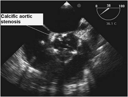
Common features on ECG include left ventricular hypertrophy with strain pattern, left bundle branch block, and left atrial hypertrophy (biphasic P waves in precordial lead V1). Chest radiography may reveal a boot-shaped heart, calcification of the aortic valve, poststenotic dilatation of the aorta, and pulmonary venous congestion. Echocardiography is the principal modality for confirming the diagnosis of AS (Fig. 123.1); the severity of AS is determined by peak gradients across the aortic valve and calculation of valve area (Table 123.2). It is important to recognize that the gradients will be lower with severe aortic stenosis if the flow across the valve is reduced by hypovolemia or by poor left ventricular function. Echocardiography also provides information about left ventricular function. Patients with AS may have preserved systolic function but with significant diastolic dysfunction. Diastolic dysfunction predisposes these patients to pulmonary edema. Coronary angiography is indicated in patients with aortic stenosis before surgery to rule out associated coronary artery disease (CAD).
Therapeutic Considerations
Patients with AS may require ICU admission because of acute cardiogenic shock, pulmonary edema, severe angina, ventricular dysrhythmias, or, less commonly, atrial fibrillation and systemic embolization.
Drugs
Drugs commonly used to treat these conditions carry significant risks in patients with AS. β-Blockers, calcium channel blockers, and other antidysrhythmics should be used with caution as patients with AS are sensitive to drugs causing myocardial depression. Pulmonary congestion should be relieved by careful administration of diuretics, digitalis, and nitroglycerin. The use of diuretics and nitroglycerin, however, may result in inadequate preload. Digitalis carries the risk of dysrhythmias. Increased left ventricular mass and intracavitary systolic pressure increase oxygen demands, thereby decreasing the patient's tolerance to vasodilatation and tachycardia. Angiotensin-converting enzyme (ACE) inhibitors and other vasodilators such as nitroprusside may precipitate syncope or even sudden death in severe AS, and are relatively contraindicated.
|
Table 123.1 Clinical signs in valvular heart disease |
||||||||||||||||||||||||||||||||||||||||||||||||||||||||||||||||||||||||
|
||||||||||||||||||||||||||||||||||||||||||||||||||||||||||||||||||||||||
Undesirable hemodynamic effects of sedative drugs such as propofol, benzodiazepines, and narcotics should also be considered. Propofol can cause significant vasodilatation and hypotension, and therefore should be avoided in patients with fixed cardiac output. Narcotics can blunt the hypertensive sympathetic responses without significant myocardial depression and are usually the agents of choice.
Fluids
Fluid is administered with extreme caution in patients with AS. Patients with diastolic dysfunction, even when systolic function and ejection fraction are preserved, are extremely sensitive to fluids and can develop pulmonary edema. Filling pressures may need to be monitored with a pulmonary artery catheter (PAC) in such critical situations.
Monitoring
Hemodynamic stabilization with drugs and fluids should be carried out with careful monitoring of arterial pressure and cardiac filling pressures. Normal central venous pressure (CVP) does not ensure adequate filling pressures in patients with a stiff left ventricle. Placement of a PAC, while helpful, is not without risks, as it may precipitate malignant dysrhythmias and sudden deterioration. However, the PAC can provide useful information about left ventricular filling pressures, cardiac output, and mixed venous oxygenation, and can be used, if needed, for transvenous pacing. It is intuitive that the risks must be weighed against the potential benefits when PAC use is considered. It is important to note that pulmonary capillary wedge pressure (PCWP) can underestimate left ventricular end-diastolic pressure (LVEDP) in patients with markedly reduced ventricular compliance; echocardiography can provide useful information in such situations. Monitoring for ischemia with continuous ECG leads V5 and II should be carried out in all patients with AS, as they are vulnerable to ischemia. However, the ECG manifestations of left ventricular hypertrophy may make detection of ischemic changes more difficult.
|
|
|
Figure 123.1 Transesophageal echocardiographic appearance of severe calcific aortic stenosis. |
Hemodynamic Goals
Left ventricular hypertrophy renders the atrial contraction—and thus sinus rhythm and preload—more crucial for diastolic filling. Atrial fibrillation should be reversed with prompt cardioversion and initiation of antidysrhythmic therapy with amiodarone and/or β-blocking drugs. Procainamide may also be used, but carries an increased risk of myocardial depression and hypotension. If cardioversion is unsuccessful, pharmacologic control of the ventricular rate is essential. It is imperative to avoid tachycardia, which will decrease the diastolic ventricular filling time and increase the risk of ischemia. Severe bradycardia also should be avoided because severe aortic stenosis results in a fixed stroke volume, therefore potentially reducing cardiac output. Adequate preload should be maintained, and one must recognize that afterload reduction may be hazardous as it can impair coronary perfusion pressure. Severe myocardial dysfunction with low blood pressure and ischemia may require administration of inotropes. Maintenance of adequate coronary perfusion pressure by vasopressors such as phenylephrine may be necessary in patients with optimized volume and myocardial contractile status. Patients who are refractory to medical management may benefit from insertion of an intra-aortic balloon counterpulsation pump (IABP) to improve coronary perfusion pressure.
|
Table 123.2 Echocardiographic assessment of aortic stenosis (AS) |
||||||||||||||||
|
Definitive Therapy
Considering the unfavorable natural history of AS, any ICU patient with severe AS who continues to deteriorate despite medical therapy should be seen by a cardiologist and a cardiac surgeon for possible balloon valvotomy or open valve replacement. Balloon valvotomy affords temporary improvement in transvalvular gradient—usually with restenosis in about 6 months—and may relieve symptoms in some patients, thus serving as a bridge to definitive surgery. Balloon valvotomy is often very effective for young adults and adolescents with bicuspid valves, although it carries a mortality of 10% in patients with calcific AS (1).
Aortic Regurgitation
Patients with aortic regurgitation (AR) may present to the critical care physician either because of decompensated chronic AR or due to acute onset of severe regurgitation.
Etiology
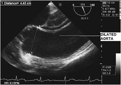
Acute AR results from infective endocarditis, with leaflet perforation, vegetations, or perivalvular fistula, and aortic dissection extending into the aortic annulus or aortic root, and from trauma, with avulsions of the annulus and tears of the cusps. Severe, acute hypertension may also cause sudden onset of AR that often reverses after control of hypertension. The causes of chronic AR are diverse; the disease may directly involve the valve or the aortic root. Primary valvular diseases include congenital bicuspid valve, prolapse of aortic cusp, rheumatic heart disease, calcific degenerative disease, connective tissue diseases, and subacute bacterial endocarditis. Diseases associated with aortic root dilatation include systemic hypertension, Marfan disease (Fig. 123.2), Ehlers-Danlos disease, granulomatous diseases of the aorta, senile and cystic medial degeneration, annuloaortic ectasia, and syphilis.
Hemodynamics
In chronic AR, the left ventricle (LV) dilates and hypertrophies when subjected to volume overload. This keeps wall stresses in check and maintains normal forward stroke volume. As the disease progresses and the compensatory limit is reached, the wall stress begins to rise and systolic function deteriorates, with decreasing forward stroke volume as well as increasing LV end-diastolic volume (LVEDV) and LVEDP, resulting in symptoms of heart failure. The presence of symptoms, systolic dysfunction, and an increase in end-systolic dimensions indicate severe and decompensated AR. During the relatively asymptomatic phase, the patient develops symptoms at a rate of 3.7% per year. In acute AR, the LV is subject to a sudden increase in volume, with no opportunity for a compensatory increase in compliance and eccentric hypertrophy to occur. Without these adaptations, the increase in end-diastolic pressures causes pulmonary venous congestion and pulmonary edema. The severity is dependent on the regurgitant orifice size, duration of diastole—as the degree of AR increases with bradycardia—and the diastolic pressure gradient between aorta and left ventricle. Thus, the patient with acute AR may present with heart failure if the AR is severe, or the initiating event—aortic dissection, trauma, or endocarditis—may dominate if AR is mild.
|
|
|
Figure 123.2 Dilatation of aortic root and ascending aorta by transesophageal echocardiography in a patient with Marfan disease. |
Diagnosis
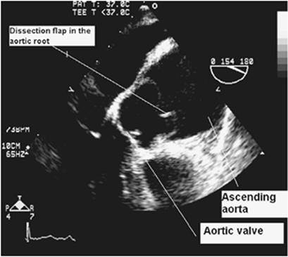
An ECG is performed to rule out ischemic heart disease in situations of acute AR. The chest radiograph will reveal cardiomegaly in chronic AR or pulmonary congestion with a normal-sized heart in acute AR. TTE is performed to define the mechanism and severity of AR. Transesophageal echocardiography (TEE) is useful in patients with limited TTE windows. TEE defines the nature of perivalvular pathology (e.g., an abscess) better than TTE. An acute aortic dissection may be diagnosed with high sensitivity and specificity with TEE. Other techniques such as computerized tomographic (CT) scanning, magnetic resonance imaging (MRI), and aortography have been used. Each modality has its own advantages and disadvantages (2). TEE is clearly superior to MRI and CT scanning to characterize the valve pathology in cases of acute dissection, and obviates the need for aortography (Fig. 123.3). Coronary angiography can rule out ischemic heart disease before surgery in chronic AR, but is rarely indicated before emergency surgery in acute AR. Echocardiography may also be helpful to rule out the coronary artery involvement in dissections.
Therapeutic Considerations
Patients with acute AR are generally ill enough to require ICU admission. While medical therapy may allow patients with mild acute AR to reach a chronic compensated state, emergency aortic valve replacement is almost always indicated in a patient with severe acute AR after medical stabilization.
|
|
|
Figure 123.3 Aortic dissection intimal flap involving aortic root. |
The principle of therapy is to optimize cardiac output and systemic perfusion, reduce pulmonary venous congestion, and initiate therapy for any underlying disorder. Invasive monitoring is initiated and volume is optimized. Tachycardia is beneficial in maintaining cardiac output, and decreases the regurgitant fraction by decreasing the duration of diastole. β-Blockers are avoided in acute severe AR before surgery, as it inhibits compensatory tachycardia and may precipitate circulatory failure. Hypertension and increased afterload are to be avoided; afterload reduction is indicated with vasodilators such as nitroprusside. Inotropic therapy is advised only in patients with depressed systolic function. The hemodynamic response to therapy is an increased cardiac output with reduction of filling pressures. An IABP is absolutely contraindicated in patients with AR, as it will increase the regurgitant fraction.
In contrast to other causes of AR, inotropic therapy is avoided in patients with aortic dissection, as it occurs as a result of long-standing, poorly controlled hypertension or trauma. In both situations, left ventricular contractility is preserved and inotropes are not indicated. β-Adrenergic blockade may be initiated to reduce the velocity of LV ejection and aortic wall stress, therefore preventing extension of the aortic dissection or aortic rupture. In patients with chronic AR who present with an acute decompensation, a search should be made for the precipitating cause, with particular attention to possible infectious endocarditis. Most patients stabilize with medical therapy, but early elective surgery should be considered, as the outlook for medically treated symptomatic patients is poor. Decompensated patients who do not improve with aggressive medical therapy should undergo emergency valve replacement. Mortality with medical therapy alone in this group approaches 100%, while many moribund patients will survive with surgery.
Mitral Regurgitation
The mitral valve apparatus is composed of the valve leaflets, mitral annulus, chorda tendineae, papillary muscles, and adjacent cardiac chambers, namely the left atrium and left ventricle. Any disruption in the integrity of the mitral valve apparatus may result in regurgitation. One of the major breakthroughs in the management of mitral regurgitation was the functional classification of mitral regurgitation (MR) by Carpentier in early 1980s (3) (Table 123.3).
|
Table 123.3 Functional classification of mitral regurgitation (MR) |
|||||||||||||||
|
Etiology
An acute presentation of MR to the critical care physician usually results from infective endocarditis with leaflet perforations, vegetations, and perivalvular leaks; connective tissue or myxomatous disorders (e.g., chordal rupture); and ischemic heart disease—infarction and rupture of papillary muscles, or transient papillary muscle dysfunction due to ischemia. Acute rheumatic mitral valvulitis as the cause of mitral regurgitation is less common today.
Hemodynamics
Chronic MR leads to adaptation of the left ventricle by dilatation and eccentric hypertrophy. Over many years of increasing regurgitant volume, systolic function may fail, resulting in decreased ejection fraction (EF) and pulmonary hypertension. Left atrial (LA) dilatation leads to atrial fibrillation. In the absence of a precipitating event such as an infection or a second hemodynamic abnormality, patients with chronic MR are rarely critically ill on presentation. In acute MR, on the other hand, the regurgitant volume is mainly ejected into the noncompliant left atrium. As a result, left atrial pressure increases, which is transmitted to the pulmonary venous system, resulting in pulmonary edema. Sudden volume overload increases the burden on the LV and it fails. Cardiac output falls and systemic vascular resistance rises, which further increases the regurgitant fraction. Patients may present with acute onset of fatigue, dyspnea, and chest pain, or can be admitted with pulmonary edema and circulatory shock, depending on the etiology. The differences in clinical signs between acute and chronic forms of MR are given in Table 123.1.
Diagnosis
The ECG may show atrial fibrillation, left ventricular hypertrophy, and right ventricular strain, while the chest radiograph may show cardiomegaly, indicating pre-existing heart disease. Pulmonary venous congestion and/or edema with a normal-sized heart indicate acute MR. An ECG may suggest the etiology in cases of ischemic MR.
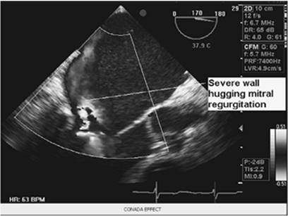
Echocardiography remains the standard for the diagnosis of mitral regurgitation. TTE is easy, safe, and quick, and can be performed at the bedside. The mechanism and type of MR should be defined. Qualitative and quantitative assessment of MR can be done by color and spectral Doppler methods. Finally, the suitability for repair and left ventricular function should be assessed. TEE may provide better detail because of superior resolution and, further, has a significant advantage in that it allows definition of anatomic details of the mitral valve apparatus and mechanism of MR; the severity of MR is better evaluated by TEE (Fig. 123.4). Indices of left ventricular function such as EF are unreliable in the presence of severe MR. EF in MR is increased when contractility is normal. Normal EF indicates a significant loss of myocardial function; when EF is reduced to 50% or less, advanced myocardial dysfunction is generally present (4). The American Heart Association/American College of Cardiology (AHA/ACC) guidelines recommend medical treatment when EF is less than 30% and surgical treatment, even in asymptomatic patients, with EF less than 60% to prevent progression of disease (5). Urgent coronary angiography is indicated if myocardial ischemia or infarction is a real consideration. Coronary angiography may delineate a culprit lesion, which may be amenable to catheter-based interventions. Severe triple-vessel disease should be referred for surgery. In the case of chordal rupture or infective endocarditis without risk factors for CAD, angiography may be deferred.
|
|
|
Figure 123.4 Transesophageal echocardiographic diagnosis of mitral regurgitation (MR) severity. Left atrial wall hugging eccentric regurgitant jet indicates severe MR. |
Therapeutic Considerations
In acute, severe MR, medical therapy has a limited role and is aimed at stabilizing the patient's hemodynamics in preparation for surgical intervention. Early valve surgery is life saving, and therefore should not be delayed. Since acute MR is often a complication of myocardial infarction, therapy should include the need to maintain coronary perfusion pressure and reduce myocardial oxygen consumption. The utilization of a PAC and invasive arterial pressure monitoring are recommended. The pulmonary artery waveform may show typical V waves of mitral regurgitation. Sinus tachycardia maintains the forward flow and, thus, should not be suppressed. Hypertension and the use of vasopressors should be avoided, as the increased afterload will increase the regurgitant fraction. Diuretics may be needed to reduce pulmonary venous congestion. While in the normotensive patient, vasodilators—such as nitroprusside—can be useful in increasing the forward flow and decreasing the regurgitant fraction, the same cannot be said when the patient is hypotensive. In this case, nitroprusside may further impair coronary perfusion pressure and should not be used except in combination with inotropes. When patients fail to respond adequately to medical management, IABP may be life saving, as it increases forward flow and blood pressure while diminishing filling pressures. It is effective in increasing coronary perfusion pressure and reducing oxygen consumption, the key factors in maintaining a favorable balance between myocardial oxygen supply and demand. Thus, IABP can be used to stabilize patients while they are prepared for surgery.
The surgical intervention is determined by the nature of the lesion. Chordal rupture or prolapse of the posterior leaflet can be successfully repaired. Revascularization alone may improve ischemic MR in selected patients with transient papillary muscle ischemia without rupture (6). Revascularization can be done using angioplasty, thrombolysis, or bypass surgery, depending on the anatomic characteristics and the severity of the lesions. However, revascularization alone may leave many of these patients with significant degrees of postoperative MR (7). Current lines of evidence suggest that a combination of complete revascularization with mitral valve repair or replacement with posterior chordal sparing is the standard of care for acute ischemic MR refractory to medical management and nonsurgical revascularization techniques (8). Mitral valve annuloplasty is the best approach for ischemic MR due to annular dilatation, diminished systolic contraction of the annulus, or papillary muscle malalignment. Mitral valve replacement should be considered if the repair is unsatisfactory. Papillary muscle rupture carries a high mortality without surgical intervention; surgery does improve the long-term outcome in functional class in these patients.
Infective endocarditis should be treated with antibiotics. However, if surgery is indicated, it should not be delayed for microbiologic clearance. Indications for surgery include congestive heart failure refractory to medical treatment, uncontrolled infection despite antibiotics, recurrent systemic embolism, perivalvular or pericardial extension of infection, fungal endocarditis, and prosthetic valve endocarditis (5).
Mitral valve prolapse (MVP) deserves special mention because MVP is the most prevalent valvular heart disease and the most common cause of MR. Patients with this condition may present to the ICU because of severe MR, atrial fibrillation with transient ischemic attacks, long QT syndrome and tachyarrhythmias, pulmonary hypertension, cerebral embolism from MVP-related fibrin, infective endocarditis, or even sudden death (9). Medical stabilization and elective surgical repair of the mitral valve are recommended therapies.
Mitral Stenosis
Etiology
Mitral stenosis (MS) is mostly related to rheumatic heart disease. Degenerative calcific stenosis, congenital stenosis, and connective tissue disorders such as systemic lupus erythematosus and Lutembacher syndrome (atrial septal defect with MS) are the other causes for MS. Atrial myxomas and left atrial ball-valve thrombus can present with intermittent obstruction to mitral inflow and mimic MS.
Hemodynamics
In rheumatic MS, inflammation of the connective tissue leads to leaflet thickening and calcification, and commissural and chordal fusion. Ultimately, many patients are left with a funnel-shaped mitral apparatus. As the condition progresses, left atrial pressure and the transmitral gradient increase, thus maintaining flow. However, the subsequent increase in pulmonary venous pressure leads to hydrostatic pulmonary edema. Pulmonary vasoconstriction increases pulmonary arterial pressure and right ventricular afterload. Over the course of rheumatic heart disease, persistently elevated pulmonary arterial pressure leads to structural changes such as intimal hyperplasia and medial hypertrophy. These structural changes are permanent and result in fixed pulmonary hypertension. Right ventricular dilatation and failure result in tricuspid insufficiency and systemic venous congestion, respectively. The impact of MS on cardiac output is initially determined by the severity of the stenosis itself and the limitation to flow across the narrowed mitral orifice. However, as the disease progresses, right ventricular failure may become severe enough to limit cardiac output. Similarly, progression of rheumatic disease with its associated inflammation, fibrosis, and calcium deposition may impair left ventricular function. Although this effect is not a major determinant of cardiac output in patients with MS, it may become important to consider in some patients after surgical repair. In such situations, the increase in transmitral flow may expose the left ventricle to a sudden increase in preload, causing failure.
Presentation
Patients with mitral stenosis often present to the ICU with acute cardiogenic pulmonary edema. Precipitating factors such as infective endocarditis, fever, anxiety, pain, atrial fibrillation, and pregnancy should be identified. Occlusion from an enlarging atrial myxoma should be ruled out. Right-sided heart failure with hepatic dysfunction, acute hemoptysis, systemic embolism, and hoarseness of voice may also be present.
|
|
|
Figure 123.5 Typical appearance of rheumatic mitral stenosis. |
Diagnosis
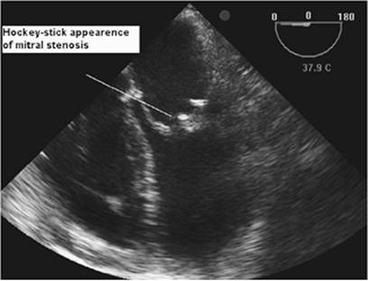
The ECG may show left atrial enlargement, right ventricular hypertrophy, and atrial fibrillation in MS. Chest radiography shows straightening of the left heart border, indicating left atrial and pulmonary artery enlargement. Kerley A and B lines indicate pulmonary venous hypertension. TTE findings include doming of the anterior leaflet, decreased leaflet mobility, increased leaflet calcification and thickness, commissural fusion, calcification of the subvalvular apparatus, increased LA size, and the presence of an LA thrombus (Fig. 123.5). Color flow will show associated mitral regurgitation. Doppler echocardiography allows calculation of pressure gradients, mitral valve area, and estimation of pulmonary artery systolic pressure. Cardiac MRI is increasingly being used in the evaluation of stenotic valvular lesions; it measures valve area by planimetry. Its advantage over echocardiography is the lack of dependence upon good echocardiographic windows.
Therapeutic Considerations
New onset atrial fibrillation with hemodynamic instability should be treated with cardioversion. Cardioversion of a patient with atrial fibrillation of unknown duration or that is known to have persisted for more than 48 hours must be preceded by 3 weeks of anticoagulation or by a TEE to exclude the presence of left atrial thrombus. Anticoagulation should be continued for 4 weeks following cardioversion because the enlarged LA remains “stunned” and does not recover a normal contractile state immediately following cardioversion (10). Anticoagulants should also be used in patients with a prior embolic event and left atrial diameter greater than 55 mm by echocardiography (5). Antidysrhythmics, such as amiodarone, may be used to maintain sinus rhythm but should not be expected to provide indefinite success.
Patients admitted with pulmonary edema should be stabilized with oxygen, morphine, anxiolytics, diuretics, and digoxin; the latter is especially useful in patients with atrial dysrhythmias and congestive heart failure. Sympathetic nervous system activity is increased in patients with mitral stenosis, and sympathetic overactivity worsens the symptom complex; β-blockers are very useful in this situation. Intravenous nesiritide, a synthetic human natriuretic peptide, is also used in the critically ill patients with acute decompensated cardiac failure and pulmonary hypertension. Short-term intravenous infusion of nesiritide is associated with hemodynamic and symptomatic improvements in patients with acutely decompensated congestive heart failure (CHF). Nesiritide may offer tolerability and practical advantages over currently used vasodilators, inodilators, and inotropes in this condition; in particular, nesiritide does not appear to have proarrhythmic effects. Nesiritide also appears to be effective and well tolerated in patients receiving concomitant β-blocker therapy and those with renal insufficiency (11,12). If the patients are hypotensive, inotropes to improve left ventricular function may not be useful, but may worsen tachycardia and pulmonary edema. Inodilators may be useful in improving right ventricular dysfunction and reducing pulmonary hypertension. Systemic blood pressure may need to be supported with vasopressors, with the caveat that they may adversely impact pulmonary vascular resistance. Assessment of volume status is difficult, as the PCWP does not correlate with LVEDP in patients with MS. However, the PCWP gives useful information about the propensity to develop pulmonary edema.
Emergency invasive intervention is rarely required to relieve MS. If the precipitating events are controlled, intervention by surgery or balloon valvotomy can be scheduled electively after medical optimization. Balloon valvotomy is indicated in patients with suitable anatomy: pliable leaflets, no commissural fusion, and minimal subvalvular calcification. LA thrombus and significant (3+ to 4+) MR should be excluded by echocardiography. Patients who are not candidates for balloon intervention should be referred for surgery. Because of extensive calcifications and marked anatomic distortion, mitral valve repair often is not possible in rheumatic mitral disease, and replacement is necessary in those patients.
Tricuspid Stenosis
Etiology
Tricuspid valve obstruction can be due to anatomic disease of the tricuspid valve or functional, causes secondary to right atrial (RA) tumors and thrombus. Anatomic disease is usually related to rheumatic heart disease, and involvement of mitral valve is common. Other anatomic causes are carcinoid syndrome, infective endocarditis, congenital stenosis or atresia, and methysergide toxicity.
Hemodynamics
Obstruction to right ventricular inflow results in systemic venous congestion: elevated jugular venous pulse (JVP), congestive hepatomegaly, and peripheral edema. Diastole is shortened by increasing heart rate, thus causing dramatic increases in transvalvular gradients. Most patients are symptomatic from coexisting mitral stenosis. The presence of systemic venous congestion out of proportion to pulmonary venous congestion should raise the suspicion for involvement of the tricuspid valve.
Clinical Signs
Physical exam will reveal an elevated JVP, a prominent a wave, and distension of veins of the upper arm and dorsum of extended hand. The JVP increases by compression of the liver—the hepatojugular reflux. A diastolic thrill may be palpated at the lower left sternal border; the S1 is increased, and an opening snap and diastolic murmur may be heard at the lower left sternal border. Assigning the murmur to the tricuspid valve is based on its location; the higher, shorter, and softer nature of tricuspid stenosis (TS) in comparison to MS; and the absence of crackles that commonly accompany the murmur of MS. Right-sided murmurs are increased during inspiration.
Diagnosis
The ECG may show evidence of RA enlargement—a P wave exceeding 2.5 mm in lead II and 1.5 mm in V1. RA enlargement may increase the distance from sinus node to atrioventricular node, causing first-degree heart block; atrial fibrillation may occur in advanced disease. The chest radiograph may show cardiomegaly and RA enlargement, and calcification of the valve may be evident. Typically, echocardiographic visualization shows thickened leaflets, limited mobility, and a dome-shaped structure in diastole. Right ventricular function and the presence of tumor, thrombus, and vegetations can also be assessed. Spectral and color Doppler methods allow calculation of pressure gradients to grade the stenosis—that is, mild less than 5 mm Hg, moderate 5 to 10 mm Hg, and severe greater than 10 mm Hg—and valve area. Catheterization is unnecessary in patients with good echocardiographic windows.
Therapeutic Interventions
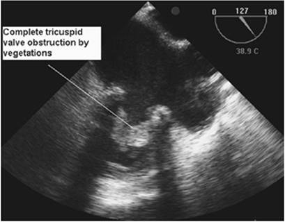
Complete obstruction of the tricuspid valve by vegetations, thrombus, and/or tumors is an indication for emergency valve surgery (Fig. 123.6). As seen in other valvular lesions, the disease progresses slowly, becoming symptomatic as obstruction to flow increases. Sodium restriction, careful diuresis, rate control, and anticoagulation are helpful, but surgical correction is required when the transvalvular gradient exceeds 5 mm Hg and the valve area falls below 2 cm2. Percutaneous valvotomy or, more often, a bioprosthetic valve replacement is necessary. Mechanical valves are avoided because of the very high risk of thromboembolism in this position.
|
|
|
Figure 123.6 Complete obstruction of prosthetic tricuspid valve by vegetations requiring emergency surgery. |
Tricuspid Regurgitation
Etiology
Tricuspid regurgitation (TR) can be classified as structural or functional. Structural diseases of the valve are caused by rheumatic disease, infective endocarditis, carcinoid syndrome, radiation therapy, Marfan syndrome, congenital heart disease, or tricuspid valve prolapse. Functional TR is usually secondary to left-sided pathology, such as left ventricular failure or mitral or—less frequently—aortic valve disease, but may also result from primary pulmonary hypertension, pulmonic stenosis, right ventricular (RV) infarction, and dilated cardiomyopathy; a small number of cases may not have an etiology identified. While most cases of TR are chronic, an acute presentation may occur following penetrating trauma, RV infarction, infective endocarditis, and, more recently, repeated endomyocardial biopsies to diagnose allograft rejection of heart transplantation.
Hemodynamics
Acutely following the development of TR, elevated RA pressure gives rise to the signs and symptoms of systemic venous congestion. Over time, the RA dilates, with the possible sequelae of atrial fibrillation and thrombus formation. Formation of an RA thrombus may lead to pulmonary and systemic emboli, the latter in patients with patent foramen ovale. RV volume overload leads to eccentric hypertrophy and dilatation. RV systolic dysfunction develops, leading to further decompensation and dilatation, which in turn causes annular dilatation and an increase in the severity of TR. The left heart also suffers from low cardiac output because the RV fails to deliver enough blood to the LV. Ventricular interdependence and paradoxical septal motion also decrease left ventricular output in patients with RV volume overload.
Clinical Symptoms and Signs
Symptoms of low cardiac output and venous congestion present in these patients. Shock and hypotension may develop following acute TR after RV infarction or papillary muscle rupture. Other patients maintain blood pressure but demonstrate signs of right-sided failure. Distended neck veins with a prominent c-v wave, pulsatile hepatomegaly, a precordial bulge, and parasternal heave from RV hypertrophy; soft S1, prominent P2, and right-sided S3 from RV dilatation; pansystolic thrill; and a murmur heard at the left lower sternal border are associated signs. Cases of isolated tricuspid valve endocarditis may disseminate emboli to the lungs, resulting in multiple septic emboli and abscess formation. Peripheral stigmata of infective emboli are usually absent, but may indicate paradoxical embolism or left-sided lesions if present.
Diagnosis
The ECG may show atrial fibrillation, right axis deviation, RV hypertrophy, and a right bundle branch block. Right-sided leads will show ST elevation in RV infarction and may be associated with LV inferior wall infarction. Enlarged RV and cardiomegaly are seen on chest radiography. Echocardiography can yield details about the structural issues with the tricuspid valve—prolapse, vegetations, annular diameter, and rheumatic disease—and helps to rule out thrombus and patent foramen ovale. The diagnosis of a patent foramen ovale is important, since the elevated right atrial pressure frequently exceeds left atrial pressure, causing a right-to-left shunt that may cause refractory hypoxemia. Severity of TR can be assessed by hepatic venous systolic flow reversal and estimation of regurgitant orifice area (greater than 40 mm2 is severe). Also, pulmonary systolic pressure can be estimated by continuous wave Doppler signal of tricuspid regurgitation. Augmenting ultrasound signal with 10% air, 10% patient's blood, and 80% saline, and estimation of pulmonary artery systolic pressures correlate well with PAC-measured PA systolic pressures (13). Right heart catheterization shows RV pressure greater than 60 mm Hg in functional TR compared to less than 40 mm Hg in structural TR.
Therapeutic Considerations
In general, the hemodynamic impact of acute TR is less severe and can be effectively managed with diuretics and inotropes. In rare instances, acute severe TR may require valvular surgery when the process is refractory to medical therapy. Once stabilized, these patients' long-term prognosis depends on the etiology and severity of the TR. Patients tolerate mild and moderate degrees of chronic TR in the presence of normal LV function. Correction of left-sided heart lesions and treatment of left-sided failure and pulmonary hypertension take priority. If functional TR is severe, surgical intervention may be needed. Ring annuloplasty can improve TR and survival in these patients (14). Cardiac resynchronization therapy with biventricular pacemakers and the Dor procedure (endoventricular circular patch plasty) help patients with functional TR due to systolic heart failure (15).
Rheumatic TR may be treated with open valvotomy or valve replacement. Valves compromised by infective endocarditis can simply be removed after an aggressive course of antibiotics. Valve replacement can be performed later if there is no recurrence of drug abuse in these patients. When acute TR is the result of RV infarction, the usual management of acute coronary insufficiency must be observed, including β-blockers, aspirin, fluids, thrombolysis, coronary angiography, angioplasty, and stenting and, in suitable patients, coronary artery bypass grafting.
Pulmonic Stenosis
Etiology
Pulmonic stenosis (PS) is usually congenital. While valvular PS does occur in isolation, subvalvular and supravalvular stenosis usually comprise part of a larger syndrome. Severe forms, or those associated with other cardiac anomalies, are generally identified and treated in childhood. Acquired forms of pulmonary stenosis include rheumatic, carcinoid disease; infundibular stenosis from pulmonary hypertension; extrinsic compression from aneurysm of the sinus of Valsalva; tumors; and scarring from previous surgery.
Pathophysiology
PS causes obstruction to right ventricular outflow. Right ventricular hypertrophy results, and, if right ventricular failure or tricuspid regurgitation develops, systemic venous hypertension can result. The degree of stenosis, once established, tends to be stable; if decompensation does not occur in childhood, subsequent deterioration is unlikely. Occasionally, patients manifest increasing right ventricular outflow obstruction, perhaps caused by secondary infundibular hypertrophy.
Presentation
Mild PS is often asymptomatic. Patients with more severe stenosis commonly develop fatigue, atypical chest pain, and syncope. When right ventricular failure or tricuspid regurgitation occurs, systemic venous hypertension may be present. Elevated right atrial pressures in the presence of a patent foramen ovale or atrial septal defect can result in a right-to-left shunt at the atrial level.
Physical examination typically reveals a large jugular venous a wave. In the absence of right ventricular failure or tricuspid regurgitation, jugular venous pressure remains normal. A left parasternal systolic lift is common with significant pulmonic stenosis. The murmur is best heard at the left upper sternal border and typically radiates to the left clavicle. A palpable thrill may be present. The intensity of the murmur does not correlate well with severity, but increasing duration and late systolic peaking are indicators of significant stenosis. An ejection click is usually present. As severity increases, P2 is increasingly delayed; thus, wide splitting of the second sound indicates more severe stenosis.
Diagnostic Studies
The ECG is normal in mild pulmonic stenosis. With increasing severity, right ventricular hypertrophy and right atrial enlargement are common. The chest radiograph may reveal poststenotic dilatation of the main and left pulmonary arteries. Echocardiography is particularly useful in assessing the valve morphology, calculating pressure gradients across the pulmonic valve, grading the severity of stenosis, and evaluating right ventricular function. The presence of other congenital abnormalities, tricuspid regurgitation, and right atrial enlargement can also be ruled out by echocardiography. Cardiac catheterization provides confirmation of pressure gradients, full hemodynamic assessment, and identification of associated pulmonary artery branch stenosis. Pulmonary stenosis is graded based on the peak pressure gradient: mild, with a gradient of 25 to 49 mm Hg; moderate, 50 to 75 mm Hg; and severe, greater than 75 mm Hg. Currently, balloon valvuloplasty is recommended for symptomatic patients and those with a peak gradient greater than 50 mm Hg. Surgical valvuloplasty is reserved for severe calcification, dysplasia, endocarditis, and previous valvuloplasty failure. Medical management includes infective endocarditis prophylaxis, treatment of right heart failure and atrial fibrillation, and anticoagulation to prevent thromboembolic complications.
Pulmonic Insufficiency
Pulmonic insufficiency (PI) generally has a benign course as an isolated abnormality. The natural history is that of the associated lesions.
Etiology
PI may be secondary to pulmonary hypertension or, rarely, to leaflet damage caused by infectious endocarditis, rheumatic fever, or carcinoid syndrome. Occasionally, PI is congenital.
Pathophysiology
In the absence of pulmonary hypertension, volume overload of the right ventricle is well tolerated. Decompensation with resulting right ventricular failure can occur when pulmonary hypertension develops from other causes.
Presentation
PI is usually an incidental auscultatory finding in patients admitted to the ICU for other reasons. Physical findings include the typical decrescendo diastolic murmur along the upper left sternal border. The intensity of the murmur does not correlate well with the severity of regurgitation.
Diagnostic Studies
The ECG is usually normal. The presence of right ventricular hypertrophy suggests pulmonary hypertension. The chest radiograph is normal in mild insufficiency. The pulmonary trunk may be prominent when the insufficiency is moderate to severe. Pulmonary hypertension may be present. Echocardiography with Doppler study can be useful for differentiating pulmonic from aortic insufficiency and for establishing right heart chamber sizes and associated abnormalities.
Therapeutic Considerations
Specific treatment is rarely required. However, therapy should be directed toward control of pulmonary hypertension when present. When the right heart fails, diuretics and sodium restriction are useful, and some clinicians suggest that cardiac glycosides are helpful. Surgical treatment (bioprosthetic valve replacement) is reserved for advanced right heart failure. In patients with a remote repair of tetralogy of Fallot and chronic PI, RV dilatation has been linked to sudden death. This has led some clinician-investigators to pursue valve replacement in early stages of RV dilatation.
Mixed Valve Lesions
Mitral Stenosis with Regurgitation
The combination of pressure and volume overload on LA favors early development of symptoms, atrial fibrillation, and congestive heart failure. Because transvalvular gradients may overestimate the degree of stenosis, Doppler measurement of valve area should be considered. Decision making is complex, and intervention is often required before either of the lesions reaches a severe degree. Moderate MR is a contraindication for balloon valvotomy.
Aortic Stenosis and Regurgitation
This combination causes both pressure and volume overload on the LV. The predominant lesion is indicated by the size of the LV: a normal-sized, but hypertrophied, LV signifies predominant AS; a dilated LV suggests dominant AR. As with combined MS and MR, transvalvular gradients may overestimate AS, so planimetry or the continuity equation method should be considered. The threshold for surgery is lowered as compared to single valve–defect patients. Those with severe AS with accompanying AR should be operated on in higher calculated valve areas or in the presence of mild symptoms. Surgery in patients with predominant AR with accompanying AS can be delayed until symptoms develop or asymptomatic LV dysfunction becomes apparent on echocardiography (enlarged ventricular dimensions).
Mitral Stenosis and Aortic Stenosis
This combination causes serial obstructions resulting in reduced cardiac output and early development of pulmonary venous congestion and hypertension. Low transvalvular gradients characterize this aortic stenosis because of low cardiac output. Mitral valvotomy is done first, followed by aortic valve replacement as indicated.
Mitral Stenosis and Aortic Regurgitation
This combination creates a challenge for the physician attempting to make a diagnosis. MS decreases the volume overload of AR, and AR attenuates antegrade mitral valve flow by increasing LV diastolic pressure, thereby decreasing transmitral gradients. Balloon mitral valvotomy followed by aortic valve replacement (AVR), as indicated, is a reasonable approach.
Mitral Regurgitation and Aortic Stenosis
Aortic stenosis aggravates MR by increasing the afterload. MR, by its pressure release effect, obscures even severe AS. Systolic function remains normal with low transaortic gradients. If both lesions are severe, AVR with mitral valve repair or replacement is necessary. Moderate or mild MR may improve after AVR for AS, especially if there is no anatomic lesion in the mitral valve. Intraoperative TEE plays an important role in this decision.
Mitral Regurgitation and Aortic Regurgitation
These lesions create additive volume loads on the LV; consequently, the sequelae of dyspnea and LV dysfunction appear sooner.
Prosthetic Valve Dysfunction
Prosthetic valves in common use are broadly divided into mechanical, bioprosthetic, and homograft valves. St. Jude bileaflet valves are commonly used mechanical valves which need life-long anticoagulation. Carpentier-Edwards and Hancock bioprosthetic valves are common tissue valves in use. They do not require long-term anticoagulation, but have a short life span and are prone to degenerative changes and failure. Acute valvular complications may result from infective endocarditis, paravalvular leak, valve ring abscess, thrombosis, pannus formation, degenerative calcification, lipid infiltration, dehiscence of the valve, and strut fracture.
Progressive congestive cardiac failure is a common presentation with stenosis and regurgitation. Acute, complete valvular obstruction may lead to sudden death in the absence of surgical intervention. Embolic phenomenon, hemolytic anemia—indicating a paravalvular leak, and a new atrioventricular block—indicating a valve ring abscess may be other presenting symptoms. Prosthetic valve thrombosis may present with nonspecific cardiac symptoms. Normally functioning prosthetic valves are associated with clicks and murmurs; hence, disappearance of clicks or a new or changing murmur is important in making the diagnosis.
Echocardiography is essential to make a diagnosis in these patients. As previously noted, TEE is more sensitive and specific in the evaluation of prosthetic valve pathologies than TTE; echocardiography has replaced cardiac catheterization in these cases. Fluoroscopy may be needed in some cases to identify the nature of the disease and assess the effects of thrombolysis. Excessive rocking motion of the valve ring or limited motion of the valve components due to thrombus or vegetation; calcification; thickening around the valve due to an abscess; and a pseudoaneurysm can be identified with two-dimensional echocardiography. The color Doppler technique may show a paravalvular leak, pseudoaneurysms, and/or fistula formation. Calculation of transvalvular gradients help in the diagnosis of prosthetic valve stenosis. Gradients depend on the type and size of the valve and dynamic conditions such as cardiac output, blood volume, heart rate, and contractility. Therefore, it is recommended that the measurements be compared to the control values obtained immediately after valve replacement. It has also been suggested that it may be more appropriate to calculate the prosthetic valve area using the continuity equation:
Area1 × velocity time integral1 = Area2 × velocity time integral2
Medical therapy is directed toward treatment of congestive heart failure—diuretics, vasodilators, and inotropes; initiation of antibiotics for infective endocarditis after obtaining blood cultures; and thrombolysis for certain cases of prosthetic valve thrombosis. Staphylococcal organisms predominate in early (less than 60 days postplacement) prosthetic valve endocarditis, whereas in late (greater than 60 days postplacement) endocarditis, there are equal percentages of infection caused by streptococcal and staphylococcal organisms (16). Empiric antibiotics are started until culture results and sensitivities are available. Fibrinolytic therapy is recommended for right-sided thrombosis with a large clot burden or New York Heart Association class III to IV symptoms (see http://www.abouthf.org/questions_stages.htm). Fibrinolysis for left-sided lesions is reserved for patients in whom emergency surgery is high risk or contraindicated because this is associated with a 12% to 15% risk of cerebral embolism (17). Ultimately, all prosthetic valve lesions require valve replacement surgery. Reoperative mortality is high in this patient population.
Important Considerations in the Treatment of Right Ventricular Failure Secondary to Valvular Heart Disease
Chronic RV failure secondary to VHD presents major therapeutic challenges to the intensive care physician. These patients are usually debilitated with low cardiac output and pulmonary, hepatic, and renal dysfunction. Patients develop hepatic failure secondary to congestive hepatic cirrhosis. Ascites, malnutrition, reduced systemic vascular resistance, jaundice, coagulopathy, and renal failure—the hepatorenal syndrome—are the manifestations of hepatic dysfunction. The management of hepatorenal syndrome is challenging and will require invasive monitoring and, often, renal replacement therapy.
Treatment of pulmonary hypertension in the critically ill patient with VHD decreases RV afterload and helps prevent and decrease RV failure. This approach, combined with maintenance of adequate coronary perfusion pressure, forms the mainstay of treatment of acute RV failure. Exacerbating factors of pulmonary hypertension, such as hypoxemia, hypercarbia, acidosis, hypothermia, hypervolemia, and increased intrathoracic pressure, should be corrected aggressively. Recent advances in pharmacology provide intensivists with a wide variety of options for selective pulmonary vasodilatation, with studies favoring the use of inhaled prostaglandins and nitric oxide (18). Inhaled pulmonary vasodilators are preferred over intravenous agents because they do not decrease systemic blood pressure and also do not increase shunt fraction. Many newer drugs—including nitric oxide donors and phosphodiesterase inhibitors—are promising and are under investigation.
References
1. Lieberman EB, Bashore TM, Hermiller JB, et al. Balloon aortic valvuloplasty in adults: failure of procedure to improve long-term survival. J Am Coll Cardiol. 1995;26:1522.
2. Shiga T, Wajima Z, Apfel CC, et al. Diagnostic accuracy of transesophageal echocardiography, helical computed tomography, and magnetic resonance imaging for suspected thoracic aortic dissection: systematic review and meta-analysis. Arch Intern Med. 2006;166:1350.
3. Carpentier A. Cardiac valve surgery—the ‘French correction.’ J Thorac Cardiovasc Surg. 1983;86:323.
4. Gillam LD, Ford-Mukkamala L. Assessment of ventricular systolic function. In: Mathew JP, Ayoub CM, eds. Clinical Manual and Review of Transesophageal Echocardiography. New York: McGraw-Hill; 2005:75.
5. Bonow RO, Carabello B, de Leon AC Jr, et al. ACC/AHA guidelines for management of patients with valvular heart disease. J Am Coll Cardiol. 2006;48:598.
6. Durate IG, Shen Y, McDonald MJ, et al. Treatment of mitral regurgitation and coronary disease by coronary bypass alone: late results. Ann Thorac Surg. 1999;68:426.
7. Prifti E, Bonacchi M, Frati G, et al. Ischemic mitral valve regurgitation grade II-III: correction in patients with impaired left ventricular function undergoing simultaneous coronary revascularization. J Heart Valve Dis. 2001;10:754.
8. Edmunds LHJ. Ischemic mitral regurgitation. In: Edmunds LH Jr, ed. Cardiac Surgery in the Adult. New York: McGraw-Hill, Co.; 1997:657
9. Fontana ME, Sparks EA, Boudoulas H, et al. Mitral valve prolapse and the mitral valve prolapse syndrome. Curr Probl Cardiol. 1991;16:309.
10. Dabek J, Gasior Z, Monastyrska-Cup B, et al. Cardioversion and atrial stunning. Pol Merkur Lekarski. 2007;22:224–228.
11. Maisel AS. Nesiritide; a new therapy for the treatment of heart failure. Cardiovasc Toxicol. 2003;3:37.
12. Keating GM, Goa KL. Nesiritide: a review of its use in acute decompensated heart failure. Drugs. 2003;63:47.
13. Jeon D, Luo H, Iwami T, et al. The usefulness of a 10% air-10%blood-80% saline mixture for contrast echocardiography: Doppler measurement of pulmonary artery systolic pressure. J Am Coll Cardiol. 2002;39:124.
14. Onoda K, Yasuda F, Takao M, et al. Long-term follow-up after Carpentier-Edwards ring annuloplasty for tricuspid regurgitation. Ann Thorac Surg. 2000;70:796.
15. Trichon BH, O'Connor CM. Secondary mitral and tricuspid regurgitation accompanying left ventricular systolic dysfunction: is it important and how is it treated?. Am Heart J. 2002;144:373.
16. Karchmer AW. Infective endocarditis. In: Braunwald E, Zipes DP, Lippy P, eds. Heart Disease: A Textbook of Cardiovascular Medicine. 6th ed. Philadelphia: WB Saunders; 2001:1723.
17. Roudaut R, Labbe T, Lorient-Roudaut MF, et al. Mechanical cardiac valve thrombosis. Is fibrinolysis justified? Circulation. 1992;86(Suppl5):II8.
18. Blaise G, Langleben D, Hubert B. Pulmonary arterial hypertension: pathophysiology and anesthetic approach. Anesthesiology. 2003;99:1415.