Michael R. Pinsky
This work was supported in part by the NIH grants HL67181, HL07820, and HL073198.
Key Points
1. Spontaneous ventilation is exercise.
a. Failure to wean may connote cardiovascular insufficiency.
b. Weaning is a cardiovascular stress test.
c. Breathing loads both the heart and lungs.
2. Changes in lung volume alter autonomic tone, pulmonary vascular resistance, and at high lung volumes compress the heart in the cardiac fossa in a fashion analogous to cardiac tamponade.
a. Low lung volumes increase pulmonary vasomotor tone by stimulating hypoxic pulmonary vasoconstriction.
b. High lung volumes increase pulmonary vascular resistance by increasing transpulmonary pressure.
3. Spontaneous inspiration and spontaneous inspiratory efforts decrease intrathoracic pressure.
a. Increasing venous return
b. Increasing left ventricular afterload
4. Positive pressure ventilation increases intrathoracic pressure.
a. Decreasing venous return
b. The decrease in venous return is mitigated by the associated increase in intra-abdominal pressure
c. Decreasing left ventricular afterload
d. Abolishing negative swings in intrathoracic pressure selectively reduces left ventricular (LV) afterload without reducing venous return.
Overview
Perhaps the most obvious and least understood aspect of cardiopulmonary disease is the profound and intimate relation between cardiac and pulmonary dysfunction. Heart–lung interactions go in both directions. They include the effect of the circulation on ventilation wherein acute ventricular failure causes hypoxemia and ischemic respiratory failure; and the effect of ventilation on circulation where hyperinflation can induce tamponade and spontaneous inspiration acute heart failure. Although most references to heart–lung interactions usually refer to the effect of ventilation on the circulation, the opposite interactions also exist and are relevant to the bedside clinician.
Heart–lung interactions can be grouped into interactions that involve three basic concepts that usually coexist (1,2). First, spontaneous ventilation is exercise, requiring O2 and blood flow, thus placing demands on cardiac output, and producing CO2, adding additional ventilatory stress on CO2 excretion. Second, inspiration increases lung volume above resting end-expiratory volume. Thus, some of the hemodynamic effects of ventilation are due to changes in lung volume and chest wall expansion. Third, spontaneous inspiration decreases intrathoracic pressure (ITP) whereas positive pressure ventilation increases ITP. Thus the differences between spontaneous ventilation and positive pressure ventilation primarily reflect the differences in ITP swings and the energy necessary to produce them.
The Effects of Cardiovascular Dysfunction on ventilation
Cardiogenic shock can induce hydrostatic pulmonary edema, impairing acute hypoxic respiratory failure. Circulatory shock, by limiting blood flow to the respiratory muscles, can induce respiratory muscle failure and respiratory arrest. These points underscore a fundamental aspect of ventilation, namely that it is exercise, and like any form of exercise, it must place a certain metabolic demand on the cardiovascular system (3). If cardiovascular reserve is limited, this metabolic demand may exceed the heart's ability to deliver O2 to meet the increased metabolic activity associated with spontaneous ventilation. Thus, ventilator-dependent patients with cardiovascular insufficiency may not be able to wean from mechanical ventilation because the metabolic demand of spontaneous ventilation is too great. Since this increased stress occurs only during the weaning trial, such insufficiency may not be apparent prior to weaning attempts.
Under normal conditions, respiratory muscle blood flow is not the limiting factor determining maximal ventilatory effort even with marked respiratory efforts. Although ventilation normally requires less than 5% of total O2 consumption (3), if the work of breathing is increased, such as in pulmonary edema, pulmonary fibrosis, or bronchospasm, the work cost of breathing can increase to 25% of total O2consumption (3,4,5,6). If cardiac output is limited, then blood flow to all organs including the respiratory muscles may be compromised, inducing both tissue hypoperfusion and lactic acidosis (7,8,9,10). Under these severe heart failure conditions respiratory muscle failure may develop despite high central neuronal drive (11). Supporting spontaneous ventilation by the use of mechanical ventilation will reduce O2 consumption increasing SvO2 for a constant cardiac output and arterial O2 content. Thus, intubation and mechanical ventilation in patients in severe heart failure will not only decrease the work of breathing but increase the available O2 delivery to other vital organs, decreasing serum lactate levels. These cardiovascular benefits are not limited to intubated patients but can also be seen with the noninvasive continuous positive airway pressure (CPAP) ventilation mask (12).
Ventilator-dependent patients who fail to wean during spontaneous breathing trials often have impaired baseline cardiovascular performance that may not be apparent when they are on at least partial ventilatory support (13). However, such at-risk patients can develop overt signs of heart failure only during spontaneous breathing trials. The spontaneous ventilation-induced heart failure can present abruptly with the development of acute pulmonary edema (13,14), myocardial ischemia (15,16,17,18), tachycardia, and gut ischemia (19). Since breathing is exercise, all subjects will increase their cardiac outputs in response to a spontaneous breathing trial. However, those who subsequently fail to wean demonstrate a reduction in mixed venous O2, consistent with a failing cardiovascular response to an increased metabolic demand (20). Importantly, the increased work of breathing may come from the endotracheal tube flow resistance (21). Thus, some subjects who fail a spontaneous breathing trial may actually be able to breathe on their own if extubated. Weaning from mechanical ventilatory support is a cardiovascular stress test. In fact, numerous studies have documented weaning-associated ischemic electrocardiogram (ECG) changes and thallium cardiac blood flow ventilation-related signs of ischemia in both subjects with known coronary artery disease (15) and those with normal coronaries (17,18). Using this same logic in reverse, placing patients with severe heart failure and/or ischemia on mechanical ventilatory support by either intubation and ventilation (22) or noninvasive continuous positive airway pressure (23) often reverses myocardial ischemia.
Hemodynamic effects of ventilation and ventilatory maneuvers
Ventilation can profoundly alter cardiovascular function. The specific response will be dependent on myocardial contractile and preload reserve, circulating blood volume, blood flow distribution, autonomic tone, endocrinologic responses, lung volume, intrathoracic pressure (ITP), and the surrounding pressures for the remainder of the circulation (24,25).
To understand this issue better one must understand, at least in part, the relation between airway pressure (Paw) and ITP: the transpulmonary pressure. Paw is relatively easy to measure (26,27), whereas ITP is not. Positive pressure ventilation–induced increases in Paw do not necessarily equate to proportional increases in ITP. The primary determinants of the hemodynamic responses to ventilation are due to changes in ITP and lung volume (28), not Paw. The relation between Paw, ITP, pericardial pressure (Ppc) and lung volume varies with spontaneous ventilatory effort, as well as lung and chest wall compliance. Lung expansion during positive pressure inspiration pushes on the surrounding structures, distorting them and causing their surface pressures to increase, increasing both Ppl and Ppc (29). Only lung and thoracic compliance determine the relation between end-expiratory Paw and lung volume in the sedated and paralyzed patient. However, if a ventilated patient actively resists lung inflation or sustains expiratory muscle activity at end-inspiration, then end-inspiratory Paw will exceed resting Paw for that lung volume. Similarly, if the patient activity prevents full exhalation by expiratory breaking, then for the same end-expiratory Paw, lung volume may be higher than predicted from end-expiratory Paw values. At end-expiration, if the respiratory system is at rest, Paw equals alveolar pressure and lung volume is at functional residual capacity. If incomplete exhalation occurs, then alveolar pressure will exceed Paw. The difference between measured Paw and alveolar pressure is called intrinsic positive end-expiratory pressure (PEEP). Finally, if chest wall compliance decreases, as may occur with increased abdominal pressure, both Paw and ITP will increase for the same tidal breath.
Since the heart is fixed within a cardiac fossa and cannot be displaced in any direction, juxtacardiac Ppl will increase more than lateral chest wall or diaphragmatic Ppl during inspiration. Ppc is the outside pressure to LV intraluminal ventricular pressure determining LV filling. Ppc and ITP may not be similar nor increase by similar amounts with the application of positive Paw, if the pericardium acts as a limiting membrane (30,31). With pericardial restraint, as in tamponade, Ppc exceeds juxtacardiac Ppl (32). With progressive increases in PEEP, juxtacardiac Ppl will increase toward Ppc levels, whereas Ppc will initially remain constant. Once these two pressures equalize, further increases in PEEP by increasing lung volume will increase both juxtacardiac Ppl and Ppc in parallel. Thus, if pericardial volume restraint exists, as may occur with acute cor pulmonale or tamponade, then juxtacardiac Ppl will underestimate Ppc.
The presence of lung parenchymal disease, airflow obstruction, and extrapulmonary processes that directly alter chest wall-diaphragmatic contraction or intra-abdominal pressure may also alter these interactions. Static lung expansion occurs as Paw increases because the transpulmonary pressure (Paw relative to ITP) increases. If lung injury induces alveolar flooding or increased pulmonary parenchyma stiffness, then greater increases in Paw will be required to distend the lungs to a constant end-inspiratory volume (9,29,33). Thus, the primary determinants of the increase in Ppl and Ppc during positive pressure ventilation are lung volume change and chest wall compliance, not Paw change (34). Since acute lung injury (ALI) is often nonhomogeneous, with aerated areas of the lung displaying normal specific compliance, increases in Paw above approximately 30 cm H2O will overdistend these aerated lung units (35). Vascular structures that are distended will have a greater increase in their surrounding pressure than collapsible structures that do not distend (36). Despite this nonhomogeneous alveolar distention, if tidal volume is kept constant, then Ppl increases equally, independent of the mechanical properties of the lung (33,37,38). Thus, under constant tidal volume conditions, changes in peak and mean Paw will reflect changes in the mechanical properties of the lungs and patient coordination, but may not reflect changes in ITP. Thus, one cannot predict the amount of change in ITP or Ppc that will occur in a given patient as PEEP is varied. Accordingly, assuming some constant fraction of Paw transmission to the pleural surface as a means of calculating the effect of increasing Paw on ITP is inaccurate and potentially dangerous if used to assess transmural intrathoracic vascular pressures. However, if the patient has a pulmonary artery catheter in situ, then one can estimate end-expiratory ITP. The clinician has the ability to measure on-PEEP intrathoracic vascular pressures by calculating the airway pressure transmission index to the pleural space (39) or by briefly removing PEEP while these pressures are directly measured (38). The ratio of end-inspiratory to end-expiratory pulmonary artery diastolic pressure (reflecting ITP changes) to Paw (reflecting alveolar pressure changes) defines the pulmonary transmission index. If one assumes that lung compliance is linear over the given tidal volume, then the product of this transmission index and PEEP represents the end-expiratory ITP.
Hemodynamic effects of Changes in Lung Volume
Changing lung volume alters autonomic tone, pulmonary vascular resistance, and at high lung volumes, compresses the heart in the cardiac fossa, limiting absolute cardiac volumes analogous to cardiac tamponade. However, unlike tamponade where Ppc selectively increases in excess of Ppl, with hyperinflation both juxtacardiac Ppl and Ppc increase together.
Autonomic Tone
Cyclic changes in lung volume induce cyclic changes in autonomic inflow. The lungs are richly enervated with integrated somatic and autonomic fibers that originate, traverse through, and end in the thorax. These neuronal pathways mediate many homeostatic processes through the autonomic nervous system that alter both instantaneous cardiovascular function and steady-state cardiovascular status (40,41). Lung inflation to normal tidal volumes (<10 mL/kg) induces parasympathetic withdrawal, increasing heart rate. This inspiration-induced cardioacceleration is referred to as respiratory sinus arrhythmia (42). The presence of respiratory sinus arrhythmia connotes normal autonomic control (43) and is used in diabetics with peripheral neuropathy to assess peripheral dysautonomia (44). Inflation to larger tidal volumes (>15 mL/kg) decreases heart rate by a combination of both increased vagal tone (45) and sympathetic withdrawal. The sympathetic withdrawal also creates arterial vasodilation (40,46,47,48,49,50). This inflation–vasodilatation response induces expiration-associated reductions in LV contractility in healthy volunteers (51), and in ventilator-dependent patients with the initiation of high-frequency ventilation (40) or hyperinflation (48). Humeral factors, including compounds blocked by cyclo-oxygenase inhibition (52), released from pulmonary endothelial cells during lung inflation may also induce this depressor response (53,54,55). However, these interactions do not appear to grossly alter cardiovascular status (56). Although overdistention of aerated lung units in patients with acute lung injury (ALI) may induce such cardiovascular depression, unilateral lung hyperinflation (unilateral PEEP) does not appear to influence systemic hemodynamics (57). Thus, these cardiovascular effects are of uncertain clinical significance.
Ventilation also compresses the right atrium and through this mechanical effect alters control of intravascular fluid balance. Both positive pressure ventilation and sustained hyperinflation decrease right atrial stretch stimulating endocrinologic responses that induce fluid retention. Plasma norepinephrine, plasma renin activity (58,59), and atrial naturietic peptide (60) increase during positive pressure ventilation owing to right atrial collapse. Potentially, one of the benefits of the use of nasal CPAP in patients with congestive heart failure (CHF) is to decrease plasma atrial naturietic peptide activity in parallel with improvements in blood flow (61,62). Thus, some of the observed benefit of CPAP therapy in heart failure patients may be mediated through humoral mechanisms.
Pulmonary Vascular Resistance
Ventilation alters pulmonary vascular resistance, and thus pulmonary arterial pressure. Right ventricular (RV) ejection performance is markedly limited by increases in RV ejection pressure because the right ventricle has thin walls that cannot distribute increased wall stress. Sudden increases in pulmonary arterial pressure can induce cardiovascular collapse. This is the common cause of cardiovascular collapse, for example, with massive pulmonary embolism. The mechanisms inducing changes in pulmonary vascular resistance with changing lung volume are often complex, often conflicting, and include both humoral and mechanical interactions. Increasing lung volume occurs because transpulmonary pressure increases. For example, although obstructive inspiratory efforts, as occur during obstructive sleep apnea, are usually associated with increased RV afterload, the increased afterload is due primarily to either increased vasomotor tone (hypoxic pulmonary vasoconstriction) or backward LV failure and not lung volume–induced changes in pulmonary vascular resistance (63,64). RV afterload, like LV afterload, can be defined as the maximal RV systolic wall stress during contraction (65). Thus, it is a function of the maximal product of the RV free wall radius of curvature (a function of end-diastolic volume) and transmural pressure (a function of systolic RV pressure) during ejection (66). Systolic RV pressure equals transmural pulmonary artery pressure. Increases in transmural Ppa impede RV ejection (67), decreasing RV stroke volume (68) and inducing RV dilation, and passively impede venous return (52,54). If not relieved quickly, acute cor pulmonale rapidly develops (69). Furthermore, if RV dilation and RV pressure overload persist, RV free wall ischemia and infarction can develop (70). Importantly, rapid fluid challenges in the setting of acute cor pulmonale can precipitate profound cardiovascular collapse due to excessive RV dilation, RV ischemia, and compromised LV filling.
The pulmonary vasculature constricts if alveolar PO2 (PAO2) decreases to below 60 mm Hg (71). This process of hypoxic pulmonary vasoconstriction is mediated, in part, by variations in the synthesis and release of nitric oxide by endothelial nitric oxide synthase localized on pulmonary vascular endothelial cells and in part by an NAD/NADH voltage-dependent calcium channel in the pulmonary vasculature. Hypoxic pulmonary vasoconstriction, by reducing pulmonary blood flow to hypoxic lung regions, minimizes shunt blood flow. However, if generalized alveolar hypoxia occurs, then pulmonary vasomotor tone increases, increasing pulmonary vascular resistance and impeding RV ejection (65). Importantly, at low lung volumes, alveoli spontaneously collapse as a result of loss of interstitial traction and closure of the terminal airways. This collapse causes both absorption atelectasis and alveolar hypoxia. Patients with acute hypoxemic respiratory failure have small lung volumes and are prone to spontaneous alveolar collapse (72,73). Therefore, pulmonary vascular resistance is often increased in patients with acute hypoxemic respiratory failure due to small lung volumes and atelectasis (e.g., ALI).
Mechanical ventilation may reduce pulmonary vasomotor tone, reducing pulmonary artery pressure and RV afterload by any one of many related processes. First, hypoxic pulmonary vasoconstriction can be inhibited if O2-enriched inspired gas increases PaO2 (74,75,76,77) or if the mechanical breaths and PEEP, by recruiting collapsed alveolar units, increases PaO2 in those local alveoli (28,78,79,80). Second, mechanical ventilation often reverses respiratory acidosis by increasing alveolar ventilation, which itself stimulates pulmonary vasoconstriction (77). Finally, decreasing central sympathetic output by sedation during mechanical ventilation will also reduce vasomotor tone (81,82,83).
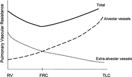
Increases in lung volume directly increase pulmonary vascular resistance by compressing the alveolar vessels (72,79,80). The actual mechanisms by which this occurs have not been completely resolved, but appear to reflect differential extraluminal pressure gradient–induced vascular compression. The pulmonary circulation can be conceptually viewed as existing in two distinct compartments based on the pressure outside the blood vessels, which will be either alveolar pressure (alveolar vessels) or extra-alveolar or ITP (extra-alveolar vessels) (79). The small pulmonary arterioles, venules, and alveolar capillaries sense alveolar pressure as their surrounding pressure while the large pulmonary arteries and veins, as well as the heart and intrathoracic great vessels of the systemic circulation, sense interstitial pressure or ITP as their surrounding pressure. Since alveolar pressure minus ITP is the transpulmonary pressure, and increasing lung volume requires transpulmonary pressure to rise, increases in lung volume augments the extraluminal pressure gradient from extra-alveolar vessels to alveolar vessels. Increases in lung volume progressively raise alveolar vessel resistance, becoming most noticeable above functional residual capacity (FRC) (75,84) (Fig. 127.1). Since the intraluminal pressure in the pulmonary arteries is generated by RV ejection relative to ITP, but the outside pressure of the alveolar vessels is alveolar pressure, if transpulmonary pressure exceeds intraluminal pulmonary arterial pressure, then the pulmonary vasculature will collapse where extra-alveolar vessels pass into alveolar loci, reducing the vasculature cross-sectional area and increasing pulmonary vascular resistance. Hyperinflation can create significant pulmonary hypertension and may precipitate acute RV failure (acute cor pulmonale) (85) and RV ischemia (70), especially in patients prone to hyperinflation (e.g., chronic obstructive pulmonary disease [COPD]). Thus, PEEP may increase pulmonary vascular resistance if it induces lung overdistention (86). Similarly, if lung volumes are reduced, increasing lung volume back to baseline levels by the use of PEEP decreases pulmonary vascular resistance by reversing hypoxic pulmonary vasoconstriction (87).
|
|
|
Figure 127.1. Schematic diagram of the relation between changes in lung volume and pulmonary vascular resistance, where the extra-alveolar and alveolar vascular components are separated. Note that pulmonary vascular resistance is minimal at resting lung volume or functional residual capacity (FRC). As lung volume increases toward total lung capacity (TLC) or decreases toward residual volume (RV), pulmonary vascular resistance also increases. However, the increase in resistance with hyperinflation is due to increased alveolar vascular resistance, whereas the increase in resistance with lung collapse is due to increased extra-alveolar vessel tone. |
Ventricular Interdependence
Although LV preload must eventually be altered by changes in RV output because the two ventricles are in series, changes in RV end-diastolic volume can also alter LV preload by altering LV diastolic compliance by the mechanism of ventricular interdependence (88). Ventricular interdependence functions through two separate processes. First, increasing RV end-diastolic volume will induce an intraventricular septal shift into the LV, decreasing LV diastolic compliance (89). Thus, for the same LV filling pressure, RV dilation will decrease LV end-diastolic volume and, therefore, cardiac output. Second, if pericardial restraint limits absolute biventricular filling, then RV dilation will increase Ppc without septal shift (2,90). This ventricular interaction is believed to be the major determinant of the phasic changes in arterial pulse pressure and stroke volume seen in tamponade, and is referred to as pulsus paradoxus. Pulsus paradoxus can be demonstrated during loaded spontaneous inspiration in normal subjects as an inspiration-associated decrease in pulse pressure. Pulse pressure is defined as systolic minus the diastolic blood pressure. If the pulse pressure change is greater than 10 mm Hg or 10% of the mean pulse pressure, then it is referred to as pulsus paradoxus (2). Maintaining a constant rate of venous return, either by volume resuscitation (91) or vasopressor infusion (27), will minimize hyperinflation-induced cardiac compression.
Hyperinflation-induced Cardiac Compression
As lung volume increases, the heart is compressed between the expanding lungs (92), raising juxtacardiac ITP. This compressive effect of the inflated lungs can be seen with either spontaneous (93) or positive pressure–induced hyperinflation (5,38,94,95,96). As described above, both Ppc and ITP are increased and no pericardial restraint exists. This decrease in apparent LV diastolic compliance (91) was previously misinterpreted as impaired LV contractility, because LV stroke work for a given LV end-diastolic pressure or pulmonary artery occlusion pressure is decreased (97,98). However, when such patients are fluid resuscitated to return LV end-diastolic volume to its original level, both LV stroke work and cardiac output also returned to their original levels (91,99) despite the continued application of PEEP (100).
Hemodynamic Effects of Changes in Intrathoracic Pressure
The heart within the thorax is a pressure chamber within a pressure chamber. Therefore, changes in ITP must affect the pressure gradients for both systemic venous return to the RV and systemic outflow from the LV, independent of the heart itself. Increases in ITP, by increasing right atrial pressure and decreasing transmural LV systolic pressure, will reduce the pressure gradients for venous return and LV ejection, decreasing intrathoracic blood volume. Conversely, decreases in ITP will augment venous return and impede LV ejection, increasing intrathoracic blood volume. Everything else below follows from these two simple truths.
Venous Return
Blood flows back from the systemic venous reservoirs into the right atrium through low-pressure, low-resistance venous conduits (101). Right atrial pressure is the back pressure for venous return. Ventilation alters both right atrial pressure and venous reservoir pressure. It is these changes in right atrial and venous capacitance vessel pressure that induce most of the observed cardiovascular effects of ventilation. Pressure in the upstream venous reservoirs is called mean systemic pressure, and is, itself, a function of blood volume, peripheral vasomotor tone, and the distribution of blood within the vasculature (102). Usually mean systemic pressure does not change rapidly during positive pressure ventilation, whereas right atrial pressure does owing to concomitant changes in ITP. Thus, variations in right atrial pressure represent the major factor determining the fluctuation in pressure gradient for systemic venous return during ventilation (103,104). The positive pressure inspiration increases in right atrial pressure decrease the pressure gradient for venous return, decreasing RV filling (68) and RV stroke volume (68,103,105,106,107,108,109,110,111,112,113). During normal spontaneous inspiration, the opposite occurs (2,27,68,69,107,110,114,115). The detrimental effect of positive pressure ventilation on cardiac output can be minimized by either fluid resuscitation to increase mean systemic pressure (27,105,116,117) or by keeping both mean ITP and swings in lung volume as low as possible. Accordingly, prolonging expiratory time, decreasing tidal volume, and avoiding PEEP all minimize this decrease in systemic venous return to the RV (4,24,103,107,108,109,110,111,118).
However, if positive pressure ventilation–induced increases in right atrial pressure always proportionally decreased venous return, then most mechanically ventilated patients would display profound cardiovascular insufficiency, especially at higher levels of PEEP. Fortunately, when lung volumes increase the diaphragm descend, compressing the abdominal compartment and increasing intra-abdominal pressure (119,120). Since a large proportion of venous blood exists in intra-abdominal vasculature, this venous blood is pressurized as well, increasing mean systemic pressure. Accordingly, the pressure gradient for venous return is often not reduced by PEEP (116). Inspiration-induced abdominal pressurization by diaphragmatic descent is probably the primary mechanism by which the decrease in venous return is minimized during positive pressure ventilation (121,122,123,124,125). However, laparotomy, by abolishing the inspiration-associated increases in intra-abdominal pressure, makes surgery patients especially sensitive to mechanical ventilation, requiring increased fluid resuscitation to sustain a constant cardiac output. This is one of the reasons why abdominal surgery patients often leave the operating room many liters positive.
Spontaneous inspiratory efforts usually increase venous return because of the combined decrease in right atrial pressure (2,26,108,109,110) and increase in intra-abdominal pressure (119,120). However, this augmentation of venous return is limited (126,127,128) because as ITP decreases below atmospheric pressure, central venous pressure also becomes subatmospheric, collapsing the great veins as they enter the thorax and creating a flow-limiting segment (101).
Ventricular Interdependence
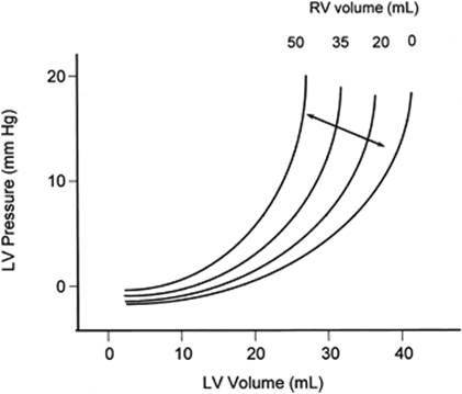
Changes in RV volume induce reciprocal changes in LV diastolic compliance. Although decreasing RV volume during positive pressure inspiration increases LV diastolic compliance by decreasing RV filling, the hemodynamic impact of this effect is usually minimal (88,127,128,129,130,131,132) (Fig. 127.2).
However, with spontaneous inspiration RV volumes increase, causing an immediate reduction in LV diastolic compliance. This process is the primary cause for the inspiration-associated decrease in LV stroke volume and pulse pressure (87,89,132,133). If the pulse pressure change is greater than 10 mm Hg or 10% of the mean pulse pressure, then it is referred to as pulsus paradoxus (2). Since spontaneous inspiratory efforts can also occur during positive pressure ventilation, the use of ventilation-associated pulse pressure variation during positive pressure ventilation can reflect ventricular interdependence. Presently, positive pressure–induced changes in pulse pressure and LV stroke volume have been advocated to be a useful parameter of preload responsiveness (134). However, to assess volume responsiveness using pulse pressure variation, it is essential that no spontaneous inspiratory efforts be present.
|
|
|
Figure 127.2. Schematic diagram of the effect of increasing right ventricular (RV) volumes on the left ventricular (LV) diastolic pressure–volume (filling) relationship. Note that increasing RV volumes decrease LV diastolic compliance, such that a higher filling pressure is required to generate a constant end-diastolic volume. (Adapted from Taylor RR, Corell JW, Sonnenblick EH, et al. Dependence of ventricular distensibility on filling the opposite ventricle. Am J Physiol. 1967;213:711–718.) |
LV Afterload
Changes in ITP can directly and indirectly alter LV afterload by altering both LV end-diastolic volume and ejection pressure. LV ejection pressure can be estimated as arterial pressure relative to ITP. Since baroreceptor mechanisms located in the extrathoracic carotid body maintain arterial pressure constant relative to atmosphere, if arterial pressure were to remain constant as ITP increased, then transmural LV pressure and thus LV afterload would decrease. Similarly, if transmural arterial pressure were to remain constant as ITP decreased, then LV wall tension would increase (135). Thus, under steady-state conditions increases in ITP decrease LV afterload and decreases in ITP increase LV afterload (136,137). The spontaneous inspiration–associated decrease in ITP-induced increase in LV afterload is one of the major mechanisms thought to be operative in the wean-induced LV ischemia described in the first part of this chapter since increased LV afterload must increase myocardial O2 consumption (MVO2). Thus, spontaneous ventilation not only increases global O2 demand by its exercise component (3,4,5), but also increases MVO2.
Profoundly negative swings in ITP commonly occur during forced spontaneous inspiratory efforts in patients with bronchospasm and obstructive breathing. This condition may rapidly deteriorate into acute heart failure and pulmonary edema (63) as has been described for airway obstruction (asthma, upper airway obstruction, vocal cord paralysis). Stiff lungs (interstitial lung disease, pulmonary edema, and ALI) selectively increase LV afterload and may be the cause of their LV failure and pulmonary edema (1,49,63,64), especially if LV systolic function is already compromised (13,138). Clearly, weaning from mechanical ventilation is a selective LV stress test (135,139,140). Similarly, improved LV systolic function is observed in patients with severe LV failure placed on mechanical ventilation if the mechanical breaths abolish negative swings in ITP (140).
The observed improvement in LV function seen with positive-pressure ventilation in subjects with severe heart failure is self-limited because venous return also decreases limiting total blood flow. However, the effect of removing large negative swings of ITP on LV performance will also act to reduce LV afterload but will not be associated with a change in venous return because until ITP becomes positive, venous return remains constant. Thus, removing negative ITP swings on LV afterload will selectively reduce LV afterload in a fashion analogous to increasing ITP but without the effect on cardiac output (27,101,141,142,143,144). This concept has been validated to be a very important clinical approach for patients with obstructive sleep apnea. For example, the cardiovascular benefits of positive airway pressure in nonintubated patients can be seen with CPAP therapy (145,146). Even low levels of CPAP, if they inhibit airway obstruction, will be beneficial (147,148,149). Prolonged nighttime nasal CPAP can selectively improve respiratory muscle strength, as well as LV contractile function if the patients had pre-existent heart failure (150,151). These benefits are associated with reductions of serum catecholamine levels (152).
Using Heart–Lung Interactions to Diagnose Cardiovascular Insufficiency
Since the cardiovascular response to positive pressure breathing is determined by the baseline cardiovascular state, ventilation-associated changes in arterial pulse pressure and stroke volume should monitor dynamic changes in venous return and the responsiveness of the heart to these transient and cyclic changes in preload (153). Systolic pressure variations during positive pressure ventilation nicely describe both preload responsiveness if the systolic pressure decreases below an apneic baseline value and also predict heart failure with volume overload if the systolic pressure increases above apneic baseline values (154,155,156,157). However, it is often difficult to assess if the variation in systolic arterial pressure is primarily up or down in clinical settings. A more physiologic approach is to measure arterial pulse pressure and assess pulse pressure variation (134,158). This technique can be modified to assess stroke volume variation (159) and has profound clinical potential as newer monitoring devices allow for the bedside display of both pulse pressure and stroke volume variation. In subjects on controlled mechanical ventilation, a pulse pressure variation of >13% or a stroke volume variation of >10% accurately predict preload responsiveness. This novel and exciting application of heart–lung interactions has been validated in many studies and is presently being assessed in prospective clinical trials, Assuming that this practical application of heart–lung interactions becomes commonplace, then a basic understanding of the principals described in this chapter will be an essential part of the training of acute care physicians.
References
1. Bromberger-Barnea B. Mechanical effects of inspiration on heart functions: a review. Fed Proc. 1981;40:2172–2177.
2. Wise RA, Robotham JL, Summer WR. Effects of spontaneous ventilation on the circulation. Lung. 1981;159:175–192.
3. Roussos C, Macklem PT. The respiratory muscles. N Engl J Med. 1982;307:786–797.
4. Grenvik A. Respiratory, circulatory and metabolic effects of respiratory treatment. A clinical study in postoperative thoracic surgical patients. Acta Anaesth Scand. 1966;(19 Suppl):1–122.
5. Shuey CB, Pierce AK, Johnson RL. An evaluation of exercise tests in chronic obstructive lung disease. J Appl Physiol. 1969;27:256–261.
6. Stock MC, David DW, Manning JW, et al. Lung mechanics and oxygen consumption during spontaneous ventilation and severe heart failure. Chest. 1992;102:279–283.
7. Kawagoe Y, Permutt S, Fessler HE. Hyperinflation with intrinsic PEEP and respiratory muscle blood flow. J Appl Physiol. 1994;77:2440–2448.
8. Aubier M, Vires N, Sillye G, et al. Respiratory muscle contribution to lactic acidosis in low cardiac output. Am Rev Respir Dis. 1982;126:648–652.
9. Frazier SK, Stone KS, Schertel ER, et al. A comparison of hemodynamic changes during the transition from mechanical ventilation to T-piece, pressure support, and continuous positive airway pressure in canines. Biol Res Nurs. 2000;1:253–264.
10. Magder S, Erian R, Roussos C. Respiratory muscle blood flow in oleic acid-induced pulmonary edema. J Appl Physol. 1986;60:1849–1856.
11. Vires N, Sillye G, Rassidakis A, et al. Effect of mechanical ventilation on respiratory muscle blood flow during shock. Physiologist. 1980;23:1–8.
12. Baratz DM, Westbrook PR, Shah K, et al. Effects of nasal continuous positive airway pressure on cardiac output and oxygen delivery in patients with congestive heart failure. Chest. 1992;102:1397–1401.
13. Lemaire F, Teboul JL, Cinoti L, et al. Acute left ventricular dysfunction during unsuccessful weaning from mechanical ventilation. Anesthesiology. 1988;69:171–179.
14. Richard C, Teboul JL, Archambaud F, et al. Left ventricular dysfunction during weaning in patients with chronic obstructive pulmonary disease. Intensive Care Med. 1994;20:171–172.
15. Hurford WE, Lynch KE, Strauss HW, et al. Myocardial perfusion as assessed by thallium-201 scintigraphy during the discontinuation of mechanical ventilation in ventilator-dependent patients. Anesthesiology. 1991;74:1007–1016.
16. Abalos A, Leibowitz AB, Distefano D, et al. Myocardial ischemia during the weaning period. Am J Crit Care. 1992;1:32–36.
17. Chatila W, Ani S, Guaglianone D, et al. Cardiac ischemia during weaning from mechanical ventilation. Chest. 1996;109:1421–1422.
18. Srivastava S, Chatila W, Amoateng-Adjepong Y, et al. Myocardial ischemia and weaning failure in patients with coronary disease: an update. Crit Care Med. 1999;27:2109–2112.
19. Mohsenifar Z, Hay A, Hay J, et al. Gastric intramural pH as a predictor of success or failure in weaning patients from mechanical ventilation. Ann Intern Med. 1993;119:794–798.
20. Jabran A, Mathru M, Dries D, et al. Continuous recordings of mixed venous oxygen saturation during weaning from mechanical ventilation and the ramifications thereof. Am J Respir Crit Care Med. 1998;158:1763–1769.
21. Straus C, Lewis B, Isebey D, et al. Contribution of the endotracheal tube and the upper airway to breathing workload. Am J Respir Crit Care Med. 1998;157:23–30.
22. Rasanen J, Nikki P, Heikkila J. Acute myocardial infarction complicated by respiratory failure. The effects of mechanical ventilation. Chest. 1984;85:21–28.
23. Rasanen J, Vaisanen IT, Heikkila J, et al. Acute myocardial infarction complicated by left ventricular dysfunction and respiratory failure. The effects of continuous positive airway pressure. Chest. 1985;87:156–162.
24. Cournaud A, Motley HL, Werko L, et al. Physiologic studies of the effect of intermittent positive pressure breathing on cardiac output in man. Am J Physiol. 1948;152:162–174.
25. Tyberg JV, Grant DA, Kingma I, et al. Effects of positive intrathoracic pressure on pulmonary and systemic hemodynamics. Repsir Physiol. 2000;119:171–179.
26. Milic-Emili J, Mead J, Turner JM. Improved method for assessing the validity of the esophageal balloon technique. J Appl Physiol. 1964;19:207–211.
27. Braunwald E, Binion JT, Morgan WL, et al. Alterations in central blood volume and cardiac output induced by positive pressure breathing and counteracted by metaraminol (Aramine). Circ Res. 1957;5:670–675.
28. Whittenberger JL, McGregor M, Berglund E, et al. Influence of state of inflation of the lung on pulmonary vascular resistance. J Appl Physiol. 1960;15:878–882.
29. Novak RA, Matuschak GM, Pinsky MR. Effect of ventilatory frequency on regional pleural pressure. J Appl Physiol. 1988;65:1314–1323.43. Taha BH, Simon PM, Dempsey JA, et al.
30. Kingma I, Smiseth OA, Frais MA, et al. Left ventricular external constraint: relationship between pericardial, pleural and esophageal pressures during positive end-expiratory pressure and volume loading in dogs. Ann Biomed Eng. 1987;15:331–346.
31. Tsitlik JE, Halperin HR, Guerci AD, et al. Augmentation of pressure in a vessel indenting the surface of the lung. Ann Biomed Eng. 1987;15:259–284.
32. Pinsky MR, Guimond JG. The effects of positive end-expiratory pressure on heart-lung interactions. J Crit Care. 1991;6:1–11.
33. Romand JA, Shi W, Pinsky MR. Cardiopulmonary effects of positive pressure ventilation during acute lung injury. Chest. 1995;108:1041–1048.
34. O'Quinn RJ, Marini JJ, Culver BH, et al. Transmission of airway pressure to pleural pressure during lung edema and chest wall restriction. J Appl Physiol. 1985;59:1171–1177.
35. Gattinoni L, Mascheroni D, Torresin A, et al. Morphological response to positive end-expiratory pressure in acute respiratory failure. Intensive Care Med. 1986;12:137–142.
36. Globits S, Burghuber OC, Koller J, et al. Effect of lung transplantation on right and left ventricular volumes and function measured by magnetic resonance imaging. Am J Respir Crit Care Med. 1994;149:1000–1004.
37. Scharf SM, Ingram RH Jr. Effects of decreasing lung compliance with oleic acid on the cardiovascular response to PEEP. Am J Physiol. 1977;233:H635–H641.
38. Pinsky MR, Vincent JL, DeSmet JM. Estimating left ventricular filling pressure during positive end-expiratory pressure in humans. Am Rev Respir Dis. 1991;143:25–31.
39. Teboul JL, Pinsky MR, Mercat A, et al. Estimating cardiac filling pressure in mechanically ventilated patients with hyperinflation. Crit Care Med. 2000;28:3631–3636.
40. Glick G, Wechsler AS, Epstein DE. Reflex cardiovascular depression produced by stimulation of pulmonary stretch receptors in the dog. J Clin Invest. 1969;48:467–472.
41. Painal AS. Vagal sensory receptors and their reflex effects. Physiol Rev. 1973;53:59–88.
42. Anrep GV, Pascual W, Rossler R. Respiratory variations in the heart rate, I: the reflex mechanism of the respiratory arrhythmia. Proc R Soc Lond B Biol Sci. 1936;119:191–217.
43. Respiratory sinus arrhythmia in humans: an obligatory role for vagal feedback from the lungs. J Appl Physiol. 1995;78:638–645.
44. Bernardi L, Calciati A, Gratarola A, et al. Heart rate-respiration relationship: computerized method for early detection of cardiac autonomic damage in diabetic patients. Acta Cardiol. 1986;41:197–206.
45. Persson MG, Lonnqvist PA, Gustafsson LE. Positive end-expiratory pressure ventilation elicits increases in endogenously formed nitric oxide as detected in air exhaled by rabbits. Anesthesiology. 1995;82:969–974.
46. Cassidy SS, Eschenbacher WI, Johnson RL Jr. Reflex cardiovascular depression during unilateral lung hyperinflation in the dog. J Clin Invest. 1979;64:620–626.
47. Daly MB, Hazzledine JL, Ungar A. The reflex effects of alterations in lung volume on systemic vascular resistance in the dog. J Physiol (London). 1967;188:331–351.
48. Shepherd JT. The lungs as receptor sites for cardiovascular regulation. Circulation. 1981;63:1–10.
49. Stalcup SA, Mellins RB. Mechanical forces producing pulmonary edema in acute asthma. N Engl J Med. 1977;297:592–596.
50. Vatner SF, Rutherford JD. Control of the myocardial contractile state by carotid chemo- and baroreceptor and pulmonary inflation reflexes in conscious dogs. J Clin Invest. 1978;63:1593–1601.
51. Karlocai K, Jokkel G, Kollai M. Changes in left ventricular contractility with the phase of respiration. J Auton Nerv Syst. 1998;73:86–92.
52. Said SI, Kitamura S, Vreim C. Prostaglandins: release from the lung during mechanical ventilation at large tidal ventilation. J Clin Invest. 1972;51:83a.
53. Bedetti C, Del Basso P, Argiolas C, Carpi A. Arachidonic acid and pulmonary function in a heart-lung preparation of guinea-pig. Modulation by PCO2. Arch Int Pharmacodyn Ther. 1987;285:98–116.
54. Berend N, Christopher KL, Voelkel NF. Effect of positive end-expiratory pressure on functional residual capacity: role of prostaglandin production. Am Rev Respir Dis. 1982;126:641–647.
55. Pattern MY, Liebman PR, Hetchman HG. Humorally mediated decreases in cardiac output associated with positive end-expiratory pressure. Microvasc Res. 1977;13:137–144.
56. Berglund JE, Halden E, Jakobson S, et al. PEEP ventilation does not cause humorally mediated cardiac output depression in pigs. Intensive Care Med. 1994;20:360–364.
57. Fuhrman BP, Everitt J, Lock JE. Cardiopulmonary effects of unilateral airway pressure changes in intact infant lambs. J Appl Physiol. 1984;56:1439–1448.
58. Payen DM, Brun-Buisson CJL, Carli PA, et al. Hemodynamic, gas exchange, and hormonal consequences of LBPP during PEEP ventilation. J Appl Physiol. 1987;62:61–70.
59. Frage D, de la Coussaye JE, Beloucif S, et al. Interactions between hormonal modifications during PEEP-induced antidiuresis and antinatriuresis. Chest. 1995;107:1095–1100.
60. Frass M, Watschinger B, Traindl O, et al. Atrial natriuretic peptide release in response to different positive end-expiratory pressure levels. Crit Care Med. 1993;21:343–347.
61. Wilkins MA, Su XL, Palayew MD, et al. The effects of posture change and continuous positive airway pressure on cardiac natriuretic peptides in congestive heart failure. Chest. 1995;107:909–915.
62. Shirakami G, Magaribuchi T, Shingu K, et al. Positive end-expiratory pressure ventilation decreases plasma atrial and brain natriuretic peptide levels in humans. Anesth Analg. 1993;77:1116–1121.
63. Fletcher EC, Proctor M, Yu J, et al. Pulmonary edema develops after recurrent obstructive apneas. Am J Respir Crit Care Med. 1999;160:1688–1696.
64. Chen L, Shi Q, Scharf SM. Hemodynamic effects of periodic obstructive apneas in sedated pigs with congestive heart failure. J Appl Physiol. 2000;88:1051–1060.
65. Maughan WL, Shoukas AA, Sagawa K, et al. Instantaneous pressure-volume relationships of the canine right ventricle. Circ Res. 1979;44:309–315.
66. Sibbald WJ, Driedger AA. Right ventricular function in disease states: pathophysiologic considerations. Crit Care Med. 1983;11:339.
67. Piene H, Sund T. Does pulmonary impedance constitute the optimal load for the right ventricle? Am J Physiol. 1982;242:H154–H160.
68. Pinsky MR. Determinants of pulmonary arterial flow variation during respiration. J Appl Physiol. 1984;56:1237–1245.
69. Theres H, Binkau J, Laule M, et al. Phase-related changes in right ventricular cardiac output under volume-controlled mechanical ventilation with positive end-expiratory pressure. Crit Care Med. 1999;27:953–8.
70. Johnston WE, Vinten-Johansen J, Shugart HE, et al. Positive end-expiratory pressure potentates the severity of canine right ventricular ischemia-reperfusion injury. Am J Physiol. 1992;262:H168–H176.
71. Madden JA, Dawson CA, Harder DR. Hypoxia-induced activation in small isolated pulmonary arteries from the cat. J Appl Physiol. 1985;59:113–118.
72. Hakim TS, Michel RP, Chang HK. Effect of lung inflation on pulmonary vascular resistance by arterial and venous occlusion. J Appl Physiol. 1982;53:1110–1115.
73. Quebbeman EJ, Dawson CA. Influence of inflation and atelectasis on the hypoxic pressure response in isolated dog lung lobes. Cardiovas Res. 1976;10:672–677.
74. Brower RG, Gottlieb J, Wise RA, et al. Locus of hypoxic vasoconstriction in isolated ferret lungs. J Appl Physiol. 1987;63:58–65.
75. Hakim TS, Michel RP, Minami H, et al. Site of pulmonary hypoxic vasoconstriction studied with arterial and venous occlusion. J Appl Physiol. 1983;54:1278–1302.
76. Marshall BE, Marshall C. A model for hypoxic constriction of the pulmonary circulation. J Appl Physiol. 1988;64:68–77.
77. Marshall BE, Marshall C. Continuity of response to hypoxic pulmonary vasoconstriction. J Appl Physiol. 1980;49:189–196.
78. Dawson CA, Grimm DJ, Linehan JH. Lung inflation and longitudinal distribution of pulmonary vascular resistance during hypoxia. J Appl Physiol. 1979;47:532–536.
79. Howell JBL, Permutt S, Proctor DF, et al. Effect of inflation of the lung on different parts of the pulmonary vascular bed. J Appl Physiol. 1961;16:71–76.
80. West JB, Dollery CT, Naimark A. Distribution of blood flow in isolated lung; relation to vascular and alveolar pressures. J Appl Physiol. 1964;19:713–724.
81. Fuhrman BP, Everitt J, Lock JE. Cardiopulmonary effects of unilateral airway pressure changes in intact infant lambs. J Appl Physiol. 1984;56:1439–1448.
82. Fuhrman BP, Smith-Wright DL, Kulik TJ, et al. Effects of static and fluctuating airway pressure on the intact, immature pulmonary circulation. J Appl Physiol. 1986;60:114–122.
83. Thorvalson J, Ilebekk A, Kiil F. Determinants of pulmonary blood volume. Effects of acute changes in airway pressure. Acta Physiol Scand. 1985;125:471–479.
84. Lopez-Muniz R, Stephens NL, Bromberger-Barnea B, et al. Critical closure of pulmonary vessels analyzed in terms of Starling resistor model. J Appl Physiol. 1968;24:625–635.
85. Block AJ, Boyson PG, Wynne JW. The origins of cor pulmonale, a hypothesis. Chest. 1979;75:109–114.
86. Vieillard-Baron A, Loubieres Y, Schmitt JM, et al. Cyclic changes in right ventricular output impedance during mechanical ventilation. J Appl Physiol. 1999;87:1644–1650.
87. Canada E, Benumnof JL, Tousdale FR. Pulmonary vascular resistance correlated in intact normal and abnormal canine lungs. Crit Care Med. 1982;10:719–723.
88. Taylor RR, Corell JW, Sonnenblick EH, et al. Dependence of ventricular distensibility on filling the opposite ventricle. Am J Physiol. 1967;213:711–718.
89. Brinker JA, Weiss I, Lappe DL, et al. Leftward septal displacement during right ventricular loading in man. Circulation. 1980;61:626–633.
90. Takata M, Harasawa Y, Beloucif S, et al. Coupled vs. uncoupled pericardial restraint: effects on cardiac chamber interactions. J Appl Physiol. 1997;83:1799–1813.
91. Marini JJ, Culver BN, Butler J. Mechanical effect of lung distention with positive pressure on cardiac function. Am Rev Respir Dis. 1980;124:382–386.
92. Butler J. The heart is in good hands. Circulation. 1983;67:1163–1168.
93. Cassidy SS, Wead WB, Seibert GB, et al. Changes in left ventricular geometry during spontaneous breathing. J Appl Physiol. 1987;63:803–811.
94. Hoffman EA, Ritman EL. Heart-lung interaction: effect on regional lung air content and total heart volume. Ann Biomed Eng. 1987;15:241–257.
95. Olson LE, Hoffman EA. Heart-lung interactions determined by electron beam X-ray CT in laterally recumbent rabbits. J Appl Physiol. 1995;78:417–427.
96. Jayaweera AR, Ehrlich W. Changes of phasic pleural pressure in awake dogs during exercise: potential effects on cardiac output. Ann Biomed Eng. 1987;15:311–318.
97. Cassidy SS, Robertson CH, Pierce AK, et al. Cardiovascular effects of positive end-expiratory pressure in dogs. J Appl Physiol. 1978;4:743–749.
98. Conway CM. Hemodynamic effects of pulmonary ventilation. Br J Anaesth. 1975;47:761–766.
99. Jardin F, Farcot JC, Boisante L. Influence of positive end-expiratory pressure on left ventricular performance. N Engl J Med. 1981;304:387–392.
100. Berglund JE, Halden E, Jakobson S, et al. Echocardiographic analysis of cardiac function during high PEEP ventilation. Intensive Care Med. 1994;20:174–180.
101. Guyton AC, Lindsey AW, Abernathy B, et al. Venous return at various right atrial pressures and the normal venous return curve. Am J Physiol. 1957;189:609–615.
102. Goldberg HS, Rabson J. Control of cardiac output by systemic vessels: circulatory adjustments of acute and chronic respiratory failure and the effects of therapeutic interventions. Am J Cardiol. 1981;47:696.
103. Pinsky MR. Instantaneous venous return curves in an intact canine preparation. J Appl Physiol. 1984;56:765–771.
104. Kilburn KH. Cardiorespiratory effects of large pneumothorax in conscious and anesthetized dogs. J Appl Physiol. 1963;18:279–283.
105. Chevalier PA, Weber KC, Engle JC, et al. Direct measurement of right and left heart outputs in Valsalva-like maneuver in dogs. Proc Soc Exper Biol Med. 1972;139:1429–1437.
106. Guntheroth WC, Gould R, Butler J, et al. Pulsatile flow in pulmonary artery, capillary and vein in the dog. Cardiovascular Res. 1974;8:330–337.
107. Guntheroth WG, Morgan BC, Mullins GL. Effect of respiration on venous return and stroke volume in cardiac tamponade. Mechanism of pulsus paradoxus. Circ Res. 1967;20:381–390.
108. Guyton AC. Effect of cardiac output by respiration, opening the chest, and cardiac tamponade. In: Circulatory Physiology: Cardiac Output and Its Regulation. Philadelphia, PA: WB Saunders; 1963:378–386.
109. Holt JP. The effect of positive and negative intrathoracic pressure on cardiac output and venous return in the dog. Am J Physiol. 1944;142:594–603.
110. Morgan BC, Abel FL, Mullins GL, et al. Flow patterns in cavae, pulmonary artery, pulmonary vein and aorta in intact dogs. Am J Physiol. 1966;210: 903–909.
111. Morgan BC, Martin WE, Hornbein TF, et al. Hemodynamic effects of intermittent positive pressure respiration. Anesthesiology. 1960;27:584–590.
112. Scharf SM, Brown R, Saunders N, et al. Hemodynamic effects of positive pressure inflation. J Appl Physiol. 1980;49:124–131.
113. Jardin F, Vieillard-Baron A. Right ventricular function and positive-pressure ventilation in clinical practice: from hemodynamic subsets to respirator settings. Intensive Care Med. 2003;29:1426–1434.
114. Scharf SM, Brown R, Saunders N, et al. Effects of normal and loaded spontaneous inspiration on cardiovascular function. J Appl Physiol. 1979;47:582–590.
115. Groeneveld AB, Berendsen RR, Schneider AJ, et al. Effect of the mechanical ventilatory cycle on thermodilution right ventricular volumes and cardiac output. J Appl Physiol. 2000;89:89–96.
116. Van den Berg P, Jansen JRC, Pinsky MR. The effect of positive-pressure inspiration on venous return in volume loaded post-operative cardiac surgical patients. J Appl Physiol. 2002;92:1223–1231.
117. Magder S, Georgiadis G, Cheong T. Respiratory variation in right atrial pressure predict the response to fluid challenge. J Crit Care. 1992;7:76–85.
118. Harken AH, Brennan MF, Smith N, et al. The hemodynamic response to positive end-expiratory ventilation in hypovolemic patients. Surgery. 1974;76:786–793.
119. Fessler HE, Brower RD, Wise RA, et al. Effects of positive end-expiratory pressure on the canine venous return curve. Am Rev Respir Dis. 1992;146:4–10.
120. Takata M, Robotham JL. Effects of inspiratory diaphragmatic descent on inferior vena caval venous return. J Appl Physiol. 1992;72:597–607.
121. Matuschak GM, Pinsky MR, Rogers RM. Effects of positive end-expiratory pressure on hepatic blood flow and hepatic performance. J Appl Physiol. 1987;62:1377–1383.
122. Chihara E, Hasimoto S, Kinoshita T, et al. Elevated mean systemic filling pressure due to intermittent positive-pressure ventilation. Am J Physiol. 1992;262:H1116–H1121.
123. Takata M, Wise RA, Robotham JL. Effects of abdominal pressure on venous return: abdominal vascular zone conditions. J Appl Physiol. 1990;69:1961–1972.
124. Barnes GE, Laine GA, Giam PY, et al. Cardiovascular responses to elevation of intra-abdominal hydrostatic pressure. Am J Physiol. 1985;248:R208–R213.
125. Lichtwarck-Aschoff M, Zeravik J, Pfeiffer UJ. Intrathoracic blood volume accurately reflects circulatory volume status in critically ill patients with mechanical ventilation. Intensive Care Med. 1992;18:142–145.
126. Brecher GA, Hubay CA. Pulmonary blood flow and venous return during spontaneous respiration. Circ Res. 1955;3:210–214.
127. Terada N, Takeuchi T. Postural changes in venous pressure gradients in anesthetized monkeys. Am J Physiol. 1993;264:H21–H25.
128. Scharf S, Tow DE, Miller MJ, et al. Influence of posture and abdominal pressure on the hemodynamic effects of Mueller's maneuver. J Crit Care. 1989;4:26–34.
129. Rankin JS, Olsen CO, Arentzen CE, et al. The effects of airway pressure on cardiac function in intact dogs and man. Circulation. 1982;66:108–120.
130. Robotham JL, Rabson J, Permutt S, et al. Left ventricular hemodynamics during respiration. J Appl Physiol. 1979;47:1275–1303.
131. Ruskin J, Bache RJ, Rembert JC, et al. Pressure-flow studies in man: effect of respiration on left ventricular stroke volume. Circulation. 1973;48:79–85.
132. Olsen CO, Tyson GS, Maier GW, et al. Dynamic ventricular interaction in the conscious dog. Circ Res. 1983;52:85–104.
133. Janicki JS, Weber KT. The pericardium and ventricular interaction, distensibility and function. Am J Physiol. 1980;238:H494–H503.
134. Michard F, Boussat S, Chemla D, et al. Relation between respiratory changes in arterial pulse pressure and fluid responsiveness in septic patients with acute circulatory failure. Am J Respir Crit Care Med. 2000;162:134–138.
135. Beyar R, Goldstein Y. Model studies of the effects of the thoracic pressure on the circulation. Ann Biomed Eng. 1987;15:373–383.
136. Buda AJ, Pinsky MR, Ingels NB, et al. Effect of intrathoracic pressure on left ventricular performance. N Engl J Med. 1979;301:453–459.
137. Pinsky MR, Summer WR, Wise RA, et al. Augmentation of cardiac function by elevation of intrathoracic pressure. J Appl Physiol. 1983;54:950–955.
138. Beach T, Millen E, Grenvik A. Hemodynamic response to discontinuance of mechanical ventilation. Crit Care Med. 1973;1:85–90.
139. Cassidy SA, Wead WB, Seibert GB, et al. Geometric left-ventricular responses to interactions between the lung and left ventricle: positive pressure breathing. Ann Biomed Eng. 1987;15:285–295.
140. Scharf SM, Brown R, Warner KG, et al. Intrathoracic pressure and left ventricular configuration with respiratory maneuvers. J Appl Physiol. 1989;66:481–491.
141. Sharpey-Schaffer EP. Effects of Valsalva maneuver on the normal and failing circulation. Br Med J. 1955;1:693–699.
142. Khilnani S, Graver LM, Balaban K, et al. Effects of inspiratory loading on left ventricular myocardial blood flow and metabolism. J Appl Physiol. 1992;72:1488–1492.
143. Garpestad E, Parker JA, Katayzama H, et al. Decrease in ventricular stroke volume at apnea termination is independent of oxygen desaturation. J Appl Physiol. 1994;77:1602–1608.
144. Sibbald WH, Calvin J, Driedger AA. Right and left ventricular preload, and diastolic ventricular compliance: implications of therapy in critically ill patients. In: Critical Care State of the Art. Fullerton, CA: Society of Critical Care; 1982:Vol. 3.
145. De Hoyos A, Liu PP, Benard DC, et al. Haemodynamic effects of continuous positive airway pressure in humans with normal and impaired left ventricular function. Clin Sci (Lond). 1995;88:173–178.
146. Naughton MT, Rahman MA, Hara K, et al. Effect of continuous positive airway pressure on intrathoracic and left ventricular transmural pressures in patients with congestive heart failure. Circulation. 1995;91:1725–1731.
147. Joet-Philip FF, Paganelli FF, Dutau HL, et al. Hemodynamic effects of bi-level nasal positive airway pressure ventilation in patients with heart failure. Respiration. 1999;66:136–143.
148. Lin M, Yang Y-F, Chiang H-T, et al. Reappraisal of continuous positive airway pressure therapy in acute cardiogenic pulmonary edema. Chest. 1995;107:1379–1386.
149. Buckle P, Millar T, Kryger M. The effect of short-term nasal CPAP on Cheyne-Stokes respiration in congestive heart failure. Chest. 1992;102:31–35.
150. Granton JT, Naughton MT, Benard DC, et al. CPAP improves inspiratory muscle strength in patients with heart failure and central sleep apnea. Am J Respir Crit Care Med. 1996;153:277–282.
151. Kaneko Y, Floras JS, Usui K, et al. Cardiovascular effects of continuous positive airway pressure in patients with heart failure and obstructive sleep apnea. N Engl J Med. 2003;348:1233–1241.
152. Naughton MT, Benard DC, Liu PP, et al. Effects of nasal CPAP on sympathetic activity in patients with heart failure and central sleep apnea. Am J Respir Crit Care Med. 1995;152:473–479.
153. Denault AY, Gasior TA, Gorcsan J 3rd, et al. Determinants of aortic pressure variation during positive-pressure ventilation in man. Chest. 1999;116:176–86.
154. Abel JG, Salerno TA, Panos A, et al. Cardiovascular effects of positive pressure ventilation in humans. Ann Thorac Surg. 1987;43:36–43.
155. Baeaussier M, Coriat P, Perel A, et al. Determinants of systolic pressure variation in patients ventilated after vascular surgery. J Cardiothorac Vasc Anesth. 1995;9:547–551.
156. Coriat P, Vrillon M, Perel A, et al. A comparison of systolic blood pressure variations and echocardiographic estimates of end-diastolic left ventricular size in patients after aortic surgery. Anesth Analg. 1994;78:46–53.
157. Szold A, Pizov R, Segal E, Perel A. The effect of tidal volume and intravascular volume state on systolic pressure variation in ventilated dogs. Intensive Care Med. 1989;15:368–371.
158. Michard F, Boussat S, Chemla D, et al. Relation between respiratory changes in arterial pulse pressure and fluid responsiveness in septic patients with acute circulatory failure. Am J Respir Crit Care Med. 2000;162(1):134–138.
159. Monnet X, Rienzo M, Osman D, et al. Esophageal Doppler monitoring predicts fluid responsiveness in critically ill ventilated patients. Intensive Care Med. 2005;31(9):1195–1201.