Andreas H. Kramer
Thomas P. Bleck
Definitions
Acute hypertension is a common issue in the intensive care unit (ICU). The settings in which blood pressure elevation occur are highly variable, and optimal care must be tailored to the pathophysiology of the specific circumstances in which it is encountered. The terminology used in the literature to classify this heterogeneous group of disorders has been somewhat inconsistent and confusing (Table 126.1). The terms, hypertensive emergency or hypertensive crisis, are commonly defined as a marked increase in blood pressure associated with target-organ damage, implying that the blood pressure should be lowered emergently.
Some authors have reserved the definition of hypertensive emergency for the situation where blood pressure elevation itself is directly responsible for causing end-organ damage. However, clinicians more often need to rapidly lower blood pressure in situations where hypertension, although not necessarily directly responsible for causing the condition, may contribute to deterioration. For example, acute hypertension is usually the result of, rather than the immediate cause of, an acute ischemic stroke. If the patient is to be treated with thrombolytics, it becomes imperative to maintain the blood pressure within certain narrow limits to minimize the risk of hemorrhagic transformation while at the same time not compromising cerebral blood flow (CBF). Thus, in this chapter, we define a hypertensive emergency broadly—as any condition in which blood pressure should be lowered immediately. Although the term, malignant hypertension, has been discouraged by some, it is still widely used in the literature to describe the syndrome where organ dysfunction is a direct consequence of the elevated blood pressure, rather than an epiphenomenon. The presence of papilledema is not necessarily required for this diagnosis to be made (1,2).
In contrast, a hypertensive urgency is defined as a condition with severe blood pressure elevation and no target-organ damage, such that the blood pressure can be decreased more gradually over the course of several hours, often with oral medications. It is therefore the presence or absence of organ dysfunction, rather than the absolute degree of blood pressure elevation, that determines whether a patient is classified as having a hypertensive emergency or urgency. It is not always clear how clinicians distinguish between hypertensive urgencies and the situation where a patient simply has severe, poorly controlled, chronic hypertension. The most recent Joint National Committee Guidelines (JNV 7) classify patients with hypertension into stage 1 (systolic blood pressure [SBP] 140–159 mm Hg or diastolic blood pressure [DBP] 90–99 mm Hg) and stage 2 (SBP exceeding 160 mm Hg or DBP exceeding 100 mm Hg). The previously used category of “stage 3 hypertension” (SBP exceeding 180 mm Hg or DBP exceeding 110 mm Hg) has been combined with stage 2 (3).
Epidemiology and Etiology
Hypertension is extraordinarily common, with over 1 billion individuals affected worldwide, and only a minority of these having adequate blood pressure control (4). The incidence of hypertensive emergencies and urgencies has not been assessed in population-based studies. Less than 1% of persons with chronic hypertension ever present in this fashion (2). Nevertheless, because hypertension is so common, and because such a wide variety of conditions other than malignant hypertension can be categorized as hypertensive emergencies, acutely elevated blood pressure is still a factor in a substantial number of medical visits to emergency departments and a frequent problem in the ICU (5,6) (Table 126.2).
Malignant Hypertension
The peak incidence of malignant hypertension occurs between the ages of 40 and 50, and risk factors include poor long-term blood pressure control, lack of a primary care physician, noncompliance with antihypertensive medications, male gender, African American ethnicity, illicit drug use, and lower socioeconomic class (7,8,9,10). Prior to the availability of effective antihypertensive therapy, the mortality of malignant hypertension was very high, with approximately 80% of patients dying within a year (hence, the term malignant) (11).
At least 90% to 95% of patients with chronically elevated blood pressure can be classified as having “essential” hypertension, meaning that the underlying cause is multifactorial and not specifically known. A small proportion of patients have “secondary” hypertension, where there is an identifiable and sometimes treatable condition that is responsible for raising blood pressure (3). In contrast, among patients who present with malignant hypertension, as many as 50% to 80% may have a secondary etiology (12). Other clues that should alert clinicians to the possibility of secondary hypertension include a history of blood pressure that is resistant to medical therapy, sudden worsening in a previously well-controlled patient, and the onset of hypertension at an unusually young or old age (13).
Renovascular disease, the most common cause of secondary hypertension, may be present in as many as 45% of patients with severe or malignant hypertension, although the proportion is higher in Caucasians than African Americans (14). Features that suggest renovascular hypertension include atherosclerotic vascular disease in other organ systems, systolic-diastolic abdominal bruits, a history of deterioration in renal function with exposure to angiotensin-converting enzyme (ACE) inhibitors or angiotensin II receptor blockers (ARBs), recurrent flash pulmonary edema, and small kidneys (determined by ultrasound or other imaging).
|
Table 126.1 Definitions |
|
|
Hypertension is almost universally present in patients with acute or chronic kidney disease, especially when the etiology is a glomerulonephropathy (15). Hypertension is a common manifestation of obstructive sleep apnea, and can be improved by the administration of noninvasive positive airway pressure (16). Various rare endocrine causes, including primary aldosteronism, Cushing syndrome, hypercalcemia, hypothyroidism, and pheochromocytoma, are also responsible for a small proportion of cases. Several illicit drugs can cause malignant hypertension in addition to other hypertensive emergencies. Sympathomimetics, such as cocaine and methamphetamine, have been implicated in causing intracerebral and subarachnoid hemorrhage, ischemic stroke, and aortic dissection (17,18,19). However, various drugs used in clinical practice have also been reported to cause severe hypertension, or even hypertensive emergencies. The most commonly implicated are erythropoietin and various immunosuppressants, most notably cyclosporine, tacrolimus, interferon, and high-dose corticosteroids, although it is sometimes difficult to separate the hypertension-inducing effects of these drugs from the complications of the diseases they are intended to treat (20,21). A careful history, physical examination, and appropriate diagnostic testing to exclude causes of secondary hypertension are indicated during the hypertensive emergency and after it has resolved (Table 126.3).
|
Table 126.2 Types of Hypertensive Emergencies |
||||||||||||||||||||||
|
||||||||||||||||||||||
|
Table 126.3 Etiologies of Malignant Hypertension |
||||||||||||||||||||||
|
Neurologic Hypertensive Emergencies
Acute stroke is one of the most common indications for which emergent blood pressure control may be necessary, and often the most frequent form of end-organ damage in a consecutive series of patients with hypertensive emergencies (5). Stroke is the second leading cause of death worldwide, and is also a major reason for long-term disability. Cerebral ischemia is responsible for 70% to 80% of strokes, while intracerebral hemorrhage (ICH) and subarachnoid hemorrhage account for 5% to 20% and 1% to 7%, respectively (22). Overall, more than 80% of patients with ischemic or hemorrhagic strokes are initially hypertensive to some degree (23). Even without treatment, the blood pressure usually declines gradually over the first several hours or days in-hospital. In the well-known National Institute of Neurological Diseases (NINDS) trial evaluating recombinant tissue plasminogen activator (rt-PA) for the management of acute ischemic stroke, 19% of patients had an initial blood pressure of more than 185/110 mm Hg, and 60% had a blood pressure of more than 180/105 mm Hg during the first 24 hours in-hospital (24). Whether or not it is appropriate (see below), more than half of these patients receive antihypertensive medications during the first few days of hospitalization (25). An even larger proportion of patients with intracerebral hemorrhage (ICH) develop, and are treated for, acute hypertension (26). The incidence of hypertension with subarachnoid hemorrhage (SAH) is somewhat lower, but aggressive treatment is sometimes advocated in order to minimize the initial risk of rebleeding from the aneurysm (27).
Perioperative Hypertension
Depending on the patient population, type of surgery, and how hypertension is defined, 3% to 34% of operations are complicated by postoperative hypertension in the recovery room or ICU, with the most important risk factors being inadequately controlled pain and a pre-existing history of hypertension, especially if antihypertensives were withdrawn preoperatively (28,29). Elevations in blood pressure occur most often during the initial 30 to 60 minutes after surgery, may last for several hours, and have been associated with higher rates of postoperative hemorrhage and myocardial ischemia in certain patient populations (30). Hypertension is particularly frequent following neurosurgical and cardiovascular procedures, occurring in approximately 54% to 91% and 30% to 80% of patients undergoing craniotomy and coronary artery bypass grafting, respectively (31,32). A common dilemma faced by anesthetists involves patients who present for surgery with poorly controlled blood pressure. Preoperative hypertension is predictive of intra- and postoperative blood pressure fluctuations, and has been associated with the occurrence of complications and worse outcomes in some studies (33,34). The American Heart Association recommends that nonemergent surgery be deferred if the blood pressure exceeds 180/110 mm Hg (35). However, it remains controversial whether delaying surgery ultimately modifies perioperative risks, with one randomized controlled trial arguing that it does not (36).
Pregnancy-induced Hypertension
Hypertension complicates approximately 12% of pregnancies, and is responsible for 18% of maternal deaths in the United States. When blood pressure elevation occurs prior to 20 weeks' gestation, it is considered chronic hypertension. If it occurs without complication after 20 weeks, it is referred to as gestational hypertension. In contrast, pre-eclampsia is a form of pregnancy-induced hypertension that is associated with vasoconstriction, endothelial dysfunction, platelet aggregation, and increased coagulation. Although it is usually defined by the concomitant presence of hypertension and proteinuria (more than 300 mg of protein per 24 hours), some patients can develop severe symptoms in the absence of proteinuria. Pre-eclampsia occurs in 2% to 10% of pregnancies, with important risk factors including nulliparity, antiphospholipid antibodies, diabetes mellitus, obesity, family history, multiple (twin) pregnancies, maternal age over 40, and a previous history during other pregnancies. Maternal complications can include progression to eclampsia, pulmonary edema, microangiopathy, and renal failure. The most common neonatal complications are prematurity and growth restriction. Eclampsia is defined as the development of severe neurologic manifestations, including seizures and a depressed level of consciousness, in women with pre-eclampsia (37,38).
Cardiovascular Hypertensive Emergencies
Acute heart failure is responsible for 5% to 10% of hospital admissions. Hospital mortality is about 4%, but increases to more than 50% by 1 year. In a large American registry, 73% of patients had a history of chronic hypertension, and 50% were hypertensive at admission (39). In patients presenting with flash pulmonary edema, acute hypertension is particularly common, and is likely to be both a consequence and contributing cause.
A history of chronic hypertension exists in about 40% to 70% of patients with acute coronary syndromes, and about 30% have an elevated blood pressure when initially assessed (40,41). Severe uncontrolled hypertension at admission (greater than 180/110 mm Hg) is a relative contraindication to thrombolysis for ST elevation myocardial infarction (STEMI) (42).
Aortic dissection is a relatively rare condition, with an annual incidence of about 3 to 4 cases per 100,000 persons per year. With modern medical and surgical therapy, the mortality has decreased from as high as 90% to about 20%–35%. More than 70% of patients have a history of chronic hypertension, but blood pressure can be highly variable at presentation. Most patients (70%) with type B dissections (descending aorta) have an admission systolic pressure of more than 150 mm Hg compared with just over a third of those with type A dissections (ascending aorta), of whom approximately 25% actually present with hypertension or in frank shock (43).
Pathophysiology of Hypertension-Induced End-Organ Dysfunction
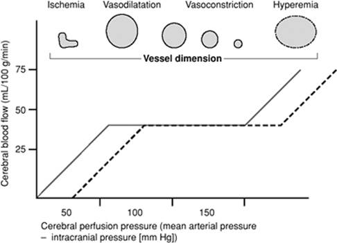
Blood flow to organs is kept relatively constant despite variations in blood pressure. This process is called autoregulation, and its limits are usually between mean arterial pressure (MAP) values of about 60 and 150 mm Hg. Increases in blood pressure induce arteriolar smooth muscle contraction and vasoconstriction, while reductions lead to vasodilatation. Extreme hypertension exceeding the upper range of autoregulation causes edema, hemorrhage, and organ dysfunction, while reductions in blood pressure beyond the lower limits of autoregulation result in tissue hypoperfusion and ischemia (Fig. 126.1). In addition to a widespread, systemic myogenic response, there are also more organ-specific vascular regulatory mechanisms to protect against the effects of acute hypertension. For example, increased delivery of filtrate to the distal nephron stimulates a tubuloglomerular feedback system that promotes afferent arteriolar vasoconstriction (44). The likelihood of end-organ damage increases not only with the absolute degree of blood pressure elevation, but also with the rate at which this occurs (1). With chronic hypertension, there is hypertrophy in the walls of small arteries and arterioles, and the autoregulation curve is shifted toward the right, such that blood flow can be maintained constant, even at unusually high blood pressures. Conversely, ischemia may occur when blood pressure falls to levels that would otherwise be well tolerated. In the setting of neurologic injury, autoregulation is often impaired, and cerebral blood flow becomes directly dependent on blood pressure (Fig. 126.1).
|
|
|
Figure 126.1. Cerebral blood flow autoregulation: Effect of changes in cerebral perfusion pressure on cerebral blood flow and vascular caliber in normal and hypertensive patients (dashed line). |
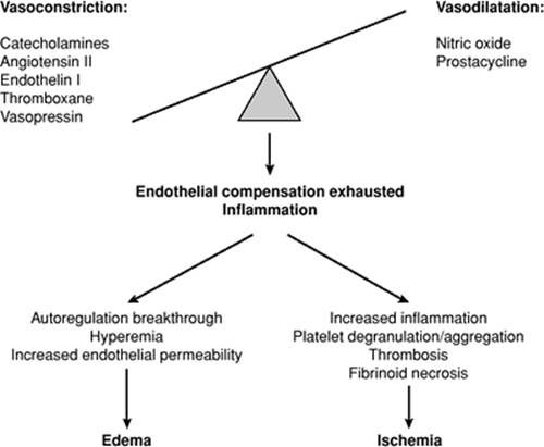
Normal endothelial function is necessary for the regulation of vascular tone, blood pressure, and regional blood flow. The endothelium is involved in maintaining a delicate balance between vasodilating substances (e.g., nitric oxide, bradykinin, prostacyclin) and vasoconstrictors (e.g., endothelin), as well as between coagulation and fibrinolysis. When blood pressure is elevated, natriuretic peptides are released from the endothelium, which in turn induce sodium and water loss, with decreased intravascular volume (1). Excessive activation of the renin-angiotensin system causes vasoconstriction and inflammation, and has been demonstrated to cause hypertensive emergencies in animal models, an effect that can be inhibited with the use of ACE inhibitors (45). Angiotensin II levels are elevated in most cases of malignant hypertension, particularly when the etiology is a renal condition (46). Increased wall stress and prolonged vasoconstriction in the face of severe hypertension eventually causes endothelial compensatory mechanisms to fail, such that a vicious cycle ensues, with consequent hyperemia, increased permeability, inflammation (endarteritis), platelet aggregation, coagulation, and thrombosis. Transmural necrosis and the entry of blood components into the vessel wall lead to obliteration of the lumen and replacement of smooth muscle with fibrous tissue, a process called fibrinoid necrosis (47). Patients with malignant hypertension may therefore paradoxically develop both hyperemia (due to endothelial dysfunction and loss of autoregulation) and ischemia (due to thrombosis and fibrinoid necrosis) (Fig. 126.2).
Manifestations
Hypertensive Encephalopathy/Reversible Posterior Leukoencephalopathy Syndrome
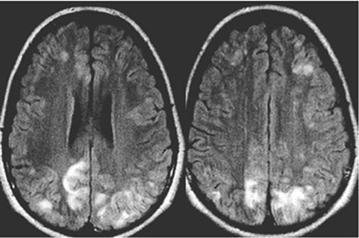
Severe elevations in blood pressure eventually cause a breakdown of the blood–brain barrier, with subsequent development of vasogenic cerebral edema. White matter is less tightly packed than the overlying cerebral cortex, making it more vulnerable to the spread of edema (48). Swelling occurs predominantly, but not exclusively, in the posterior regions of the brain. This is thought to be due to a larger concentration of sympathetic fibers around arterioles in the anterior brain, which results in a greater degree of vasoconstriction and relative protection against the effects of severe hypertension (49). The characteristic clinical features and magnetic resonance imaging (MRI) findings of vasogenic edema in posterior white matter led to the description of a clinical radiologic syndrome, now most commonly termed reversible posterior leukoencephalopathy syndrome (RPLS) or posterior reversible encephalopathy syndrome (PRES) (50). Although occurring most commonly in association with severe hypertension, there are other conditions that may at least predispose to, if not directly cause, the development of RPLS, perhaps because they induce endothelial toxicity. RPLS has been described in association with certain medications—most notably cyclosporine (21)—and other immunosuppressive agents, as well as in the setting of microangiopathies (51), connective tissue diseases, vasculitis, and pre-eclampsia (52). Although cerebral edema can sometimes be seen on computed tomography (CT) scans, RPLS is best visualized using T2 and fluid-attenuated inversion recovery (FLAIR) MRI sequences. Diffusion-weighted MRI has confirmed that vasogenic edema is much more prominent than cytotoxic edema and, although RPLS is usually “reversible,” some patients do develop ischemic strokes (53). Many patients do not adhere perfectly to the typical patterns of RPLS: gray matter involvement of the cerebral cortex and basal ganglia, as well as edema occurring in the frontal lobes, posterior fossa, and brainstem have all been described. It is rare for only one vascular territory to be involved (Fig. 126.3).
The clinical manifestations of RPLS in the setting of acute blood pressure elevation are collectively described by the term, hypertensive encephalopathy, which is characterized by the subacute development of neurologic signs and symptoms and may include headache, altered mental status, seizures, and visual disturbances (54,55). Headaches are usually generalized, severe, and poorly responsive to analgesics and improve rapidly with treatment of hypertension (56). Altered mental status can range from lethargy and confusion to stupor and coma, although the latter is unusual. The posterior predominance of RPLS is reflected by the frequent occurrence of unilateral or bilateral visual disturbances, including hemianopsia, visual neglect, cortical blindness, Anton syndrome (patient is not aware of blindness), and visual hallucinations. Focal seizures originating in the occipital regions have also been described, although they most often generalize, and may be recurrent (57).
|
|
|
Figure 126.2. Simplified pathophysiology of acute hypertension-induced organ dysfunction. (Adapted from Vaughan CJ, Delanty N. Hypertensive emergencies. Lancet. 2000;356:411–417.) |
Retinopathy
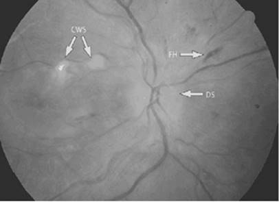
Endothelial damage and leakage of plasma proteins into the retina lead to edema and the formation of hard exudates (Fig. 126.4). Focal areas of ischemia and infarction within the nerve fiber layer cause white areas, called cotton-wool spots, to appear. Breakdown of the blood–retinal barrier results in the emergence of flame-shaped hemorrhages within the retina. The development of papilledema has historically been used to differentiate “accelerated” from “malignant” hypertension. However, the presence or absence of papilledema has little impact on the natural history and prognosis of hypertensive emergencies, nor should it significantly alter management (58). The mechanism of papilledema may include raised intracranial pressure (ICP), which is known to be present in some patients with hypertensive encephalopathy (59), as well as ischemia of the optic nerve head (60). It should be noted that ophthalmoscopic examination for hypertensive retinopathy has relatively high rates of inter- and intraobserver variability, particularly among nonophthalmologists (61).
|
|
|
Figure 126.3. Fluid-attenuated inversion recovery magnetic resonance imaging sequences of a 15-year-old patient with a history of Wegener granulomatosis presenting with seizures and altered mental status. The findings are consistent with reversible posterior leukoencephalopathy syndrome (RPLS). Although vasogenic edema is seen predominantly posteriorly, anterior changes are also present. |
Nephropathy and Microangiopathy
Certain conditions causing acute renal failure may cause hypertensive emergencies, but severely elevated blood pressure can also cause renal dysfunction, a condition called malignant nephrosclerosis. Renal biopsies reveal fibrinoid necrosis, hyperplastic arteriolitis, neutrophilic infiltration, and thrombosis of glomerular capillaries. The histologic appearance can be difficult to distinguish from other microangiopathies, like hemolytic uremic syndrome and thrombotic thrombocytopenic purpura (62). It is therefore not surprising that more than a quarter of patients with malignant hypertension, especially those with acute renal failure, have typical clinical features of microangiopathy, including thrombocytopenia, elevated lactate dehydrogenase, and schistocytes on blood smear (63). Impaired renal perfusion leads to greater activation of the renin-angiotensin system, which further augments vasoconstriction, fluid retention, and blood pressure elevation. The earliest evidence of renal involvement is the presence of abnormal urine sediment, with proteinuria, hematuria, and the appearance of red and white blood cell casts. This is followed by the development of acute renal failure, which is sometimes severe enough for patients to require dialysis, and occasionally results in end-stage renal disease. In one series, more than 20% of patients with malignant hypertension required dialysis (64), but this is more common when the etiology is secondary rather than essential hypertension, particularly with renal causes (12). The degree of renal dysfunction is an important prognostic variable (64).
|
|
|
Figure 126.4. Severe hypertensive retinopathy with evidence of disc swelling (DS, papilledema), cotton wools spots (CWS), and flame-shaped hemorrhage (FH). (From Wong T, Mitchell P. The eye in hypertension. Lancet. 2007;369:425–435, used with permission.) |
Cardiovascular Complications
Approximately 20% of patients with malignant hypertension present with cardiac complications, which may include myocardial ischemia or pulmonary edema (65). Many patients have a pre-existing history of chronic hypertension, and already have left ventricular hypertrophy and diastolic dysfunction, while a smaller subset also has impaired systolic function. Left ventricular hypertrophy increases myocardial oxygen requirements while also outstripping vascular supply (66) and compressing coronary arterioles. The increased afterload associated with severe hypertension further increases myocardial wall stress and oxygen demand, such that ischemia occurs, especially if there is also concomitant coronary artery disease. Increased wall thickness and changes to the extramyocardial collagen network impair myocardial relaxation, and cause pressure within the left ventricle to rise at relatively lower volumes during diastole. As a result, even small increases in intravascular volume and afterload can produce pulmonary edema (67).
Treatment
Pharmacologic Agents
An ideal pharmacologic agent (Table 126.4) for treatment of hypertensive emergencies should have a rapid onset in order to immediately reduce the progression of organ failure, but should also be short-acting and easy to titrate to avoid excessively lowering the blood pressure for long periods of time. The following agents are the most commonly used.
|
Table 126.4 Intravenous Pharmacologic Agents for Management of Hypertensive Emergencies |
||||||||||||||||||||||||||||||||||||||||||||||||||
|
||||||||||||||||||||||||||||||||||||||||||||||||||
Sodium Nitroprusside
Sodium nitroprusside (NTP) has been used in the management of hypertensive emergencies for over 40 years, and continues to be considered first-line therapy, largely because of its rapid onset, short duration of action, affordability, familiarity, and efficacy (1,68). NTP is a parenterally administered, potent arterial and venous vasodilator, which has an onset within less than 30 seconds and a duration of action of only 2 to 3 minutes, such that cessation of the infusion allows blood pressure to rise back to previous levels within 1 to 10 minutes. Cardiac output is either preserved or increased, and cardiac filling pressures decrease (69,70). The mechanism of action involves the release of nitric oxide into the bloodstream, activation of guanylate cyclase, and subsequent conversion of guanosine triphosphate (GTP) to cyclic guanosine monophosphate (cGMP) in vascular smooth muscle, which in turn inhibits the intracellular movement of calcium. The usual starting dose of NTP is 0.25 to 0.5 µg/kg/minute, and it is increased in increments of 0.5 µg/kg/minute every 5 to 10 minutes until the goal blood pressure is achieved.
NTP has the potential for several serious, life-threatening complications. First, despite lowering preload and afterload, NTP may induce coronary steal, whereby excessive vasodilatation of coronary arterioles shunts blood away from ischemic regions. This effect may help explain the findings of a large clinical trial in which patients with acute myocardial infarction and high left ventricular filling pressures had a worse outcome when treated with nitroprusside within the first 8 hours but improved outcomes thereafter (71).
Second, NTP is often avoided in neurologic emergencies because of the observation in several studies that it vasodilates cerebral vasculature, increases cerebral blood volume, and therefore raises ICP (72). The demonstration of this phenomenon is not entirely consistent in the literature (73), and it may be avoidable with a somewhat slower administration (74).
Third, each nitroprusside molecule has five cyanide moieties, such that cyanide makes up 44% of its molecular weight. Cyanide is metabolized by transulfuration in the liver to thiocyanate, which is 100 times less toxic than cyanide, and is cleared in the urine and stool. When this pathway is overwhelmed, cyanide accumulates and causes toxicity. Patients receiving NTP are particularly vulnerable when treated with prolonged, high-dose infusions, especially if hepatic and renal function are impaired, or if sulfur stores are depleted because of malnutrition (68,75). Cyanide blocks oxidative phosphorylation and essentially causes tissue anoxia and lactic acidosis despite adequate oxygen delivery and high venous oxygen saturation levels. Manifestations of cyanide poisoning include depressed mental status, seizures, and, eventually, bradycardia and hemodynamic collapse. Cyanide levels are not easily monitored, and the development of lactic acidosis is a late finding. The development of tachyphylaxis to nitroprusside, with consequent increasing dose requirements, may be a harbinger of toxicity (68,76).
Treatment of cyanide poisoning consists of discontinuation of nitroprusside, supportive care with the delivery of 100% oxygen, and the administration of sodium nitrite (300 mg IV), followed by sodium thiosulfate (12.5 g IV). The use of sodium nitrite is controversial, as it actually causes the production of methemoglobin, which, although also potentially toxic, binds avidly to cyanide. Sodium thiosulfate acts as a sulfur donor to promote the formation of thiocyanate, and has been used effectively as monotherapy. Hydroxycobalamin combines with cyanide to form cyanocobalamin, and may provide synergy with sodium thiosulfate (77). Both agents have been used prophylactically with infusions of nitroprusside to prevent cyanide accumulation (78,79). Accumulation of thiocyanate, although far less toxic than cyanide, may also cause complications (68,79).
Despite these concerns, NTP has an extensive track record, and many prospective studies have not found evidence of clinical toxicity even after using it for several days. Still, NTP should be administered at as low a dose as possible, and for as short a duration as possible. Newer, alternative agents are increasingly being used as they become available, largely because of concerns about cyanide toxicity with NTP.
Nitroglycerin
Because nitroglycerin produces more venous than arterial vasodilatation, it is usually not used as first-line therapy for hypertensive emergencies, unless there is concurrent pulmonary edema or myocardial ischemia. It is usually started at 5 to 10 µg/minute, and can be titrated up every 5 to 10 minutes to doses as high as 200 to 300 µg/minute. Particular caution must be exercised in the setting of hypovolemia. Patients using drugs for erectile dysfunction should not be given nitroglycerin or nitroprusside, as this may induce profound hypotension (80). As with nitroprusside, there are case reports of nitroglycerin increasing ICP, such that it should be used with caution in brain-injured patients (81).
β-Blockers
Labetalol is the most commonly used intravenous β-blocker for the management of hypertensive emergencies. One of its unique properties is that it blocks both α and β receptors, although the β-blocking effect is more prominent (82). Labetalol is usually administered as 10- to 20-mg boluses, which can be repeated every 15 minutes until the desired effect is achieved. Blood pressure lowering begins within 2 to 5 minutes, with maximal effect after about 10 to 15 minutes. Labetalol can also be delivered as an infusion, beginning at a dose of 1 to 2 mg/minute. It has been demonstrated to be safe and effective in the management of severe hypertension, with advantages including that it does not cause reflex tachycardia, and has little effect on cerebral blood flow or ICP (83). Labetalol does not cross the placenta well, and there is extensive experience using it in pregnancy, making it one of the preferred agents in the management of pre-eclampsia and eclampsia (84).
Esmolol is an extremely short-acting, relatively cardioselective β-blocker, has an onset within less than a minute, and is uniquely metabolized by red blood cell esterases, such that its duration of action is only 10 to 20 minutes. A loading dose of 0.5 mg/kg is administered over 1 minute, followed by an infusion of 50 µg/kg/minute, which can be adjusted as often as every 5 minutes to a maximum dose of 200 µg/kg/minute (85). It is a useful agent when blood pressure is elevated and cardiac output is preserved, especially when there are concerns about myocardial ischemia.
Nicardipine
Nicardipine is a dihydropyridine calcium channel blocker that inhibits calcium influx through L-type channels, thereby preventing smooth muscle contraction, particularly in vascular smooth muscle, rather than cardiac myocytes. Thus, nicardipine causes arterial vasodilatation, with minimal venodilatation or change in cardiac output. It is most often administered as an intravenous infusion, beginning at 3 to 5 mg/hour, to a maximum of 15 mg/hour, but it can also be given as 0.5- to 3-mg boluses. Because of a distribution half-life of less than 3 minutes and an intermediate elimination half-life of less than 45 minutes, nicardipine has a relatively rapid onset and offset (86). It has compared favorably with NTP in clinical trials, with therapeutic targets achieved in a similar amount of time, fewer episodes of severe hypotension, and less frequent dose adjustment (86,87). Nicardipine has been used in a variety of settings, including malignant hypertension, perioperative hypertension, pre-eclampsia, and acute heart failure. It is generally well tolerated, with few adverse effects when used with caution. Given that nicardipine increases cerebral blood flow, one might expect that it would raise ICP. Although this has been reported, it does not appear to be a major concern in most studies (86,88).
Fenoldopam
Fenoldopam is a selective dopamine-1 (DA-1)-receptor agonist, which produces peripheral, renal, splanchnic, and, to a lesser degree, coronary vasodilatation. Stimulation of DA-1 receptors also promotes natriuresis. Unlike dopamine, fenoldopam does not have any effect on DA-2 receptors, nor does it act at the α- or β-adrenergic receptor level. It does not cross the blood–brain barrier, and therefore has little effect on cerebral blood flow and ICP. It is typically administered in doses of 0.025 to 1 µg/kg/minute, and begins to lower blood pressure after as little as 2 minutes, although the maximal effect is not seen for at least 20 to 30 minutes or more. With discontinuation of an infusion, the elimination half-life is less than 10 minutes. Reflex tachycardia, thought to be related to activation of the baroreflex, may occur and can be attenuated with concomitant use of a β-blocker (89). A theoretical advantage of fenoldopam is improved renal blood flow, but despite a slight increase in creatinine clearance, it remains unclear that this will translate into clinically important prevention of renal failure (90). Fenoldopam has been demonstrated to be effective at controlling blood pressure in hypertensive emergencies, with similar efficacy to nitroprusside (91), but has shown no clear improvement in outcomes; it is also considerably more expensive (92). Overall, fenoldopam seems to have few adverse effects. It does raise intraocular pressure, and should therefore probably be avoided in patients with a known history of glaucoma (93).
Treatment for Specific Hypertensive Emergencies
Malignant Hypertension
Because of the shift in the autoregulatory curve that occurs with prolonged hypertension, rapid reductions in blood pressure may cause organ ischemia (94). With hypertensive urgencies, the blood pressure should therefore be reduced carefully and gradually with oral medications over the course of several days. It is not certain how long it takes for the autoregulatory curve to recover and shift back toward the left, such that the initial goal should never be a normal blood pressure.
With hypertensive emergencies, the blood pressure must be reduced immediately, with an initial goal of no more than a 15% reduction. Specific treatment goals should be individualized to ensure that the pressure is reduced sufficiently for organ failure to resolve without compromising perfusion. To facilitate keeping blood pressure within a narrow range, placement of an arterial catheter and careful observation in the ICU are recommended.
Neurologic
Acute Ischemic Stroke
Although acute hypertension is common in patients with ischemic stroke, optimal treatment remains uncertain. The greatest priority in these patients is to preserve as much of the ischemic penumbra as possible. Because autoregulation is usually impaired within the penumbra, pressure reductions may cause blood flow to fall, which in turn may increase infarct size (95). Accordingly, there are several observational studies demonstrating that lowering blood pressure may cause clinical deterioration (96). Preliminary studies have even suggested that transcranial Doppler middle cerebral artery (MCA) flow velocities, cerebral perfusion as determined by MRI, and neurologic examination may improve with supranormal augmentation of blood pressure using a vasopressor (97). The International Stroke Trial of more than 17,000 patients found a U-shaped relationship, where the best outcomes occurred in patients with a presenting systolic blood pressure of about 150 mm Hg (98). The American Stroke Association guidelines currently recommend not treating hypertension unless the systolic pressure exceeds 220 mm Hg or the diastolic pressure exceeds 120 mm Hg (99).
The exception to this rule is if the patient is a candidate for intravenous, or possibly intra-arterial, thrombolysis. Of patients who receive intravenous rt-PA for treatment of acute ischemic stroke, approximately 5% to 6% will develop ICH (100). Whether lowering blood pressure helps to limit this risk is not certain. However, patients in whom systolic and diastolic pressure could not easily be lowered to less than 185 mm Hg and 110 mm Hg, respectively, were excluded from the definitive clinical trial (101). Patients who did receive thrombolysis had a goal blood pressure of less than 180/105 mm Hg. Approximately 10% of patients require intravenous antihypertensive therapy prior to receiving thrombolysis, while 25% to 30% require treatment in the 24 hours after thrombolysis (24).
Intracerebral Hemorrhage
Good neurologic recovery is very uncommon if the volume of ICH exceeds 30 to 60 mL. Hematoma growth is a dynamic process that occurs over several hours, with a significant proportion of patients having detectable expansion even within the first few hours after presentation to the emergency department or ICU (102). Patients with early hematoma enlargement have substantially worse outcomes, and it is likely that therapy that can limit this early growth will improve outcomes. Numerous studies have suggested that patients with higher blood pressure at presentation are more likely to develop hematoma expansion (103) and have a higher mortality (104). However, a randomized controlled trial of lowering blood pressure for acute ICH has not yet been performed. At present, the American Heart Association guidelines are conservative, and recommend the following goals: MAP less than 130 mm Hg, SBP less than 180 mm Hg, and DBP less than 105 mm Hg (105). The main reason for concern has been the observation that there is reduced blood flow around areas of ICH and the belief that this represents an ischemic penumbra that could be compromised with lower blood pressure. However, recent studies using positive emission tomography and MRI have suggested that decreased flow surrounding ICH is appropriate for the corresponding reduction in cerebral metabolic rate (106). Furthermore, CBF autoregulation in this region appears to be preserved, such that lowering blood pressure does not adversely impact regional blood flow (107).
Subarachnoid Hemorrhage
Nontraumatic subarachnoid hemorrhage results from a ruptured intracranial aneurysm in more than 80% of cases (27). The risk of recurrent hemorrhage from the aneurysm is about 4% on the first day, followed by 1% to 2% per day for the next 2 to 3 weeks (108). Although the risk of rebleeding has decreased with the practice of securing aneurysms early, with either surgical clipping or endovascular coil embolization, the incidence of this devastating complication remains approximately 7%, with a mortality of over 50% (109). Many centers attempt to decrease the risk of recurrent hemorrhage by reducing blood pressure prior to the aneurysm being secured, with review articles often citing goal systolic pressures of less than 140 to 180 mm Hg (27). However, the efficacy of this practice remains uncertain, with conflicting results from observational studies. Particular caution should be exercised when treating patients with high-grade SAH who are stuporous or comatose, because these patients may have a raised ICP, and excessive reduction in blood pressure may compromise cerebral perfusion and promote ischemia.
After the aneurysm has been treated, the most concerning complication is the development of cerebral vasospasm and delayed ischemic neurologic deficits, which occur in approximately 20% to 30% of patients, with maximum risk between days 4 and 12 after the event. Hypertension should not routinely be treated during this time, even at relatively high levels, since this practice increases the risk of delayed infarction. If clinical vasospasm develops, first-line therapy actually involves hemodynamic augmentation, using vasopressors to raise blood pressure in order to increase CBF and improve cerebral perfusion.
Perioperative Neurosurgery
Hypertension occurs in the setting of craniotomy more often than with any other type of surgery (31). Neurosurgery results in the release of large amounts of vasoactive substances that raise blood pressure, including catecholamines, endothelin, and renin (110). Importantly, severe hypertension occurring intraoperatively or during the first 12 postoperative hours has been associated with a higher risk of ICH-complicated craniotomy (31).
There are certain neurosurgical procedures where tight postoperative blood pressure control is particularly important. In most patients undergoing carotid stenting or endarterectomy, the sudden resolution of carotid stenosis results in a sudden, asymptomatic, 20% to 40% increase in ipsilateral CBF. However, in some patients, the increase can be much more profound, to the degree that it overcomes the autoregulatory capacity of the corresponding, previously hypoperfused territory. The resulting cerebral hyperperfusion syndrome (CHS) is characterized by the presence of vasogenic edema, which resembles RPLS in that there is a posterior predilection. Patients who are most vulnerable are those with relatively severe carotid stenosis and poor collateral circulation, but postoperative hypertension is also an important risk factor. CHS is treated with tight blood pressure control, but agents that cause cerebral vasodilatation, including nitroprusside and perhaps calcium channel blockers, ideally should be avoided (111).
Mild intraoperative hypotension is sometimes used for vascular neurosurgical procedures, such as microsurgical excision or endovascular obliteration of arteriovenous malformations (AVMs) in order to reduce the risk of hemorrhage. With AVMs, the sudden “repressurization” of previously hypotensive arterioles may contribute to the development of regional hyperemia, edema, and bleeding, a condition sometimes referred to as “normal perfusion pressure breakthrough.” Consequently, hypertension should be avoided in the immediate postoperative period. Conversely, the sacrifice of vascular branches during the procedure, or vasospasm from surgical manipulation and retraction, may also create areas of relative underperfusion, such that hypotension would also be deleterious (112).
Cardiovascular Hypertensive Emergencies
Aortic Dissection
The purpose of emergently lowering blood pressure in aortic dissection is to decrease shear stress in the aorta and limit propagation of the intimal tear and false lumen. In order to concomitantly reduce both blood pressure and the force of left ventricular contraction, first-line therapy consists of a β-blocker or, if contraindicated, a calcium channel blocker with negative inotropic and chronotropic properties (e.g., diltiazem or verapamil). Pure vasodilators should not be used in isolation. If possible, the heart rate should be lowered to less than 60 beats per minute, and the blood pressure reduced as much as can be tolerated, ideally below a systolic pressure of 120 mm Hg. Because patients often have substantial chest discomfort, the use of opiates to ameliorate pain may greatly reduce antihypertensive requirements. If the blood pressure remains elevated despite adequate β-blockade, a vasodilator can be added. There is extensive experience with nitroprusside in this setting, although other agents have also been used (86,113).
Acute Pulmonary Edema
Acute hypertension exists in the majority of patients presenting with flash pulmonary edema, and is likely to be both a consequence and contributing cause (39,114). The venodilating properties of nitroglycerin make it an excellent initial choice as an antihypertensive in both normotensive and hypertensive patients, especially if there is failure to improve after administration of a loop diuretic. In titrating the dose of intravenous nitroglycerin, clinicians should be aware that relatively large doses, often in excess of 100 µg/minute, may be required to significantly lower cardiac filling pressures and improve symptoms (115). Patients who benefit from aggressive afterload reduction, such as those with acute aortic or mitral regurgitation (if not hypotensive), may require a more potent arterial vasodilator than nitroglycerin, such as nitroprusside or nicardipine. Although ACE inhibitors are standard care for chronic heart failure, there is little evidence of benefit for acute decompensated heart failure. The only intravenous preparation, enalaprilat, was potentially harmful when routinely administered to patients within the first 24 hours following STEMI, and is therefore not recommended (116). Considerable caution must be exercised when intubating hypertensive patients with acute pulmonary edema, since it is very common for blood pressure to drop precipitously with sedation and positive pressure ventilation. Another agent that has been proposed for use in acute heart failure is nesiritide (recombinant human brain natriuretic peptide). However, it has not been demonstrated to improve clinically important outcomes when compared with standard therapy (117), may worsen renal function (118), and has been linked to a possible increased risk of short-term death (119).
Acute Coronary Syndromes
Although definitive therapy for acute coronary syndromes (ACSs) involves revascularization, tachycardia and hypertension increase myocardial oxygen requirements, and can potentially worsen ischemia and increase infarct size. β-Blockers are recommended for all ACS patients without a contraindication. An intravenous β-blocker is therefore a good initial choice as an antihypertensive (42). Intravenous β-blockers must be used with caution in patients with reduced left ventricular systolic function and in those in whom there is concern about impaired cardiac conduction (e.g. inferior myocardial infarction). A large clinical trial of early β-blockade in patients with STEMI has demonstrated an increased risk of cardiogenic shock (41). If the blood pressure remains significantly elevated, especially with ongoing chest pain or pulmonary edema, then nitroglycerin should be used. In addition to reducing preload and afterload, nitrates also vasodilate coronary arteries, especially at the site of plaque disruption. If three sublingual tablets (0.4 mg over 5 minutes) are ineffective, then an intravenous infusion should be started and adjusted to alleviate chest pain and reduce blood pressure (10% reduction in normotensive patients, 30% reduction in hypertensive patients) (42).
Pre-eclampsia and Eclampsia
Although hypertensive encephalopathy and eclampsia have largely been considered separate entities, they have a similar pathophysiology and essentially the same MRI findings (RPLS) (120). Definitive treatment for severe pre-eclampsia and eclampsia is delivery, but careful intravenous blood pressure control is frequently also necessary.
Treatment of hypertension in severe pre-eclampsia has not been shown to improve perinatal outcomes, and may actually contribute to a decrease in neonatal birth weight (121). Thus, pharmacologic therapy is not recommended unless the degree of blood pressure elevation is severe (defined as SBP exceeding 160 mm Hg or DBP exceeding 105–110 mm Hg) or there are end-organ complications. Oral medications, with a goal pressure of 140 to 155/90 to 105 mm Hg, may be sufficient in the absence of organ dysfunction, but more rapid and tighter control is necessary for severe pre-eclampsia and eclampsia. Although there has been extensive experience with intravenous hydralazine (5–10 mg every 15–20 minutes to a maximum dose of 30 mg), this agent has a relatively slow onset, has not commonly been used as an infusion, may overshoot blood pressure goals, and has recently been linked to worse outcomes, including more placental abruption, adverse effects on fetal heart rate, lower Apgar scores, and a greater need for cesarean section (122). Other intravenous agents that have been successfully and safely used include labetalol and nicardipine (84,86). In addition to the above, magnesium sulfate should be given to prevent the development of seizures in patients with severe pre-eclampsia (123), and should be used to treat acute seizures when eclampsia occurs (124).
Summary
The occurrence of acute hypertension in critically ill patients is not uncommon and can have serious consequences with regard to outcome. Careful evaluation of the underlying causes is important in selecting the best treatment options. Furthermore, the severity of the condition is determined by the magnitude of the acute increase in blood pressure from baseline more than by the absolute blood pressure level. Thus, in the presence of primarily normotensive baseline values (such as those in pre-eclampsia), a minor increase of blood pressure may lead to a life-threatening condition. Organ manifestations in the course of a hypertensive emergency concern the cardiovascular system and brain, and greatly affect therapeutic options and goals. With few exceptions from the rule (aortic dissection or severe pulmonary edema), the patient's blood pressure should be reduced in a stepwise approach and with precision by intravenous medications rapidly delivered, all the while monitoring the cardiovascular and central nervous systems. The selection of the antihypertensive agent, therefore, depends on the existing organ failure as well as its reliable effectiveness.
References
1. Vaughan CJ, Delanty N. Hypertensive emergencies. Lancet. 2000;356:411–417.
2. Calhoun DA, Oparil S. Treatment of hypertensive crisis. N Engl J Med. 1990;323:1177–1183.
3. Chobanian AV, Bakris GL, Black HR, et al. The seventh report of the Joint National Committee on prevention, detection, evaluation, and treatment of high blood pressure: the JNC 7 report. JAMA. 2003;289:2560–2572.
4. Hajjar I, Kotchen TA. Trends in prevalence, awareness, treatment, and control of hypertension in the United States, 1988–2000. JAMA. 2003;290:199–206.
5. Zampaglione B, Pascale C, Marchisio M, et al. Hypertensive urgencies and emergencies. Prevalence and clinical presentation. Hypertension. 1996;27:144–147.
6. Karras DJ, Ufberg JW, Heilpern KL. Elevated blood pressure in urban emergency department patients. Acad Emerg Med. 2005;12:835–843.
7. Bennett NM, Shea S. Hypertensive emergency: case criteria, sociodemographic profile, and previous care of 100 cases. Am J Public Health. 1988; 78:636–640.
8. Shea S, Misra D, Ehrlich MH, et al. Predisposing factors for severe, uncontrolled hypertension in an inner-city minority population. N Engl J Med. 1992;327:776–781.
9. Bender SR, Fong MW, Heitz S, et al. Characteristics and management of patients presenting to the emergency department with hypertensive urgency. J Clin Hypertens. 2006;8:12–18.
10. Tisdale JE, Huang MB, Borzak S. Risk factors for hypertensive crisis: importance of out-patient blood pressure control. Fam Pract. 2004;2:420–424.
11. Keith NM, Wagener HP, Kernohan JW. The syndrome of malignant hypertension. Arch Intern Med. 1928;41:141–188.
12. Scarpelli PT, Livi R, Caselli GM, et al. Accelerated (malignant) hypertension: a study of 121 cases between 1974 and 1996. J Nephrol. 1997;10:207–215.
13. Hemmelgran BR, Zarnke KB, Campbell NR, et al. The 2004 Canadian hypertension education program recommendations for the management of hypertension: part I—blood pressure measurement, diagnosis and assessment of risk. Can J Cardiol. 2004;20:31–40.
14. Mann SJ, Pickering TG. Detection of renovascular hypertension. State of the art: 1992. Ann Intern Med. 1992;117:845–853.
15. Buckalw VM Jr, Berg RL, Wang SR, et al. Prevalence of hypertension in 1795 subjects with chronic renal disease: the modification of diet in renal disease study baseline cohort. Modification of Diet in Renal Disease Study Group. Am J Kidney Dis. 1996;28:811–821.
16. Becker HF, Jerrentrup A, Ploch T, et al. Effect of nasal continuous positive airway pressure treatment on blood pressure in patients with obstructive sleep apnea. Circulation. 2003;107:68–73.
17. Levine SR, Brust JC, Futrell N, et al. Cerebrovascular complications of the use of the “crack” form of alkaloidal cocaine. N Engl J Med. 1990;323:699–704.
18. Green RM, Kell KM, Gabrielsen T, et al. Multiple intracerebral hemorrhages after smoking “crack” cocaine. Stroke. 1990;21:957–962.
19. Hsue PY, Salinas CL, Bolger AF, et al. Acute aortic dissection related to crack cocaine. Circulation. 2002;105:1592–1595.
20. Novak BL, Force RW, Mumford BT, et al. Erythropoietin-associated hypertensive posterior leukoencephalopathy. Neurology. 1997;49:686–689.
21. Schwartz RB, Bravo SM, Klufas RA, et al. Cyclosporine neurotoxicity and its relationship to hypertensive encephalopathy: CT and MR findings in 16 cases. Am J Roentgenol. 1995;165:627–631.
22. Feigin VL, Lawes CM, Bennett DA, et al. Stroke epidemiology: a review of population-based studies of incidence, prevalence and case fatality in the late 20th century. Lancet Neurol. 2003;2:43–53.
23. Wallace JD, Levy LL. Blood pressure after stroke. JAMA. 1981;246:2177–2180.
24. Brott T, Lu M, Kothari R, et al. Hypertension and its treatment in the NINDS rt-PA stroke trial. Stroke. 1998;29:1504–1509.
25. Lindenauer PK, Mathew MC, Ntuli TS, et al. Use of antihypertensive agents in the management of patients with acute ischemic stroke. Neurology. 2004;63:318–323.
26. Jauch EC, Lindsell CJ, Adeoye O, et al. Lack of evidence for an association between hemodynamic variables and hematoma growth in spontaneous intracerebral hemorrhage. Stroke 2006;37:2061–2065.
27. Suarez J, Tarr RW, Selman WR. Aneurysmal subarachnoid hemorrhage. N Engl J Med. 2006;354:387–396.
28. Gal TJ, Cooperman LH. Hypertension in the immediate postoperative period. Br J Anaesth. 1975;47:70–74.
29. Kutz JD, Cronau LH, Barash PG. Postoperative hypertension hazard of abrupt cessation of antihypertensive medication in preoperative period. Am Heart J. 1976;92:79.
30. Towne JB, Bernhard VM. The relationship of postoperative hypertension to complications following carotid endarterectomy. Surgery. 1980;88:575–580.
31. Basali A, Mascha EJ, Kalfas I, et al. Relation between perioperative hypertension and intracranial hemorrhage after craniotomy. Anesthesiology. 2000;93:48–54.
32. Leslie JB. Incidence and etiology of perioperative hypertension. Acta Anaesthesiol Scand Suppl. 1993;99:5–9.
33. Prys-Roberts C, Meloche R, Foex P. Studies of anesthesia in relation to hypertension. I: cardiovascular responses of treated and untreated patents. Br J Anesth. 1971;43:122.
34. Aronson S, Boisvert D, Lapp W. Isolated systolic hypertension is associated with adverse outcomes from coronary artery bypass grafting surgery. Anesth Analg. 2002;94:1079–1084.
35. Eagle KA, Berger PB, Calkins H, et al. ACC/AHA guideline update for perioperative cardiovascular evaluation for noncardiac surgery—executive summary: A report of the American College of Cardiology/American Heart Association Task Force on practice guidelines (committee to update the 1996 guidelines on perioperative cardiovascular evaluation for noncardiac surgery). Circulation. 2002;105:1257–1267.
36. Weksler N, Klein M, Szendro G, et al. The dilemma of immediate preoperative hypertension: to treat and operate, or to postpone surgery? J Clin Anesth. 2003;15:179–183.
37. Vidaeff A, Carroll MA, Ramin SM. Acute hypertensive emergencies in pregnancy. Crit Care Med. 2005;33:S307–S312.
38. Sibai B, Dekker G, Kupfermink M. Pre-eclampsia. Lancet. 2005;365:785–799.
39. Adams Jr KF, Fonarow GC, Emerman CL, et al. Characteristics and outcomes of patients hospitalized for heart failure in the United States: rationale, design, and preliminary observations from the first 100,000 cases in the Acute Decompensated Heart Failure National Registry (ADHERE). Am Heart J. 2005;149:209–216.
40. Fifth Organization to Assess Strategies in Acute Ischemic Syndromes Investigators; Yusuf S, Mehta SR, Chrolvaicius S, et al. Comparison of fondaparinux and enoxaparin in acute coronary syndromes. N Engl J Med. 2006;354:1464–1476.
41. Chen ZM, Pan HC, Chen YP, et al. Early intravenous then oral metoprolol in 45,852 patients with acute myocardial infarction: a randomised placebo-controlled trial. Lancet. 2005;366:1622–1632.
42. Antman EM, Anbe DT, Armstrong PW, et al. ACC/AHA guidelines for the management of patients with ST elevation myocardial infarction: a report of the American College of Cardiology/American Heart Association Task Force on Practice Guidelines. Circulation. 2004;110:e82– 292.
43. Hagan PG, Nienaber CA, Isselbacher EM, et al. The international registry of acute aortic dissection (IRAD). JAMA. 2000;283:897–903.
44. Louzenhiser R, Griffin KA, Bidani AK. Systolic blood pressure as the trigger for the renal myogenic response: protective or autoregulatory? Curr Opin Nephrol Hypertens. 2006;15:41–49.
45. Montgomery HE, Kiernan LA, Whitworth CE, et al. Inhibition of tissue angiotensin converting enzyme activity prevents malignant hypertension in TGR(mREN2)27. J Hypertens. 1998;16:635–643.
46. Davies DL, Schalekamp MA, Beevers DG, et al. Abnormal relationship between exchangeable sodium and the renin-angiotensin system in malignant hypertension. Lancet. 1973;1:683–687.
47. Fleming S. Malignant hypertension—the role of the paracrine renin-angiotensin system. J Pathol. 2000;192:135–139.
48. Kalimo H, Fredriksson K, Nordberg C, et al. The spread of brain injury in hypertensive injury. Med Biol. 1986;64:133–137.
49. Beausang-Linder M, Bill A. Cerebral circulation in acute arterial hypertension—protective effects of sympathetic nervous activity. Acta Physiol Scand. 1981;111:193–199.
50. Hinchey J, Chaves C, Appignani B, et al. A reversible posterior leukoencephalopathy syndrome. N Engl J Med. 1996;334:494–500.
51. Bakshi R, Shaikh ZA, Bates VE, et al. Thrombotic thrombocytopenic purpura. Brain CT and MRI findings in 12 patients. Neurology. 1999;52:1285–1288.
52. Schwartz RB, Feske SK, Polak JK, et al. Preeclampsia-eclampsia: clinical and neuroradiologic correlates and insights into the pathogenesis of hypertensive encephalopathy. Radiology. 2000;217:371–376.
53. Covarrubius DJ, Luetmer PH, Campeau NG. Posterior reversible encephalopathy syndrome: prognostic utility of quantitative diffusion-weighted MR images. Am J Neuroradiol. 2002;23:1038–1048.
54. Chester EM, Agamanolis DP, Banker BQ, et al. Hypertensive encephalopathy: a clinicopathologic study of 20 cases. Neurology. 1978;28:928–939.
55. Healton EB, Brust JC, Feinfeld DA, et al. Hypertensive encephalopathy and the neurologic manifestations of malignant hypertension. Neurology. 1982;32:127–132.
56. Stott VL, Hurrell MA, Anderson TJ. Reversible posterior leukoencephalopathy syndrome: a misnomer reviewed. Intern Med J. 2005;35:83–90.
57. Bakshi R, Bates VE, Mechtler LL, et al. Occipital lobe seizures as the major clinical manifestation of reversible posterior leukoencephalopathy syndrome: magnetic resonance imaging findings. Epilepsia. 1998;39:295– 299.
58. Ahmed ME, Walker JM, Beevers DG, et al. Lack of difference between malignant and accelerated hypertension. BMJ. 1986;292:235–237.
59. Griswold WR, Viney J, Mendoza SA, et al. Intracranial pressure monitoring in severe hypertensive encephalopathy. Crit Care Med. 1981;9:573–576.
60. Hammond S, Wells JR, Marcus DM, et al. Ophthalmoscopic findings in malignant hypertension. J Clin Hypertens (Greenwich). 2005;8:222–223.
61. Dimmitt SB, West JN, Eames SM, et al. Usefulness of ophthalmoscopy in mild to moderate hypertension. Lancet. 1989;1:1103–1106.
62. Alpers CE. The kidney. In: Kumar V, Fausto N, Abbas A, eds. Robbins and Cotran: Pathological Basis of Disease. 7th ed. Philadelphia: Saunders; 2005.
63. van den Born BJH, Honnebier UPF, Koopmans RP, et al. Microangiopathy hemolysis and renal failure in malignant hypertension. Hypertension. 2004;45:246.
64. James SH, Meyers AM, Milne FJ, et al. Partial recovery of renal function in black patients with apparent end-stage renal failure due to primary malignant hypertension. Nephron. 1995;71:29–34.
65. Lip GY, Beevers M, Beevers G. The failure of malignant hypertension to decline: a survey of 24 years' experience in a multiracial population in England. J Hypertens. 1994;12:1297–1305.
66. Nixenberg A, Antony I. Epicardial coronary arteries are not adequately sized in hypertensive patients. J Am Coll Cardiol. 1996;27:1115–1123.
67. Aurigemma GP, Gaasch WH. Diastolic heart failure. N Engl J Med. 2005; 351:1097–1105.
68. Friederich JA, Butterworth JF. Sodium nitroprusside: twenty years and counting. Anesth Analg. 1995;81:152–162.
69. Armstrong PW, Walker DC, Burton JR, et al. Vasodilator therapy in acute myocardial infarction. A comparison of sodium nitroprusside and nitroglycerin. Circulation. 1975;52:1118–1122.
70. Campbell CS, Wallwork J, Taylor KM, et al. The hemodynamic effects of sodium nitroprusside following cardiopulmonary bypass: a clinical study. J Cardiovasc Surg. 1982;23:41–48.
71. Cohn JN, Franciosa JA, Francis GS, et al. Effect of short-term infusion of sodium nitroprusside on mortality rate in cute myocardial infarction complicated by left ventricular failure: results of a Veterans Administration cooperative study. N Engl J Med. 1982;306:1129–1135.
72. Davis RF, Douglas ME, Heenan TJ, et al. Brain tissue pressure measurement during sodium nitroprusside infusion. Crit Care Med. 1981;9:17–21.
73. Ekhart E, Bayer H, Auer L, et al. Sodium nitroprusside in neurosurgery and intensive care of neurosurgical patients. Aneaesthetist. 1978;27:527–532.
74. Marsh ML, Aidinis SJ, Naughton KV, et al. The technique of nitroprusside administration modifies the intracranial pressure response. Anesthesiology. 1979;51:538–541.
75. Ivankovich AD, Miletich DJ, Tinker JH. Sodium nitroprusside: metabolism and general considerations. Int Anesthesiol Clin. 1978;16:1–29.
76. Robin ED, McCauley R. Nitroprusside-related cyanide poisoning. Time (long past due) for urgent, effective interventions [special report]. Chest. 1992;102:1842–1845.
77. Hall AH, Rumack BH. Hydroxocobalamin/sodium thiosulfate as a cyanide antidote. J Emerg Med. 1987;5:115–121.
78. Cottrell JE, Casthely P, Brodie JD, et al. Prevention of nitroprusside-induced cyanide toxicity with hydroxocobalamin. N Engl J Med. 1978;15:809–811.
79. Hall VA, Guest JM. Sodium nitroprusside-induced cyanide intoxication and prevention with sodium thiosulfate prophylaxis. Am J Crit Care. 1992;1: 19–27.
80. Ishikura F, Beppu S, Hamada T, et al. Effects of sildenafil citrate (Viagra) combined with nitrate on the heart. Circulation. 2000;102:2516–2521.
81. Ghani GA, Sung YF, Weinstein MS, et al. Effects of intravenous nitroglycerin on the intracranial pressure and volume pressure response. J Neurosurg. 1983;58:562–565.
82. Lund-Johansen P. Pharmacology of combined alpha-beta blockade. II. Hemodynamic effects of labetalol. Drugs. 1984;Suppl 2:35–50.
83. Orlowski JP, Shiesley D, Vidt DG, et al. Labetalol to control blood pressure after cerebrovascular surgery. Crit Care Med. 1988;16:765–768.
84. Mabie WC, Gonzalez AR, Sibai BM, et al. A comparative trial of labetalol and hydralazine in the acute management of severe hypertension complication pregnancy. Obstet Gynceol. 1987;70:328–333.
85. Gray RJ. Managing critically ill patients with esmolol. An ultra short-acting beta blocker. Chest. 1988;93:398–403.
86. Curran MP, Robinson DM, Keating GM. Intravenous nicardipine: its use in the short-term treatment of hypertension and various other indications. Drugs. 2006;66:1755–1782.
87. Neutel JM, Smith DH, Wallin D, et al. A comparison of intravenous nicardipine and sodium nitroprusside in the immediate treatment of severe hypertension. Am J Hypertens. 1994;7:623–628.
88. Nishiyama T, Omote K, Namiki A, et al. The effects of nicardipine on cerebrospinal fluid pressure in humans. Anesth Analg. 1986;65:507–510.
89. Murphy MB, Murray C, Shorten GD. Fenoldopam: a selective peripheral dopamine-receptor agonist for the treatment of severe hypertension. N Engl J Med. 2001;345:1548–1557.
90. Elliott WJ, Weber RR, Nelson KS, et al. Renal and hemodynamic effects of intravenous fenoldopam versus nitroprusside in severe hypertension. Circulation. 1990;81:970–977.
91. Panacek EA, Bednarczyk EM, Dunbar LM, et al. Randomized, prospective trial of fenoldopam vs sodium nitroprusside in the treatment of acute severe hypertension. Fenoldopam Study Group. Acad Emerg Med. 1995;2:959–965.
92. Devlin JW, Seta ML, Kanji S, et al. Fenoldopam versus nitroprusside for the treatment of hypertensive emergency. Ann Pharmacother. 2004;38:755–759.
93. Elliott WJ, Karnezis TA, Silverman RA, et al. Intraocular pressure increases with fenoldopam, but not nitroprusside, in hypertensive humans. Clin Pharacmol Ther. 1991;49:285–293.
94. Reed WG, Anderson RJ. Effects of rapid blood pressure reduction on cerebral blood flow. Am Heart J. 1986;111:226–222.
95. Powers WJ. Acute hypertension after stroke: the scientific basis for treatment decisions. Neurology. 1993;43:461–467.
96. Oliveira-Filho J, Silva SC, Trabuco CC, et al. Detrimental effect of blood pressure reduction in the first 24 hours of acute stroke onset. Neurology. 2003;61:1047–1051.
97. Rordorf G, Koroshetz WJ, Ezzeddine MA, et al. A pilot study of drug-induced hypertension for treatment of acute stroke. Neurology. 2001;56:1210–1213.
98. Leonardi-Bee J, Bath PM, Phillips SJ, et al. Blood pressure and clinical outcomes in the International Stroke Trial. Stroke. 2002;33:1315–1320.
99. Adams HP Jr, Adams RJ, Brott T, et al. Guidelines for the early management of patients with ischemic stroke: a scientific statement from the stroke council of the American Stroke Association. Stroke. 2003;34:1056–1083.
100. Hill MD, Buchan AM. Thrombolysis for acute ischemic stroke: results of the Canadian Alteplase for Stroke Effectiveness Study. CMAJ. 2005;172:1307–1312.
101. The National Institute of Neurological Disorders and Stroke rt-PA Stroke Study Group. Tissue plasminogen activator for acute ischemic stroke. N Engl J Med. 1995;333:1581–1587.
102. Brott T, Broderick J, Kothari R, et al. Early hemorrhage growth in patients with intracerebral hemorrhage. Stroke. 1997;28:1–5.
103. Willmot M, Leonardi-Bee J, Bath PM. High blood pressure in acute stroke and subsequent outcome: a systematic review. Hypertension. 2004;43:18–24.
104. Leira R, Davalos A, Silva Y. Early neurologic deterioration in intracerebral hemorrhage: predictors and associated factors. Neurology. 2004;63:461–467.
105. Broderick JP, Adams HP Hr, Barsan W, et al. Guidelines for the management of spontaneous intracerebral hemorrhage: a statement for healthcare professionals from a special writing group of the Stroke Council, American Heart Association. Stroke. 1999;30:905–915.
106. Zazulia AR, Diringer MN, Videen TO, et al. Hypoperfusion without ischemia surrounding acute intracerebral hemorrhage. J Cereb Blood Flow Metab. 2001;21:804–810.
107. Powers WJ, Zazulia AR, Videen TO, et al. Autoregulation of cerebral blood flow surrounding acute (6 to 22 hours) intracerebral hemorrhage. Neurology. 2001;57:18–24.
108. Mayberg MR, Batjer HH, Dacey R, et al. Guidelines for the management of aneurysmal subarachnoid hemorrhage: a statement for healthcare professionals from a special writing group of the Stroke Council, American Heart Association. Circulation. 1994;90:2592–2605.
109. Naidech AM, Janjua N, Kreiter KT, et al. Predictors and impact of aneurysm rebleeding after subarachnoid hemorrhage. Arch Neurol. 2005; 62:410–416.
110. Olsen K, Pedersen CB, Madsen JB, et al. Vasoactive modulators during and after craniotomy: relation to postoperative hypertension. J Neurosurg Anesthesiol. 2002;14:171–179.
111. van Mook W, Rennenberg R, Schurink GW, et al. Cerebral hyperperfusion syndrome. Lancet Neurol. 2005;4:877–888.
112. Hashimoto T, Young WL. Anesthesia-related considerations for cerebral arteriovenous malformations. Neurosurg Focus. 2001;11:1–6.
113. Tsai TT, Nienaber CA, Eagle KA. Acute aortic syndromes. Circulation. 2005;112:3802–3813.
114. Kramer K, Kirkman P, Kitzman D, et al. Flash pulmonary edema: association with hypertension and reoccurrence despite coronary revascularization. Am Heart J. 2000;140:451–455.
115. Elkayam U, Akhter MW, Singe H, et al. Comparison of effects on left ventricular filling pressure of intravenous nesiritide and high-dose nitroglycerin in patients with decompensated heart failure. Am J Cardiol. 2004;93:237–240.
116. Sigurdsson A, Swedberg K. Left ventricular remodelling, neurohormonal activation and early treatment with enalapril (CONSENSUS II) following myocardial infarction. Eur Heart J. 1994;15:14–19.
117. Publication Committee for VMAC Investigators. Intravenous nesiritide vs nitroglycerin for treatment of decompensated congestive heart failure: a randomized controlled trial. JAMA. 2002;287:1531–1540.
118. Sackner-Bernstein JD, Skopicki HA, Aaronson KD. Risk of worsening renal function with nesiritide in patients with acutely decompensated heart failure. Circulation. 2005;111:1487–1491.
119. Sackner-Bernstein JD, Kowalski M, Fox M, et al. Short-term risk of death after treatment with nesiritide for decompensated heart failure: a pooled analysis of randomized controlled trials. JAMA. 2005;293:1900–1905.
120. Zeeman GC, Hatab MR, Twickler DM. Increased cerebral blood flow in preeclampsia with magnetic resonance imaging. Am J Obstet Gynec. 2004; 191:1425–1429.
121. von Dadelszen P, Magee LA. Fall in mean arterial pressure and fetal growth restriction in pregnancy hypertension: an updated metaregression analysis. J Obstet Gynaec Can. 2002;24:941–945.
122. Magee LA, Cham C, Waterman EJ, et al. Hydralazine for treatment of severe hypertension in pregnancy: a meta-analysis. BMJ. 2003;327:955–960.
123. Altman D, Carroli G, Duley L, et al. Do women with pre-eclampsia, and their babies, benefit from magnesium sulphate? The Magpie Trial: a randomised placebo-controlled trial. Lancet. 2002;359:1877–1890.
124. Lucas MJ, Leveno KJ, Cunningham FG. A comparison of magnesium sulphate with phenytoin for the prevention of eclampsia. N Engl J Med. 1995; 333:201–205.
125. Wong T, Mitchell P. The eye in hypertension. Lancet. 2007;369:425–435.