Harrinarine Madhosingh
Denise Schain
Introduction
Infection control is a key factor in reducing the transmission of nosocomial infections. It involves the use of specific measures in an attempt to reduce the spread of infectious agents from health care workers (HCWs) and the hospital environment to the patient, and from the patient back into the hospital environment to other patients and to HCWs. Increased length of stay and total hospital cost have been associated with nosocomial methicillin-resistant Staphylococcus aureus (MRSA) infections (1,2). Through prevention, costs associated with treating these infections, as well as length of hospitalization, can be reduced. In this chapter, we will take a brief look at the history of infection control, define the various infection control measures, and explore the application of these measures as they apply to common bacterial and viral agents in the health care setting.
Historical Perspective
One of the pioneers of infection control measures was undoubtedly the Hungarian obstetrician Ignaz Semmelweis (3). Bored with his study of law, Semmelweis switched to medicine and graduated from the Second Vienna Medical School in 1844. After his graduation, he worked on the obstetric wards at Allegemeines Krankenhaus in Vienna. It was there that he became alarmed by the high rates of puerperal sepsis (“childbed fever”). It was noted that infection rates were much lower on the teaching ward for midwives compared to that of the medical students and physicians. Semmelweis postulated that women likely became infected when cared for by medical students and physicians who were often coming directly to the obstetric ward after performing autopsies on women who had died of puerperal fever. The death of Jacob Kolletschka, a friend of Semmelweis, who sustained a scalpel injury while performing an autopsy on a patient who died of childbed fever, cemented the association between autopsies and puerperal fevers, especially with the autopsy of his friend showing similar pathologic features to those patients who had died of puerperal fever. Semmelweis then implemented a simple measure by asking the medical students and physicians to wash their hands with a chlorine solution after performing autopsies prior to attending to patients. With this simple measure of handwashing, the rates of puerperal fever drastically dropped on the obstetric teaching ward for the women cared for by the medical students and physicians. Semmelweis' methods failed to gain favor due to a combination of several factors, not the least of which was that chlorine solution was not gentle on the hands. It was not until much later that Semmelweis got credit for his astute observation and intervention. Another pioneer of infection control was the British surgeon Joseph Lister (4). Lister used the results of earlier experiments done by Louis Pasteur, who had used an animal model to demonstrate transmission of infection by microbes, and then applied these to humans. He showed that chemicals could be used to prevent infection, and used carbolic acid spray on wounds to prevent gangrene, also spraying it on surgical instruments and incisions, thus achieving lower infection rates. Lister also encouraged his surgical colleagues to use a 5% solution of carbolic acid on their hands before and after operations. Further improvements and modifications were made to these early advances and ultimately led to the modern era of infection control.
Precautions
There are several types of precautions for use in patient care settings that have been described; making sense of them and knowing when to implement each can be confusing. The Centers for Disease Control and Prevention (CDC) has published guidelines outlining the recommended approach to infection control, as well as the definition and application of the various precautions. It is noteworthy that implementation of standard precautions (previously termed universal) does not negate the need for further specialized infection control precautions if necessary, such as droplet precautions for influenza, airborne isolation for pulmonary tuberculosis, or contact isolation for MRSA. There are four types of precautions recommended in the CDC guidelines (5):
1. Standard (previously termed universal)
2. Contact
3. Droplet
4. Airborne
Standard Precautions
Standard precautions are those that are applied to which the practitioner comes into contact, regardless of the diagnosis. Standard precautions include handwashing before and after contact with every patient, and between anatomic sites on the same patients. Based on the clinical task and setting, additional precautions may be required to care for patients who do not have a diagnosis requiring other specific categories of isolation. These precautions apply to blood and body fluids (including secretions and excretions, excluding sweat), broken skin, and mucous membranes. If indicated, the HCW may also require use of (a) gloves; (b) mask, eye protection, and face shield; and/or (c) gowning.
Other considerations include special handling of patient care equipment, including sharps and other instruments, and environmental control with correct disposal and cleaning of linen and other contaminated items. It is important to note that, while not all of these additional precautions apply in every circumstance and are only used if indicated, the standard precaution of handwashing is truly universal and must be done before and after every patient contact.
Handwashing
Hands must be washed before and after every patient contact, even in clinical situations where there has been no visual contamination with blood, body fluids, secretions, excretions, and contaminated items. Further, hands must be washed immediately after gloves are removed if they were worn. In addition, washing hands should be done whenever indicated to avoid transfer of microorganisms to other patients and the environment, such as after contact with IV tubing or a monitor even when there has been no direct patient contact. A plain, nonantimicrobial soap could be used for routine handwashing. An antimicrobial soap or a waterless antiseptic agent can be used for specific circumstances, such as control of outbreaks or hyperendemic infections, or may be chosen by some facilities to be used on a daily basis in all patient areas. Alcohol-based hand sanitizers have been shown to be more convenient, faster, and more efficient than handwashing (6,7), and many institutions have these available throughout patient care areas. However, alcohol-based regimens are not recommended in all situations.
Gloves
Clean, nonsterile gloves are appropriate when contact with blood, body fluids, secretions, excretions, and contaminated items is expected. In addition, clean gloves should be used if there is expected contact with broken skin or mucous membranes. Recommendations also include changing of gloves between tasks and procedures on the same patient if contact with material containing a high concentration of microorganisms is made. Further, it has been recommended that gloves be changed between contact with different anatomic sites on the same patient. Gloves should be removed immediately after use and before contact with the environment to avoid surfaces and items becoming contaminated. Proper disposal of gloves prior to going to another patient should be ensured. It is essential to change gloves between patient contacts, as the failure to do so is an infection control hazard (8).
Handwashing or use of a hand sanitizer should always be performed after removal of gloves and prior to seeing the next patient. According to published CDC guidelines, the use of gloves does not replace the need for handwashing. The rationale is that gloves may have small, inapparent defects or may be torn during use, and hands can become contaminated during removal of gloves (8).
Mask, Eye Protection, and Face Shield
Many procedures can generate aerosols or droplets and may create splashes or sprays of blood or other body fluids. The mucous membranes of the HCW, including those of the eyes, nose, and mouth, are at risk for exposure when performing procedures or patient care tasks that may generate aerosols or droplets. Masks, goggles (eye protection), or face shields are universally available and should be worn to protect the mucous membranes of the HCW. Bronchoscopy, suctioning, and intubation are examples of procedures that may generate aerosols.
Gown
Gowns should be worn if there is expected direct contact with an infected patient or environment during procedures or routine patient care. A clean, nonsterile gown is adequate for protecting skin and preventing soiling of clothing during such procedures and patient care activities. Gowns should be removed immediately after patient contact and should not be worn outside of the patient's room. Hands should be washed after removal of gowns to avoid contamination of the environment or other patients.
Patient Care Equipment
Patient care equipment soiled with blood, body fluids, secretions, and excretions should be handled in a manner that prevents skin and mucous membrane exposures, contamination of clothing, and transfer of microorganisms to other patients and environments. Multiuse equipment should be appropriately cleaned or processed prior to being used for the care of another patient. Single-use items should be disposed of in the appropriate manner, including use of a puncture-resistant sharps container if indicated.
Environmental Control and Linen
Adequate procedures for the routine care, cleaning, and disinfection of environmental surfaces, beds, bedrails, bedside equipment, and other frequently touched surfaces should be used. Soiled linen should be handled and transported in a manner that prevents skin and mucous membrane exposure and contamination of clothing (5).
Patient Placement
Patients who may contaminate an environment or who do not—or cannot be expected to—assist in maintaining appropriate hygiene or environmental control should be placed in a private room.
Occupational Health and Bloodborne Pathogens
Handling of sharps (needles, scalpels, and other sharp instruments or devices) during use, cleaning, or disposal should be done with extreme care to avoid percutaneous injury. Many institutions have training programs for new staff in order to avoid such injuries. These involve directions on never recapping used needles, avoiding manipulation using both hands, or using other techniques that involve the point of a needle being directed toward any part of the user's body. Used needles should not be removed from disposable syringes by hand, and bending or breaking, or otherwise manipulating used needles by hand, should not be done. Used disposable syringes and needles, scalpel blades, and other sharp items should be placed in an appropriate puncture-resistant container, which should be located as close as practical to the area in which the items are used. Reusable sharps can be placed in a puncture-resistant container for transport to the reprocessing area. Many facilities, based on CDC recommendations, have converted to needleless or self-covering sharps.
Contact Precautions
Contact precautions are designed to reduce the risk of transmission of epidemiologically important microorganisms such as antibiotic-resistant Gram-positive or Gram-negative organisms. Transmission of these organisms may take place either by direct or indirect contact. Direct contact transmission from patient to staff includes physical transfer of microorganisms to the HCW from an infected or colonized patient. This usually takes place via skin-to-skin contact. Activities that are a risk for direct contact include turning and bathing patients, assisting patients with personal hygiene, or performing patient transfers, dressing changes, or other patient care activities that require physical contact. Direct contact transmission can also occur between two patients sharing common areas in the same room, such as sinks and chairs.
Indirect contact transmission involves contact of a susceptible host or HCW with a contaminated intermediate object, usually inanimate, in the patient's environment. These inanimate objects can harbor pathogenic microorganisms, and thus serve as agents of transmission; they are referred to as fomites. Fomites have been described as sources of MRSA, vancomycin-resistant enterococcus (VRE), and Gram-negative organisms such as Pseudomonas and Acinetobacter because these organisms can potentially survive for months (9).
Contact precautions are applied to patients with known or suspected infection or colonization (presence of microorganism in or on patient but without clinical signs and symptoms of infection) with epidemiologically important microorganisms that can be transmitted by direct or indirect contact. Contact precautions involve patient placement, gloves and handwashing, gowning, and precautions with patient transport and patient care equipment. Patients should be placed in a private room if possible; however, the door to the room may be left open. When a private room is not available, the patient can be placed in a room with a patient who has colonization or active infection with the same microorganism but no other infection (cohorting). If a private room is not available and cohorting is not achievable, the epidemiology of the microorganism should be considered and the patient population taken into consideration when determining patient placement. An example of this would include the avoidance of placing an immunocompromised patient in the same room as a patient with a resistant organism.
Gloves and handwashing should be used as with standard precautions, and gowns should be used if there will be substantial contact with the patient, environmental surfaces, or items in a patient's room. A gown should also be worn if the patient is incontinent, has diarrhea, or has an ileostomy, colostomy, or wound drainage not covered or contained by a dressing. Patient transport should be limited only to essential purposes, and if the patient is transferred out of the room, precautions should be taken to ensure minimal risk of transmission to other patients and environmental surfaces or equipment. Patient care equipment such as stethoscopes, thermometers, and IV pumps should be dedicated to a single patient (or cohort) to avoid sharing between patients, leading to transfer of organisms.
Droplet Precautions
Droplet precautions aim to reduce the risk of spreading infectious agents by droplet transmission. Transmission in this manner involves contact between the conjunctiva and other mucous membranes (nose, mouth) of either a patient or HCW with large droplets (greater than 5 µm in size) containing microorganisms generated from a person who is either infected or colonized. Droplets can be generated in various ways including coughing, sneezing, or talking and during procedures such as suctioning or bronchoscopy. Transmission via large particle droplets requires close contact between source and recipient persons, as droplets do not remain suspended in the air and generally travel only short distances—usually less than 3 feet (5). According to CDC recommendations, because droplets do not remain suspended in the air, special air handling and ventilation are not required to prevent droplet transmission, and the door to the patient's room may remain open.
Droplet precautions apply to any patient known or suspected to be infected with epidemiologically important pathogens that can be transmitted by infectious droplets, such as MRSA, meningococcal infection, and Acinetobacter pneumonia. Invasive Haemophilus influenzae type B, influenza, mycoplasma pneumonia, and parvovirus B19 also require droplet precautions. In addition to the standard precautions, patients should be placed in a private room or cohorted. If cohorting is not possible, spatial separation of at least 3 feet between other patients and visitors should be maintained. A mask should be worn if working within 3 feet of the patient, and transport should be limited to essential purposes only. If transport becomes necessary, the patient should be masked to minimize dispersal by droplets.
Airborne Precautions
Airborne precautions are designed to reduce the risk of airborne transmission of infectious agents. Airborne transmission occurs by dissemination of either airborne droplet nuclei—small particle residue, 5 µm or smaller in size, of evaporated droplets that may remain suspended in the air for long periods of time—or dust particles containing the infectious agent. Microorganisms carried in this manner can be dispersed widely by air currents or may travel long distances through ventilation systems. They may be inhaled by a susceptible host within the same room or by a patient several rooms or floors away from the source patient. Therefore, special air handling and ventilation are required to prevent airborne transmission. Airborne precautions apply to patients known or suspected to be infected with epidemiologically important pathogens such as tuberculosis or varicella zoster virus (10) that can be transmitted by the airborne route. The patient should be placed in a private room that has (a) monitored negative air pressure in relation to the surrounding areas, (b) 6 to 12 air exchanges per hour, and (c) appropriate discharge of air outdoors or monitored high-efficiency filtration of room air before the air is circulated to other areas in the hospital. The room door should be kept closed and the patient should be kept in the room.
If a private room is not available, the patient should be placed in a room with a patient who has an active infection with the same microorganism, unless otherwise recommended, but with no other infection. When a private room is not available and cohorting is not possible, consultation with infection control professionals is advised before patient placement. Respiratory protection should be worn; the N95 respiratory mask provides an adequate barrier to various airborne organisms including tuberculosis (11,12). Susceptible persons should not enter the room of patients known or suspected to have rubeola (measles) or varicella (chickenpox) if other immune caregivers are available. If susceptible persons must enter the room of a patient known or suspected to have measles or chickenpox, they should wear adequate respiratory protection—the N95 respirator; persons immune to measles or chickenpox need not wear respiratory protection. Patient transport should be limited to the movement and transport of the patient from the room for essential purposes only. If transport or movement is necessary, minimize patient dispersal of droplet nuclei by placing an N95 mask on the patient. A summary of the recommended precautions is provided in Table 13.1.
|
Table 13.1 Recommended isolation precautions for selected pathogens |
||||||||||||||||||
|
Viruses
The major viruses of concern in the health care setting are human immunodeficiency virus (HIV), influenza virus, and the hepatitis viruses, hepatitis B and C (HBV, HCV). Nosocomial outbreaks of herpes simplex virus I (HSV type I), pneumonia (13), and varicella zoster virus have been reported (14,15,16); however, these events are generally rare (17). We will discuss HIV first and then move on to a discussion of the hepatitides. The herpes viruses will be briefly discussed with specific regard to precautions. Again, the application of standard (universal) precautions is recommended in all patients, regardless of the etiology of infection.
HIV
HIV is the virus that causes acquired immunodeficiency syndrome (AIDS). Two species of HIV—HIV-1 and HIV-2—infect humans. These are thought to have originated in southern Cameroon as a species-jumping event from wild chimpanzees (HIV-1) and an old-world monkey, the Sooty Magabey (HIV-2), to humans. The virus was first discovered in France in 1983 and named the lymphadenopathy-associated virus (LTAV). However, U.S. scientists later confirmed the discovery, naming the virus human T-cell lymphotropic virus 3 (HTLV-3). In 1986, the name was changed to HIV. The AIDS epidemic was documented to begin in the early 1980s when the CDC reported a cluster of Pneumocystis jiroveci (formerly carinii) pneumonia in homosexual men. The virus is a single-stranded RNA virus classified in the genus Lentivirus of the family Retroviridae. Upon entry into the cell, the virus uses reverse transcriptase to convert its genome into a double-stranded DNA that then integrates itself into host nuclear DNA for transcription of its genome using host cellular machinery. The transcribed virus then enters the cytoplasm where it undergoes translation and later, under the influence of the enzyme protease, becomes cleaved to be incorporated into mature virions.
HIV is transmitted primarily by exposure to blood and other body fluids. The three primary methods of transmission are (a) via unprotected sexual intercourse, (b) vertical transmission (mother to child), and (c) with contaminated needles (either occupational exposure or with the use of IV drugs). Blood products are now screened routinely for HIV, and transfusion-associated transmission has been, for the most part, eliminated. Animal models of mucosal transmission have shown that after initial exposure, HIV replicates within dendritic cells of the skin and mucosa. The virus later spreads via lymphatics, infecting CD4+ cells, and the process ultimately becomes a chronic disseminated infection. The delay in systemic spread leaves a “window of opportunity” for postexposure prophylaxis (PEP) using antiretroviral drugs designed to block replication of HIV. PEP aims to inhibit the replication of the initial inoculum of virus and thereby prevent establishment of chronic HIV infection. Several studies have investigated the rates of exposure in HCWs, and nurses have been found to be at highest risk. The most common exposure was via percutaneous injury. Several other types of exposures among HCWs have been described and include (a) mucous membrane exposure, (b) nonintact skin exposure, and (c) bites resulting in blood exposure.
Studies have assessed the average risk of HIV transmission to HCWs after a percutaneous injury and estimated the risk to be approximately 0.3% (95% confidence interval [CI], 0.2%–0.5%) (18,19) and after a mucous membrane exposure to be 0.09% (95% CI, 0.006%–0.5%) (20). Transmission of HIV via nonintact skin exposure has been documented (21); however, the average risk for transmission via this route is much less than for mucous membrane exposures (22). The risk for transmission after exposure to fluids or tissues other than HIV-infected blood also has not been quantified but is also considerably lower than for blood exposures.
Several factors may affect the risk of HIV transmission after an occupational exposure. Increased risk was associated with (a) a larger quantity of blood from the source, (b) a procedure that involves placing a needle directly into a vein or artery, (c) a deep tissue injury, and (d) blood exposure from a patient with terminal disease, as there is usually a higher viral load in AIDS. Studies have shown that more blood is transferred by deeper injuries and hollow-bore needles, which support the observation that risk is related to blood quantity.
Postexposure Management
Postexposure prophylaxis for HIV has been investigated with various regimens. However, the administration of antiretroviral medications is the only active outcome of postexposure evaluation. One approach to postexposure prophylaxis has been published by the AIDS Education and Training Center (AETC). The initial step is prompt treatment of the exposure site including washing wounds and skin sites with soap and water, and flushing mucous membranes with water. This should be followed by immediate reporting to facilitate rapid evaluation of the HCW, including testing for HIV and hepatitis B and C, evaluation of the source patient, and initiation of medications for the HCW if indicated. The use of local antiseptics at the injury site is not contraindicated, although there is no evidence of efficacy. The types of exposure, as outlined above, as well as the type and amount of fluid/tissue should be assessed. Potentially infectious fluids include blood and blood-containing fluids; fluids from other sites such as semen, vaginal secretions, and cerebrospinal fluid (CSF); and synovial, pleural, peritoneal, pericardial, and amniotic fluids. The source patient also needs to be evaluated, including serologic studies for HIV using antibody testing, hepatitis B surface antigen, and hepatitis C antibody. Direct viral assays, usually polymerase chain reaction (PCR), for routine screening of source patients are not recommended. If the source patient is not found to be infected with a bloodborne pathogen, further testing of the HCW may not be warranted. However, if the infection status of the source remains unknown, the comorbidities, clinical symptoms, and high-risk behaviors of the source patient should be considered. If the source patient is considered high risk, postexposure prophylaxis should be initiated.
Both basic and expanded PEP regimens have been described. Ideally, PEP regimens should be started within hours of exposure. If there is a question regarding which regimen—basic or expanded—to use, the basic regimen should be started in order to avoid delay. The exact course duration of PEP has not been determined. However, 4 weeks is recommended for HCWs and other occupational exposures, such as law enforcement encounters. PEP has been shown to be protective in animal studies. A basic regimen consists of two nucleoside reverse transcriptase inhibitors (NRTIs). Examples of basic regimens include a combination of lamivudine or emtricitabine with either stavudine or tenofovir. Combivir (zidovudine and lamivudine) or Truvada (tenofovir and emtricitabine) have also been recommended. Expanded regimens include the addition of another drug class such as protease inhibitors (PIs) or nonnucleoside reverse transcriptase inhibitors (NNRTIs). The preferred expanded regimen is one that includes the PI lopinavir/ritonavir (Kaletra) with two NRTIs.
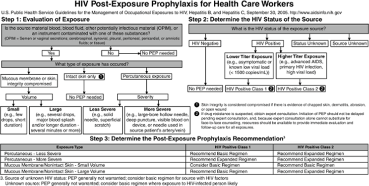
The AETC has published algorithms derived from the U.S. Public Health Service guidelines for management of occupational exposures to HIV, hepatitis B, and hepatitis C (5) (Figs. 13.1 and 13.2). HIV-positive class 1 is defined as a source patient with asymptomatic HIV infection or known low viral load. HIV-positive class 2 refers to a source patient with symptomatic HIV infection, AIDS, acute seroconversion syndrome, or known high viral load.
Influenza Virus
Influenza viruses are RNA viruses belonging to the family Orthomyxoviridae. Infection with these viruses causes an acute febrile illness usually occurring during the winter months and characterized by sudden onset of high fever, headache, myalgia, arthralgia, and cough. Those at risk for more severe disease include young children, the elderly, and immunocompromised patients. Although the infection can be self-limited, it may also result in severe prostration in elderly patients, primary influenza pneumonia, and secondary bacterial pneumonia. Transmission of the virus occurs person to person via large virus-laden droplets. Therefore, droplet precautions are used for patients who are admitted to the hospital with active or suspected influenza infection. While droplet precautions prevent spread, the mainstay of disease prevention is annual immunization of both HCWs and the at-risk patient population, which is defined as:
|
|
|
Figure 13.1. HIV postexposure prophylaxis (PEP) algorithm for health care workers. (From U.S. Public Health Service Guidelines for the Management of Occupational Exposures to HIV, Hepatitis B, and Hepatitis C, September 30, 2005. Available at: www.aidsinfo.nih.gov. Accessed November, 2006.) |
|
|
|
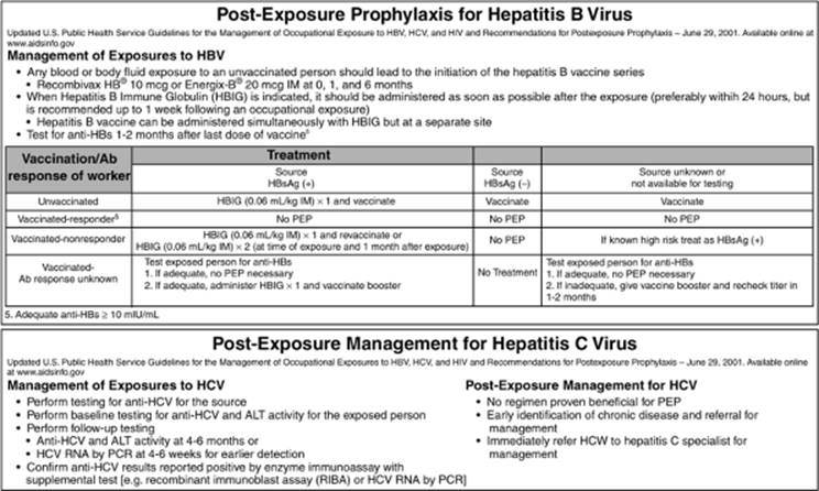
Figure 13.2. Postexposure recommendations for hepatitis B and hepatitis C in health care workers. (From U.S. Public Health Service Guidelines for the Management of Occupational Exposures to HIV, Hepatitis B, and Hepatitis C, September 30, 2005. Available at: www.aidsinfo.nih.gov. Accessed November, 2006.) |
· Age greater than 65
· Residents of nursing homes or long-term care facilities
· Pregnant women in the second or third trimester
· Patients with a chronic pulmonary or cardiac disease
· Patients with diabetes
· Individuals on dialysis
· Immunosuppressed patients
· Patients on long-term aspirin therapy
· Children aged 6 to 23 months
Vaccination can be done with a live, attenuated influenza vaccine resulting in virus replication in the respiratory epithelium. Because this can result in active viral shedding, the inactivated influenza vaccine is recommended for HCWs with direct patient contact.
Hepatitides
Several hepatitis viruses have been described, including hepatitis A, B, C, D, E, and G. Hepatitis A is caused by a picornavirus and is transmitted by the oral–fecal route, usually by contaminated food. It causes only an acute form of hepatitis that is generally self-limited and confers immunity to future infections. Hepatitis A is not usually a concern in the health care setting. Hepatitis D is caused by a delta virus and can only replicate in the presence of hepatitis B. Hepatitis E is like hepatitis A in that it causes an acute, usually self-limited hepatitis and is also transmitted via the oral–fecal route. In a small percent of cases, hepatitis E can develop into an acute severe liver disease that is often fatal. Pregnant women can develop severe disease with fulminant hepatic failure due to hepatitis E. By far, hepatitis B and C pose the greatest threat to health care workers.
Hepatitis B
Hepatitis B is a hepadnavirus that is endemic in certain parts of the world. Hepatitis B causes both an acute and chronic hepatitis, often with cirrhosis, and still remains a major cause of hepatocellular carcinoma in various parts of the world, especially Asia. The virus is transmitted through exposure to blood and body fluids. Routes of transmission include unprotected sexual contact—in which 16% to 40% of unimmunized partners will become infected—blood transfusions, use of contaminated needles and syringes, vertical transmission—20% risk of transmission from mother to child without intervention in a hepatitis B surface antigen (HBsAg)-positive mother—and occupational exposure including needlesticks. As with many viral infections, the risk of transmission from a bloodborne exposure is closely related to the volume of blood exposure and the number of copies of virus present in the blood of the source.
Per the CDC guidelines, risk of transmission of hepatitis B is also related to the hepatitis B envelope antigen (HBeAg) status of the source patient. In patients who were both HBsAg and HBeAg positive, the risk of developing clinical hepatitis from a needle injury was 22% to 31%. The risk of developing serologic evidence of infection was 36% to 62%. If the source patient was HBsAg positive with a negative HBeAg, the risk of developing clinical hepatitis from a needle injury was 1% to 6%, and the risk of developing serologic evidence of hepatitis B infection was 23% to 37% (23). Blood exposure and percutaneous injuries with contaminated blood are among the most efficient modes of transmitting hepatitis B since blood has the highest titers of hepatitis B compared to other body fluids. Interestingly, some studies suggest that most infected HCWs could not recall a percutaneous injury but rather recalled caring for a patient who was HBsAg positive when investigations of outbreaks were performed (24,25,26,27). Hepatitis B has been shown to survive in dried blood at room temperature for at least 1 week (28) and it is possible that contact with environmental surfaces is a potential risk for hepatitis B transmission, as has been shown in patients and staff of hemodialysis units (29,30,31).
The key factor in preventing hepatitis B infection in the health care setting is vaccination, and most facilities require that employees who come into direct contact with patients or with risk of exposure to blood and body fluids undergo the hepatitis B vaccination series. In fact, hepatitis B vaccination is part of the routine immunization schedule for children in the United States; by the age of 18 months, children who are up to date on their vaccinations have been fully immunized for hepatitis B. As with childhood vaccination, the protocol for adult immunization consists of three doses of the vaccine. For those whose hepatitis B vaccination series is interrupted, there is no need to restart. Vaccination can resume based on where in the series the patient was at the time of the interruption.
In the event that a health care worker is not immunized, postexposure prophylaxis (Fig. 13.2) is available in the form of hepatitis B immune globulin (HBIG). When indicated, HBIG should be given as soon as possible, preferably within 24 hours. Data on efficacy when HBIG was given after 7 days are not available. However, multiple doses of HBIG within 1 week of exposure are 75% effective in preventing hepatitis B infection. Postexposure hepatitis B vaccination is recommended in addition to HBIG because unimmunized HCWs continue to be at risk for exposure. In addition, data derived from vertical transmission regarding concurrent vaccination and HBIG administration show a better rate of prevention—85% to 95% with combined therapy as opposed to either therapy alone—70% to 75%.
Hepatitis C
Hepatitis C is an RNA virus in the family Flaviviridae. The virus replicates mainly in the hepatocytes after binding to specific receptors and entering the cells. Like hepatitis B, hepatitis C causes both acute and chronic hepatitis and is a risk factor for development of hepatocellular carcinoma. The virus is transmitted by direct contact with blood and body fluids containing blood. Various routes of transmission have been identified. Of note, IV drug abusers seem to have the highest incidence of developing hepatitis C due to the sharing of contaminated needles. While sexual transmission is possible, it is largely due to the possibility of blood contact and not other body fluids such as semen or vaginal secretions. Vertical transmission is also possible, although this occurs infrequently. In the health care setting, hepatitis C is not transmitted efficiently through occupational exposure to blood, and thus has a low incidence after accidental percutaneous exposure. Mucous membrane or skin exposures, both intact and nonintact, rarely result in transmission of hepatitis C. To date, there is no available vaccine for hepatitis C, and studies have shown no beneficial effect of giving immune globulin. Instead, postexposure management is aimed at the early detection of hepatitis C infection and the development of chronic disease for which treatment can be given.
The risk of acquiring hepatitis C also varies depending on the nature of exposure. After accidental percutaneous exposure from a known HCV-positive source, the incidence of seroconversion is 1.8% (32). Transmission from mucous membranes rarely occurs, and no cases of transmission in health care workers have been described from intact or nonintact skin exposures to blood (33,34). Furthermore, the risk of transmission from exposure to fluids other than blood has not been determined, but is postulated to be low. Although data are limited on the survival of HCV in the environment, one study has suggested that HCV-RNA is resistant to drying at room temperature for at least 48 hours (35).
Herpes Viruses
Human herpes viruses (HHVs) are DNA viruses that cause a variety of diseases in humans. Several human herpes viruses have been described including HHV-1 (also known as herpes simplex virus [HSV-1]), HHV-2 (herpes simplex virus [HSV-2]), HHV-3 (varicella-zoster virus [VZV]), HHV-4 (Epstein-Barr virus [EBV]), HHV-5 (cytomegalovirus [CMV]), HHV-6 (Roseolovirus), HHV-7, and HHV-8 (Kaposi sarcoma–associated virus). The seroprevalence of CMV is relatively high by adolescence (36) and has been documented to be up to 60% to 90% in adult populations. EBV generally causes a self-limited mild disease, but can cause infectious mononucleosis in teenagers and young adults. Thus, the herpes viruses of most importance in the health care setting are HSV-1, HSV-2, and VZV.
HSV-1 and HSV-2 cause blisters or sores either in the oral or genital area and can be transmitted to the health care worker by direct contact with the lesions, when they are present, and when appropriate use of precautions is forgone. VZV is the causative agent of chickenpox as well as shingles. Like other herpes viruses, VZV lies dormant in the dorsal root ganglia of the nervous system and can reactivate to produce zoster (“shingles”). VZV can be transmitted both by direct contact with a patient who has active skin lesions (vesicles) as well as via respiratory secretions in which the virus is shed during active infection, such as disseminated zoster. The recommended approach to patients with varicella-zoster disease includes both contact and airborne isolation.
One issue that arises in the clinical setting is the exposure of the nonimmune pregnant HCW to patients with CMV or VZV disease, as primary infection with either virus during pregnancy can be devastating to both mother and child. Some studies have shown that up to 50% of pregnant women are seropositive for CMV (37). In addition, the incidence of primary CMV during pregnancy is 1% to 4%, depending on certain variables. The transmission of CMV requires prolonged or recurrent close contact and can also be transmitted sexually. Since there are no effective therapies for treatment of CMV in pregnancy, prevention is the best method to avoid the complications of disease. Vaccines for CMV are available and, although there has been no change in the rate of CMV infection, there has been a reduction in disease severity in those who are vaccinated prior to primary infection. While there have been no conclusive recommendations regarding the precautions to be used by nonimmune pregnant HCWs with respect to patients with CMV, it would seem prudent to identify patients with active CMV infection so that these HCWs can be aware of the risk or be assigned to another patient. It must be recognized, however, that any patient can actively shed CMV without clinical signs or symptoms. This is the foundation for the CDC's strong recommendation of meticulous adherence to handwashing before and after patient care as the best way to prevent disease transmission in all settings. In patients with proven or suspected CMV pneumonitis, mask and eye protection may be a consideration in the nonimmune or seronegative pregnant HCW. As with CMV, a vaccine is available for VZV. The vaccine is a live, attenuated vaccine that is given in two doses spaced 4 to 8 weeks apart; it is 70% to 90% effective in preventing infection and 95% effective in preventing severe disease up to 10 years after administration. The vaccine is recommended for nonpregnant women of childbearing age and is not recommended for pregnant women, with a further stipulation being that women should not become pregnant for at least 1 month after each dose of the vaccine. In the event that a pregnant HCW who is nonimmune becomes exposed to VZV, postexposure prophylaxis is available. Varicella-zoster immunoglobulin is recommended within 96 hours of exposure and has been reported be up to 90% effective in preventing severe disease (38). It is recommended that nonimmune HCWs be vaccinated, especially female HCWs of childbearing age. If a pregnant HCW is not immune and has had exposure to VZV, prophylaxis should be instituted. In addition, contact and airborne precautions should be used at all times in patients with VZV disease.
Bacteria
Having discussed the major viruses of importance in the health care setting, we will now turn to a discussion of bacterial organisms including MRSA, VRE, Clostridium difficile, selected Gram-negative organisms, and Mycobacterium tuberculosis. Standard precautions are recommended for all patients, but additional precautions include contact precautions (MRSA, VRE, C. difficile) and airborne precautions (tuberculosis).
Methicillin-resistant Staphylococcus aureus
Methicillin-resistant S. aureus has become a major problem both in hospital-acquired (HA-MRSA) infections as well as community-acquired (CA-MRSA) infections. MRSA infections in the hospital are associated with both longer stays and higher costs. Data from the U.S. National Nosocomial Infections Surveillance Systems indicate that MRSA accounts for 55% of S. aureus–related infections in the intensive care setting in the United States. CA-MRSA is a microbiologically distinct isolate with a specific sensitivity pattern and the presence of the Panton-Valentine leukocidin (PVL) exotoxin. While it was previously thought that the PVL exotoxin was the virulence factor for CA-MRSA, studies in PVL-negative and PVL-positive mice failed to support this assertion; thus, the factor responsible for the virulence of CA-MRSA remains uncharacterized. Methicillin resistance is related to the acquisition of a staphylococcal cassette chromosome (SCC) that is known as the mecA gene. CA-MRSA can be distinguished from hospital-acquired MRSA by the presence of type 4 SCC (39). Expression of the mecA gene leads to an altered penicillin-binding protein, PBP2a, which has a reduced affinity for β-lactam rings.
CA-MRSA has been associated with significant skin and soft tissue infections, often requiring surgical drainage, usually in hospital emergency departments, as well as more severe infections such as necrotizing pneumonia, necessitating hospital admission. Nosocomial infections with HA-MRSA include catheter-related bacteremia, postsurgical wound infections, postoperative neurosurgical meningitis, ventilator-associated pneumonia, and device and graft infections. Risk factors for MRSA infection include patients with open wounds or pressure ulcers, invasive devices—such as catheter, tracheostomy, gastrostomy, nasogastric tube, and indwelling bladder catheter—recent antibiotic therapy, hospitalization or significant health care contact within the past 6 months, increased age, and male gender. Knowing the MRSA infection status of patients is helpful in determining who should be isolated or cohorted. While nasal swabs for MRSA culture are a way to identify those who are colonized, the time delay in identifying these patients based on cultures makes this option less useful. PCR has been proposed as a means of rapid identification of MRSA-colonized patients; however, this may not be cost effective. Although there are differences in the ways that hospitals approach the issue of screening patients for MRSA, once it is isolated from a culture, the patient should be immediately placed on contact precautions.
While there are antibiotic options available to treat MRSA infections, as well as to decolonize carriers of MRSA, prevention of spread is the most important method in combating MRSA infections. Patients with MRSA infection in the hospital should be placed on contact precautions. However, handwashing remains a critical factor in preventing spread. Health care workers must wash their hands before and after any contact with a patient, even when gloves are worn as part of MRSA contact precautions. Recommendations for preventing the spread of MRSA also include cohorting of patients if isolation in a single room is not possible. Contact precautions should be observed meticulously, especially with any anticipated contact with an open wound or ulcer, mucous membranes, or any blood or body fluid contact. Mask and eye protection are indicated if exposure to aerosols generated by the coughing patient is likely or when irrigating wounds. Precautions for MRSA-infected or -colonized patients remain the same regardless of the strain (HA-MRSA or CA-MRSA).
Fomites have been implicated in the transmission of MRSA. Environmental surfaces that have been described as vectors of MRSA transmission include a plastic patient chart (survived 11 days), a laminated tabletop (survived 12 days), and a cloth curtain (survived 9 days) (40). Further, even in the outpatient setting, studies have shown that environmental surfaces are important sources of transmission including a patient examination table, a computer keyboard, a pulse oximeter, and multiple patient chairs located in the triage station, the waiting room, and the examination room (41). Daily routine cleaning should be done with a disinfectant and performed in a sanitary manner as is done in all rooms regardless of the presence of MRSA. Equipment should be routinely cleaned, disinfected, or sterilized per institution policy.
Clostridium difficile
Members of the genus Clostridium are Gram-positive rods that are anaerobic and spore forming. C. difficile is the causative organism of pseudomembranous colitis and C. difficile–associated diarrhea (CDAD), which occurs with the use of antibiotics that eradicate normal gut flora. The organism produces two toxins, enterotoxin (toxin A) and cytotoxin (toxin B), which are responsible for diarrhea and inflammation. While growing C. difficile in culture is the gold standard for diagnosis, enzyme-linked immunosorbent assay (ELISA) testing for toxin A or B has a high sensitivity and specificity when performed on three separate stool specimens. In addition to watery diarrhea, computed tomography (CT) scan of the abdomen demonstrating colonic wall thickening is a key finding. Complications of untreated infection include toxic megacolon and bowel perforation. A major risk factor for the development of C. difficile diarrhea is the use of antibiotics—especially penicillins, clindamycin, and cephalosporins, particularly third-generation cephalosporins. Repeated enemas, prolonged nasogastric tube insertion, and gastrointestinal surgery also increase the risk of developing a disease.
The disease is spread from person to person by spores that are shed in the stool. Such spores can survive up to 70 days in the environment and can be carried on the hands of health care workers, who then have direct contact with uninfected patients or with environmental surfaces—floors, bedpans, toilets—thus contaminating them with C. difficile.
The treatment of C. difficile colitis includes oral metronidazole or oral vancomycin. A newer agent, nitazoxanide, has not received Food and Drug Administration (FDA) approval for this indication, but shows promise in small clinical trials. As with MRSA, longer hospital stays and increased costs have been directly related to infection with C. difficile. Recommendations for preventing spread include standard precautions as well as contact precautions if soiling of clothes is likely. It is important to note that handwashing is the only method to be used in preventing spread of C. difficile, as the alcohol substitutes do not kill the spores. However, the mechanical action of applying the hand sanitizer may eliminate some spores from the hand of the HCW. Waste material should also be handled in a proper way. Furthermore, limiting the use of inappropriate antibiotics is central to lowering the risk of developing C. difficile–associated diarrhea.
Vancomycin-resistant Enterococcus
Enterococci are Gram-positive, facultatively anaerobic organisms that colonize the gastrointestinal tract. Two species, Enterococcus faecalis (90% to 95%) and Enterococcus faecium (5% to 10%), are commensal in the intestines of humans. While the organism is not highly invasive, infections caused by enterococci include catheter-related bacteremia, urinary tract infections, diverticular abscess, cholangitis, and endocarditis. While many strains of enterococci remain susceptible to ampicillin, penicillin, and vancomycin, there has been an alarming increase in the incidence of VRE, which has implications both for therapy and prevention. In addition, there is concern regarding resistance in other bacteria, such as S. aureus, as VRE appears to have an enhanced ability to pass resistant genes (such as vanA) to other Gram-positive organisms.
Risk factors for VRE colonization and infection have not been clearly identified, although there seems to be a higher incidence in patients who are immunosuppressed—transplant and chemotherapy patients in an intensive care setting who have renal insufficiency. In addition, a higher risk of colonization and infection is logically associated with intra-abdominal surgery or gastrointestinal tract manipulation such as endoscopic retrograde cholangiopancreatography (ERCP), indwelling urinary catheters, enteral feeding tubes, and central venous catheters, as these are all—with the exception of the central venous catheter—associated with the natural reservoir of the organism. Patients on broad-spectrum antibiotics, and especially those who have received oral vancomycin, are also at higher risk for colonization or infection with a VRE species. The use of intravenous vancomycin is associated to a lesser extent with VRE colonization and infection.
Transmission generally occurs from a colonized or infected patient to the HCW via direct contact with either the patient or contaminated environmental surfaces (fomites), such as toilets, doorknobs, and even Yankauer suction devices (42). In general, the lack of proper hand hygiene appears to be the major factor in the spread of VRE from patient to HCW and back to other patients. Studies have shown a dramatic decrease in the incidence of VRE with the enforcement of proper hand hygiene, either with alcohol-based solutions or with routine handwashing (43). Proper environmental cleaning has been shown to also decrease the transmission of VRE (44). Patients who are known to be colonized or infected with VRE should be placed in contact isolation to prevent spread of the organism.
As with MRSA, the issue of screening for VRE has been addressed by several studies. One study has shown advantage in a clinical active surveillance strategy (culture of a rectal swab on admission, weekly while the patient was in the intensive care unit [ICU], and at discharge) with a cost savings ranging from $56,258 to $303,334 per month (45). A decision should be made at the institutional level regarding the cost effectiveness of a screening program. The available data support strong consideration for a routine screening program with resultant preventive measures (contact isolation) to help control the spread of VRE.
Tuberculosis
Mycobacterium tuberculosis (MTB) is the causative agent of all forms of tuberculosis—pulmonary, central nervous system (CNS), and disseminated. The disease is spread by aerosol droplets from persons with active infection when they cough, sneeze, speak, or spit. Infectious droplets are 0.5 to 5 µm in diameter; about 40,000 can be produced in a single sneeze and 3,000 in a single cough. The probability of transmission from person to person depends on several factors, including (a) quantity of infectious droplets expelled, (b) effectiveness of ventilation, (c) duration of exposure, and (d) the virulence of the Mycobacterial strain. Persons who are in direct contact with an infected patient, either frequently or for a prolonged time, have the highest risk of developing tuberculosis, with an estimated infection rate of 22%. A person with untreated, active tuberculosis can infect 10 to 15 people per year. Others at risk for infection include persons living in endemic areas—some parts of Asia, Haiti, and South America; immunocompromised patients—those with HIV/AIDS and those on immunosuppressive medications; health care workers serving high-risk patients; and IV drug abusers. Single males, alcoholics, the urban poor—especially the homeless—migrant farm workers, and prison inmates have been associated with a higher frequency of tuberculosis.
The rate of tuberculosis in the United States has steadily declined since a resurgence between 1985 and 1992 that correlated with the AIDS epidemic prior to the development of highly active antiretroviral therapy. Since then, the majority of MTB cases in the United States are now diagnosed in persons who are immigrants from endemic areas. There has been a recent deceleration in the yearly percentage of decline, from an average of 7.1% per year (1993 through 2000) to 3.8% per year (2001 through 2005). This has raised concerns regarding the progress toward the goal of eliminating MTB in the United States. According to the most recent CDC data, a total of 14,093 cases of TB were reported in the United States in 2005, which was down from 14,516 in 2004; this decrease represents the smallest decline in over a decade. A CDC study has also shown racial/ethnic disparities, as well as a disparity between U.S.-born and foreign-born MTB rates. In 2005, Hispanics, African Americans, and Asians were respectively 7.3, 8.3, and 19.6 times more likely than Caucasians to become infected with the disease. Furthermore, MTB rates were 8.7 times higher for foreign-born individuals as compared to their U.S. counterparts. More than half of foreign-born cases were reported in patients from Mexico, the Philippines, Vietnam, India, and China. The more alarming data from the most recent CDC report is the finding of higher rates of multidrug- and extended drug-resistant tuberculosis. In developing countries, rates of tuberculosis have been increasing in concordance with the rise of HIV as well as the neglect of TB control programs.
The most effective way to prevent the spread of tuberculosis to the HCW is to identify patients at high risk of active infection, which includes an assessment of symptoms—for example, fever, night sweats, shortness of breath, hemoptysis, and weight loss—as well as demographic factors including questioning about immigration from an endemic area, recent incarceration, and contact with patients known to have tuberculosis, and then isolating these patients. The patient must be placed in a negative pressure isolation room with airborne precautions. Patients should have three expectorated or induced sputum specimens sent for acid-fast staining and culture for acid-fast bacilli (AFB). Patients with suspected MTB infection cannot be removed from isolation until three adequate sputum specimens—or an equivalent, such as specimens obtained by bronchoscopy or bronchoalveolar lavage—have been obtained and are negative for AFB by smear. AFB culture may take up to 6 weeks to grow organisms, and is therefore not used to decide on discontinuation of isolation unless there is a very high index of suspicion. Newer diagnostic techniques including MTB DNA PCR can be used on a variety of AFB-negative body fluids or can be used to determine if early growth of AFB in liquid media is MTB or a nontuberculous mycobacteria.
Gram-Negative Organisms
Recently, the incidence of multidrug-resistant, Gram-negative infections has stirred debate as to whether isolation precautions such as those in place for resistant Gram positives should be instituted. The National Nosocomial Infections Surveillance System has reported increases in the prevalence of multidrug-resistant Gram-negative infections including Pseudomonas, Enterobacter, and Klebsiella, as well as extended-spectrum β-lactamase (ESBL)–producing organisms. In addition, some institutions have reported outbreaks of highly resistant Acinetobacter baumannii infections (46,47). Furthermore, interest in Acinetobacter is growing now that the organism has been found in soldiers returning from Iraq and Afghanistan.
A recent review of the data related to Pseudomonas, Enterobacteriaceae (Escherichia coli and Klebsiella) and Acinetobacter baumannii infection led the authors to conclude that there was not sufficient evidence to determine that infection control measures would be effective in controlling the spread of multidrug-resistant Gram-negative bacteria (48). However, outbreaks of resistant Gram-negative infections have become all too common and have been associated with catheters (49) and other intravascular devices (50), including one report of Stenotrophomonas prosthetic valve endocarditis (51). In addition, fatalities have been reported with certain outbreaks (52). Control of outbreaks has included both antibiotic restriction as well as institution of contact isolation. When dealing with multidrug-resistant, virulent, Gram-negative organisms, especially in such settings as the ICU, contact isolation should be strongly considered for use in colonized and infected patients as suggested by CDC guidelines (5).
References
1. Cosgrove SE, Qi Y, Kaye KS, et al. The impact of methicillin resistance in Staphylococcus aureus bacteremia on patient outcomes: mortality, length of stay, and hospital charges. Infect Control Hosp Epidemiol. 2005;26:166–174.
2. Chaix C, Durand-Zaleski I, Alberti C, et al. Control of endemic methicillin-resistant Staphylococcus aureus: a cost-benefit analysis in an intensive care unit. JAMA. 1999;282:1745–1751.
3. Wiklicky H, Skopec M. Ignaz Philipp Semmelweis, the prophet of bacteriology. Infect Control. 1983;4(5):367–370.
4. Herr HW. Ignorance is bliss: the Listerian revolution and education of American surgeons. J Urol. 2007;177(2):457–460.
5. Centers for Disease Control and Prevention. Guidelines for the management of occupational exposures to hepatitis B, hepatitis C, and HIV and recommendations for postexposure prophylaxis. MMWR Morb Mortal Wkly Rep. 2001;50(RR11):1–42. Available at http://www.cdc.gov/ncidod/dhqp/gl_occupational.html.
6. Widmer AF, Conzelmann M, Tomic M, et al. Introducing alcohol-based hand rub for hand hygiene: the critical need for training. Infect Control Hosp Epidemiol. 2007;28(1):50–54. Epub 2006 Dec 29.
7. Boyce JM, Pittet D. Guideline for hand hygiene in health-care settings. Recommendations of the healthcare infection control practices advisory committee and the ICPAC/SHEA/APIC/IDSA Hand hygiene task force. MMWR Recomm Rep. 2002;51:1–45.
8. Garner JS, Hospital Infection Control Practices Advisory Committee. Guidelines for Isolation Precautions in Hospitals, Am J Infect Control. 1996;24:24–31.
9. Kramer A, Schwebke I, Kampf G. How long do nosocomial pathogens persist on inanimate surfaces? A systematic review. BMC Infect Dis. 2006;16:130.
10. Menkhaus NA, Lanphear B, Linnemann CC. Airborne transmission of varicella-zoster virus in hospitals. Lancet. 1990;336(8726):1315.
11. Li Y, Wong T, Chung J, et al. In vivo protective performance of N95 respirator and surgical facemask. Am J Ind Med. 2006;49(12):1056–1065.
12. Fennelly KP. Personal respiratory protection against Mycobacterium tuberculosis. Clin Chest Med. 1997;18(1):1–17.
13. Mohan S, Hamid NS, Cunha BA. A cluster of nosocomial herpes simplex virus type 1 pneumonia in a medical intensive care unit. Infect Control Hosp Epidemiol. 2006;27(11):1255–1257. Epub 2006 Oct 17.
14. Gustafson TL, Lavely GB, Brawner ER, et al. An outbreak of airborne nosocomial varicella. Pediatrics. 1982;70(4):550–556.
15. Hyams PJ, Stuewe MC, Heitzer V. Herpes zoster causing varicella (chickenpox) in hospital employees: cost of a casual attitude. Am J Infect Control. 1984;12(1):2–5.
16. Gustafson TL, Shehab Z, Brunell PA. Outbreak of varicella in a newborn intensive care nursery. Am J Dis Child. 1984;138(6):548–550.
17. Daubin C, Vincent S, Vabret A, et al. Nosocomial viral ventilator-associated pneumonia in the intensive care unit: a prospective cohort study. Intens Care Med. 2005;31(8):1116–1122. Epub 2005 Jul 6.
18. Bell DM. Occupational risk of human immunodeficiency virus infection in healthcare workers: an overview. Am J Med. 1997;102(suppl 5B):9–15.
19. Henderson DK, Fahey BJ, Willy M, et al. Risk for occupational transmission of human immunodeficiency virus type 1 (HIV-1) associated with clinical exposures: a prospective evaluation. Ann Intern Med. 1990;113:740–746.
20. Ippolito G, Puro V, De Carli G. Italian Study Group on Occupational Risk of HIV Infection. The risk of occupational human immunodeficiency virus in health care workers. Arch Int Med. 1993;153:1451–1458.
21. Centers for Disease Control and Prevention. Update: human immunodeficiency virus infections in health-care workers exposed to blood of infected patients. MMWR Morb Mortal Wkly Rep. 1987;36:285–289.
22. Fahey BJ, Koziol DE, Banks SM, et al. Frequency of nonparenteral occupational exposures to blood and body fluids before and after universal precautions training. Am J Med. 1991;90:145–153.
23. Werner BG, Grady GF. Accidental hepatitis-B-surface-antigen-positive inoculations: use of e antigen to estimate infectivity. Ann Intern Med. 1982;97:367–369.
24. Garibaldi RA, Hatch FE, Bisno AL, et al. Nonparenteral serum hepatitis: report of an outbreak. JAMA. 1972;220:963–966.
25. Rosenberg JL, Jones DP, Lipitz LR, et al. Viral hepatitis: an occupational hazard to surgeons. JAMA. 1973;223:395–400.
26. Callender ME, White YS, Williams R. Hepatitis B virus infection in medical and health care personnel. BMJ. 1982;284:324–326.
27. Chaudhuri AKR, Follett EAC. Hepatitis B virus infection in medical and health care personnel [Letter]. BMJ. 1982;284:1408.
28. Bond WW, Favero MS, Petersen NJ, et al. Survival of hepatitis B virus after drying and storage for one week [Letter]. Lancet. 1981;1:550–551.
29. Hennekens CH. Hemodialysis-associated hepatitis: an outbreak among hospital personnel. JAMA. 1973;225:407–408.
30. Garibaldi RA, Forrest JN, Bryan JA, et al. Hemodialysis-associated hepatitis. JAMA. 1973;225:384–389.
31. Snydman DR, Bryan JA, Macon EJ, et al. Hemodialysis-associated hepatitis: a report of an epidemic with further evidence on mechanisms of transmission. Am J Epidemiol. 1976;104:563–570.
32. Alter MJ. The epidemiology of acute and chronic hepatitis C. Clin Liver Dis. 1997;1:559–568.
33. Sartori M, La Terra G, Aglietta M, et al. Transmission of hepatitis C via blood splash into conjunctiva [Letter]. Scand J Infect Dis. 1993;25:270–271.
34. Ippolito G, Puro V, Petrosillo N, et al. Simultaneous infection with HIV and hepatitis C virus following occupational conjunctival blood exposure [Letter]. JAMA. 1998;280:28.
35. Piazza M, Borgia G, Piccioto L, et al. HCV-RNA survival as detected by PCR in the environment. Boll Soc Ital Biol Sper. 1994;70(5-6):167–170.
36. Staras SA, Dollard SC, Radford KW, et al. Seroprevalence of cytomegalovirus infection in the United States, 1988–1994. Clin Infect Dis. 2006;43(9):1143–1151. Epub 2006 Oct 2.
37. Griffiths PD, Baboonian C, Rutter D. Congenital and maternal cytomegalovirus infection in a London population. Br J Obstet Gynaecol. 1991;98:135–140.
38. Enders G. Management of varicella-zoster contact and infection in pregnancy using a standardized varicella-zoster ELISA test. Postgrad Med J. 1985;61:23.
39. Kowalski TJ, Berbari EF, Osmon DR. Epidemiology, treatment, and prevention of community-acquired methicillin-resistant Staphylococcus aureus infections. Mayo Clin Proc. 2005;80(9):1201–1207.
40. Huang R, Mehta S, Weed D, et al. Methicillin-resistant Staphylococcus aureus survival on hospital fomites. Infect Control Hosp Epidemiol. 2006;27(11):1267–1269. Epub 2006 Sep 28.
41. Johnston CP, Cooper L, Ruby W, et al. Epidemiology of community-acquired methicillin-resistant Staphylococcus aureus skin infections among healthcare workers in an outpatient clinic. Infect Control Hosp Epidemiol. 2006;27(10):1133–1136. Epub 2006 Aug 31.
42. Brown M, Willms D. Colonization of Yankauer suction catheters with pathogenic organisms. Am J Infect Control. 2005;33(8):483–485.
43. Gordin FM, Schultz ME, Huber RA, et al. Reduction in nosocomial transmission of drug-resistant bacteria after introduction of an alcohol-based handrub. Infect Control Hosp Epidemiol. 2005;26(7):650–653.
44. Hayden MK, Bonten MJ, Blom DW, et al. Reduction in acquisition of vancomycin-resistant enterococcus after enforcement of routine environmental cleaning measures. Clin Infect Dis. 2006;42(11):1552–1560. Epub 2006 Apr 27.
45. Shadel BN, Puzniak LA, Gillespie KN, et al. Surveillance for vancomycin-resistant enterococci: type, rates, costs, and implications. Infect Control Hosp Epidemiol. 2006;27(10):1068–1075. Epub 2006 Sep 21.
46. Longo B, Pantosti A, Luzzi I, et al. An outbreak of Acinetobacter baumannii in an intensive care unit: epidemiological and molecular findings. J Hosp Infect. 2006;64(3):303–305. Epub 2006 Sep 14.
47. Bogaerts P, Naas T, Wybo I, et al. Outbreak of infection by carbapenem-resistant Acinetobacter baumannii producing the carbapenemase OXA-58 in Belgium. J Clin Microbiol. 2006;44(11):4189–4192. Epub 2006 Sep 6.
48. Harris AD, McGregor JC, Furuno JP. What infection control interventions should be undertaken to control multidrug-resistant gram-negative bacteria? Clin Infect Dis. 2006;43(Suppl 2):S57–61.
49. Wolfenden LL, Anderson G, Veledar E, et al. Catheter-associated bloodstream infections in 2 long-term acute care hospitals. Infect Control Hosp Epidemiol. 2007;28(1):105–106.
50. Siegman-Igra Y, Golan H, Schwartz D, et al. Epidemiology of vascular catheter-related bloodstream infections in a large university hospital in Israel. Scand J Infect Dis. 2000;32(4):411–415.
51. Mehta NJ, Khan IA, Mehta RN, et al. Stenotrophomonas maltophilia endocarditis of prosthetic aortic valve: report of a case and review of literature. Heart Lung. 2000;29(5):351–355.
52. Niu MT, Knippen M, Simmons L, et al. Transfusion-transmitted Klebsiella pneumoniae fatalities, 1995 to 2004. Transfus Med Rev. 2006;20(2):149–157.