Marcelo Amato
Chronic obstructive pulmonary diseases (COPDs) are a group of disorders characterized by airflow limitation that is not fully reversible (1). There are several diseases under this designation (Table 141.1), the most common of which are chronic bronchitis and emphysema. These two disorders represent the extremes of the COPD spectrum and usually coexist in COPD patients. Bronchitis is predominantly a disease of the airways and presents as a chronic productive cough for at least 3 months during 2 consecutive years, while emphysema is a disease of the parenchyma and consists of permanent airspace enlargement associated with rupture of the alveolar septa.
The common final pathway leading to COPD is an increased inflammatory response to inhaled particles or gases, of which the most common is cigarette smoke. This inflammatory process involves the airways and the lung parenchyma, leading to mucosal gland hypertrophy and disruption of alveolar septa with loss of elastic recoil. These alterations ultimately lead to the obstructive ventilatory defect that defines COPD (2,3). Some patients develop pulmonary hyperinflation caused by the loss of elastic recoil and increased airway resistance. During exacerbations, there might be a secondary dynamic pulmonary hyperinflation (2,4) caused by the increased ventilatory requirement and shortened expiratory time (5). The capacity of the respiratory muscles to generate inspiratory pressure is limited by their shortened operating length and impaired geometric arrangement (6). Long-term steroid use and/or malnutrition also contribute to strength impairment in many patients with severe chronic disease (7).
|
Table 141.1 Diseases Associated with Chronic Obstructive Pulmonary Disease |
||||||||
|
Only about 15% of all smokers will develop the full-blown syndrome with overt clinical symptoms, although a much higher proportion will develop some degree of airway obstruction. Rarely, the disease results from an inborn imbalance between the proteases and antiproteases present in the lung, as occurs in the autosomal recessive α1-antitrypsin deficiency (8).
Clinical Findings
The clinical manifestations of COPD appear late in the course of the disease. There is initially a slow decline in lung function that goes unnoticed over the years (9). Cough is the first finding, usually after the patient has been a smoker for many years. After about 20 years of smoking, some patients begin to notice shortness of breath on exertion, reflecting the progressive airflow limitation that is characteristic of the disease. The dyspnea worsens slowly over time, although sometimes patients deny the deterioration of lung function because they slowly adapt their level of activity to their exercise capacity. The decrease in lung function might become steeper during exacerbations, with a slow recovery to baseline levels after resolution of the decompensation.
Spirometry
Spirometry is the most important functional test for the diagnosis and the classification of severity of the disease. It consists of a forced exhalation after a deep inspiration while the patient is connected to a pneumotachograph. The ratio of the forced expiratory volume in the first second of the exhalation (FEV1) to the forced vital capacity is diagnostic of an obstructive ventilatory defect if <0.7 (1). The FEV1is a useful marker of the disease severity (Table 141.2) and is well suited as a longitudinal monitor of lung function (9).
Lung Volume
Lung volumes can be measured using whole-body plethysmography or gas dilution (helium or nitrogen washout) techniques. In emphysema, both total lung capacity and residual volume may be increased because of loss of lung elastic recoil. The carbon monoxide diffusing capacity may be diminished with the progression of the disease, reflecting the loss of the functional parenchyma.
|
Table 141.2 Classification of Severity according to GOLD |
||||||||||||||
|
||||||||||||||
Chest Radiographic Findings
Chest radiographic alterations usually occur late in the course of the disease, and there is no alteration pathognomonic of COPD. The radiograph is usually normal in mild disease; changes reflecting airway disease and hyperinflation may appear with progression of COPD. Sometimes it is possible to see enlarged bronchial walls reflected as an increase in bronchovascular markings. Emphysema is manifested by an increased lucency of the lungs. In smokers, these changes are more prominent in the upper lobes, while in α1-antitrypsin deficiency, they are more likely in basal zones. With hyperinflation, the chest becomes vertically elongated with low, flattened diaphragms. The heart shadow is also vertical and narrow. The retrosternal airspace is increased on the lateral view, and the sternal-diaphragmatic angle exceeds 90 degrees. Radiographic computerized tomography is more sensitive and specific for the presence of emphysema, but it is rarely required. It is most useful in the preoperative evaluation for lung volume reduction surgery.
Exacerbation
COPD exacerbation can be defined as an increase in dyspnea, cough, or sputum production that requires therapy (10). The two most commonly identified precipitating factors are infection—viral, such as Rhinovirus spp. or influenza, and bacterial, such as Haemophilus influenzae, Streptococcus pneumoniae, Moraxella catarrhalis, Enterobacteriaceae spp., or Pseudomonas spp.—as well as environmental exposure to air pollutants. However, in about one third of cases, no underlying cause is identified. Infectious agents can also be recovered from some patients with stable COPD, indicating that in some instances, their presence in decompensated COPD represents an epiphenomenon. On the other hand, Sethi et al. (11) have recently recognized that the acquisition of a new strain of a bacterial species colonized with a pathogenic bacteria might lead to an exacerbation in stable COPD patients. All exacerbations should be evaluated carefully for their potential to lead to or worsen respiratory failure that requires hospitalization. In the following section, we discuss the hospital treatment of COPD exacerbations.
Treatment of Exacerbations
The goals of the treatment of COPD exacerbations are to eliminate or control the cause of the exacerbation, provide optimum bronchodilator therapy, assure adequate oxygenation, and correct respiratory acidemia, all the while avoiding tracheal intubation when possible. Most patients with mild exacerbations can be treated at home, but those with a more severe presentation require hospitalization.
Admission criteria according to the American Thoracic Society/European Respiratory Society guidelines (12) include:
· High-risk comorbidities including pneumonia, cardiac arrhythmia, congestive heart failure, diabetes mellitus, renal failure, or liver failure
· Inadequate response of symptoms to outpatient management
· Marked increase in dyspnea
· Inability to eat or sleep because of symptoms
· Worsening hypoxemia
· Worsening hypercapnia
· Changes in mental status
· Inability to care for oneself (i.e., lack of home support)
· Uncertain diagnosis
Pharmacologic
The mainstay of pharmacologic treatment is the use of bronchodilators, corticosteroids, and antibiotics which are discussed below.
Bronchodilators
1. β2-Agonists: The bronchodilators most commonly used are the inhaled short-acting β2-agonists because of their rapid onset of action. They can be administered via a nebulizer or through metered dose inhalers (MDIs). Typically, two puffs of albuterol or salbutamol are given every 4 hours, or an equivalent dose via nebulizer. During mechanical ventilation, the use of a spacer interposed in the circuit between the tube and the Y-piece is recommended. An unresolved issue relates to dosage when MDIs are used with intubated patients. Fernandez et al. (13) used two puffs, Gay et al. (14) used three puffs, and Fuller et al. (15) used four puffs in their studies. Because the MDI dose deposited in the lungs of intubated patients is, at best, half of the dose deposited in the lungs of ambulatory patients, it seems reasonable to at least double the number of MDI puffs in intubated patients (i.e., at least four puffs). In some patients, this dose will be inadequate, and a greater number of puffs (e.g., 10 to 20) can be safely and effectively used. Long-acting β2-agonists can also be considered. Subcutaneous or intravenous administration should not be used unless there is contraindication for the inhaled route because of their increased systemic effects.
2. Anticholinergics: Ipratropium bromide can be used in association with the β2-agonists as needed. It is available both via nebulization (500 µg) or MDI (two puffs every 2 to 4 hours). There are no clinical studies that have evaluated the use of the long-acting anticholinergic tiotropium bromide during COPD exacerbations.
3. Methylxanthines: Methylxanthines are currently not indicated in the treatment of exacerbations of COPD.
Corticosteroids
Steroids are usually recommended for exacerbations of COPD. If feasible, prednisone can be given orally at a dose of 30 to 40 mg/day for 10 to 14 days. If the oral route is not an option, hydrocortisone or methylprednisolone can be substituted in equivalent doses. Some investigators advocate the use of much higher doses (methylprednisolone, 125 mg intravenously 4 times daily) (16), but as no studies have been designed to find the optimal dose, we favor the lower dose. More recent studies (17,18) have proposed the use of inhaled steroids for the treatment of acute exacerbations. The combination of salmeterol, 50 µg, and fluticasone, 500 µg, given twice daily, has been compared with placebo and resulted in a reduction in mortality of 3 years (p = 0.052), fewer exacerbations, and improved health status and lung function. Nebulized budesonide, 1,500 µg 4 times daily, was compared with prednisolone 40 mg and demonstrated equal efficacy and potentially fewer side effects, especially less hyperglycemia.
Antibiotics
Antibiotics decrease mortality during exacerbations. These agents are indicated when there is increased production or change in the color of the sputum. For mild exacerbations, amoxicillin, sulfamethoxazole-trimethoprim, or doxycycline for 7 to 10 days is usually adequate. Patients requiring hospitalizations should receive penicillin/penicillinase (e.g., amoxicillin/clavulanate), a respiratory quinolone (levofloxacin, gatifloxacin, moxifloxacin), or a third-generation cephalosporin together with a macrolide (e.g., ceftriaxone plus clarithromycin). In addition to their antimicrobial activity, macrolides possess anti-inflammatory and mucoregulatory properties that may confer beneficial effects to patients with COPD (19).
Respiratory Support
The goal of respiratory support in patients with exacerbations of COPD is to correct hypoxemia/acidemia and reduce the respiratory work, thus avoiding respiratory muscle fatigue (20,21). In the acute setting, oxygen therapy alone is able to revert hypoxemia, but not acidemia and respiratory distress. For this reason, invasive or noninvasive mechanical ventilation are frequently needed (2,20).
Oxygen Therapy
To improve the hypoxemia commonly present in exacerbations of COPD, controlled oxygen therapy is the cornerstone of hospital treatment (2). Long-term oxygen therapy is established as the standard of care for selected patients with advanced chronic stable hypoxemia due to COPD (22,23). However, in the acute setting, some patients have an impaired response to hypercapnia when treated with supplementary oxygen, leading to worsening of CO2 retention (24,25). The precise mechanism of this impairment is not well understood, but ventilation/perfusion (26,27,28,29) and respiratory drive (30,31) disturbances have been implicated. Some evidence suggests that the relief of hypoxic vasoconstriction due to the higher oxygen content in poorly ventilated areas may be the culprit of the acute CO2 retention. The increased perfusion of such poorly ventilated, previously hypoxic areas might suddenly increase the shunt effect, transferring a great part of the venous CO2 content directly to the arterial compartment.
There is no individual risk factor that identifies patients with COPD who will evolve to hypercapnia after oxygen exposure (2,24,25); therefore, the National Heart, Lung, Blood Institute/World Health Organization Global Initiative for Chronic Obstructive Lung Disease (GOLD) Workshop summary has recommended controlled oxygen therapy for the exacerbations, where adequate levels of oxygenation—PaO2 ≥60 mm Hg or SaO2 ≥90%—are easy to achieve in uncomplicated exacerbations. Notwithstanding, CO2 retention can occur insidiously with little change in symptoms; hence, measuring arterial blood gases 30 minutes after oxygen therapy, as recommended, is started. Venturi masks are more accurate sources of oxygen than are nasal prongs, but are more likely to be removed by the patient (2). Controlled oxygen therapy must be started at a low inspiratory oxygen fraction—0.24 to 0.28—and titrated upward to reach a PaO2 ≥60 mm Hg or SaO2 ≥90% without significant retention of CO2. A clinically significant increase in PaCO2 has been arbitrarily defined as a raise in CO2 of 6.5 mm Hg, especially if clinical mental deterioration occurs (24).
One must always remember that most of these patients have some degree of chronic vascular disease associated with their smoking history, and cardiovascular complications may be frequent during prolonged hypoxic episodes; for example, acute coronary syndromes, atrial fibrillation, cerebral ischemia, and pulmonary congestion. Therefore, the quick reversal of severe hypoxemia is frequently a priority.
Noninvasive Mechanical Ventilation
Patients with COPD are prone to acute hypercapnic respiratory failure, often resulting in emergency admission to the hospital. Between 20% and 30% of patients admitted with hypercapnic respiratory failure secondary to acute exacerbation of COPD will die in the hospital (32,33,34,35). Traditionally, patients who do not respond to conventional treatment are given invasive mechanical ventilation despite its well-known risks. Tracheal intubation and assisted ventilation have been associated with high morbidity and mortality rates, in addition to the difficulties during the weaning process from the ventilator (36,37). Many clinical complications seem to arise from the intubation procedure itself, or during the course of mechanical ventilation. The most common complications have been nosocomial infections, aspiration, pulmonary embolism, muscle atrophy, polyneuropathies, electrolyte imbalance, and gastrointestinal bleeding, as well as prolonging the stay in the intensive care unit (38,39).
In view of such difficulties, noninvasive positive pressure ventilation is an alternative treatment for patients admitted to the hospital with hypercapnic respiratory failure secondary to acute exacerbation of COPD. With this ventilatory modality, the patient receives air, or a mixture of air and oxygen, from a flow generator or a special ventilator through a facial/nasal mask, thus avoiding the need for tracheal intubation (32,33,34,40,41,42). Many studies have shown that noninvasive positive pressure ventilation increases pH, reduces PaCO2, reduces the severity of breathlessness in the first 4 hours of treatment, and decreases the length of hospital stay (32,33,34). More importantly, mortality and the intubation rate are consistently reduced by this intervention (32,33,34). Some studies suggest that the use of proper noninvasive ventilation can reduce the chances of an eventual endotracheal intubation to less than half (0.42, 95% confidence interval of 0.31 to 0.59) when compared with the conventional treatment with oxygen mask. This alternative has been also associated to a reduced mortality rate (0.41, 95% confidence interval [CI] of 0.26 to 0.64). In clinical-physiological terms, the expected elevation of pH after one hour of treatment should be around 0.03 (95% CI 0.02 to 0.04) and the expected reduction in PaCO2 in the same interval around -3.0 mm Hg (95% CI -5.1 to -0.2) (43).
Unfortunately, noninvasive ventilation is not appropriate for all patients (2). Failure rates between 9% and 50% have been reported (44,45). One important signal that this procedure is not working for a patient is the progression—even slight—of hypercapnia or acidosis 30 to 60 minutes after the procedure, and deterioration of the mental status.
The classic indications for noninvasive mechanical ventilation in exacerbated COPD patients are (1) respiratory distress with respiratory rate above 30 to 35 breaths per minute; (2) respiratory acidosis with a pH <7.35, and with normal or high standard base excess; and (3) a PaO2 below 45 mm Hg. These measurements are made after the patient has been breathing room air for at least 10 minutes (32,33,46). Noninvasive mechanical ventilation is contraindicated for patients with profound bradypnea, defined as a respiratory rate below 12 breaths per minute, severe hypercapnic encephalopathy with Glasgow Coma Scale score below 10, cardiac and/or respiratory arrest, and hemodynamic instability (32,33,46). Some authors, however, have successfully applied noninvasive mechanical ventilation in comatose COPD patients with a Glasgow Coma Scale score below 8, with other causes of coma being ruled out (47). This latter use of noninvasive mechanical ventilation is not widely accepted (46).
Adjustments of noninvasive ventilation
Among the studies on noninvasive mechanical ventilation and COPD exacerbation, some used an exclusive inspiratory pressure support (33) or inspiratory volume support (32); most used an associated positive end-expiratory pressure (PEEP) in consonance with the rationale that the use of PEEP/continuous positive airway pressure (CPAP) further reduces the inspiratory work in patients with COPD exacerbation, especially the extra load generated by high levels of intrinsic PEEP (48,49,50).
There are many approaches to set the noninvasive ventilation. An easy way is to set the expiratory pressure at 5 cm H2O, the inspiratory pressure at 10 cm H2O—resulting in a “delta P” of 5 cm H2O—and to increase the delta P in increments of 5 cm H2O, up to 20 to 25 cm H2O or the maximum tolerated, over 1 hour (42). An alternative approach is to adjust the inspiratory pressure in order to obtain a tidal volume of 6 to 8 mL/kg and a respiratory rate of 25 to 30 breaths per minute, setting the end-expiratory pressure to 5 cm H2O to offset the inspiratory threshold induced by intrinsic PEEP.
If inspiratory comfort is not achieved, especially when wasted inspiratory efforts are visible without a prompt response from the ventilator, meaning that the patient effort is not enough to easily trigger the assisted breath, trials of 2 cm H2O elevations in the PEEP/CPAP levels must be performed in order to further reduce the extra load imposed by the intrinsic PEEP. During these trials of augmentation of external PEEP, the minimum inspiratory pressure should be provided that maintains a stable tidal volume (51). Oxygen should be offered to keep oxygen saturation above 85% to 90% (2,42,51).
Theoretically, pure CPAP support in these patients might be of some help, offsetting part of the inspiratory threshold load imposed on COPD patients. The appeal of such a strategy is the possibility of using low-cost CPAP systems. This approach, however, has not been tested systematically and should be reserved for very special conditions under close supervision. Whenever possible, some level of inspiratory support should always be added to a CPAP strategy.
Taking all the studies into account, there is evidence to support the use of noninvasive mechanical ventilation in these patients, with documented success rates of 80% to 85% (52). Nevertheless, it is important that those patients who will fail the noninvasive ventilation trial be recognized early. Confalonieri et al. (53) evaluated the risk of failure of noninvasive ventilation in 1,033 consecutive patients with exacerbation of COPD admitted to experienced hospital units. The identified risk factors were Glasgow Coma Scale score <11, Acute Physiology and Chronic Health Evaluation (APACHE) II score ≥29, respiratory rate ≥30 breaths per minute, and arterial pH on admission of <7.25. The presence of all these risk factors resulted in a predicted risk of failure >70%. An arterial pH <7.25 after 2 hours of ventilation greatly increased the risk of failure to >90%. All these numbers and thresholds should be taken as relative reference points, because the success of noninvasive mechanical ventilation depends on a learning curve of the whole staff. The less experienced the staff, the more conservative we should be with these limits, not waiting for further deterioration of the patient before deciding on invasive ventilation.
After hospital admission, the correct timing for starting noninvasive ventilatory support is either immediately or at any time the patient shows worsening of the respiratory distress, a fall in PaO2, or an increase in PaCO2 (42,51). Noninvasive ventilatory support can be applied in any area of the hospital where close monitoring of the patient by trained personnel is available, such as intensive care units, emergency departments, high-dependency units, and respiratory wards. The duration of the noninvasive ventilation and the number of possible interruptions for oral and facial cleaning varies according to the patient need. Ventilatory periods lasting at least 40 minutes are warranted (33), and some patients will require uninterrupted use (42,51).
The choice for an appropriate mask is an important aspect of noninvasive mechanical ventilation. In general, patients benefit from a facial mask that covers the mouth and the nose; this is more efficient than the nasal type to deliver effective inspiratory pressures. Leaks directed at the eyes, sores in the nasal area, and a dry mouth are frequent causes of extreme discomfort to patients. The total face mask may be better tolerated by some patients, but not all, and greatly reduces the skin sores. However, one has to be aware that the anatomic dead space may increase a bit with this mask, which also imposes some challenges to the mechanical ventilator in terms of synchrony and PEEP maintenance.
The helmet interface is not yet appropriate to use in COPD patients; it warrants further technical improvements. Although efficient in maintaining the end-expiratory pressure, this device usually results in poor inspiratory support because of the high compliance of the whole system.
Invasive Mechanical Ventilation
Invasive mechanical ventilation can be either the initial choice in patients with COPD exacerbation or the strategy to be applied after failure of a trial of noninvasive ventilation (2). Mechanical ventilation can reduce or eliminate the work of breathing and improve gas exchange, while allowing the respiratory function to return to baseline through the treatment of the precipitating causes of the acute decompensation (4).
Assuming that all appropriate measures to improve airflow obstruction have already been taken (see previous discussion), minimization of dynamic hyperinflation is a key objective of the ventilatory support of these COPD patients. At the bedside, dynamic hyperinflation is typically detected by the presence of nonzero end-expiratory flow at the flow-time curve, or by effectively measuring the end-expiratory pressure (auto-PEEP) after an expiratory pause. Precise quantification of the auto-PEEP, however, is problematic in patients with spontaneous breathing efforts (54).
In some patients, especially in those with predominant emphysema, the airway obstruction in the expiratory phase is disproportionally higher than in the inspiration. In these patients, the measured auto-PEEP is higher than expected when considering the calculated inspiratory airway resistance. This situation can be anticipated by looking at the flow-volume curve available on most ventilators. The slope of this curve is proportional to the time constant of the respiratory system, and the differences between inspiratory and expiratory airway resistances can thus be determined (Fig. 141.1) (55).
|
|
|
Figure 141.1. Example of a flow-volume curve of a patient mechanically ventilated with severe and equivalent inspiratory and expiratory flow limitation. The dashed lines represent the slope of the curves and are proportional to the inspiratory and expiratory time constants. |
To reduce the hyperinflation, several concepts that have emerged from the recently published literature should be employed (4,56). The most effective strategy is controlled hypoventilation (57), which decreases dynamic hyperinflation through the reduction of the minute volume. Hypoventilation, with a fixed inspiratory time, decreases the expiratory flow requirement and consequently reduces air trapping and plateau pressures (57,58,59). An appropriate clinical goal at present is to keep the plateau pressures ≤30 cm H2O, a strategy associated with lower rates (4%) of barotrauma (60,61). Adequate sedation and analgesia help by lowering the production of CO2 and allowing further reduction of the minute volume (4). At the bedside, the general rules to minimize hyperinflation are (1) keep the minute volume ≤8 L/min, and (2) keep the expiratory time ≥4 seconds; low respiratory rates should be used, for example, 8 to 12 breaths per minute, with 5 to 8 mL/kg of tidal volume. Once these goals have been achieved, there is probably little gain from further adjusting the ventilator. For example, Leatherman et al. (62) showed that halving minute ventilation from 7.4 to 3.7 L/min, and more than doubling expiratory time from 4.5 to 9.5 seconds had no significant effect on auto-PEEP and plateau pressure (56). Although controlled hypoventilation is the most effective measure to decrease hyperinflation, this ventilatory strategy frequently worsens CO2 retention. The hypercapnia and acidosis are generally well tolerated and considered acceptable by most clinicians (4,56,57), provided that such levels of hypoventilation are essential to keep plateau pressures below 30 cm H2O.
Metabolic acidosis may also accompany the respiratory acidosis seen in COPD exacerbations, resulting in amplification of the acidemia (63). The underlying mechanism of such acidosis is not clear (63), but its buffering may accentuate coexistent pulmonary injury in hypoxemic patients (64). The treatment of metabolic acidosis in these hypercapnic patients should be directed to the etiology of the process, and not to the metabolic acidosis per se.
There is no optimal ventilation modality to support exacerbated COPD patients (56). The peak airway pressure may reach high values in the volume-controlled mode (62), but this is of limited clinical relevance because most of this pressure is dissipated in the large airways and, consequently, it does not reflect the alveolar pressure (60). One limitation of the volume-controlled mode is the impossibility of continuously monitoring the plateau pressure, as the use of an inspiratory pause every breath may worsen dynamic hyperinflation. On the other hand, in the pressure-controlled mode, conditions of fluctuating airway resistance and auto-PEEP entail the risk of variable tidal volume, with sometimes unacceptably low alveolar ventilation. Furthermore, with this latter mode, severe respiratory alkalosis may develop if airway obstruction subsides rapidly (56).
Although there is no clear advantage of one over the other, volume-controlled ventilation is currently the preferred mode by most investigators (56,57,59,60,62). When using volume-controlled ventilation, the flow waveform should also be adjusted. The square waveform usually results in higher peak pressures, frequently triggering the high pressure alarm, which should not be of much concern, as previously discussed (60,65). On the other hand, the decelerating-flow waveform usually minimizes peak pressure, allowing full delivery of the tidal volume, with less interruption by opening of the pop-off safety valve (56). By forcing a slower flow at the end of inspiration, this flow waveform could result in two theoretical benefits: (1) less overdistention of alveoli distal to the least obstructed airways, and (2) slightly better CO2 exchange. Whenever possible, and provided that peak pressures are effectively reduced (this must be tested), this flow pattern thus should be preferred. During controlled mechanical ventilation with volume-controlled ventilation, the inspiratory pause should be used with extreme caution, and mainly for monitoring purposes to check whether hyperinflation is improving.
When initiating mechanical ventilation in the pressure-controlled mode, one must keep in mind that the inspiratory time should be set in proportion to the inspiratory time constant in order to deliver the desired tidal volume with the lowest possible plateau pressure (66). Thus, patients with increased airways resistance will need a longer inspiratory time. For a fixed respiratory rate, the increase in inspiratory time always occurs at the expense of a shortening of the expiratory time, which might aggravate pulmonary hyperinflation. Therefore, the ideal inspiratory time would optimize delivery of tidal volume without increasing air trapping. That will occur if, when looking at the flow-volume curve, both end-inspiratory and end-expiratory flows are equal or close to zero (4,56,60).
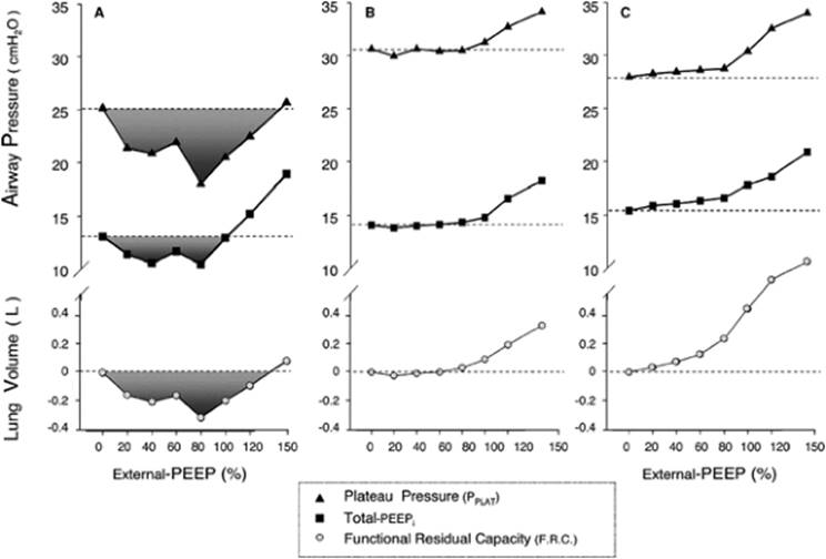
After choosing the best respiratory rate and inspiratory and expiratory times, the physician has to decide on how much PEEP to apply. During controlled mechanical ventilation, PEEP can be detrimental to paralyzed patients with severe airflow obstruction, raising the functional pulmonary capacity (67). Based on this information, some authors have advocated the use of zero PEEP or no more than 5 cm H2O (56,67). However, some patients show a paradoxical response to an increase in PEEP with relief of the air trapping (Fig. 141.2) (68). There are two ways to identify these patients:
1. Using volume-control mode, with an inspiratory pause of 1 to 2 seconds, tidal volume of 5 to 8 mL/kg, and a respiratory rate of 10 breaths per minute (with the lowest inspiratory:expiratory ratio possible, e.g. <1:4), increase the external PEEP in steps of 2 cm H2O every 10 to 20 respiratory cycles, starting from ZEEP (baseline). The best PEEP is the highest level associated with a plateau pressure equal to or less than the baseline.
2. In pressure-control mode, start with ZEEP, driving pressure, and inspiratory time to achieve a tidal volume of 5 to 8 mL/g. Raise the external PEEP in steps of 2 cm H2O every 10 to 20 respiratory cycles, keeping the plateau pressure constant. If there is an increase in tidal volume during the upward PEEP titration, it means there was recruitment of the airway. PEEP should be kept at the highest level before the tidal volume begins to fall (4).
As soon as possible, the ventilation mode should be switched from controlled to assisted ventilation in order to decrease muscle atrophy. There are no objective indicators of the best moment to start assisted ventilation; therefore, at least daily trials of assisted ventilation should be made, with close monitoring of patient comfort and plateau pressure (4,56). Adding external PEEP during assisted ventilation can reduce the inspiratory work by means of eliminating the offset of inspiratory pressure threshold induced by auto-PEEP (4). Appendini et al. (69) demonstrated that 41% of the inspiratory muscle effort was expended to overcome auto-PEEP in patients with COPD during spontaneous breathing. Adding external PEEP at an average of 80% of the measured auto-PEEP is well tolerated, with no increase of total PEEP or plateau pressure (70), improving synchrony between patient and ventilator (48,49,50). Frequent reassessments are essential because the adequate level of external PEEP is subject to change as lung mechanics and ventilatory requirements change.
|
|
|
Figure 141.2. Three of the possible responses observed in plateau pressure (PPLAT), total intrinsic positive end-expiratory pressure (PEEPi), and functional residual capacity (F.R.C.) with the application of external-PEEP (represented as percentage of PEEPi measured at zero external-PEEP). The FRC measured at zero external-PEEP was considered as the reference. A, Paradoxical response (patient 4), observed with a tidal volume (VT) of 6 mL/kg and respiratory rate (RR) of 9 breaths/min; B, biphasic response (patient 7), observed with VT of 9 mL/kg and RR of 6 breaths/min; C, classic overinflation response (patient 5), observed with a VT of 9 mL/kg and RR of 9 breaths/min. (From Caramez MP, Borges JB, Tucci MR, et al. Paradoxical responses to positive end-expiratory pressure in patients with airway obstruction during controlled ventilation. Crit Care Med. 2005;33:1519–1528, with permission.) |
Humidification should be achieved with a heated humidifier, not with heat and moisture exchangers. The latter devices are undesirable for three reasons: (1) they increase expiratory airway resistance, which would hardly be of any help to reduce hyperinflation (71); (2) when inserted between the tracheal tube and the Y-piece of ventilator tubing, they increase dead space and therefore contribute unnecessarily to hypercapnia (72,73); and (3) the efficacy of any inhalational medication will be blunted by the heat and moisture exchanger (56).
Weaning from the ventilator should be initiated as soon as possible in order to avoid mechanical ventilator-associated complications (74). According to recent published experiences of two specialized weaning units, 19% of patients with COPD exacerbation remained partially dependent on the ventilator (75,76). The classic rapid shallow breathing criterion—<80 breaths per minute per liter—was met by 56% of COPD patients who failed the weaning trial (77). General patient condition and subjective dyspnea seemed to be more effective predictors of success of extubation than quantifiable indexes (78). Using spontaneous breathing trials or progressive reduction in pressure support is equally effective to wean the patient from the ventilator (79). Automatic algorithms for pressure support reduction are available today, which resulted in a faster weaning process when compared with the physician-driven approach (80,81).
If the spontaneous breathing trial is chosen, it can be applied for at least 30 minutes, up to 2 hours once a day (36). After tracheal decannulation, the use of intermittent or continuous support with noninvasive mechanical ventilation for at least 24 hours is strongly recommended, using settings similar to those used during conventional ventilation weaning. The last technique is associated with higher rates of extubation success, lower length of stay in the intensive care unit and/or hospital, and a lower mortality at 60 days (82,83). Nava et al. (84) have described a strategy in which patients are ventilated and sedated for 6 to 8 hours after intubation; an assisted mode is subsequently started using pressure support. After 48 hours of conventional mechanical ventilation, if the patient is not hypersecretory or hemodynamically unstable, he or she is extubated and supported with noninvasive mechanical ventilation using the same ventilatory settings as before tracheal decannulation. This approach is associated with less ventilator-associated pneumonia, a shorter length of stay in the intensive care unit, and a lower mortality rate.
Sedation and Neuromuscular Blockade
Benzodiazepines, often given with narcotics, are widely accepted (4,56,60). Benzodiazepines, especially when associated with opiates, are effective in facilitating the controlled hypoventilation in hypercapnic patients without the need of high doses (85). Propofol may also be used because of its bronchodilating action (56), but generally requires high infusion rates (85). When patient-ventilator asynchrony cannot be suppressed by increasing the opioid dose, neuromuscular blocking agents should be given as intermittent intravenous boluses rather than as a continuous infusion in order to reduce the dose and duration of administration (4,56,60). Muscle resting is not currently recommended (86); however, muscle unloading for a short time—about 12 hours—has been advocated for muscular recuperation after the fatigue of acute stress (56).
Summary
Respiratory failure in COPD is an exceptionally difficult disorder to manage. Nonetheless, with appropriate and timely diagnostic and therapeutic interventions, including noninvasive ventilation in the proper clinical setting, our hope is that the mortality rate can be decreased from its approximate 20% to 30%.
References
1. Rabe KF, Hurd S, Anzueto A, et al. Global Strategy for the Diagnosis, Management, and Prevention of Chronic Obstructive Pulmonary Disease: GOLD Executive Summary. Am J Respir Crit Care Med. 2007;176:532–555.
2. Pauwels RA, Buist AS, Calverley PM, et al. Global strategy for the diagnosis, management, and prevention of chronic obstructive pulmonary disease. NHLBI/WHO Global Initiative for Chronic Obstructive Lung Disease (GOLD) Workshop summary. Am J Respir Crit Care Med. 2001;163:1256–1276.
3. Barnes PJ. Chronic obstructive pulmonary disease. N Engl J Med. 2000;343:269–280.
4. Peigang Y, Marini JJ. Ventilation of patients with asthma and chronic obstructive pulmonary disease. Curr Opin Crit Care. 2002;8:70–76.
5. Rossi A, Ganassini A, Polese G, et al. Pulmonary hyperinflation and ventilator-dependent patients. Eur Respir J. 1997;10:1663–1674.
6. Marchand E, Decramer M. Respiratory muscle function and drive in chronic obstructive pulmonary disease. Clin Chest Med. 2000;21:679–692.
7. Perez T, Becquart LA, Stach B, et al. Inspiratory muscle strength and endurance in steroid-dependent asthma. Am J Respir Crit Care Med. 1996;153:610–615.
8. Eriksson S. Pulmonary emphysema and alpha1-antitrypsin deficiency. Acta Med Scand. 1964;175:197–205.
9. Burchfiel CM, Marcus EB, Curb JD, et al. Effects of smoking and smoking cessation on longitudinal decline in pulmonary function. Am J Respir Crit Care Med. 1995;151:1778–1785.
10. Rodriguez-Roisin R. Toward a consensus definition for COPD exacerbations. Chest. 2000;117(5 Suppl 2):398S–401S.
11. Sethi S, Evans N, Grant BJ, et al. New strains of bacteria and exacerbations of chronic obstructive pulmonary disease. N Engl J Med. 2002;347:465–471.
12. Celli BR, Macnee W. Standards for the diagnosis and treatment of patients with COPD: a summary of the ATS/ERS position paper. Eur Respir J. 2004;23:932–946.
13. Fernandez A, Lazaro A, Garcia A, et al. Bronchodilators in patients with chronic obstructive pulmonary disease on mechanical ventilation. Utilization of metered-dose inhalers. Am Rev Respir Dis. 1990;141:164–168.
14. Gay PC, Patel HG, Nelson SB, et al. Metered dose inhalers for bronchodilator delivery in intubated, mechanically ventilated patients. Chest. 1991;99:66–71.
15. Fuller HD, Dolovich MB, Turpie FH, et al. Efficiency of bronchodilator aerosol delivery to the lungs from the metered dose inhaler in mechanically ventilated patients. A study comparing four different actuator devices. Chest. 1994;105:214–218.
16. Erbland ML, Deupree RH, Niewoehner DE. Systemic Corticosteroids in Chronic Obstructive Pulmonary Disease Exacerbations (SCCOPE): rationale and design of an equivalence trial. Veterans Administration Cooperative Trials SCCOPE Study Group. Control Clin Trials. 1998;19:404–417.
17. Calverley PM, Anderson JA, Celli B, et al. Salmeterol and fluticasone propionate and survival in chronic obstructive pulmonary disease. N Engl J Med. 2007;356:775–789.
18. Gunen H, Hacievliyagil SS, Yetkin O, et al. The role of nebulised budesonide in the treatment of acute exacerbations of COPD. Eur Respir J. 2007;30:399–400.
19. Gotfried MH. Macrolides for the treatment of chronic sinusitis, asthma, and COPD. Chest. 2004;125(2 Suppl):52S–60S.
20. Wouters EF. Management of severe COPD. Lancet. 2004;364(9437):883–895.
21. Calverley PM, Walker P. Chronic obstructive pulmonary disease. Lancet. 2003;362(9389):1053–1061.
22. Continuous or nocturnal oxygen therapy in hypoxemic chronic obstructive lung disease: a clinical trial. Nocturnal Oxygen Therapy Trial Group. Ann Intern Med. 1980;93:391–398.
23. Long term domiciliary oxygen therapy in chronic hypoxic cor pulmonale complicating chronic bronchitis and emphysema. Report of the Medical Research Council Working Party. Lancet. 1981;1(8222):681–686.
24. Moloney ED, Kiely JL, McNicholas WT. Controlled oxygen therapy and carbon dioxide retention during exacerbations of chronic obstructive pulmonary disease. Lancet. 2001;357(9255):526–528.
25. Calverley PM. Oxygen-induced hypercapnia revisited. Lancet. 2000;356(9241):1538–1539.
26. Aubier M, Murciano D, Milic-Emili J, et al. Effects of the administration of O2 on ventilation and blood gases in patients with chronic obstructive pulmonary disease during acute respiratory failure. Am Rev Respir Dis. 1980;122:747–754.
27. Robinson TD, Freiberg DB, Regnis JA, et al. The role of hypoventilation and ventilation-perfusion redistribution in oxygen-induced hypercapnia during acute exacerbations of chronic obstructive pulmonary disease. Am J Respir Crit Care Med. 2000;161:1524–1529.
28. Hanson CW, III, Marshall BE, Frasch HF, et al. Causes of hypercarbia with oxygen therapy in patients with chronic obstructive pulmonary disease. Crit Care Med. 1996;24:23–28.
29. Sassoon CS, Hassell KT, Mahutte CK. Hyperoxic-induced hypercapnia in stable chronic obstructive pulmonary disease. Am Rev Respir Dis. 1987;135:907–911.
30. Dunn WF, Nelson SB, Hubmayr RD. Oxygen-induced hypercarbia in obstructive pulmonary disease. Am Rev Respir Dis. 1991;144(3 Pt 1):526–530.
31. Dick CR, Liu Z, Sassoon CS, et al. O2-induced change in ventilation and ventilatory drive in COPD. Am J Respir Crit Care Med. 1997;155:609–614.
32. Bott J, Carroll MP, Conway JH, et al. Randomised controlled trial of nasal ventilation in acute ventilatory failure due to chronic obstructive airways disease. Lancet. 1993;341(8860):1555–1557.
33. Brochard L, Mancebo J, Wysocki M, et al. Noninvasive ventilation for acute exacerbations of chronic obstructive pulmonary disease. N Engl J Med. 1995;333:817–822.
34. Foglio C, Vitacca M, Quadri A, et al. Acute exacerbations in severe COLD patients. Treatment using positive pressure ventilation by nasal mask. Chest. 1992;101:1533–1538.
35. Ambrosino N, Foglio K, Rubini F, et al. Non-invasive mechanical ventilation in acute respiratory failure due to chronic obstructive pulmonary disease: correlates for success. Thorax. 1995;50:755–757.
36. Esteban A, Frutos F, Tobin MJ et al. A comparison of four methods of weaning patients from mechanical ventilation. Spanish Lung Failure Collaborative Group. N Engl J Med. 1995;332:345–350.
37. Brochard L, Rauss A, Benito S, et al. Comparison of three methods of gradual withdrawal from ventilatory support during weaning from mechanical ventilation. Am J Respir Crit Care Med. 1994;150:896–903.
38. Guerin C, Girard R, Chemorin C, et al. Facial mask noninvasive mechanical ventilation reduces the incidence of nosocomial pneumonia. A prospective epidemiological survey from a single ICU. Intensive Care Med. 1997;23:1024–1032.
39. Nourdine K, Combes P, Carton MJ, et al. Does noninvasive ventilation reduce the ICU nosocomial infection risk? A prospective clinical survey. Intensive Care Med. 1999;25:567–573.
40. Meduri GU, Abou-Shala N, Fox RC, et al. Noninvasive face mask mechanical ventilation in patients with acute hypercapnic respiratory failure. Chest. 1991;100:445–454.
41. Celikel T, Sungur M, Ceyhan B, et al. Comparison of noninvasive positive pressure ventilation with standard medical therapy in hypercapnic acute respiratory failure. Chest. 1998;114:1636–1642.
42. Plant PK, Owen JL, Elliott MW. Early use of non-invasive ventilation for acute exacerbations of chronic obstructive pulmonary disease on general respiratory wards: a multicentre randomised controlled trial. Lancet. 2000;355(9219):1931–1935.
43. Lightowler JV, Wedzicha JA, Elliott MW, et al. Non-invasive positive pressure ventilation to treat respiratory failure resulting from exacerbations of chronic obstructive pulmonary disease: Cochrane systematic review and meta-analysis. BMJ. 2003;326(7382):185.
44. Kramer N, Meyer TJ, Meharg J, et al. Randomized, prospective trial of noninvasive positive pressure ventilation in acute respiratory failure. Am J Respir Crit Care Med. 1995;151:1799–1806.
45. Soo Hoo GW, Santiago S, Williams AJ. Nasal mechanical ventilation for hypercapnic respiratory failure in chronic obstructive pulmonary disease: determinants of success and failure. Crit Care Med. 1994;22:1253–1261.
46. Evans TW. International Consensus Conferences in Intensive Care Medicine: non-invasive positive pressure ventilation in acute respiratory failure. Organised jointly by the American Thoracic Society, the European Respiratory Society, the European Society of Intensive Care Medicine, and the Societe de Reanimation de Langue Francaise, and approved by the ATS Board of Directors, December 2000. Intensive Care Med. 2001;27:166–178.
47. Diaz GG, Alcaraz AC, Talavera JC, et al. Noninvasive positive-pressure ventilation to treat hypercapnic coma secondary to respiratory failure. Chest. 2005;127:952–960.
48. Goldberg P, Reissmann H, Maltais F, et al. Efficacy of noninvasive CPAP in COPD with acute respiratory failure. Eur Respir J. 1995;8:1894–1900.
49. Appendini L, Patessio A, Zanaboni S, et al. Physiologic effects of positive end-expiratory pressure and mask pressure support during exacerbations of chronic obstructive pulmonary disease. Am J Respir Crit Care Med. 1994;149:1069–1076.
50. Smith TC, Marini JJ. Impact of PEEP on lung mechanics and work of breathing in severe airflow obstruction. J Appl Physiol. 1988;65:1488–1499.
51. Conti G, Antonelli M, Navalesi P, et al. Noninvasive vs. conventional mechanical ventilation in patients with chronic obstructive pulmonary disease after failure of medical treatment in the ward: a randomized trial. Intensive Care Med. 2002;28:1701–1707.
52. International Consensus Conferences in Intensive Care Medicine: noninvasive positive pressure ventilation in acute respiratory failure. Am J Respir Crit Care Med. 2001;163:283–291.
53. Confalonieri M, Garuti G, Cattaruzza MS, et al. A chart of failure risk for noninvasive ventilation in patients with COPD exacerbation. Eur Respir J. 2005;25:348–355.
54. Gladwin MT, Pierson DJ. Mechanical ventilation of the patient with severe chronic obstructive pulmonary disease. Intensive Care Med. 1998;24:898–910.
55. Dhand R. Ventilator graphics and respiratory mechanics in the patient with obstructive lung disease. Respir Care. 2005;50:246–261.
56. Oddo M, Feihl F, Schaller MD, et al. Management of mechanical ventilation in acute severe asthma: practical aspects. Intensive Care Med. 2006;32:501–510.
57. Darioli R, Perret C. Mechanical controlled hypoventilation in status asthmaticus. Am Rev Respir Dis. 1984;129:385–387.
58. Darioli R, Domenighetti G, Perret C. Mechanical ventilation in the treatment of acute respiratory insufficiency in asthma [in German]. Schweiz Med Wochenschr. 1981;111:194–196.
59. Tuxen DV. Permissive hypercapnic ventilation. Am J Respir Crit Care Med. 1994;150:870–874.
60. Leatherman JW. Mechanical ventilation in obstructive lung disease. Clin Chest Med. 1996;17:577–590.
61. Leatherman J. Life-threatening asthma. Clin Chest Med. 1994;15:453–479.
62. Leatherman JW, McArthur C, Shapiro RS. Effect of prolongation of expiratory time on dynamic hyperinflation in mechanically ventilated patients with severe asthma. Crit Care Med. 2004;32:1542–1545.
63. Mountain RD, Heffner JE, Brackett NC Jr, et al. Acid-base disturbances in acute asthma. Chest. 1990;98:651–655.
64. Laffey JG, Engelberts D, Kavanagh BP. Buffering hypercapnic acidosis worsens acute lung injury. Am J Respir Crit Care Med. 2000;161:141–146.
65. Guerin C, Lemasson S, La Cara MF, et al. Physiological effects of constant versus decelerating inflation flow in patients with chronic obstructive pulmonary disease under controlled mechanical ventilation. Intensive Care Med. 2002;28:164–169.
66. Marini JJ, Crooke PS III, Truwit JD. Determinants and limits of pressure-preset ventilation: a mathematical model of pressure control. J Appl Physiol. 1989;67:1081–1092.
67. Tuxen DV. Detrimental effects of positive end-expiratory pressure during controlled mechanical ventilation of patients with severe airflow obstruction. Am Rev Respir Dis. 1989;140:5–9.
68. Caramez MP, Borges JB, Tucci MR, et al. Paradoxical responses to positive end-expiratory pressure in patients with airway obstruction during controlled ventilation. Crit Care Med. 2005;33:1519–1528.
69. Appendini L, Purro A, Patessio A, et al. Partitioning of inspiratory muscle workload and pressure assistance in ventilator-dependent COPD patients. Am J Respir Crit Care Med. 1996;154:1301–1309.
70. Guerin C, Milic-Emili J, Fournier G. Effect of PEEP on work of breathing in mechanically ventilated COPD patients. Intensive Care Med. 2000;26:1207–1214.
71. Verkerke GJ, Geertsema AA, Schutte HK. Airflow resistance of heat and moisture exchange filters with and without a tracheostoma valve. Ann Otol Rhinol Laryngol. 2002;111:333–337.
72. Hinkson CR, Benson MS, Stephens LM, et al. The effects of apparatus dead space on P(aCO2) in patients receiving lung-protective ventilation. Respir Care. 2006;51:1140–1144.
73. Moran I, Bellapart J, Vari A, et al. Heat and moisture exchangers and heated humidifiers in acute lung injury/acute respiratory distress syndrome patients. Effects on respiratory mechanics and gas exchange. Intensive Care Med. 2006;32:524–531.
74. Pingleton SK. Complications of acute respiratory failure. Am Rev Respir Dis. 1988;137:1463–1493.
75. Dasgupta A, Rice R, Mascha E, et al. Four-year experience with a unit for long-term ventilation (respiratory special care unit) at the Cleveland Clinic Foundation. Chest. 1999;116:447–455.
76. Bagley PH, Cooney E. A community-based regional ventilator weaning unit: development and outcomes. Chest. 1997;111:1024–1029.
77. Purro A, Appendini L, De Gaetano A, et al. Physiologic determinants of ventilator dependence in long-term mechanically ventilated patients. Am J Respir Crit Care Med. 2000;161(4 Pt 1):1115–1123.
78. Afessa B, Hogans L, Murphy R. Predicting 3-day and 7-day outcomes of weaning from mechanical ventilation. Chest. 1999;116:456–461.
79. Vitacca M, Vianello A, Colombo D, et al. Comparison of two methods for weaning patients with chronic obstructive pulmonary disease requiring mechanical ventilation for more than 15 days. Am J Respir Crit Care Med. 2001;164:225–230.
80. Bouadma L, Lellouche F, Cabello B, et al. Computer-driven management of prolonged mechanical ventilation and weaning: a pilot study. Intensive Care Med. 2005;31:1446–1450.
81. Lellouche F, Mancebo J, Jolliet P, et al. A multicenter randomized trial of computer-driven protocolized weaning from mechanical ventilation. Am J Respir Crit Care Med. 2006;174:894–900.
82. Ferrer M, Valencia M, Nicolas JM, et al. Early noninvasive ventilation averts extubation failure in patients at risk: a randomized trial. Am J Respir Crit Care Med. 2006;173:164–170.
83. Nava S, Gregoretti C, Fanfulla F, et al. Noninvasive ventilation to prevent respiratory failure after extubation in high-risk patients. Crit Care Med. 2005;33:2465–2470.
84. Nava S, Ambrosino N, Clini E, et al. Noninvasive mechanical ventilation in the weaning of patients with respiratory failure due to chronic obstructive pulmonary disease. A randomized, controlled trial. Ann Intern Med. 1998;128:721–728.
85. Vinayak AG, Gehlbach B, Pohlman AS, et al. The relationship between sedative infusion requirements and permissive hypercapnia in critically ill, mechanically ventilated patients. Crit Care Med. 2006;34:1668–1673.
86. Vassilakopoulos T, Zakynthinos S, Roussos C. Bench-to-bedside review: weaning failure—should we rest the respiratory muscles with controlled mechanical ventilation? Crit Care. 2006;10:204.