Eelco F. M. Wijdicks
Impaired consciousness is a very common neurologic problem in the Intensive Care Unit (ICU). Neurologists are commonly consulted for “altered mental status,” often after sedation has been discontinued, after cardiopulmonary resuscitation and failure to awaken, and in patients with multisystem trauma to address the extent of traumatic brain injury and assist in management. With the emergence of transplantation intensive care units, neurologic complications have surfaced with many involving altered awareness.
The assessment of impaired consciousness or coma is a complex undertaking. Obviously, there are multiple factors in play, but simply denoting the cause of impaired consciousness as multifactorial would be counterproductive and, most likely, inaccurate. Some patterns, however, are apparent. First, prolonged accumulation of sedative agents in patients with impaired renal or hepatic function is common, and allowance of time for these agents to metabolize would lead to improvement. Second, hypoxic/ischemic injury to the brain is probably more likely than commonly appreciated. The circumstances for episodic hypoxemia and shock are commonly present, and therefore, the brain is at risk. Third, the clinical spectrum of the neurologic aspect of critical illness has become better defined. In many circumstances, with a plethora of potential causes of coma, it is possible to localize the lesion and diagnose the cause of coma. The more frequent use of MRI technology has increased, providing not only important diagnostic findings, but also prognostic pointers. This chapter provides the essentials of neurologic examination and places it into the context of the different ICUs.
Anatomy of Coma
In general, structural lesions interrupt the ascending reticular activating system. This system consists of the reticular formation, a network of neurons that signals the thalamus and cortex. The thalamus, with its complex circuitry connecting the cortex and basal forebrain, participates in arousal. Without this neuronal network, the person cannot be aroused. The thalamus, through the interlaminar nuclei, maintains arousal and relays sensory, motor, and critical cortical circuits. As a general principle, altered consciousness is caused by lesions that are dorsally located in the pons and thalamus, or are bihemispheric (1,2). Bilateral lesions of the thalamus, bilateral white matter lesions, or bilateral cortical lesions are necessary to impair consciousness. Alternatively, a mass in one hemisphere could produce bilateral injury when brain shift occurs or when the ventricular system become obstructed, resulting in an acute hydrocephalus.
The mechanism of acute physiologic derangement of the brain in acute metabolic abnormalities is not as well understood, and not uncommonly, a structural lesion is at play. Examples are patients with rapid sodium correction that causes osmotic demyelination, central pontine myelinolysis, or extensive white matter disease. Cerebral edema may also occur in patients with acute ketoacidosis or in those with fulminant hepatic failure. In these circumstances, it would appear that the laboratory derangement is the cause of coma, but, in fact, a structural abnormality is present.
Definitions
The major categories of altered level of consciousness can be arbitrarily defined to facilitate interphysician communication. Many patients in the ICU are in an acute confusional state. This is a condition in which there is impairment of attention, inability to retain memory, and incoherent conversation. The patient is not following any commands, including simply tracking a finger. When hallucinations occur and are accompanied by signs of autonomic hyperactivity that may include tachycardia, hypertension, and sweating, the term delirium is used. Some patients may be combative, noisy, and have a markedly abnormal sleep pattern (3,4,5,6). Hallucinations can be vivid and often visual, particularly in cyclosporin toxicity. A delirium tremens should be recognized and is probably more common than appreciated. The warning signs are increased pulse to more than 120 beats per minute, systolic blood pressure more than 160 mm Hg, respiratory rate of more than 30 breaths per minute, fever, new-onset seizures, and a need for incremental doses of lorazepam or other benzodiazepines to control confusion.
Patients who are comatose are unresponsive, have their eyes closed, and do not respond to painful stimuli. In this condition, painful stimuli should not produce a localizing response to the pain stimulus, nor eye opening, grimacing, nor—in the absence of an endotracheal tube—speech. Additionally, the patient is unable to fixate on an object. Patients who remain in a comatose state for a prolonged period will eventually open their eyes, start to fixate on persons around the bedside, and then, in a gradual fashion, show improvement by following commands and uttering a few words.
Patients with pathologic motor responses, such as extensor (decerebrate posturing) or flexor (decorticate posturing) motor responses, who remain comatose may open their eyes, but then typically start to develop sleep-and-wake cycles. There will be periods during which the patient has the eyes open without fixation and periods in which the eyes are closed and the patient is in a deep sleep. These patients do not recognize any external stimuli, and there is no meaningful expression and no visual tracking, but brainstem function, including respiration, is preserved. These patients are termed as being in a persistent vegetative state. If a vegetative state persists for a sufficient amount of time—a year after traumatic brain injury and 3 months after anoxic ischemic injury, the adjective permanent has been used, indicating the virtual impossibility of recovery and awakening (7). Anoxic-ischemic encephalopathy after cardiopulmonary resuscitation or asphyxia spares the brainstem but damages the cortex, and is a common cause of the vegetative state. After a traumatic brain injury, there is often some improvement in the level of consciousness; for these patients, the term minimally conscious state has been used. Some communication is possible, but the disability is profound.
When all brain function is lost, the patient fulfills the clinical criteria for brain death (8). Irreversible loss of all brain and brainstem function occurs in patients with catastrophic neurologic injury, often with a sudden increase in intracranial pressure due to a traumatic injury or cerebral hemorrhage. Using certain criteria, the clinical diagnosis of brain death can be made, allowing for the legal declaration of the patient's death.
Physicians should recognize these different comatose states but should also be aware of the states that mimic coma, the most common of which is known as locked-in syndrome (9). In this state, patients have normal consciousness but complete body paralysis, except for vertical eye movements. These patients cannot move their limbs, grimace, or swallow but are able to look up and down and blink. The lesion is typically in the base of the pons—often an embolus to the basilar artery—but due to sparing of the ascending reticular formation, consciousness is not impaired, and patients are fully alert.
Clinical Examination
The neurologic examination consists of two parts. The first is to evaluate the patient in a broad manner using coma scales. The Glasgow coma scale has been in use for many years and continues to be a useful means of conducting such an examination; however, in the ICU, with a high proportion of patients who are ventilated, the scale becomes largely useless. This has been recognized, and a new scale—the FOUR score—has been devised and validated both in emergency departments, medical and surgical ICUs (10,11,12).
Four Score
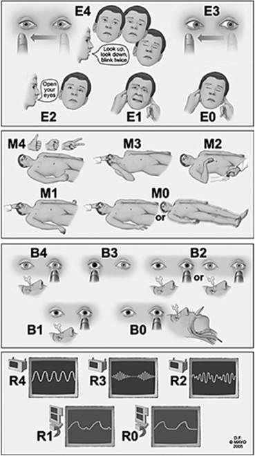
The FOUR (Full Outline of UnResponsiveness) score, not surprisingly, has four components, each of which has 4 as the maximal grade. The FOUR score is shown in Figure 147.1.
This score is simple to use and has significant advantages. It is able to recognize signs that suggest increased intracranial pressure and uncal herniation. It also recognizes respiratory patterns that not only indicate a need for ventilatory support but are also indicative of a declining level of consciousness and abnormal respiratory pattern. A recent validation study also found that the score is predictive of in-hospital death (10). This score can be used to communicate between nurses, staff and residents, and indicates the depth of coma.
|
|
|
Figure 147.1. The FOUR score components are eye response, motor response, brainstem reflexes, and respiration. The eye responses are E0 for eyelids that remain closed with pain; E1 for eyelids closed but open to pain; E2 for eyelids closed but open to loud voice; E3 for eyelids open but not tracking; E4 for eyelids open or open to tracking or blinking to commands. The motor responses are M0 for no response to pain or a generalized myoclonic status epilepticus; M1 for extension response to pain; M2 for flexion response to pain; M3 for localizing to pain; M4 for thumbs up, fist, or peace sign. The brainstem reflexes are B0 for absent pupil, corneal, or cough reflex; B1 for pupil and corneal reflexes absent; B2 for pupil or corneal reflexes absent; B3 for one pupil wide and fixed; B4 for pupil and corneal reflexes present. And finally, respiration responses are R0 for breathes at a ventilator rate or apnea; R1 for breathes above ventilator rate; R2 for not intubated and irregular breathing; R3 for not intubated and Cheyne-Stokes breathing pattern; and R4 for not intubated, regular breathing pattern. |
Neurological Examination
The next step in the neurologic examination is the examination of the brainstem reflexes (11,12). The pupillary responses are typically present; very few patients in the ICU have an abnormality in pupil diameter. Bilateral myosis is often seen because most patients are treated with opioids. A unilateral myosis represents a Horner syndrome when ptosis is present. In the ICU, it commonly reflects damage to the sympathetic pathway after placement of an internal jugular vein catheter. Extensive thoracic surgery or brachial plexopathies are also causes for unilateral myosis; (it is surprisingly common that the consult is for a “dilated eye” on the opposite side). Bilateral mydriasis with good light responses is due to delirium or anxiety. A fixed, dilated pupil indicates a third cranial nerve involvement. This could be due to direct compression from herniated brain tissue, but more likely is due to shift of the brainstem in which the third nerve is tethered. This is an important clinical sign that indicates brainstem distortion and immediately indicates the presence of a new structural mass. However, a major pitfall in the ICU is that unilateral pupillary dilatation can be seen with aerosolized anticholinergics and may cause alarm. The mist condenses on eyelids and then touches the cornea, causing a dilated pupil.
Gaze deviation is an important clinical sign, although it has very little localizing value, and can be seen in acute hepatic or renal failure without a structural brain lesion. Downward gaze indicates a lesion in the thalamus or dorsal midbrain and is frequently seen after hypoxic/ischemic injury. Traditionally, gaze preference indicates a lesion in one hemisphere, which, most of the time, is a lesion of the frontal lobe. The eyes turn toward the abnormality because one eye field is unopposed. Skew deviation, in which the eyes at rest are not lined up correctly, is a classic sign of a midbrain or pons lesion.
Spontaneous eye movement abnormalities have been noted. Common eye movements are the following:
· Ping pong: Horizontal conjunctive deviation of the eyes alternating every few seconds
· Convergence nystagmus: Ocular divergence in slow motion followed by rapid convergent jerk
· Ocular bobbing: Rapid downward conjugate movement with slow return to baseline
· Ocular dipping: Slow downward conjunctive movements with rapid return
These spontaneous eye movement abnormalities are indicative of a bihemispheric lesion and are rarely observed in toxic encephalopathie.
The motor tone is then investigated, and the patient is observed for the presence of rigidity or flaccidity. Marked flaccidity often indicates the presence of poisoning or drug intoxication. In patients who have been in the ICU for many months, a critical illness polyneuropathy should be considered. Twitching of the face and mouth may indicate seizures but, more often, will represent myoclonus status epilepticus. Myoclonus status epilepticus, with repetitive twitching in face, arm, and legs, is an important clinical sign. It has been seen in drug intoxications but also after cardiopulmonary resuscitation, and is a poor prognostic indication.
Localization Principles
It is important to make several decisions after certain neurologic findings have presented themselves. First, is the lesion structural or metabolic? Second, is the lesion in the cerebral hemispheres or brainstem? To achieve this objective, it is important to have a systematic diagnostic approach to coma in critical illness. The major categories of coma are as follows: hemispheric lesions with brain shift; diffuse bihemispheric structural lesion; diencephalon lesion involving both thalami; cerebellar lesion with brainstem compression or ischemia; primary brainstem lesion in the mesencephalon or pons; or a diffuse physiologic brain dysfunction from acute metabolic derangement, drugs, or intoxication; and, much less likely, a psychogenic unresponsiveness. Table 147.1 shows the common causes of these major categories of coma; a systematic diagnostic approach is shown in Table 147.2.
|
Table 147.1 Classification and Major Causes of Coma |
||
|
||
|
Table 147.2 Diagnostic Approach to Coma in Critical Illness |
||
|
|
Table 147.3 Frequent Abnormalities on Neuroimaging Studies in Coma |
||||||||||||||||||||||||||||||||||||||||
|
||||||||||||||||||||||||||||||||||||||||
After the cause of the coma state has been localized on examination, computed tomography (CT), magnetic resonance imaging (MRI) scan, cerebrospinal fluid testing, or electrodiagnostic tests may be helpful. An MRI scan is able to document certain abnormalities and can be very helpful in determining not only the cause but also the immediate action to undertake. The MRI scan has become extremely important in the diagnosis of coma, but it requires anesthesia support in many patients and, therefore, is cumbersome in certain circumstances. A summary of the abnormalities seen on neuroimaging studies of comatose patients is shown in Table 147.3. This can be used as a reference of the most common causes of coma seen in the ICU. The electroencephalograph (EEG) has lost much of its value, largely because MRI scans are able to clearly show the lesion. EEGs can be performed to detect nonconvulsive status epilepticus (NCSE), a rare condition in which the patient has subtle eye blinking or eye movements or gaze preference that is associated with epileptic discharges. The EEG can detect these abnormalities, and the patient can sometimes be successfully treated. Most often, the EEG is the reflection of significant brain injury, and aggressive treatment has not led to a marked improvement in outcome (13). The major EEG abnormalities include delta or theta activity, in which there is marked slowing of the activity that is typically seen in sedation, meningitis, encephalitis, or other diffuse injuries. Triphasic waves are typically associated with hepatic or renal failure. Burst suppression patterns are seen most likely after anoxic ischemic injury. EEG-defined patterns, such as alpha, theta, or spindle coma, have little prognostic value.
Laboratory tests are typically available on a daily basis in a critically ill patient, and therefore, these parameters should be known. Nevertheless, several essential laboratory studies are needed to exclude acute metabolic derangements in coma and certainly should be obtained if acute new changes have occurred in neurologic examination (Table 147.4).
Failure to Awaken After Surgery
A common problem is patients who fail to awaken after a general surgical or vascular surgical procedure. In the vast majority of patients, anoxic/ischemic injury or multiple infarcts are detected. Failure to awaken after surgery may also be due to multiple emboli to the brain. The risk of embolization is most significant in patients with severe atherosclerotic disease of the ascending aorta and thus is applicable to patients with major cardiovascular surgeries (14). However, not uncommonly, patients do not awaken because of the accumulation of narcotic and sedative agents. Many of these patients have underlying liver dysfunction that further increases accumulation of these drugs. Finally, acute postoperative hyponatremia, due to administration of a large volume of hypertonic fluid to patients, may be a cause of failure to awaken.
|
Table 147.4 Laboratory Tests in the Evaluation of Coma |
||
|
Failure to Awaken After Transplantation
Changes in consciousness have been recognized in patients having undergone major organ transplantation (15). This is most commonly seen with cardiac or liver transplantation. Although in the vast majority of patients, a delirium that may evolve to coma develops due to the toxicity of immunosuppressive agents, other reasons for loss of consciousness are hypoxic ischemic encephalopathy, central pontine myelinolysis, air embolization, acute uremia, acute graft failure, or multiple intracranial abscesses from aspergillosis. Immunosuppressive agents such as cyclosporin and tacrolimus can produce a marked neurotoxicity in which patients evolve from rambling speech and visual hallucinations into a stuporous stage that is associated with marked MRI abnormalities. The MRI scan often shows diffuse lesions in the white matter that are fully reversible with discontinuation of the medication. Both tacrolimus and cyclosporin are known offenders in this context; a new drug, sirolimus, has been found to be much less frequently associated with neurotoxicity (15).
Central nervous system infections are often serious and may be caused by opportunistic infections. Cytomegalovirus is the most common viral infection after organ transplantation but rarely results in cytomegalovirus encephalitis. In most of these patients, there is slow onset of abnormalities. Serious complications result from infection with Cryptococcus neoformans or Aspergillus fumigatus that leave not only multiple brain abscesses but also multiple intracranial hemorrhages. The outcome from these infections is very poor.
Sudden loss of consciousness may indicate a new onset of seizures in transplant recipients; the incidence in liver transplantation is recognized to be about 20%. An acute metabolic derangement—such as hypernatremia or hyperglycemia—may be implicated, but in a large proportion of patients, an intracranial hemorrhage or bacterial abscess is the causative factor. Seizures in cardiac transplantation occur in 10% to 15% of transplanted patients and most of the time are associated with an ischemic stroke.
Failure to Awaken in Multisystem Trauma
Trauma units commonly admit patients with not only multiple fractures but also with traumatic brain injury (TBI). The initial attention paid to the patient with TBI may be less in patients with severe shock due to blood loss and, since many of these patients require urgent exploration, they are taken to the operating room with an inadequate evaluation of their brain function. Multitrauma patients are at high risk of TBI, so close observation is important. In fact, these patients may need intracranial pressure monitoring, particularly if they do not localize to pain or open their eyes to pain, or have brainstem reflex abnormalities. Many traumatic parenchymal lesions are localized in the frontal and temporal lobes, and there is a significant risk of secondary deterioration from swelling. In the most severe circumstances, diffuse axonal injury is present, in which the injury itself causes swelling with multiple so-called shear lesions in the brain. These are typically in the frontal cortex, white matter, basal ganglia, thalamus, and internal capsule. Rarely, failure to awaken after trauma is due to the fat embolization syndrome; these patients often have axillary or subconjunctival petechiae, are markedly hypoxemic, and frequently have pulmonary edema. These patients have large bone fractures and often present 12 to 75 hours after the initial traumatic injury.
Management of Coma
Initial management is determined by the presence or absence of increased intracranial pressure (ICP). Patients with a supratentorial mass lesion or diffuse edema are best treated with placement of an ICP monitor, followed by the brief (hours) use of hyperventilation, with a target of a PaCO2 of 30 mm Hg (Table 147.5). Mannitol is a more effective method. Mannitol is typically administered at a dose of 1 gm/kg using a 20% solution, with the goal of increasing plasma osmolality to 310 mOsmols/L. Mannitol decreases brain volume by extracting water from brain tissue, which is generated through an osmotic gradient. It is an important initial intervention and can bridge the patient to neurosurgical evacuation. Hypertonic saline (7.5 or 23.4%) may be more effective (30 cc bolus) but requires infusion through a central line. Surgical evacuation of the mass is indicated before further brainstem compression occurs and all brainstem function is lost. The use of corticosteroids is highly questionable and may have a role only in patients who have edema surrounding a metastatic lesion or primary brain tumor. It has no role in closed-head injury, cerebral infarction, or cerebral abscess. Patients presenting in coma from an infectious disease such as acute bacterial meningitis need immediate IV infusion with antibiotics. The empiric treatment is discussed elsewhere but includes cefotaxime (2 g intravenously (IV) every 6 hours), vancomycin (2 g IV every 12 hours), and corticosteroids. If herpes simplex encephalitis is considered, acyclovir (10 mg/kg IV every 8 hours) is administered. In practice, in most patients with an infectious cause but no clear etiologic factors, both antibacterial and antiviral treatments are initiated until the spinal fluid examination reveals the true cause of infection.
|
Table 147.5 Management of Acute Supratentorial Mass with Brain Shift |
||
|
The neurosurgeon's role in the treatment of comatose patients includes removal of the mass and placement of a ventriculostomy, particularly in patients with a subtentorial mass or brainstem lesion. A craniotomy to remove a bone flap can be performed to allow for swelling. This is an option in patients with massive middle cerebral artery infarction or those with a swollen traumatic contusion. In patients with a brain abscess in the setting of organ transplantation, a drain can be placed in the abscess. The management of patients in coma from a subtentorial lesion is noted in Table 147.6.
Prognosis
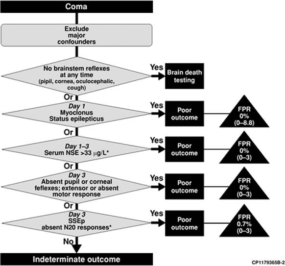
Prognosis may be determined before hospital admission. Time without circulation and poor airway control may all play an important role in outcome; in TBI, outcome is related to prehospital hypoxemia. Many guidelines have been proposed to determine the futility of care (16,17,18,19,20). Certain neurologic conditions may be associated with no hope for good recovery. These include patients with myoclonus status epilepticus and brain swelling after cardiac arrest; patients with multiple territorial infarcts and brain swelling after cardiac surgery; occlusion of the basilar artery and coma; multiple intracranial hemorrhages associated with tissue plasminogen activator (tPA); pontine hemorrhages with hypertension and extension to the midbrain and thalamus; multiple hemorrhagic contusion and associated extradural hematomas; patients with gunshot wounds; and intraventricular extension. In any of these circumstances, outcome is poor and the level of care should be discussed with family members. Major reasons for consultation in the ICU are prognostication in a comatose patient after cardiopulmonary resuscitation. Recent guidelines have been proposed by the American Academy of Neurology (17). An evidence-based evaluation has determined that myoclonus status epilepticus, the absence of corneal and pupillary reflexes, and absent N20 potentials on somatosensory evoked potentials (SSEP) are important indicators of poor outcome (Fig. 147.2). MRI and CT scan are useful diagnostic tests but have not been investigated thoroughly to be used as prognosticating tools. Outcome in TBI is very difficult to assess with certainty in young individuals, who can make a substantial improvement despite neuroimaging indicators of severe brain damage.
|
Table 147.6 Management of Acute Subtentorial Mass or Brain Stem Lesion |
||
|
Brain Death
Loss of all brain function is not frequently noted but may occur after traumatic brain injury, severe anoxic/ischemic injury, and cardiac resuscitation. Patients with catastrophic intracranial hemorrhages and massive hemispheric infarcts, cerebral edema, and fulminant hepatic failure are other major examples (8). Brain death is equivalent to death, where the time of its determination is the time of death. Definition of brain death implies documentation of loss of consciousness, no motor response to painful stimuli, no brainstem reflexes, and apnea. A structural lesion on CT scan is commonly found.
The diagnosis of brain death is made in the following steps:
1. First, the cause has to be identified, and coma has to be irreversible.
2. Major confounding factors have to be excluded, and there should be no reversible medical illness.
3. The core temperature should be at least 32°C, and no confounding pharmaceutical agents should have been administered or lingering on.
4. On examination, there is no motor response to pain applied to the face and limbs. Any movement that is seen must be attributable only to a spinal cord response.
5. There should be absent pupil response, absent cold caloric oculovestibular response, absent corneal reflex, absent cough to bronchial suctioning, and
6. Apnea with a PaCO2 of 60 mm Hg—or a 20 mm Hg increase from pretest baseline. The apnea test is done under controlled circumstances using the oxygen diffusion method.
|
|
|
Figure 147.2. Algorithm for determination of prognosis of coma after cardiopulmonary resuscitation. FPR, false positive ratio; NSE, neuron-specific enolase; SSEP, somatosensory evoked potentials. *These laboratory tests may not be available on a timely basis. |
If a patient fulfills all these criteria, brain death can be diagnosed. It is useful to have two examinations 6 hours apart, although the American Academy of Neurology considers this optional. In younger children, electrophysiologic tests are required.
Withdrawal of Care
In the discussion of withdrawal of care, it is important to have a neurologist involved to explain to the family members the findings and how they play a role in their assessment. Palliative care includes use of lorazepam and phosphenytoin to prevent seizures but also involves the use of propofol to reduce myoclonus status epilepticus, which can be a dreadful sight to family members. Neurologic complications in critically ill patients are often major setbacks, certainly when the patient has lapsed into coma. Failure to awaken after 1 week in a structural coma is often associated with a high mortality rate due to withdrawal of support, either instigated by directives of the patient or by family members.
References
1. Young GB, Pigott SE. Neurobiological basis of consciousness. Arch Neuro. 1999;56:153–157.
2. Wÿdicks EFM. The Comatose Patient. Oxford, England: Oxford University Press; 2008.
3. van der Mast RC, Roest FH. Delirium after cardiac surgery: a critical review. J Psychosom Res. 1996;41:13–30.
4. Dyer CB, Ashton CM, Teasdale TA. Postoperative delirium. A review of 80 primary data-collection studies. Arch Intern Med. 1995;155:461–465.
5. Brown TM. Drug-induced delirium. Semin Clin Neuropsychiatry. 2000;5:113–124.
6. O'Keeffe ST, Ni Chonchubhair A. Postoperative delirium in the elderly. Br J Anaesth. 2004;73:673–687.
7. Wÿdicks EFM, Crawford RE. Clinical diagnosis of prolonged states of impaired consciousness in adults. Mayo Clinic Proceedings. 2005;80:1037–1045.
8. Wijdicks EFM. The diagnosis of brain death. N Engl J Med. 2001;344:1215.
9. Smith E, Delargy M. Locked-in syndrome. BMJ. 2005;330:406–409.
10. Wijdicks EF, Bamlet WR, Maramattom BV, et al. Validation of a new coma scale: The FOUR score. Ann Neurol. 2005;58(4):585–93.
11. Wijdicks EFM. Neurologic Complications of Critical Illness. Contemporary Neurology Series 64. 2nd ed. Oxford, England: Oxford University Press; 2002.
12. Wijdicks EF. Neurologic complications in critically ill patients. Anesth Analg. 1996;83(2):411–419.
13. Kaplan PW. The EEG in metabolic encephalopathy and coma. J Clin Neurophysiol. 2004;21:307–318.
14. Gootjes EC, Wijdicks EF, McClelland RL. Postoperative stupor and coma. Mayo Clin Proc. 2005;80(3):350–354.
15. Wijdicks EF. Neurotoxicity of immunosuppressive drugs. Liver Transpl. 2001;7(11):937–942.
16. Wijdicks EF, Rabinstein AA. Absolutely no hope? Some ambiguity of futility of care in devastating acute stroke. Crit Care Med. 2004;32(11):2332–2342.
17. Wijdicks EF, Hijdra A, Young GB, et al; Quality Standards Subcommittee of the American Academy of Neurology. Practice parameter: prediction of outcome in comatose survivors after cardiopulmonary resuscitation (an evidence-based review): report of the Quality Standards Subcommittee of the American Academy of Neurology. Neurology. 2006;67(2):203–210.
18. Bates D. The prognosis of medical coma. J Neurol Neurosurg Psychiatry. 2001;71(Suppl 1):i20–i23.
19. Bateman DE. Neurological assessment of coma. J Neurol Neurosurg Psychiatry. 2001;71(Suppl 1):i13–i17.
20. Chi JH, Knudson MM, Vassar MJ, et al. Prehospital hypoxia affects outcome in patients with traumatic brain injury: a prospective multicenter trial. J Trauma. 2006;61(5):1134–1141.