Hsiu-Po Wang
Immediate Concerns
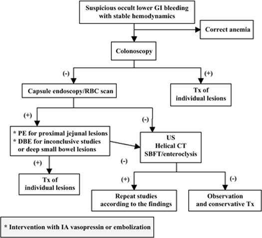
Lower gastrointestinal (LGI) bleeding (LGIB) may be the primary cause of admission to the intensive care unit (ICU) or occurs during the course of ICU care for disorders other than LGIB. The initial approach to patients with LGIB, whether occult or acutely overt, is resuscitation to maintain organ perfusion. ICU physicians must maintain adequacy of airway and circulation, including ensuring an open airway and adequate intravenous access. Thereafter, the severity of bleeding should be graded to determine whether urgent diagnostic and interventional procedures need be initiated immediately. The consequences of LGIB should be prevented or treated, just as would an acute myocardial infarction (AMI) induced by anemia or unstable hemodynamic status. A summary approach to ICU patients is noted in Table 153.1 and Figure 153.1.
Evaluation
1. Is there true gastrointestinal (GI) bleeding (for suspicious cases with anemia in the ICU)?
2. Is the bleeding from the lower or upper GI tract?
3. Are there any adverse consequences caused by the LGIB?
4. What are the effective diagnostic and interventional tools to use for the workup of patients with LGIB?
Essential Diagnostic Tests and Procedures
1. History taking, including previous operative interventions and medications
2. Physical examination, including digital examination
3. Stool guaiac for anemic patients with suspicious GI blood loss
4. Complete blood count
5. Endoscopic procedures: sigmoidoscopy, colonoscopy, enteroscopy, and capsule endoscopy
6. Barium study
7. Bedside ultrasound (US)
8. Radiologic procedures: angiography
9. Nuclear scintigraphy: technetium-labeled red blood cell scan
10. Other examinations for affected organs due to unstable hemodynamics, such as electrocardiogram (ECG) for AMI
Treatment
1. Ensure stable hemodynamic status, using fluids and transfusion of blood components.
2. Endoscopic therapies include local injection, electrocoagulation, hemoclipping, argon plasma coagulation, and elastic banding according to the nature of the bleeding.
3. Radiologic interventions include angiography with intra-arterial vasopressin infusion, embolization, and glue.
4. The adverse events caused by treatment modalities, such as perforation caused by endoscopic thermocoagulation or bowel infarction due to angiographic embolization, should be monitored and managed.
5. Surgical consultation is warranted for severe LGIB in which the patient cannot be resuscitated, or if all available therapeutic modalities fail.
Overview
Gastrointestinal diseases are often encountered in the ICU setting, either as the major cause of admission to the ICU or as a comorbid complication of another primary disease process. The consequences of LGIB in the ICU—anemia and hypovolemia—may delay the weaning or extubation of patients, which can prolong the ICU course. LGIB is defined as a bleeding source distal to the ligament of Treitz, thus involving the small bowel and colon, and accounts for an estimated 20% to 24% of all major GI bleeding (1,2). It has been estimated that the annual incidence of LGIB is approximately 0.03% in the adult population (3). Longstreth estimated the annual incidence of hospitalization for LGIB to be 20 to 30 cases per 100,000 persons (4). LGIB is more common in men than women, and the incidence increases with age, with a greater than 200-fold increase from the third to ninth decades of life (2). The true incidence of LGIB during ICU hospitalization is not precisely clear; the incidence for ICU patients with acquired hemorrhage (not primarily due to LGIB) was reported as 0.94% (5).
LGIB is clinically distinct from upper gastrointestinal (UGI) bleeding (UGIB) in epidemiology, management, and outcome. LGIB is approximately 20% to 33% as common as UGIB (2,6,7). LGIB generally has a lower mortality rate than does UGIB (8), but mortality is markedly higher in patients who begin bleeding after admission: 2.4% versus 23% (4). Most deaths are not the direct result of uncontrolled bleeding, but rather exacerbation of an underlying disorder or development of a nosocomial complication. Lin et al. (5) noted a 53% mortality in 55 patients, but this outcome was attributable to LGIB in only two patients. In another study, Lin et al. noted that patients with LGIB and comorbid illness had a higher mortality rate than those without: 29.5% versus 4.3% (9). No matter whether reported in the general population or in an ICU-based study, LGIB remains a difficult diagnostic and treatment problem for several reasons:
|
Table 153.1 Summary of an Approach to ICU Patients with Lower Gastrointestinal Bleeding |
||
|
1. Bleeding can originate from any part of the lower GI tract.
2. Blood loss is often intermittent in nature, and it is difficult to identify the source in the absence of active bleeding, especially angiographically.
3. The colon preparation before urgent colonoscopy is, obviously, needed but often incomplete (10).
4. Recurrent bleeding due to angiodysplasia (7) or diverticula (8,11) may be seen with LGIB.
5. Unlike UGIB, there are no evidence-based and effective pharmacologic therapies for LGIB.
Among the many causes of LGIB (Table 153.2), diverticular bleeding, angiodysplasia, colitis, and neoplasm have been reported to be the most frequent (4,12). While the data from most reports are mixed with both non-ICU and ICU patients, the spectrum of LGIB from ICU patients should be different from others. Data from a study limited to medical ICU patients have shown ischemic colitis and acute hemorrhagic rectal ulcers to be the most frequent causes of LGIB, followed by colitis and diverticular bleeding (5). Our unpublished data—from surgical, trauma, and medical ICUs—showed acute hemorrhagic rectal ulcers followed by ischemic colitis to be the most frequently encountered causes (Table 153.3).
Patient age is a very important factor in the differential diagnosis of GI bleeding. Patients younger than 40 years are more likely to suffer from small bowel tumors, such as lymphomas, carcinoid tumors, and adenocarcinomas; anatomic anomalies such as Meckel diverticulum and Dieulafoy lesions; genetic problems such as polyps from a hereditary polyposis syndrome; or Crohn disease and ulcerative colitis, which are common in Western countries and, recently, increasing in Asia. Patients older than 40 years are more prone to bleeding from vascular lesions and neoplasm (12). Lewis et al., while evaluating small bowel bleeding, noted that in patients between 30 and 50 years, tumors were the most common abnormalities; in patients younger than 25 years, Meckel diverticulum was the most common source of small bowel bleeding, whereas vascular ectasias predominated in the elderly (13).
|
|
|
Figure 153.1. General approach to lower gastrointestinal bleeding. GI, gastrointestinal; CVP, central venous pressure; BP, blood pressure; ECG, electrocardiogram; Pt, patient; CAD, coronary artery disease; Hb, hemoglobin; PT, prothrombin time; PTT, partial thromboplastin time. |
Presentation
Specific groups may have specific causes of LGIB. In the immunosuppressed patient, such as those with human immunodeficiency virus (HIV) infection and renal or pancreatic transplant patients, LGIB is often caused by cytomegalovirus (CMV) ulcers. Renal failure is a well-known risk factor for angiodysplasia (14); this is also prone to occur in patients with aortic valvular stenosis (15). Radiation colitis should be considered in patients with LGIB and a history of radiation therapy for cervical or prostate malignancy. Although some lesions (i.e., diverticula) are common in the left colon, most bleeding sites are noted in the right colon. In approximately 76% of patients, a definitive site of bleeding will be diagnosed (16). Bleeding in most patients with LGIB will stop spontaneously, although continued or recurrent bleeding during an acute episode occurs in 10% to 40% of patients (17,18,19). Finally, 2% to 15% of patients with presumed LGIB will have UGIB (20); the cause of overt GI bleeding remains undetermined in 4% to 15% of cases, even after upper GI endoscopy and colonoscopy (21).
|
Table 153.2 Causes of Lower Gastrointestinal Bleeding |
||||||||||||||||||||||||||||||
|
||||||||||||||||||||||||||||||
|
Table 153.3 Causes of Lower Gastrointestinal Bleeding in Surgical, Trauma, and Medical ICUs |
||||||||||||||||||||||||||||||
|
||||||||||||||||||||||||||||||
LGIB may present in multiple ways, including occult fecal blood; iron deficiency anemia; melena; intermittent scant hematochezia; or acute, massive, and overt bleeding (22). Patients with chronic LGIB may only present with anemia, whereas those with acute LGIB may complain of passing bright red blood per rectum, dark blood with clots, or, less commonly, melena. Patients with brown or infrequent stools are unlikely to have brisk bleeding, and those with frequent passage of red or maroon stool may have aggressive ongoing bleeding (23). Pallor, fatigue, chest pain, palpitations, dyspnea, tachypnea, tachycardia, posture-related dizziness, and syncope are suggestive of hemodynamic compromise and demand aggressive care. The severity of LGIB may be underestimated due to compensatory mechanisms that delay the onset of hypotension. Careful history taking and physical examination are essential to the care of LGIB patients.
Modern management of LGIB encompasses emergency medicine, intensive care medicine, gastroenterology, interventional radiology, and surgery. The approach to LGIB patients is initially aimed at immediate assessment and stabilization of the hemodynamic status, followed by identifying the source of bleeding, stopping any active bleeding, treating any underlying lesions, monitoring and managing comorbid illness, and preventing recurrent bleeding.
The first phase of management adheres to the ABCs of resuscitation, governed by the same priorities that apply to all acutely ill patients. Of course, resuscitation and diagnosis must proceed simultaneously. To identify the source of bleeding, endoscopy, mesenteric arteriography, and radionuclide scintigraphy are the major tools, in addition to history taking and physical examination. Endoscopy, the gold-standard procedure for LGIB, includes:
· Sigmoidoscopy
· Colonoscopy
· Capsule endoscopy
· Push enteroscopy
· Double-balloon enteroscopy
Endoscopic local hemostasis can be performed at the same time the diagnosis is made. Mesenteric arteriography and radionuclide scintigraphy may be used in difficult LGIB cases, with the former providing the ability for embolization or delivering pharmacotherapy. Advances in endoscopic and radiologic hemostasis techniques appear to decrease the rates of rebleeding and surgical intervention (24,25). Transabdominal ultrasound (US) has become popular as a first-line diagnostic tool in a GI emergency. Details of these procedures are noted below. The limited pharmacologic treatments available for LGIB—estrogen and octreotide for angiodysplasia, and topical formalin for intractable bleeding from radiation colitis—are strongly supported (26,27). Even with the advances in localization studies, approximately 10% of patients still require surgical intervention without having their bleeding source identified (28).
Anatomic Considerations of Lower Gastrointestinal Bleeding
LGIB is anatomically defined as bleeding located from the ligament of Treitz to the anus, and may include the jejunum, ileum, ileocecal valve, colon (ascending, transverse, descending, sigmoid, rectum), and anus. Given the length of lower GI tract (the small bowel averages 6.7 meters), as well as the special anatomic problem for the small bowel, such as its free intraperitoneal location, multiple overlying loops, and active contractility, there are several management issues for LGIB; these include choice of tests for fecal occult blood, the necessity of nasogastric (NG) tube placement, and the application of endoscopy, radiologic procedures, and US.
Fecal Occult Blood
This is the most common form of GI bleeding. In the ICU, patients suspected of having GI blood loss, secondary to findings of persistent anemia, will have a fecal occult blood study as the first investigative step. While there are numerous types of fecal occult blood tests, the test used determines the location in the GI tract where blood is likely to be detected (29).
Guaiac-based Tests
Guaiac-based tests, the classic fecal occult blood study, utilizes hemoglobin's pseudoperoxidase activity. Guaiac turns blue after oxidation by oxidants or peroxidases in the presence of an oxygen donor such as hydrogen peroxide. Because hemoglobin is degraded in the GI tract, guaiac-based tests are more sensitive for detecting bleeding in the lower than upper GI tract (21). However, the characteristics of specific guaiac-based tests from different companies vary. Whether a guaiac-based test will be positive or not is related to the quantity of blood present in the stool, which is related to the size and location of the bleeding lesion (30,31). Because bleeding colonic lesions are more likely to lead to undegraded blood and heme in the stool, guaiac-based tests are best at detecting these distal lesions (29). Various factors influence guaiac test results; for example, fecal rehydration affects the reactivity of guaiac-based tests, and may raise sensitivity but reduce specificity (32). Additionally, foods that contain peroxidases or animal hemoglobin can cause false-positive guaiac test results. False-negative guaiac-based tests may be seen with hemoglobin degradation, sample storage, and vitamin C ingestion (21). Orally administered iron, even in large amounts, does not cause a positive guaiac reaction (33). Bismuth-containing antacids and antidiarrheals cause dark stool, which should not be confused with a positive guaiac reaction. Guaiac-based tests are rapid bedside studies.
Immunochemical Fecal Occult Blood Tests
Immunochemical fecal occult blood tests detect human globin epitopes, and are highly sensitive for the detection of stool blood (34). These tests do not detect UGI blood because globin molecules are degraded by UGI tract enzymes. Theoretically, these tests have a higher specificity for the detection of colonic lesions than guaiac-based tests. However, they are limited by technical problems and the need for more intensive laboratory processing. A false negative may occur from hemoglobin degradation and sample storage.
Heme-porphyrin Test
The heme-porphyrin test provides a highly accurate determination of total stool hemoglobin based on a spectrofluorometric method that measures porphyrin derived from heme. The heme-porphyrin test is the most sensitive method of detecting occult blood loss of either the upper or lower GI tract. The results of the heme-porphyrin test are neither affected by intraluminal degradation of hemoglobin nor by the interference of peroxidase-producing substances. False positives result from animal hemoglobin and red meats, which contain myoglobin, a heme-containing protein; false negatives are a consequence of sample storage. In summary, guaiac-based tests and immunochemical fecal occult blood tests focus on LGIB, especially from the colon. The heme-porphyrin tests cannot discriminate between bleeding from the upper and lower GI tract.
In the cardiac ICU, warfarin or low-dose aspirin is used frequently. Neither of these alone appears to cause positive guaiac-based, fecal occult blood tests (35). A positive fecal occult blood test in this setting should raise the possibility of a GI tract abnormality, and requires appropriate evaluation. Jaffin et al., prospectively evaluating the GI tract in anticoagulated patients with positive guaiac-based fecal occult blood tests, showed that 20% of these results were associated with malignancy (36).
Nasogastric Tube Placement
The necessity of NG tube placement and gastric lavage for acute LGIB to exclude a UGI source has not been studied prospectively. Theoretically, blood originating from the LGI tract will not retropulse across the ligament of Treitz to the duodenum or stomach, and there should be no blood content in the nasogastric aspirate. Nonetheless, approximately 2% to 15% of patients presenting with acute severe hematochezia have an upper GI source of bleeding identified on upper GI endoscopy (20). Therefore, some favor the routine use of the NG tube to exclude the possibility of a UGI bleed, although this is controversial and, overall, nasogastric aspiration localizes bleeding accurately only in 66% of attempts (37). In the American Society for Gastrointestinal Endoscopy (ASGE) guideline, NG tube placement to rule out a UGI source of bleeding may be considered if a source is not identified on colonoscopy, particularly if there is a history of UGI symptoms or anemia (38). Patients with hemodynamic compromise and hematochezia should have an NG tube placed (39). The absence of blood in NG aspirate is not sufficient to exclude UGI bleeding—16% of patients with bleeding secondary to a duodenal ulcer have a negative NG tube lavage. The presence of bile without blood is considered as the most reassuring result of a negative NG aspirate, and indicates the absence of active UGIB (40).
In spite of the length of the LGI tract, there are anatomically fixed portions, such as the ascending and descending colon and rectum, which makes a US survey of part of the LGI tract possible. The proximal jejunum and terminal ileum can be traced from the fixed duodenum and cecum, respectively (41). US may be considered as a screening tool for LGIB, or applied when the bleeding source cannot be identified by other modalities.
The small bowel is a problematic area to evaluate because of the limitations of standard upper GI endoscopy and colonoscopy. The length of the small intestine and its loosely supported and looped structure on the mesentery make conventional endoscopic techniques difficult and, frequently, inadequate. Push enteroscopy can reach only the proximal jejunum. Even with the maximal depth of insertion, approximately 160 cm beyond the ligament of Treitz, most of the small intestine still remains unexamined. The distal ileum, the small intestine closest to the colon, can be surveyed occasionally with traditional colonoscopy retrograde examination via the ileocecal valve. Initially introduced by Yamamoto in 2001, double-balloon enteroscopy (DBE) has been implemented as a method of deeper intubation of the small bowel (42). By combining oral intubation and anal intubation, a complete study of the small bowel can be achieved.
Blood supply to the LGI tract is separated into the superior mesentery artery (SMA) and inferior mesentery artery (IMA) territories. The SMA territory includes the jejunum, ileum, ascending colon, and transverse colon, while the IMA territory covers the descending colon, sigmoid colon, and rectum. The territory of the mesentery vessels is very important for interventional angiography or second-look endoscopy if the first endoscopic survey fails.
Approach to Lower Gastrointestinal Bleeding
Resuscitation
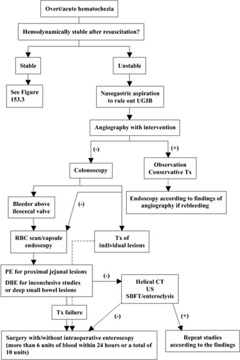
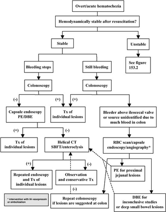
Approximately 85% of GI bleeding episodes stop spontaneously, whereas the remainder require aggressive resuscitation, diagnostic modalities, and often intense medical and/or surgical management. The first management step for a patient presenting with overt LGIB is the ABCs of resuscitation. Resuscitation is imperative to restore euvolemia and prevent complications of blood loss in the cardiac, pulmonary, renal, or neurologic systems. This takes place in parallel with the initial evaluation of the patient; resuscitation must not be withheld or delayed for diagnostic procedures. The patient's respiratory and heart rates, and blood pressure, including orthostatic measurements, should be assessed. Attention to the airway is important when the LGIB is caused by an obstructive lesion, which may lead to vomiting with the consequent high risk of aspiration. Postural hemodynamic changes, chest pain, palpitations, syncope, pallor, dyspnea, and tachycardia suggest hemodynamic compromise (43); the severity of bleeding is easy to underestimate due to compensatory mechanisms. An orthostatic decrease in systolic blood pressure greater than 10 mm Hg or an increase in heart rate greater than 10 beats/minute indicates an acute loss of at least 15% of blood volume (44). With hemodynamic compromise, two 16-gauge or larger intravenous (IV) catheters should be secured immediately; central venous access can be established in unstable patients. Packed red blood cells should be utilized in hemodynamically unstable patients, with the goal of maintaining a hematocrit of approximately 30% in the elderly and in those with heart disease or who are otherwise compromised physiologically, and 20% to 25% in younger patients (45). The initial hematocrit may not be the true value, requiring up to 72 hours for equilibration with the intravascular space (46). The presence of coagulopathy (international normalized ratio [INR] greater than 1.5) or thrombocytopenia (less than 50,000 cells/µL) should be corrected with fresh frozen plasma or platelet transfusions, respectively. Oxygen should be administered to keep the SpO2 between 93% and 95% at a minimum, and vital signs and urine output should be closely monitored. In the elderly or those with a history of cardiac disease, an ECG and cardiac enzyme analysis should be considered. Approaches for LGIB with hemodynamic instability and hemodynamically stable LGIB are noted, respectively, in Figures 153.2 and 153.3.
Determination of Bleeding Site: Noninstrumental
History
Careful history taking is helpful for determining the level of bleeding from the GI tract and to work out a differential diagnosis of the LGIB. Included should be the duration, frequency, and color of the stool; related symptoms of the GI tract such as constipation, fever, or location of pain; medical or surgical history; and history of medications. Except for asymptomatic patients with only anemia due to GI bleeding, the color of the stool is always queried. The patient presenting with acute LGIB may complain of passing bright red blood per rectum, dark blood with clots, or, less commonly, melena. The stool appearance, largely dependent on blood transit time, may be suggestive of the location of bleeding. Blood that has been in the GI tract for <5 hours is usually red, whereas blood present for >20 hours is usually melenic. Upper GI, small bowel bleeding, or slow oozing from the right colon usually produces melena, whereas patients with hematochezia typically have left colonic or rectal lesions (47). If the blood coats the surface of stool, the left colon—especially the rectum and anus—is more likely to be the bleeding source. Blood dripping into the toilet may occur with hemorrhoidal bleeding. If the stool is mixed with blood, the source may be in the right colon. Of course, massive UGIB can occasionally masquerade as lower GI bleeding in 10% to 15% of patients presenting with severe hematochezia (20). This may happen when massive bleeding originates from esophageal or gastric varices; insertion of an NG tube may be helpful in this situation if the patient does not have hematemesis.
The duration and frequency of bleeding may help in determining the severity of the problem. Related GI tract symptoms may lead to the diagnosis of LGIB. Severe constipation should prompt an investigation for colon cancer or a stercoral ulcer; diarrhea may indicate enterocolitis; and fever or local pain may indicate an inflammation-related bleeding source, such as infectious colitis or inflammatory bowel disease. The medical or surgical history may provide clues to the causes of LGIB. A previous history of colonic polyps, diverticulosis, or colonic tumor should be considered as a possible source of LGIB during the initial evaluation. Renal failure is a well-known risk factor for angiodysplasia or arteriovenous malformation (AVM) (48), as is aortic stenosis (15). Note should be made of patients with renal impairment or who are being dialyzed, as these patients may have platelet abnormalities, resulting in a tendency to bleed if a lesion is present. Ischemic bowel may be present when severe abdominal pain and bloody stool occur in patients with severe atherosclerotic vascular disease, atrial fibrillation, or hypotension. Radiation therapy for prostate or pelvic cancer induces inflammatory changes of the rectum, and can produce radiation proctitis, presenting months or even years after the radiation exposure. A history of recent colonoscopy with polypectomy indicates postpolypectomy bleeding as the likely source. In patients who have undergone aortoiliac reconstructive surgery, the frequency of significant postoperative colonic ischemia ranges between 1% and 7% (49,50,51).
|
|
|
Figure 153.2. Approach for lower gastrointestinal bleeding with hemodynamic instability. UGIB, upper gastrointestinal bleeding; Tx, treatment; RBC, red blood cell; PE, push-type enteroscopy; DBE, double-balloon enteroscopy; CT, computed tomography; US, ultrasound; SBFT, small bowel follow-through. |
Medication history is also important. Medications that can damage the GI mucosa or exacerbate bleeding include nonsteroidal anti-inflammatory drugs (NSAIDs), alendronate, potassium chloride, and anticoagulants. Patients admitted with GI bleeding were more likely to be taking selective serotonin reuptake inhibitors (SSRIs) than controls; this association exists for LGIB as well as UGIB (52). Concurrent anticoagulation or use of NSAIDs may be important cofactors in potentiating bleeding (53). The use of aspirin or NSAIDs is strongly associated with both LGIB—chiefly from diverticula—and UGIB (54,55). NSAID enteropathy has been increasingly reported, and can be a potential cause of LGIB (56,57). A family history of colon cancer increases the likelihood of a colorectal neoplasm, and generally calls for a complete colonic examination in patients with hematochezia (58,59).
Physical Examination
A thorough physical examination is essential to assess loss of blood volume, a possible bleeding source, and comorbid conditions (especially for ICU patients). The comorbid conditions may affect the suitability for interventions, such as urgent colonoscopy. The physical examination should also include complete vital signs and heart, lung, and abdominal assessment, as well as an examination of the conjunctiva and skin. Pale conjunctiva indicate anemia. Cutaneous manifestations may suggest disorders causing GI bleeding; those caused by celiac sprue may indicate dermatitis herpetiformis. The polyposis syndromes (Peutz-Jeghers syndrome, Gardner syndrome, and Cronkite-Canada syndrome) often have cutaneous abnormalities, such as lip pigmentation in Peutz-Jeghers syndrome. Orthostatic vital signs are an important complement to standard monitoring in a patient with severe bleeding but without overt hemodynamic instability. Abdominal tenderness on examination may indicate an inflammatory process, such as ischemic colitis or inflammatory bowel disease (60). The rectal examination serves to identify anorectal lesions and confirm the stool color described by the patient. In addition, approximately 40% of rectal carcinomas are palpable during a digital rectal examination (61). Regardless of the presenting features and findings on physical examination, most patients with LGIB still warrant a full examination of the colon.
|
|
|
Figure 153.3. Approach for hemodynamically stable lower gastrointestinal bleeding. PE, push-type enteroscopy; DBE, double-balloon enteroscopy; Tx, treatment; SBFT, small bowel follow-through. |
Laboratory Examination
Initial laboratory studies should include a complete blood count, serum urea nitrogen, creatinine, coagulation profile (prothrombin time, INR, and partial thromboplastin time), liver tests, blood type and cross-match, and electrolytes. As discussed above, positive fecal occult tests (guaiac-based tests and immunochemical fecal occult blood tests) may favor LGIB. The blood urea nitrogen–to–creatinine ratio has been used as a noninvasive test to help distinguish UGIB from colonic sources of bleeding (62,63,64). In the study of Chalasani et al., a ratio of 33 or higher had a sensitivity of 96% for UGIB, although overlap was observed with LGIB, especially in patients with UGIB without hematemesis (62).
Determination of the Bleeding Site: Diagnostic Approach
Endoscopic Approach: General
Safety of Endoscopic Procedures for Critically Ill Patients
Colonoscopy remains the procedure of choice for evaluating patients with acute LGIB, and enteroscopy is considered for proximal small bowel bleeding. However, for ICU patients with acquired bleeding and hemodynamic instability, questions remain due to the possibility of severe comorbid illness, including respiratory distress, aortic aneurysm, life-threatening dysrhythmias, AMI, history of GI tract perforation or operation, severe lower GI obstruction, and bleeding tendency, to list only a few conditions. Colonoscopy complication rates in LGIB of the general population or patients admitted to the ICU primarily for hemorrhage are low, and the bowel preparation itself appears to be safe (65,66,67). Zuckerman and Prakash reviewed 13 studies and found an overall complication rate of 1.3% (68). A study of 55 ICU patients with acquired LGIB demonstrated an acceptable diagnostic rate of 67% (37 of 55 cases) without procedure-related complications, suggesting that bedside colonoscopy after preparation is safe for the critically ill (5).
Preparation before Endoscopic Procedures
In patients with massive active bleeding, colonoscopy is often frustrating and nonproductive, and sometimes dangerous. However, some clinicians still consider it the first diagnostic maneuver, even in the patient with severe ongoing bleeding (20,69). Traditionally, colonoscopy for LGIB was delayed because of the need for bowel preparation and the fear of increased procedural complications. Indeed, urgent colonoscopy in an unprepared colon can be challenging, or even dangerous. Good bowel preparation is important for an adequate and sensitive colonoscopy. Studies of urgent colonoscopies performed in an unprepared colon to evaluate for LGIB revealed completion rates as low as 35% (70,71). Jensen et al. studied patients with severe diverticular hemorrhage, noting that completion rates may reach up to 100% if aggressive bowel preparation is performed before urgent colonoscopy (24). Because of variable comorbid conditions, such as decreased bowel motility, obstruction, and electrolyte disturbance, colon preparation in ICU patients may not be as thorough as in the general population. There are limited studies of the effectiveness of colon preparation for LGIB in ICU patients. Lin et al. (5) noted that for patients with LGIB occurring after admission to ICU, the reach rate of the cecum with a colonoscope was 58% after enemas or an oral polyethylene glycol solution were administered.
In spite of providing a relatively feces-free colon, old methods for colon preparation with clear liquids, laxatives, and enemas or peroral gut lavage 48 to 72 hours before colonoscopy are time consuming, uncomfortable, and inconvenient for patients. For those in the ICU or with active LGIB, these methods are clearly not useful. Peroral gut lavage with saline or balanced electrolyte solutions have been proposed and were found to provide rapid, effective cleansing of the colon; however, the method is not tolerated in 11% of patients due to the high fluid volume—7 to 12 L—and might cause fluid and electrolyte disturbances (72). Recently, development of osmotically balanced solutions may provide minimal water absorption or secretion into the bowel lumen. Generally speaking, there are isoosmotic preparations, hyperosmotic preparations, and stimulant laxatives for colon cleansing. Polyethylene glycol electrolyte lavage solution (PEG-ELS) is an isotonic, nonabsorbable electrolyte solution that clears the bowel by washing out ingested fluid without significant fluid and electrolyte shifts. Rapid purge is best accomplished with polyethylene glycol-based solutions. They can be administered by a nasogastric tube or by drinking 1 L every 30 to 45 minutes, to a median dose of 5.5 L (range 4–14 L); 3 to 4 hours are required to cleanse the colon (73). Two liters of PEG-ELS plus bisacodyl or magnesium citrate have also been suggested for colon preparation (74). The studies comparing a standard 4-L PEG-ELS with 2 PEG-ELS with either a magnesium citrate or bisacodyl preparation have shown equal efficacy for colon cleansing (75,76,77). With PEG-ELS, 5% to 15% of patients have difficulty drinking the large amounts of fluid, or they develop symptoms such as nausea, vomiting, abdominal fullness, and cramps, leading to incomplete colon preparation (78,79). Hyperosmotic preparations, including monobasic and dibasic sodium phosphate (NaP) and magnesium citrate, draw plasma water into the bowel lumen to promote evacuation. In addition to hyperosmotic action, magnesium citrate also stimulates fluid secretion and intestinal motility through the action of cholecystokinin. Fleet Phospho-Soda (C. B. Fleet Co., Lynchburg, VA) is a poorly absorbed salt that produces intestinal fluid retention due to the osmotic load, and causes fluid evacuation from the bowel. Patients often prefer NaP to PEG-ELS because there is much less fluid to drink. Liquid and tablet forms of NaP are currently available. Stimulant laxatives include bisacodyl and senna extract. The former is a poorly absorbed diphenylmethane that stimulates colonic peristalsis, and the latter contains anthracene derivatives that may be metabolized by colonic bacteria into substances that enhance colonic motility.
The timing of colon preparation still varies among institutions. Early methods required preparation 48 to 72 hours before the procedure; of course, this is not ideal for ICU patients who need urgent interventions. Some institutions administer the solution on the day of the examination, even though the manufacturer recommends taking the medication the day before the procedure (74). For the critically ill patient with LGIB, there are no data on the “proper timing” of colon preparation. Further, preparation may be incomplete due to poor motility or ileus. Chiu et al. have suggested that PEG-ELS on the day of procedure may be adequate (74).
The safety of colon preparation is important for critically ill patients with comorbid illness; of particular concern are fluid overload and electrolyte disturbances. PEG-ELS causes no significant change in weight, vital signs, serum electrolytes, or complete blood count (80,81,82). It is a relatively safe colon preparation solution for patients with electrolyte imbalance, advanced liver disease, poorly compensated congestive heart failure, or renal failure, although in the study of Granberry et al., exacerbation of congestive heart failure after PEG-ELS administration was noted (83). NaP, a hyperosmotic preparation, may cause alterations in serum electrolytes and extracellular fluid status (84,85). Asymptomatic hyperphosphatemia is seen in up to 40% of patients, but clinically significant hyperphosphatemia is rare and usually limited to patients with renal failure (86,87,88). Twenty percent of patients had abnormally low serum potassium levels after bowel preparation with NaP (84). It is suggested that NaP is contraindicated in patients with renal failure, acute myocardial infarction or unstable angina, congestive heart failure, ileus, intestinal malabsorption, and significant ascites. Gremse et al. reported that the degree of asymptomatic hyperphosphatemia in children was greater than in adults, and recommended avoiding NaP in children with renal failure, congestive heart failure, ileus, and ascites (88).
Colonoscopy for Critically Ill Patients
Three primary diagnostic tools for LGIB are colonoscopy, radionuclide scintigraphy, and mesenteric arteriography.
Advances in endoscopic technology have brought colonoscopy to the forefront of the management of LGIB. Colonoscopy as the first choice for occult or stable LGIB is not in dispute. However, for brisk LGIB or LGIB with hemodynamic compromise, whether the attending physicians consider colonoscopy as the first diagnostic maneuver for brisk LGIB or LGIB with hemodynamic compromise is controversial. Colonoscopy in patients with severe hematochezia is impractical because of inadequate visualization caused by brisk blood loss (2,68). Some are reluctant to perform colonoscopy in hemodynamically unstable patients with ongoing bleeding, suggesting that these patients are best served by urgent angiography, perhaps in conjunction with surgical consultation (73,89). However, there is a reason to favor colonoscopy for acute LGIB. In addition to affording a rapid diagnosis, colonoscopy may indicate specific therapy (when [and if] the bleeders are found [24,90–92], although the rate for intervention in an ICU study was low [5]). Rapid endoscopic identification of a bleeding source, regardless of whether therapy is administered, may contribute to the clinical management of recurrent bleeding, if it occurs. Finally, compared with angiography, urgent colonoscopy has a higher diagnostic yield and a lower complication rate. In a retrospective study of 107 patients with severe LGIB, colonoscopy was diagnostic in 90% of patients and angiography in 48%; the former was therapeutic in 12% versus 22% for the latter (19). In another study, the diagnostic yield of colonoscopy was 82% versus 12% for angiography (20). Interestingly, most patients undergoing radiographic evaluation for LGIB—regardless of findings and interventions—will subsequently require a colonoscopy to establish the cause of bleeding. In addition to diagnosis, occasional therapy, and management planning, earlier colonoscopy does contribute to a shorter length of hospital stay (93). Hemodynamic instability, higher comorbidity, performance of a tagged red blood cell nuclear scan, and surgery for hemostasis were significantly associated with a decreased likelihood of discharge (93).
While most episodes of LGIB will stop spontaneously, 10% to 15% of patients undergoing urgent colonoscopy received endoscopic therapy. The lesions most amenable to colonoscopic treatment of LGIB, in most studies, are usually angiodysplasia or diverticulosis. Once it is identified as the source of bleeding, angiodysplasia is usually coagulated by methods including the following (5,24,94,95):
· Injection therapy (epinephrine, saline, or ethanol)
· Heater probe
· Monopolar and multipolar electrocoagulation
· Argon plasma coagulation
· Hemoclips
· Band ligation
In a study of ICU patients with nonprimary LGIB, spontaneous cessation occurred in 53% of patients; 29% achieved hemostasis with endoscopy, but had a higher rate (19%) of recurrent bleeding (5). Other studies, not limited to ICU patients, have reported rebleeding rates of 13% to 53%, and many patients may require more than one treatment (94,95).
Timing of Colonoscopy
The use of colonoscopy is controversial for critically ill patients with LGIB. Some believe that colonoscopy is best utilized in patients whose bleeding has stopped or slowed down (73,89), and others agree with early colonoscopy for LGIB. Evidence suggests that earlier intervention leads to more diagnostic and therapeutic opportunities (24,96). It has further been noted that early colonoscopy reduces the length of hospital stay, and therefore should decrease treatment costs (97,98). Urgent colonoscopy after bowel preparation with endoscopic treatment of patients with active diverticular bleeding or stigmata of bleeding has been shown to be highly effective in decreasing the need for surgical intervention (24,73).
In conclusion, prompt intervention may decrease the need for surgical exploration, as well as the rate of recurrent bleeding and length of hospital stay. Who needs urgent colonoscopy? Early identification of high-risk patients would allow the more selective delivery of urgent therapeutic interventions to those who will benefit. Clinical high-risk predictors in the first hour of evaluation in patients with severe LGIB have been proposed, and included an initial hematocrit of no more than 35%, the presence of abnormal vital signs 1 hour after initial medical evaluation, and gross blood on initial rectal examination (99). How early should the urgent colonoscopy be performed? The definition of urgent colonoscopy varies widely in the literature—from within 8 hours to 24 hours of presentation (5,20,24,100,101,102,103,104). Most definitions consider the procedure within 12 to 24 hours; more recently, the literature defines urgent colonoscopy as within 12 hours (20,24).
Sigmoidoscopy or Colonoscopy?
In studies of the general population with LGIB, diverticular and angiodysplastic bleeding are the most frequent events. Although anatomically prone to be located in the left colon, bleeding most often occurs in the right colon; thus, most endoscopists prefer a total colonoscopy. Flexible sigmoidoscopy can be performed in the initial evaluation of patients with LGIB, but the diagnostic yield of flexible sigmoidoscopy in LGIB is low, ranging from 9% to 58% (10). Regardless of presentation, flexible sigmoidoscopy may miss serious proximal pathology (105). However, in the study of ICU patients with acquired bleeding, 78% of responsible lesions were in the left colon (5). If the critical situation precludes use of total colonoscopy, sigmoidoscopy as the first maneuver may be acceptable for this patient group. Nonetheless, unless a definite and compatible bleeding source is identified with flexible sigmoidoscopy, the study of LGIB should proceed to a full colonoscopy in most patients.
In 2005, the ASGE offered the following guidelines regarding LGIB (38):
1. Colonoscopy is effective in the diagnosis and treatment of LGIB (prospective controls).
2. Colonoscopy is recommended in the early evaluation of severe acute LGIB (prospective controls).
3. Thermal contact modalities, including heat probe, and bipolar/multipolar coagulation and/or epinephrine injection can be used in the treatment of bleeding diverticula, vascular ectasia, or postpolypectomy bleeding (prospective controls).
Enteroscopy and Capsule Endoscopy for Critically Ill Patients
The small bowel has traditionally been a problematic area to evaluate because of the long length, looping, free intraperitoneal location, active contractility, and limits of standard endoscopy. It is estimated that 10% to 25% of LGIB originates in the small bowel and can pose a diagnostic dilemma for clinicians (106,107,108). Small bowel sources account for 0.9% to 7% of cases presenting with blood per rectum (19,20,109), and comprises approximately 5% of obscure GI bleeding (110). Upper endoscopy and colonoscopy appear to have limited roles in the investigation of small bowel bleeding, and are only useful when the bleeding source is the duodenum or the most distal segment of the small intestine (terminal ileum), respectively (53). Some clinicians have utilized peroral intubation with a standard colonoscope, reaching a point 20 to 60 cm distal to the ligament of Treitz; this may increase the diagnostic yield of GI bleeding of obscure origin by 17% to 46%—not an insignificant proportion (13,111,112,113). Small bowel enteroscopy is currently the best endoscopic investigative modality. Indeed, it has become the cornerstone of management in patients with obscure GI bleeding. Current tools for ruling out small bowel diseases include push-type enteroscopy (PE), double-balloon enteroscopy (DBE), intraoperative enteroscopy, and capsule endoscopy.
Small bowel evaluation may be performed with PE, which allows endoscopic evaluation of the proximal 60 cm of the jejunum or 150 cm distal to the pylorus. PE is probably the most commonly performed small bowel procedure today, and is often pursued when upper GI endoscopy and colonoscopy have failed to find the source for blood loss. PE may be performed using an enteroscope or pediatric colonoscope. The obvious limitation of push enteroscopy is the inability to reach lesions distal to the middle jejunum. PE can only examine a relatively short portion of the small bowel, even under fluoroscopy, and the true depth of insertion is unreliable. Some experts consider it complementary to capsule endoscopy, and believe that it should be performed only if capsule endoscopy or other modalities are positive for a proximal small bowel lesion. The use of an overtube to prevent looping of the instrument in the stomach increases the insertion depth by 10 to 25 cm (114). Prototype, variable stiffness enteroscopes are emerging in an attempt to achieve maximal insertion depth without the use of an overtube (115,116). Although PE cannot investigate the entire small bowel, an important benefit of PE is the ability to provide diagnostic and therapeutic capabilities with one procedure if the bleeder can be found.
The diagnostic yield of PE is between 38% and 65% of patients in whom upper and lower endoscopy are negative (117). Multivariate analysis in a retrospective, two-center study by Lepère et al. showed that melena and chronic renal failure increase the diagnostic yield of PE in patients with unexplained GI bleeding (123). The positive findings noted with PE for patients with renal failure were most often in the distal duodenum or jejunum, including ulcers (17%) and AVMs (41%). Others have shown similar results in that most small bowel lesions diagnosed by PE are vascular in nature (angiodysplasia/AVM) (119,120,121), followed by ulcerations and malignancies (122). A delay between the bleeding and PE (less than or more than 4 days) and a history of recurrent intestinal bleedings before PE were not associated with positive findings; thus, rapid performance of PE may not be necessary, except in patients with continuous active bleeding (123). Another benefit of PE is that it provides a “second look” for lesions that may have been missed on original endoscopy. Interestingly, 25% to 40% of lesions found on PE are within reach of a standard upper endoscopy (124,125). Complications of PE are infrequent, occurring in less than 1% of cases. Most complications, including bleeding and perforation, are related to the use of an overtube (126,127). The data of effectiveness and safety of PE for ICU patients are limited.
Even when PE reaches a maximal depth of insertion of 160 cm below the ligament of Treitz, there is still over 250 cm of small intestine remaining unexamined. It is possible to close this gap with a newly developed DBE system (128). The newest modality for imaging the small bowel was introduced in 2001, when Yamamoto et al. reported their results using a double-balloon method in four patients (129). The DBE represents the first successful provision of both diagnostic and therapeutic intervention to the entire small bowel. The goal is to reach the ileocecal valve, but this often is not possible. The total inspection of the small bowel is usually attainable with the peroral approach and retrograde approach per rectum. On average, approximately 250 cm is achieved via the oral route and 130 cm via the anal route, with a mean examination time of 75 min (130). If total enteroscopy is necessary, it can be achieved in 60% to 86% of cases, depending on the experience of the endoscopist (131,132). Yamamoto et al. performed 50% of the studies in an antegrade fashion, with the remainder being retrograde. The bleeding source was found in 76%, and hemostasis using electrocautery was performed successfully in 18% (131). Multicenter experience with DBE in the United States showed that the mean procedure time was 115 minutes, and the yield of DBE for a GI bleed ranged between 52% and 75% (130,133). Oral DBE requires no specific preparation other than a 6- to 8-hour fast before the procedure. If a retrograde (anal) approach is undertaken, standard colonic preparation is necessary. Conscious sedation or general anesthesia may be utilized. Endoscopic hemostasis using injection therapy, argon plasma coagulation, electrocautery, and hemoclipping may be used as is done in routine upper GI endoscopy and colonoscopy. Complications of DBE are noted in 1.1% to 8.5% (131,133), and include aspiration pneumonia, abdominal pain, perforation, and acute pancreatitis (130,131,133,134,135). The effectiveness and safety of DBE for critically ill patients are unknown.
Before the advent of capsule endoscopy and DBE, intraoperative endoscopy was the only way to detect and treat lesions beyond the reach of push enteroscopy. Now the role of diagnostic intraoperative enteroscopy is likely to decrease with introduction of the less invasive capsule endoscopy and DBE; however, it still plays a role in specific clinical situations. Intraoperative endoscopy is performed in conjunction with a surgeon in the operating room, and with the patient under general anesthesia. Intubation may be achieved transorally, transanally, or through an operative enterotomy, depending on the clinical circumstance and physicians' preference.
During laparotomy combined with intraoperative enteroscopy, the endoscopist carefully inspects the intestinal lumen with a push enteroscope, while the surgeon slowly guides the bowel over the endoscope using the air-trapping technique and examines the external wall with palpation and transillumination (136). Lesions can be treated endoscopically or marked with a tattoo for surgical resection. The terminal ileum is reached in more than 90% of patients (137). The yield in detecting bleeding lesions reaches 70% to 100% (53,138), making intraoperative endoscopy the most sensitive method of diagnosing small bowel disorders. However, the high sensitivity comes at the cost of extreme invasiveness, making it a procedure of last resort. The complication rate is estimated at about 3%, including mucosal tears and bleeding of the mesentery due to traction (133).
Capsule endoscopy is a safe and promising diagnostic tool for GI bleeding of unknown origin, focusing especially on the small bowel; it may obviate the need for angiography in some difficult patients. The idea of wireless imaging of the small intestine was conceived simultaneously by Paul Swain, a British gastroenterologist, and Gavriel Iddan, an Israeli scientist. They merged research efforts in 1998 and soon developed a pill-sized camera with sufficient battery life to image the entire small intestine (139). Capsule endoscopy was introduced into clinical practice in 2001 and made it possible for the first time to visualize intraluminal conditions throughout the entire small bowel. The first commercially available video capsule (Given) is composed of three main subsystems: an ingestible capsule endoscope, a data recorder, and a workstation. The PillCam SB capsule measures 11 × 26 mm in size, and weighs less than 4 g, with a miniaturized image-capturing system, battery, light source, and transmitter. After an overnight fast, the patient swallows the capsule, which travels through the GI tract by means of the actions of normal peristalsis. The capsule device captures two images per second and has a battery life of approximately 6 to 8 hours. Captured images are transmitted by a digital radio-frequency communication channel to an external data recorder unit.
Studies comparing capsule endoscopy with other diagnostic procedures, including enteroclysis, PE, computed tomography (CT) scan, and intraoperative enteroscopy, showed that capsule endoscopy was clearly superior in the diagnosis of occult/obscure/overt small bowel bleeding (127,140,141,142,143,144,145,146,147,148,149). Capsule endoscopy has proven superior to enteroclysis (144,145,146,147) and PE (140,142,143). In a series of 60 patients comparing the wireless capsule to PE, Saurin et al. showed that the use of the capsule raised the diagnostic yield from 38% to 69% (143). However, one study did not show the superiority of capsule endoscopy over PE. Van Gossum et al. noted that, with obscure GI bleeding, no significant difference in diagnostic yield was found between push and wireless-capsule endoscopy (150). Capsule endoscopy, compared to CT scanning, is reported to be superior in detecting small bowel lesions (144,146,147). In a study of 42 patients with obscure GI bleeding, capsule endoscopy sensitivity and intraoperative enteroscopy was not significantly different. Furthermore, no additional diagnoses were made with intraoperative enteroscopy (149).
The overall diagnostic yield rate of capsule endoscopy in patients with GI bleeding ranges from 45% to 66% (141,144,151,152,153). The timing of capsule endoscopy has been addressed in two studies and appears related to diagnostic yield. When administered to patients with ongoing overt bleeding, the diagnostic yield is higher—87% to 92%—than in those with previous overt bleeding or iron deficiency anemia—46% to 56% (151,154). Hartmann et al. noted that capsule endoscopy identified lesions in 100% of patients with ongoing overt bleeding, 67% of patients with previous overt bleeding, and 67% of patients with obscure/occult bleeding (148). To increase the diagnostic yield of capsule endoscopy, prokinetic agents (155), oral bowel preparation (oral sodium phosphate and polyethylene glycol), simethicone, and erythromycin have been recommended. Prokinetic agents prompt the passage of the capsule and prevent the exhaustion of batteries before study completion. Bowel preparation with oral sodium phosphate has been suggested to offer better visualization than overnight fasting alone, and is associated with fewer disturbances by intraluminal turbid fluid (156). However, there are concerns that increasing bowel motility may result in missing a lesion. Fireman et al. showed that erythromycin markedly reduced gastric emptying time and had a negative effect on the small bowel images. Preparation of elderly subjects with PEG or sodium phosphate also had a negative effect on small bowel transit time (157). PEG increased the visibility in the proximal small bowel in one study, but had no effect in a second investigation (158,159). Presently, bowel preparation is preferred by most practitioners. Erythromycin leads to faster gastric emptying at the expense of small bowel transit time and poorer visualization (160). Bowel preparation with simethicone, which can decrease intraluminal gas bubbles, resulted in significantly better visibility (161). Simethicone may be added to the routine preparation for capsule endoscopy to improve visualization of the small bowel mucosa. A delay in bowel transit time may result in an incomplete study due to capsule battery drainage.
Despite the higher diagnostic yield, capsule endoscopy limitations are evident: biopsy specimens cannot be obtained, therapeutic intervention cannot be performed, and localization of some lesions is imprecise (162). Capsule endoscopy, however, is regarded as a low-risk procedure that is well tolerated (163). The primary risk with capsule endoscopy is capsule entrapment within the GI tract; this occurs in 0.75% to 5% of cases. Most entrapment occurs in the small intestine, although case studies report impaction at the cricopharyngeus, tracheal aspiration, and retention in diverticula (10). Risk factors for entrapment include NSAID-induced strictures, prior abdominal radiation, Crohn enteritis, prior major abdominal surgery, and known diverticula. A trapped capsule may be retrieved endoscopically or surgically. In cases where the colon is not visualized on capsule endoscopy and the patient does not see the capsule pass, an abdominal radiograph should be obtained to document passage. Absolute contraindications to its use include GI obstruction and pseudo-obstruction—meaning ileus. Relative contraindications include a history of GI motility disorders, such as gastroparesis; history of intestinal strictures or fistulae; pregnancy; history of multiple small bowel diverticula; history of Zenker diverticulum; history of abdominal surgeries or radiation; and an active swallowing disorder or dysphagia. Although there is concern about the use of capsule endoscopy in patients with pacemakers, new evidence suggests that capsule endoscopy may be safely utilized in these patients (164).
Capsule endoscopy has been reported to change patient management in up to 75% of cases (142), although the studies focused solely on small bowel lesions. Colonic bleeding is difficult to evaluate via capsule endoscopy because of retained stool, limited battery life, and poor visual field due to the colon's large diameter. A recently developed PillCam Colon capsule endoscopy appears promising for colonic evaluation (165), although in comparison to conventional colonoscopy, false-positive findings were recorded in 33% cases (166).
The use of capsule endoscopy in critically ill patients has been limited. These patients often cannot ingest the capsule by themselves, especially if endotracheally intubated; an endoscopic technique of capsule placement has been described for such patients (167). In addition to swallowing problems, bowel transit time may be delayed due to sepsis, electrolyte imbalance, medication use, and anatomic changes due to surgeries. Abnormal bowel transit time affects the diagnostic yield of capsule endoscopy. The utilization of capsule endoscopy for small bowel or obscure bleeding must be made on a case-by-case basis.
Nonendoscopic Approach
Nuclear Medicine
As mentioned previously, for LGIB, endoscopy, nuclear medicine, and mesentery angiography are three main diagnostic modalities. The two techniques of radionuclide scanning commonly use either 99mTc-labeled red blood cells or 99mtechnetium sulfur colloid. A 99mTc-labeled red blood cell scan is the preferred technique, with images that can be detected for up to 12 to 24 hours after injection. If the rate of bleeding is insufficient to give an immediate positive test, or if the bleeding is intermittent, the labeled red blood cells can sometimes accumulate to detect the site of bleeding—when rescanned—up to 24 hours after injection. This technique can detect bleeding at a rate as low as 0.1 to 0.5 mL/minute, and is thought to be a sensitive diagnostic tool for LGIB (168). 99mTechnetium sulfur colloid is rapidly cleared by the reticuloendothelial system after injection, with a half-life of only 2 to 3 minutes. Therefore, if there is no active bleeding when administered, the 99mtechnetium sulfur colloid is quickly cleared, with a resultant nondiagnostic test.
Timing the Use of Radionuclide Scanning
Radionuclide scanning, often performed repeatedly during a hospital course, may be used as the screening test, followed by angiography, small bowel enteroscopy, or surgery to definitively localize and treat the bleeding lesion. Because of its high sensitivity—it has the ability to detect bleeding as low as 0.1 to 0.5 mL/minute—radionuclide scanning has been utilized as a guide for surgical resection, and as a screening test prior to angiography when colonoscopy fails to find the LGI bleeder. As a guide for surgical resection, localization of the bleeding site is essential. The literature suggests that the localization accuracy of radionuclide scanning is quite variable, ranging from 24% to 94% (169,170,171). Contrarily, a review by Hunter and Pezim suggested that a localization rate with the red blood cell scan was estimated to be 25% to 75% (172). In the same study, nearly half of patients studied (42%) underwent an incorrect surgical procedure based on red blood cell scan results (172); other studies have noted that radionuclide scanning did not alter surgical management in any manner (173,174,175). Thus, most clinicians use radionuclide scanning as a guide for further diagnostic studies, such as enteroscopy/colonoscopy, rather than for surgical intervention (176).
Reportedly requiring 10-fold less hemorrhage to achieve a positive study than angiography, the sensitivity of a radionuclide scan for active bleeding has been noted to be greater than 90%, and is superior to that of angiography (177,178,179). Pennoyer et al. showed that radionuclide scans increased the yield of angiography from 22% to 53% (180). Other studies have had contrary results (180,181,182), and radionuclide scanning may potentially delay therapeutic interventions (183). Therefore, although there is no strong evidence supporting radionuclide scanning prior to mesentery angiography, it may demonstrate low-flow bleeders, leading to better management.
Another role for radionuclide scanning is in the evaluation for Meckel diverticulum, especially in young patients presenting with LGIB (184). The Meckel scan uses a technetium pertechnetate tracer, which has affinity to accumulate in the gastric mucosa. It is quite useful in the pediatric population, with sensitivity as high as 81% to 90% (185,186). Due to insufficient gastric mucosa in the diverticulum, the sensitivity of the Meckel scan is much lower in the adult population, estimated to be approximately 62% (187,188). Several techniques that are reported to increase the diagnostic yield of the Meckel scan, administered before the study, include pentagastrin, histamine blockers, and saline lavage of the stomach and bladder (189,190,191,192).
Angiography
Angiography, first employed in the diagnosis of GI bleeding more than 40 years ago, provides imaging of the entire mesenteric system, localizes the sites of hemorrhage, and affords the opportunity for transcatheter interventions. It now holds an established place in dealing with difficult GI bleeding, both for diagnosis and treatment. Mesenteric angiography is more invasive than technetium-labeled red blood cell scanning, and requires a bleeding rate of at least 0.5 to 1.0 mL/minute to detect bleeding (193). Unfortunately, bleeding is frequently intermittent and may occur at a much lower rate, resulting in the inability to detect the causative lesion (194). Angiography is usually undertaken when patients have clinical indicators of severe bleeding (e.g., tachycardia and/or syncope). Although colonoscopy is the diagnostic modality of first choice for LGIB, many endoscopists are reluctant to perform colonoscopy in hemodynamically unstable patients with ongoing bleeding; these patients usually undergo radiographic studies. In addition, the bleeding of colonic lesions, such as vascular abnormalities, can be too massive for colonoscopic visualization, thus precluding the procedure. In some centers, a radionuclide scan is requested before mesenteric angiography, because a negative radionuclide scan is unlikely to have a positive angiogram. Angiography has been reported to be especially useful in patients presenting with postoperative GI hemorrhage (195).
Angiography will localize the site of bleeding in 40% to 86% of patients with LGIB (118,183,196,197). Even if a bleeding site is identified on angiography, localizing the site intraoperatively can be difficult; angiography has a specificity of 100% but a sensitivity of only 30% to 47% (198). Diverticula and angiodysplasia are the most common findings when angiography is positive, with 50% to 80% of the bowel bleeding sites being supplied by the superior mesenteric artery (199). Diverticular hemorrhage is most likely to produce extravasation on angiography (200). Following the injection of contrast media, bleeding and nonbleeding angiodysplastic lesions are characteristically seen as ectatic slowly emptying veins, vascular tufts, or small veins, with early filling in the arterial phase (201). Angiography is more sensitive than colonoscopy for detecting angiodysplasia (202), and when angiography identifies a bleeding site, treatment with embolization therapy or directed infusion of vasopressin may be performed. The overall rate of complication for mesenteric angiography is similar to most selective angiography, and is acceptable at less than 5% (203). Complications include hematoma or bleeding at the catheter site; access site thrombosis; contrast reactions; injury to the target vessels, including dissection and distal embolization; and renal failure (204). The injured vessels usually involve the SMA, IMA, and celiac artery.
In addition to its diagnostic role, angiography offers therapeutic possibilities via pharmacologic vasoconstriction or selective embolization (transcatheter arterial embolization [TAE]), and therefore may reduce the need for surgical resection. Once the bleeder is confirmed with contrast injection, embolization of the vessel is performed, usually with one of three embolic agents: microcoils, polyvinyl alcohol sponge particles, or gelatin sponge particles, alone or in combination (205). Pharmacologic vasoconstriction is achieved with intra-arterial vasopressin infusion.
TAE may be a more definitive means of controlling bleeding, but is associated with a risk of intestinal infarction. Selective embolization initially controls bleeding in up to 100% of patients, but rebleeding rates have been reported to be 15% to 40% (206,207). The major complication rate was 10% to 20% and included dysrhythmias, pulmonary edema, hypertension, and ischemia (206,207). The bowel infarction or colonic necrosis rate from embolization ranged from 10% to 20% (208,209,210). Superselective TAE may decrease the incidence of ischemia and rebleeding (209,211,212). A literature review of 144 cases by Kuo et al. showed a minor complication rate of 9% and 0% for major complications (205).
In the past, embolization has been reserved for treatment of UGIB, whereas LGIB has been controlled with vasopressin infusion. The reason is based on reports in the older literature in which infarction frequently occurred after LGI embolization. With advances in superselective embolization techniques, clinically significant bowel ischemia has become an uncommon complication (213,214). Although the efficacies of vasopressin and embolization are reasonably comparable, embolization allows more rapid completion of therapy and a decreased likelihood of systemic complications. Embolization should be considered a primary option for LGIB, although vasopressin is still preferable for diffuse lesions and cases in which superselective catheterization is not technically possible.
Pharmacologic vasoconstriction for LGIB involves an intra-arterial infusion of vasopressin, started at a rate of 0.2 units/ minute. If the bleeding continues, the rate of infusion can be increased up to maximal dose of 0.4 units/minute. A repeat angiogram can be performed after 20 to 30 minutes to assess whether the bleeding is continuing or slowing down. If the bleeding seems to stop, infusion continues at the same rate for 12 hours, and subsequently, the dose of vasopressin is decreased by 50% provided that no bleeding recurs. After 12 hours of only saline infusion, the catheters are removed (169). Success rates for hemostasis are variable, with some reports as low as 36% and others as high as 100% (183,196,215,216). Bleeding recurrence is high, and may occur in up to 50% of patients after cessation of the infusion (183). Vasopressin should not be used in patients with significant coronary artery disease or peripheral vascular disease; mesenteric thrombosis, intestinal infarction, and death have been reported with its use (196). During the vasopressin infusion, patients need to be in an ICU setting where they can be monitored for myocardial, bowel, and peripheral ischemia; hypertension; dysrhythmias; and hyponatremia. Nitroglycerin reverses the vasopressin-induced coronary vasoconstriction without affecting the therapeutic vasoconstriction of the mesenteric artery (217). If standard angiography is negative, provocative angiography has been suggested with anticoagulants, vasodilators, and thrombolytics; of course, their use may cause bleeding (122) and is not routine. The use of provocative angiography should be reserved for selected patients at competent centers with well-trained radiologists.
For critically ill patients, ensuring the adequacy of intravascular volume is very important before mesentery angiography. Dehydration may exacerbate the nephrotoxicity of the contrast medium.
Computed Tomography Scan and Magnetic Resonance Imaging
CT scans are not usually considered diagnostic tools for LGIB, except in the context of bleeding bowel tumors. Several recent reports suggest that helical CT scans may be useful (218,219,220) in that this mode of scanning has the potential to detect hemorrhage rates of 0.5 mL/minute or less (221,222)—between 72% and 79% (218,219). In the evaluation of colonic vascular lesions, helical CT has a sensitivity of 70% and specificity of 100% compared to colonoscopy and conventional angiography (223).
Helical CT angiogram is a modified form of angiography. A rapid-acquisition CT scan is performed 30 seconds after contrast is injected into the abdominal aorta. The images are taken in 10-mm slices, 5 mm apart. A positive result is seen as extravasation of contrast medium into the intestinal lumen. Helical CT angiography reports a 70% sensitivity in the diagnosis of colonic angiodysplasia through the demonstration of vessel accumulation in the colon wall, early filling vein, and enlarged supplying artery (223). Currently, there is no role for the use of magnetic resonance imaging (MRI) in the evaluation of LGIB.
Ultrasound
Ultrasonography is a convenient, noninvasive, nonradiation-emitting, and easily available diagnostic tool in the emergency department and ICU. Data regarding the use of US for LGIB have been limited. Yamaguchi et al. noted that the colonic bleeding site was localized by US in 59 of the 90 (66%) patients compared with 81% by colonoscopy. When the bleeding site was in the rectum, the US detection rate was only 30% (10 of 33 patients), but the US detection rate was 82% to 100% when the bleeding site was elsewhere. These clinicians concluded that rectal and diverticular bleeding were difficult to diagnose by US, but for the other diseases, diagnosis by US was possible in 91% to 100% of cases (224). In our experience, angiodysplasia cannot be detected by US, while diverticulitis—but not diverticular bleeding—can. Other causes of bleeding, especially due to tumors, enteropathy, and colitis, can also be detected by US. In contrast to Yamaguchi's study (224), we think rectal lesions may be visualized via US through a urinary bladder window. We have proposed the “ultrasonographic bisection approximation method” to localize and detect GI obstructive lesions. The accuracy of US in predicting obstructive levels in the gastric outlet and duodenum, the jejunum and ileum, and the colon were 100%, 74%, and 98%, respectively (225).
Other Modalities
Small bowel follow-through (SBFT) and enteroclysis are used to detect small lesions, while barium enema is the image study for the colon. All of these are less sensitive for superficial lesions such as angiodysplasia, a common bleeder of LGIB. SBFT is of little use in evaluating obscure GI bleeding, with a diagnostic yield that may be as low as 0% (226). For patients with a high index of suspicion for the presence of small bowel diseases, such as small bowel tumor or Crohn disease, the diagnostic yield will be higher; SBFT detected 83% of small bowel tumors. In patients with suspected Crohn disease, SBFT may have a sensitivity of over 90% (227,228).
Enteroclysis is a modified form of SBFT in which a 10 French catheter is inserted into the distal duodenum or proximal jejunum under fluoroscopy, followed by the infusion under high pressure of a double-contrast solution with barium and air, water, or methylcellulose. This rapid rate of infusion allows better distention and visualization of the small bowel. Studies revealed that enteroclysis seems superior to SBFT for evaluation of the small bowel (229,230,231). Small bowel tumors seem to be the most common diagnosis made by enteroclysis, followed by Meckel diverticulum and Crohn disease of the terminal ileum (232,233). Angiodysplasia is not detected by enteroclysis. With the advent of capsule endoscopy, the use of SBFT or enteroclysis for GI bleeding has declined.
Barium enema cannot detect superficial lesions or confirm a definitive bleeding source of the colon. Furthermore, it may complicate subsequent colonoscopy or angiography, and is less useful for critically ill patients with LGIB.
Treatment Of Lower Gastrointestinal Bleeding
Pharmacologic Therapy
Unlike pharmacologic therapies for UGIB, there are no medications with a strong evidence base for LGIB. The medications for the different causes of LGIB include estrogen/progesterone compounds, octreotide, aminocaproic acid (an antifibrinolytic), and tranexamic acid (an antifibrinolytic, marketed as Cyklokapron in the United States and as Transamin in Asia). Hormonal therapy with estrogen/progesterone compounds, previously used to treat bleeding associated with hereditary hemorrhagic telangiectasia, has been tried in patients with GI bleeding from angiodysplasia. For diffuse ectasias or angiodysplasia refractory to conservative and endoscopic therapy, estrogen/progesterone compound use is controversial, and has been noted to be ineffective in recent studies (234,235,236,237). Although the true mechanism is unknown, estrogen/progesterone compounds are thought to improve coagulation, alter microvascular circulation, and improve endothelial integrity. Adverse effects include breast tenderness and vaginal bleeding in women, gynecomastia and loss of libido in men, fluid retention, and stroke (238).
Octreotide has been used in patients with bleeding from diffuse vascular ectasia (238,239,240). At a dose of 0.05 to 1 mg/day subcutaneously, it was reported to be effective and without adverse effects (238,239). Nardone et al. noted that octreotide may lead to decreased transfusion requirements (239), but unfortunately, carefully controlled trials are not available. Other agents, including aminocaproic acid and tranexamic acid, may be helpful, but studies with controlled data are not forthcoming.
Steroids, 5-aminosalicylic acid compounds, and sucralfate (per mouth or per rectum) have been used to treat radiation proctitis, but there are little data supporting their effectiveness (241,242,243,244).
Ulcerative colitis (UC) and Crohn disease can cause severe LGIB (245). A recent review of acute major GI hemorrhage in inflammatory bowel disease suggests that bleeding is much more common in Crohn disease than UC (245). Bleeding from inflammatory diseases is usually self-limited and responds to medical therapy. An endoscopically treatable lesion is uncommon. Steroid and 5-aminosalicylic acid compounds are frequently used for active lesions. Infliximab, known as a “chimeric monoclonal antibody,” reduces the amount of active tumor necrosis factor-a (TNF-a) in the body. It has been used successfully to avoid emergency surgery in Crohn patients with severe bleeding (246,247).
Endoscopic Procedures
Endoscopic therapy has been the major modality for LGIB, including the small bowel and colon, as long as the endoscopes reach the lesions. Endoscopic therapy for LGIB includes injection therapy (epinephrine, saline, or ethanol), heater probe, monopolar and multipolar electrocoagulation, argon plasma coagulation (APC), hemoclips, and band ligation (Table 153.4). An alternative treatment for hemorrhagic radiation-induced proctitis, by topical application of formalin, was first described by Rubinstein et al. in 1986 (248). Topical application of formalin for hemorrhagic radiation-induced proctitis can be performed with or without endoscopy in the operating room. It is simple, effective, inexpensive, and without major systemic side effects. Monitoring and management of the adverse events caused by endoscopic treatment are essential, such as perforation caused by endoscopic thermocoagulation and abdominal distention caused by argon gas after APC.
|
Table 153.4 Endoscopic Procedures for Bleeding |
||||||||||||||||||
|
||||||||||||||||||
|
Table 153.5 Comparison between TAE and Intra-arterial Vasopressin Infusion for LGIB |
||||||||||||||||||
|
||||||||||||||||||
Radiologic Interventions (See Also Angiography)
In addition to its diagnostic role, angiography offers therapeutic possibilities via pharmacologic vasoconstriction with vasopressin or selective embolization (TAE) with microcoils, polyvinyl alcohols sponge particles, or gelatin sponge particles, alone or in combination. Comparison between embolization and intra-arterial vasopressin infusion is listed in Table 153.5. Recently, superselective intra-arterial embolization with tissue glue, N-butyl-2-cyanoacrylate, has been applied in UGIB (249); further study is needed in LGIB.
Surgery
Despite the advances of interventional modalities, an emergency surgical procedure for LGIB is ultimately required in 10% to 25% of patients (250), and is indicated for uncontrolled, massive, or recurrent bleeding. An emergency procedure is suggested for patients who require more than 6 units of blood within 24 hours, or a total of 10 units (73). Among the causes of LGIB, the most challenging are vascular ectasias and angiodysplasia. These lesions are usually multiple, and localize in different segments of bowel, making management difficult.
Surgery should be considered in patients in whom a bleeding source has clearly been identified. Blind segmental resection is contraindicated, as it is associated with a rebleeding rate of 42% and excessive rates of morbidity and mortality as high as 83% and 57%, respectively (251). If the bleeder cannot be clearly identified, intraoperative enteroscopy may be a management option. Recurrent bleeding from colon diverticula occurs in 20% to 40% of patients and is generally considered an indication for surgery (11). The operative mortality of diverticula bleeding is 10%, even with accurate localization, and up to 57% with blind subtotal colectomy (108,251,252). Most cases of colonic ischemia resolve with conservative treatment. However, 15% to 20% of patients who develop infarction will require surgical intervention, with a substantial risk of death (253). Until recently, surgery was the only effective management for Dieulafoy lesions in up to 5% of patients (254). Surgery is usually not recommended on the basis of nuclear red blood cell scans alone because of variable accuracy of nuclear red blood cell scans.
Assessment of Severity in Lower Gastrointestinal Bleeding
Acute LGIB ceases spontaneously in 80% to 85% of patients (68), but the overall mortality rate may be as high as 12% (255). A reliable predictive or scoring system can accurately and quickly forecast the severity of an episode of acute LGIB, risk of recurrent bleeding, need for therapeutic intervention, and related mortality. Moreover, a scoring system is potentially of great benefit to the clinician at the point that initial triage is performed to ensure appropriate levels of care. Unfortunately, in contrast to acute UGIB, there are few scoring systems developed and validated to predict the outcome of patients with acute LGIB.
The BLEED classification system was proposed for evaluation of acute UGIB and LGIB by Kollef et al. (256) utilizing five items, including ongoing bleeding, systolic blood pressure <100 mm Hg, prothrombin time greater than 1.2 times control, altered mental status, and unstable comorbid disease. The BLEED classification system triages patients with acute LGIB into those at high and low risk of adverse in-hospital outcome—defined as recurrent hemorrhage, need for surgery for control of hemorrhage, and death. A second study by the same group found that it could predict outcome in patients hospitalized with acute LGIB when the BLEED classification system was applied at the point of initial evaluation in the emergency department (257). In a retrospective chart review of 252 consecutive, hospitalized patients, Strate et al. analyzed 24 clinical, nonendoscopic variables that were available within 4 hours of medical evaluation (60). They identified seven independent clinical risk factors for severe, acute LGIB: tachycardia, low systolic blood pressure, syncope, nontender abdominal examination, bleeding per rectum within the first 4 hours of medical assessment, use of aspirin, and more than two active comorbid conditions. The investigators speculated that such clinical data may be used to risk-stratify patients with acute LGIB who may benefit from urgent intervention. Based on these factors, patients were stratified into three risk groups: (a) those with more than three risk factors—an 84% risk of severe bleeding; (b) those with one to three risk factors—a 43% risk; and (c) those with no risk factors—a 9% risk. Das et al. developed and validated artificial neural networks (ANNs), computer-based decision support systems, for the prediction of recurrent bleeding, need for intervention, and death with LGIB (258). ANNs performed well in predicting death (97%), recurrent bleeding (93%), and the need for intervention (94%). Velayos et al. prospectively studied patients admitted with LGIB, and identified three predictors of severity and adverse outcome in the first hour of evaluation: initial hematocrit less than 35%, presence of abnormal vital signs 1 hour after initial medical evaluation, and gross blood on initial rectal examination. These predictive tools may help guide the initial triage and approach to the patient with LGIB (259). The scoring system elements are summarized in Table 153.6; however, an ideal risk scoring system does not yet exist.
|
Table 153.6 Risk Factors Used in Different Scoring Systems for Severity of LGIB |
||||||||||
|
||||||||||
Which factors are most predictive of adverse patient outcome? The presence of comorbidity, evidence of acute hemodynamic instability, and presence of high-risk endoscopic stigmata of recent hemorrhage are considered most predictive of an adverse outcome. The initial hemoglobin level may be an inaccurate marker of the severity of GI bleeding. Which risk stratification system is better—endoscopic or nonendoscopic? It has been suggested that endoscopic triage in acute LGIB may not be as efficacious as with acute UGIB (255).
Obscure Lower Gastrointestinal Bleeding
Upper GI endoscopy and colonoscopy are the usual initial evaluation tools for GI bleeding; they will be negative in the patient with a source in the small intestine. If, despite the initial evaluation, no source is found and bleeding continues, the patient meets the definition of obscure GI bleeding, defined as ongoing or recurrent intestinal bleeding without a cause found at original endoscopic studies. Approximately 5% of patients have recurrent bleeding of unclear etiology, and need extensive and repetitive testing (260,261). Furthermore, even after extensive localization studies, approximately 10% of patients require surgical intervention without having identified the bleeding source (262). A missed diagnosis may occur secondary to bleeding that has stopped during endoscopic examination; very slow or intermittent bleeding leading to negative endoscopic and nuclear scans; significant anemia and volume contraction causing lesions to appear less obvious; and lesions in the small bowel that are not detected by routine examinations (263). When repeated endoscopy of the upper or lower GI tract is negative, investigation should rapidly focus on the small intestine. However, before surveying the small bowel, one needs to ensure that a repeat upper GI endoscopy has been performed, as 25% to 64% of patients with a negative upper GI endoscopy and colonoscopy are found to have UGI tract lesions at the time of repeat UGI endoscopy (264,265,266,267). Interestingly, the source of obscure GI bleeding may be identified in up to 58% of cases within a month from the last bleeding episode, even if previous investigations did not allow identification (268).
Clinically, the age of the patient is very important in the differential diagnosis of GI bleeding. Patients younger than 40 are more likely to suffer from small bowel tumors, anatomic anomalies, genetic problems, or Crohn disease/ulcerative colitis. Patients older than 40 are more prone to bleeding from vascular lesions and neoplasm (12). A special group of patients may have the specific causes of LGIB. In populations with immunosuppression, such as patients with HIV infection, renal transplant, or pancreatic transplant, LGIB often is caused by CMV ulcers. Renal failure and aortic valvular stenosis are well-known risk factors for angiodysplasia (14,15). Radiation colitis should be considered in patients with a history of radiation therapy for cervical or prostate cancer. Aortoenteric fistulae may be considered in patients with obscure GI bleeding and prior aortic aneurysm repair. The approach to the evaluation of the obscure bleeder is listed in Table 153.7 and Figure 153.4.
Bleeding from the small intestine that occurs between the ligament of Treitz and the ileocecal valve represents a challenging problem because of the relative inaccessibility of traditional endoscopy to the long, looping small intestine. Small bowel bleeding comprises approximately 5% of obscure GI bleeding (110). The utilization of resources was reported to be significantly higher in this group of LGI bleeders, with a higher number of diagnostic procedures and blood transfusions, longer hospitalization, and a higher cost of hospitalization when compared with patients with upper or distal lower GI bleeding (269). Small bowel examination can be divided into radiographic, endoscopic, and surgical modalities (Table 153.8). Radiographic techniques include barium studies, such as SBFT and enteroclysis; radionuclide scanning, such as tagged red blood cell scans and Meckel scan; cross-sectional imaging, such as CT and MRI; and mesenteric angiography. Endoscopic examinations include push enteroscopy, double-balloon enteroscopy, and capsule endoscopy. Surgical procedures include exploratory laparotomy, with and without assistance of intraoperative enteroscopy. The details of each diagnostic tool have been given above. Among these modalities, available data suggest that SBFT has little use in the evaluation of obscure GI bleeding unless a tumor or Crohn disease is suspected (270).
|
Table 153.7 Approach to the Evaluation of Obscure Bleeding |
||||||||||||
|
||||||||||||
|
|
||||||||||||
|
Figure 153.4. Approach for occult lower gastrointestinal bleeding. GI, gastrointestinal; RBC, red blood cell; Tx, treatment; PE, push-type enteroscopy; DBE, double-balloon enteroscopy; CT, computed tomography; SBFT, small bowel follow-through; US, ultrasound. |
||||||||||||
Lower Gastrointestinal Bleeding and Acute Myocardial Infarction
GI bleeding may occur after myocardial infarction (MI) due to both medications and interventions, or it can induce an MI secondary to hemodynamic instability and anemia. For the critical care practitioner, management of coexisting GI bleeding and coronary arterial events includes how to predict and detect an MI that occurs after GI bleeding, how to prevent and manage GI bleeding associated with interventions related to the acute MI, and the necessity/safety/timing of endoscopic procedures.
The prevalence of acute MI in patients with GI bleeding ranges from 1% to 14% (271,272,273,274). Conversely, acute MI seen with significant upper or lower GI bleeding occurs in 30% to 49% of patients admitted to the ICU, with an overall mortality rate of 5% to 10% (271,272). Significant GI bleeding deleteriously affects myocardial function, as massive blood loss may cause hypovolemia, hypoperfusion, and decreased oxygenation delivery to the myocardium, eventually leading to an acute MI. Elderly and patients with a history of coronary artery disease (CAD) are candidates for acute MI after a significant GI bleed. On occasion, the overt symptoms of GI bleeding may mask the typical symptoms of an evolving MI. Therefore, cardiac enzymes, including troponin-I, and an electrocardiogram are routinely suggested in high-risk patients with GI bleeding, even when the patient has no chest pain, to avoid a delay in diagnosis of myocardial ischemia or acute MI (273). Although GI bleeding occurring after an acute MI carries significant mortality (275), an acute MI after a GI bleed does not seem to alter the risk of in-hospital mortality (271,272).
|
Table 153.8 Imaging Studies for Small Bowel Bleeding |
||||||||||||||||||||||||||||||||
|
||||||||||||||||||||||||||||||||
GI bleeding occurs after an acute MI, owing to the use of antiplatelet agents, anticoagulants, and thrombolytic agents. In a recent meta-analysis of 6,300 patients receiving low-dose aspirin (less than 325 mg/day) for secondary prevention of coronary artery disease, it was found that the aspirin group was 2.5 times more likely to have GI bleeding than the placebo group (276). Another antiplatelet agent, clopidogrel, was noted to have a significant association with GI bleeding when used in patients with a history of GI bleeding (277); however, clopidogrel incited fewer cases of GI bleeding compared with aspirin in the Clopidogrel versus Aspirin in Patients at Risk of Ischemic Events trial (278). In the study of 3,130 patients by Abbas et al., GI bleeding was more likely to occur in patients over 70 years old (276). In spite of many studies that evaluated for GI bleeding after acute MI, most lesions were in the upper GI tract; data on LGIB after acute MI are limited. When sigmoidoscopy is performed within 30 days of an MI, ischemic colitis is the most frequent diagnosis of LGIB (279).
Whether LGIB occurs before or after the acute MI, selecting an endoscopic procedure is a dilemma. Although endoscopy has been applied widely in managing GI disorders in the general population, the risks versus benefits of endoscopy must be carefully considered in patients with a recent MI because of the potential for cardiopulmonary complications, including myocardial ischemia, hypotension, cardiac arrhythmias, and hypoxia. Underlying heart disease, lower blood pressure on arrival in the emergency department, lower hemoglobin level on arrival, and persistent shock before endoscopic examination are all associated with higher risk of MI after emergency endoscopy (280). Establishment of stable hemodynamics and oxygen delivery before emergency endoscopy may reduce the risk of procedure-related MI, especially in patients with known heart disease. Sigmoidoscopy, colonoscopy, enteroscopy, and capsule endoscopy may be arranged for LGIB. The safety of enteroscopy and capsule endoscopy for small bowel bleeding complicated by an MI is unknown, although, theoretically, capsule endoscopy should be safer than the enteroscopy, because it is less invasive. Cappell has suggested that sigmoidoscopy is relatively safe and often beneficial after MI, even in moderately ill patients, but should be undertaken with pulse oximetry and continuous electrocardiography (279). In the same study, the complication rate of sigmoidoscopy was lower than that of UGI endoscopy (7.5%) after an MI, probably because sigmoidoscopy is less invasive, is less painful, and does not affect the airway. Cappell also suggested that sigmoidoscopy should be deferred in unstable patients, such as those in shock, for several weeks after myocardial infarction unless an emergent indication for the procedure exists. Urgent sigmoidoscopy may be indicated in the patient with LGIB related to anticoagulant or thrombolytic therapy (281,282), or LGIB related to colonic ischemia from systemic hypotension due to MI, myocardial hypoperfusion, and infarction related to massive LGIB, which may be treated endoscopically (272,273).
Colonoscopy in patients with a recent MI is associated with a higher rate of minor, transient cardiovascular complications compared with control patients, but is relatively infrequently associated with major complications (283). Colonoscopy may be beneficial after an MI despite a higher risk in certain circumstances. It is also suggested that recent improvements in cardiac interventions, conscious sedation, patient monitoring, and endoscopic instrumentation may render colonoscopy even safer after MI.
Individual Causes of Lower Gastrointestinal Bleeding
Angiodysplasia
Angiodysplasia composed of ectatic, dilated submucosal veins is the most common vascular anomaly of the GI tract. It includes vascular ectasias, AVMs, or angiomas. Angiodysplasia is thought to be due to degeneration of the submucosal venules, and thus is seen predominately in the elderly (284). It has been reported as a common cause of acute major LGIB and slow intermittent blood loss (285,286). The percentage of acute LGIB that has been attributed to angiodysplasia varies from 3% to 40%, depending on the study (287,288). Angiodysplasia is also the most common cause of small bowel bleeding, accounting for 70% to 80% of episodes (289). Angiodysplasia may be a clinically challenging problem, as it frequently has multiple lesions that may be difficult to identify, and bleeding associated with angiodysplasia is more likely to be intermittent than diverticular bleeding. Furthermore, angiodysplasia is the most common cause of recurrent LGIB of the elderly, with recurrent bleeding rates reported between 10% and 30% (285,286,290). The lesions of angiodysplasia are predominantly located in the right colon (cecum and ascending colon, 54%), followed by the sigmoid colon (18%) and rectum (14%) (291), whereas angiodysplasia can be found throughout the small intestine. Overt bleeding from angiodysplasia is typically brisk, painless, and intermittent. Modalities for diagnosing angiodysplasia as a cause of LGIB include colonoscopy, mesentery angiography, enteroscopy, capsule endoscopy, and, sometimes, helical CT angiography. The sensitivity of colonoscopy for detecting angiodysplasia exceeds 80% (202). At colonoscopy, angiodysplastic lesions have a characteristic appearance: red, flat, ectatic blood vessels radiating from a central feeding vessel. A pale halo may typically be seen around the lesion. Use of narcotic medications for sedation and analgesia have been reported to decrease the sensitivity of colonoscopy for detecting angiodysplasia because of a transient decrease in mucosal blood flow. Additionally, colonoscopy can provide a therapeutic function; this mode of therapy is safe and effective for angiodysplasia. Argon plasma coagulation is increasingly popular for the treatment of bleeding colonic angiodysplastic lesions and angiodysplasia located in the small bowel (292,293). Other methods include heater probe, bipolar coagulation, and injection therapy. If no other cause of bleeding is identified in a patient with recurrent or persistent GI bleeding requiring transfusions, the presence of angiodysplasia is an indication for treatment. Angiography is the other diagnostic modality for angiodysplasia. After injection of contrast, angiodysplasia is seen as ectatic slowly emptying veins, vascular tufts, or small veins with early filling. When angiography identifies a bleeding angiodysplasia, treatment with embolization therapy or infusion of vasopressin may be performed. Helical CT angiography had 70% sensitivity in the diagnosis of colonic angiodysplasia by the demonstration of vessel accumulation in the colon wall, early filling vein, and enlarged supplying artery (259). Pharmacologic therapy for diffuse angiodysplasia includes hormonal therapy with estrogen/progesterone and octreotide analogues. Octreotide has been tried in patients with bleeding caused by diffuse angiodysplasia, at a dose of 0.05 to 1 mg/day subcutaneously, and was reported to be effective and without adverse effects (239). However, so far, no randomized, double-blinded studies have demonstrated the effectiveness of these agents. Adverse effects seen with hormonal therapy include vaginal bleeding, fluid retention, and stroke (294). Surgery should be considered in patients with a bleeding source clearly identified and in whom conservative therapies have failed.
Colonic Diverticular Bleeding
Colonic diverticular bleeding results from rupture of the intramural branches (vasa recta) of the marginal artery at the dome of a diverticulum or at the antimesenteric margin (295,296). Diverticula are the second most common source, if not the most common, of acute LGIB in some studies, and have been reported to comprise 20% to 55% of all cases of LGIB. Although greater than 75% of diverticula are found in the left colon, the right colon is the source of diverticular bleeding in 50% to 90% of patients. Most of the diverticula are not symptomatic, whereas approximately 20% develop diverticulitis and 3% to 5% develop acute severe bloody stool (11). Clinical presentation in LGIB generally is acute, painless hematochezia (295,296,297). Diverticulosis is rare in patients under 40 years of age. Age and NSAIDs have been shown to be associated with diverticular bleeding (298,299). At least 75% of diverticular bleeding will stop spontaneously, but up to 25% will require emergent intervention (300). Recurrent bleeding from diverticula occurs in 14% to 38% of patients (4,11,169,297). Colonoscopy and angiography are used to diagnose diverticular bleeding. Endoscopic therapy utilized includes epinephrine injection, bipolar coagulation, band ligation, and placement of hemoclips; the latter has been more popular in recent years. Diverticular hemorrhage is most likely to produce extravasation on angiography (200). Vasopressin infusion and embolization have also been used to stop bleeding. The traditional management of diverticular bleeding has largely been supportive. Nonsurgical therapy may be performed with angiography or colonoscopy. Surgical intervention is required when hemodynamic instability persists despite aggressive resuscitation. Surgical intervention may be necessary in 18% to 25% of cases. In the elderly patient with comorbid conditions, diverticular bleeding results in morbidity and mortality rates of 10% to 20% (301,302).
Ischemic Colitis
Ischemic colitis, resulting from a sudden, often temporary, reduction in mesenteric blood flow, is increasingly recognized as a cause of acute LGIB. Ischemic colitis accounts for approximately 1% to 19% of LGIB (303), and may be transient and reversible. Data from a study limited to medical ICU patients has shown that ischemic colitis, not angiodysplasia or diverticula, is one of the most frequent causes of LGIB (5). Ischemic colitis is usually caused by “low-flow states” and occlusion of small, rather than large, vessels. Mesenteric hemodynamics may be compromised by changes in the systemic circulation or by anatomic or functional changes in the mesenteric vasculature. In patients who have undergone aortoiliac reconstructive surgery, the frequency of postoperative colonic ischemia is 1% to 7% (304,305,306). The typical segments affected by nonocclusive colonic ischemia are the ‘watershed’ areas of the colon: the splenic flexure and the rectosigmoid junction. Clinically, ischemic colitis most frequently involves the splenic flexure, the descending colon, and the sigmoid colon. Ischemic colitis with segmental distribution has an abrupt transition between damaged and normal mucosa at colonoscopy. Clinically, ischemic colitis presents with the sudden onset of mild, left lower quadrant, crampy abdominal pain with infrequent hemodynamic alterations. The pain may be accompanied or followed by bright red blood per rectum or bloody diarrhea. Conditions that compromise colonic blood flow can lead to ischemia, and include cardiovascular insults; aortic bypass surgery; aneurysmal rupture; vasculitis; inherited or acquired hypercoagulable states, such as pregnancy and oral contraceptives; intense exercise (304); and medications or drugs that reduce colonic motility or blood flow, such as catecholamines. Among them, intense exercise results in blood being shunted from the viscera to the working muscles, resulting in decreased splanchnic blood flow by as much as 80% (305). Colonoscopy or flexible sigmoidoscopy have replaced barium enema as the choice for colonic ischemia. Edema, hemorrhage, and ulceration with a sharp demarcation between normal and abnormal mucosa can be shown at endoscopy. Histologically, submucosal hemorrhages, intravascular thrombus, and hyalinization of the lamina propria are seen, in addition to inflammatory cell infiltrates. In contrast to acute mesenteric ischemia, angiography is not necessary for ischemic colitis. Most cases of colonic ischemia resolve with conservative treatment. The 15% to 20% of patients who develop gangrene will require surgical intervention (306). A minority of patients will develop chronic ischemic colitis or stricture. Treatment is supportive with bowel rest, intravenous fluids, optimization of hemodynamic status, and correction of the precipitating conditions (307). When surgery is necessary, it is often because of transmural infarction with necrosis rather than bleeding.
Nonsteroidal Anti-inflammatory Drug–induced Enteropathy and Colonopathy
NSAID enteropathy and colonopathy are lesions related to the use of NSAIDs. NSAIDs have been demonstrated to exacerbate inflammatory bowel disease, cause colitis that resembles inflammatory bowel disease, and complicate diverticular diseases by increasing the risk of perforation and severe hematochezia (308,309). The terminal ileum and cecum are particularly susceptible to NSAID-induced injury. This is because the pills may be static for a longer period of time in the terminal ileum and cecum than in other segments of the bowel. History of use of NSAIDs and endoscopy (colonoscopy and enteroscopy) are essential for diagnosing NSAID enteropathy and colonopathy. The diaphragm-like stricture is pathognomonic of NSAID injury as a result of a scarring reaction secondary to ulceration. They are most frequently found in the midsection of the small intestine, but have also been reported to occur in the terminal ileum and colon (310,311,312,313). Treatment of NSAID-induced mucosal injury is discontinuation of the NSAIDs. Performance of a repeat colonoscopy has been suggested 6 to 8 weeks after cessation of the NSAID in order to check for resolution of the ulcers or colitis. Surgical intervention is rarely required for NSAID-induced bleeding or perforation (310).
Radiation Colitis
Radiation therapy to the colon may induce inflammatory changes and can produce radiation colitis. A history of prior radiation therapy for prostate or pelvic cancer may indicate radiation proctitis, no matter how distant from radiation exposure. Argon plasma coagulation is the most effective treatment (314,315,316). Complications of argon plasma coagulation were reported, such as severe bleeding, extensive necrosis of the rectum, or perforation, which occurred in 10% of patients. Treatment of hemorrhagic, radiation-induced proctitis by topical application of formalin can be simple, effective, and inexpensive. No major systemic side effects have been described. Other treatments of radiation colitis include steroids, hyperbaric oxygen, 5-aminosalicylic acid compounds, and sucralfate, but little data support their effectiveness.
Dieulafoy Lesions
Dieulafoy lesions are unpredictable and life threatening, because bleeding is often massive and recurrent (317,318). Dieulafoy lesions should always be included in the differential diagnosis of GI bleeding, especially when a definitive source is not found on routine investigation (i.e., in the presence of obscure GI bleeding). In the colon, solid bowel content can contribute to mucosal stercoral ulceration over an abnormally dilated submucosal arteriole and subsequent rupture and bleeding. Endoscopic diagnosis and treatment of enteric Dieulafoy lesions beyond the duodenal bulb are difficult. In colonic Dieulafoy lesions, massive bleeding makes endoscopic diagnosis and treatment more problematic; angiography can be quite useful in localizing the source of bleeding in these situations. Once the Dieulafoy lesions are found by endoscopy, they can be treated with heater probe, electrocoagulation, sclerotherapy, band ligation, or hemoclips; failure of endoscopic treatment is not uncommon. Angiography can also be therapeutic, providing access for such treatments as embolization or tissue glue. Surgery is now reserved for lesions that cannot be controlled by endoscopic or angiographic techniques, estimated at up to 5% of patients.
Postpolypectomy Bleeding
Postpolypectomy bleeding is the cause of 2% to 5% of acute LGIB. A history of recent colonoscopy with polypectomy leads to the diagnosis of postpolypectomy bleeding as the most likely source. Most of this bleeding stops spontaneously. Persistent bleeding can be treated with various endoscopic techniques, including injection of epinephrine followed by thermal therapy, band ligation of the remaining polyp stalk, and hemoclips (39). Endoscopic therapy was successful in treating over 95% of patients in a retrospective case review study (319).
Ulcerative Colitis and Crohn Disease
Bleeding due to ulcerative colitis and Crohn disease is usually self-limited and responds to medical therapy, but can sometimes cause severe LGIB (245). An endoscopically treatable lesion is uncommon, and surgical intervention may be necessary, especially in patients with recurrent bleeding. Bleeding is much more common in Crohn disease than in ulcerative colitis (245). Infliximab has been used successfully to avoid emergency surgery in Crohn patients with severe bleeding (246,247).
Less Common Causes of Lower Gastrointestinal Bleeding
There are less common causes of LGIB. Neoplastic lesions are the cause of acute LGIB in 2% to 26% of cases (2); if limited to ICU patients, the incidence may be even lower. Severe constipation should prompt an investigation for a stercoral ulcer. Hemorrhoids are common and account for 2% to 9% of cases of acute severe hematochezia (3,4). Conservative management with sitz baths, avoidance of straining, and dietary modification are usually effective. However, surgical hemorrhoidectomy and rubber band ligation are options for refractory cases. Colitis, including pseudomembranous colitis, can be caused by different diseases, each a potential cause of LGIB. Numerous infectious agents can penetrate and injure the colonic mucosa and cause acute LGIB. The major role of endoscopy is to visualize the mucosa and obtain biopsies to guide the use of antimicrobial agents. There are no reports of endoscopic therapy for bleeding due to colitis, although sometimes an actively bleeding ulcer or a visible vessel may warrant an attempt endoscopic therapy.
Acute Hemorrhagic Rectal Ulcer or Acute Hemorrhagic Rectal Ulcer Syndrome
Acute hemorrhagic rectal ulcer (AHRU) or acute hemorrhagic rectal ulcer syndrome (AHRUS) has attracted the attention of ICU practitioners. AHRUS has been reported as one of the most frequent causes of LGIB in the ICU (5). AHRUS was first introduced in 1981, and was recognized as a syndrome later; so far, there are reports only from Japan and Taiwan (320,321,322,323). AHRUS accounts for 2.8% of the patients with massive LGIB (321). AHRUS characteristically occurs suddenly, with painless, massive, fresh rectal bleeding in elderly, bedridden patients with severe comorbid illness (321,322). It is prone to occur in patients with diabetes mellitus who are using anticoagulant or antiplatelet agents (323). Lesions of AHRUS locate at the lower rectum. Endoscopically, they were characteristically solitary or multiple rectal ulcers with round, circumferential, geographic, or Dieulafoy-like lesions located within a mean distance of 4.7 ± 1.5 cm from the dentate line. Histopathologically, the lesions appear as necrosis, with denudation of the covering epithelium, hemorrhage, and multiple thrombi in the vessels of the mucosa and underlying stroma (320). Lesions of AHRUS are considered to be similar to stress-related mucosal injury. Therapies for AHRUS include injection therapy, heater probe, hemoclips, and per anal suturing (322,323). As a hemostatic strategy, hemoclipping alone showed a favorable result, with a hemostatic success rate as high as 76.9% (323). There is no established pharmacologic treatment. Risk factors associated with recurrent bleeding were severity of comorbid disease and abnormal coagulation status. The prognosis of AHRUS depends on the state of the underlying diseases and achievement of hemostasis (322).
Pearls
· The consequences of LGIB in the ICU, anemia and hypovolemia, may prevent weaning and extubation, thus prolonging ICU length of stay.
· Data indicating the true incidence of LGIB during ICU hospitalization are lacking.
· Patients who develop LGIB while hospitalized for another disease process have a higher risk of death than those admitted with LGIB.
· LGIB patients with comorbid illness have higher mortality than those without.
· Two to 15% of patients with presumed LGIB will have UGIB.
· Studies limited to ICU patients show that ischemic colitis and acute hemorrhagic rectal ulcers are the most frequent causes of LGIB, followed by colitis and diverticula.
· Pallor, fatigue, chest pain, palpitations, dyspnea, tachypnea, tachycardia, posture-related dizziness, and syncope are suggestive of hemodynamic compromise, and demand aggressive care.
· For detecting occult blood loss from the lower GI tract, guaiac-based tests and immunochemical fecal occult blood tests are optimal choices. Heme-porphyrin tests cannot discriminate between UGIB and LGIB.
· NG tube placement to rule out a UGI source of bleeding should be considered in patients with hemodynamic compromise and hematochezia, or if a source is not identified on colonoscopy.
· The absence of blood in NG aspirate is not sufficient to refute UGI bleeding, but the presence of bile without blood indicates the absence of an active UGIB.
· The past medical history may help to elucidate a specific bleeding source. Key points include antecedent constipation or diarrhea, the presence of diverticulosis, radiation therapy, recent polypectomy, and vascular disease/systemic hypotension/atrial fibrillation/aortoiliac reconstructive surgery.
· A rectal exam is essential in LGIB, serving to identify anorectal lesions and confirm the stool color described by the patient.
· Evaluation of the small bowel is indicated for those patients in whom UGI endoscopy and colonoscopy are negative.
· Current endoscopic tools for small bowel diseases include PE, DBE, intraoperative enteroscopy, and capsule endoscopy.
· The diagnostic yield of PE is between 38% to 65% in patients with negative upper and lower endoscopy.
· The diagnostic yield of DBE is between 52% and 76%.
· Intraoperative endoscopy provides the highest diagnostic and therapeutic yield (70%–100%) in patients with chronic or acute recurrent LGIB.
· The use of capsule endoscopy for small bowel or obscure bleeding must be made on a case-by-case basis.
· The timing of capsule endoscopy appears related to the diagnostic yield. A high yield may be possible in ongoing overt bleeding.
· Evidence suggests that capsule endoscopy may be safely used in patients with pacemakers.
· Radionuclide scanning or angiography may be appropriate in patients with massive bleeding that precludes colonoscopy or in whom a bleeding source is not identified on colonoscopy.
· Radionuclide scanning detects active bleeding at rates of 0.1 to 0.5 mL/minute, and is more sensitive than angiography, but less specific than a positive endoscopic or angiographic study.
· Radionuclide scanning is normally not used as a definitive study before surgical therapy, but rather as a tool to guide further diagnostic studies or therapeutic interventions.
· Angiography may be a useful diagnostic and therapeutic tool in patients with active bleeding.
· Angiography may be used in patients with massive LGIB and unstable hemodynamic status, or after a failed endoscopy or a positive radionuclide scan.
· Angiography offers therapeutic options via pharmacologic vasoconstriction with vasopressin or selective embolization (TAE) with microcoils, polyvinyl alcohol sponge particles, or gelatin sponge particles, alone or in combination.
· Intra-arterial vasopressin infusion is preferred in diffuse or multiple lesions.
· During the vasopressin infusion, patients need to be in an ICU setting.
· For critically ill patients, maintenance of adequate intravascular volume is very important to prevent contrast nephrotoxicity.
· In general, the presence of comorbidity, acute hemodynamic instability, and high-risk endoscopic stigmata of recent bleeding are considered most predictive of an adverse outcome.
References
1. Gostout CJ. Acute lower GI bleeding. In: Brandt LJ, ed. Current Medicine. Clinical Practice of Gastroenterology. Philadelphia: Churchill Livingstone; 1998:651–662.
2. Peura DA, Lanza FL, Gostout CJ, et al. The American College of Gastroenterology Bleeding Registry: preliminary findings. Am J Gastroenterol. 1997;92:924–928.
3. Bramley PN, Masson JW, McKnight G, et al. The role of an open-access bleeding unit in the management of colonic haemorrhage: a 2-year prospective study. Scand J Gastroenterol. 1996;31:764–769.
4. Longstreth GF. Epidemiology and outcome of patients hospitalized with acute lower gastrointestinal hemorrhage: a population-based study. Am J Gastroenterol. 1997;92:419–424.
5. Lin CC, Lee YC, Lee H, et al. Bedside colonoscopy for critically ill patients with acute lower gastrointestinal bleeding. Intensive Care Med. 2005;31:743–746.
6. Velayos FS, Williamson A, Sousa KH, et al. Early predictors of severe lower gastrointestinal bleeding and adverse outcomes: a prospective study. Clin Gastroenterol Hepatol. 2004;2:485–490.
7. Santos JCM, Apilli F, Guimaraes AS, et al. Angiodysplasia of the colon: endoscopic diagnosis and treatment. Br J Surg. 1988;75:256–258.
8. Farrell JJ, Friedman LS. Gastrointestinal bleeding in the elderly. Gastroenterol Clin North Am. 2001;30:377–407.
9. Lin CC, Wang HP, Wu MS, et al. The etiology and clinical characteristics of acute lower gastrointestinal bleeding in patients hospitalized for comorbid illnesses. Hepato-Gastroenterology. 2006;52:391–394.
10. Strate LL. Lower GI bleeding: epidemiology and diagnosis. Gastroenterol Clin North Am. 2005;34:643–664.
11. McGuire HH. Bleeding colonic diverticula. A reappraisal of natural history and management. Ann Surg. 1994;220:653–656.
12. Mujica VR, Barkin JS. Occult gastrointestinal bleeding: general overview and approach. Gastrointest Endosc Clin N Am 1996;6:833–845.
13. Lewis BS, Kornbluth A, Waye JD. Small bowel tumours: yield of enteroscopy. Gut. 1991;32:763–765.
14. Marcuard S, Weinstock J. Gastrointestinal angiodysplasia in renal failure. J Clin Gastroenterol. 1988;10:482–484.
15. Imperiale T, Ransohoff D. Aortic stenosis, idiopathic gastrointestinal bleeding and angiodysplasia: is there an association? A methodologic critique of the literature. Gastroenterology. 1988;95:1670–1676.
16. Rossini FP, Ferrari A, Spandre M, et al. Emergency colonoscopy. World J Surg. 1989;13:190–192.
17. Angtuaco TL, Reddy SK, Drapkin S, et al. The utility of urgent colonoscopy in the evaluation of acute lower gastrointestinal tract bleeding: a 2-year experience from a single center. Am J Gastroenterol. 2001;96:1782–1785.
18. McGuire HH. Bleeding colonic diverticula. A reappraisal of natural history and management. Ann Surg. 1994;220:653–656.
19. Richter JM, Christensen MR, Kaplan LM, et al. Effectiveness of current technology in the diagnosis and management of lower gastrointestinal hemorrhage. Gastrointest Endosc. 1995;41:93–98.
20. Jensen DM, Machicado GA. Diagnosis and treatment of severe hematochezia. The role of urgent colonoscopy after purge. Gastroenterology. 1988;95:1569–1574.
21. Rockey DC. Occult gastrointestinal bleeding. N Engl J Med. 1999;341:38–46.
22. Proctor DD. Critical issues in digestive diseases. Clin Chest Med. 2003;24:623–632.
23. Green BT, Rockey DC. Acute gastrointestinal bleeding. Semin Gastrointest Dis. 2003;14:44–65.
24. Jensen DM, Machicado GA, Jutabha R, et al. Urgent colonoscopy for the diagnosis and treatment of severe diverticular hemorrhage. N Engl J Med. 2000;342:78–82.
25. Patel TH, Cordts PR, Abcarian P, et al. Will transcatheter embolotherapy replace surgery in the treatment of gastrointestinal bleeding? Curr Surg. 2001;58:323–327.
26. Seow-Choen F, Goh H, Eu K, et al. A simple and effective treatment for hemorrhagic radiation proctitis using formalin. Dis Colon Rectum. 1993;36:135–138.
27. Mathai V, Seow-Choen F. Endoluminal formalin therapy for haemorrhagic radiation proctitis. Br J Surg. 1995;82:190.
28. Klas JV, Madoff RD. Surgical options in lower gastrointestinal bleeding. Semin Colon Rectal Surg. 1997;8:172–177.
29. Rockey DC. Occult gastrointestinal bleeding. Gastroenterol Clin North Am. 2005;34:699–718.
30. Dybdahl JH, Daae LN, Larsen S, et al. Occult faecal blood loss determined by a 51Cr method and chemical tests in patients referred for colonoscopy. Scand J Gastroenterol. 1984;19:245–254.
31. Herzog P, Holtermuller KH, Preiss J, et al. Fecal blood loss in patients with colonic polyps: a comparison of measurements with 51chromium-labeled erythrocytes and with the haemoccult test. Gastroenterology. 1982;83:957–962.
32. Mandel JS, Bond JH, Church TR, et al. Reducing mortality from colorectal cancer by screening for fecal occult blood. Minnesota Colon Cancer Control Study. N Engl J Med. 1993;328:1365–1371.
33. Laine LA, Bentley E, Chandrasoma P. Effect of oral iron therapy on the upper gastrointestinal tract. A prospective evaluation. Dig Dis Sci. 1988;33:172–177.
34. Saito H. Screening for colorectal cancer by immunochemical fecal occult blood testing. Jpn J Cancer Res. 1996;87:1011–1024.
35. Greenberg PD, Cello JP, Rockey DC. Asymptomatic chronic gastrointestinal blood loss in patients taking aspirin or warfarin for cardiovascular disease. Am J Med. 1996;100:598–604.
36. Jaffin BW, Bliss CM, LaMont JT. Significance of occult gastrointestinal bleeding during anticoagulation therapy. Am J Med. 1987;83:269–272.
37. Witting MD, Magder L, Heins AE, et al. Usefulness and validity of diagnostic nasogastric aspiration in patients without hematemesis. Ann Emerg Med. 2004;43:525–532.
38. ASGE Guideline: the role of endoscopy in the patient with lower-GI bleeding Gastrointest Endosc. 2005;62(5):656–660.
39. Green BT, Rockey DC. Lower gastrointestinal bleeding—management. Gastroenterol Clin North Am. 2005;34:665–678.
40. Cuellar RE, Gavaler JS, Alexander JA, et al. Gastrointestinal tract hemorrhage: the value of a nasogastric aspirate. Arch Intern Med. 1990;150:1381–1384.
41. Lin CK, Chiu HM, Lien WC, et al. Ultrasonographic bisection approximation method for gastrointestinal obstruction in ER. Hepato-Gastroenterology. 2006;53:547–551.
42. Yamamoto H, Sekine Y, Sato Y, et al. Total enteroscopy with a nonsurgical steerable double-balloon method. Gastrointest Endosc. 2001;53:216–220.
43. Rockey DC. Gastrointestinal bleeding. In: Feldman M, Friedman LS, Sleisenger MH, eds. Sleisenger and Fordra's Gastrointestinal and Liver Disease. 7th ed. Philadelphia: Saunders; 2002:211–248.
44. Committee on Trauma. American College of Surgeons. Advanced Trauma Life Support. 5th ed. Chicago: American College of Surgeons; 1993:84.
45. Proctor DD. Critical issues in digestive diseases Clin Chest Med. 2003;24:623–632.
46. Ebert RA, Stead EA, Gibson JG. Response of normal subjects to acute blood loss. Arch Intern Med. 1940;68:578–580.
47. Hilsman JH. The color of blood-containing feces following the instillation of citrated blood at various levels of the small intestine. Gastroenterology. 1950;15:131–134.
48. Marcuard S, Weinstock J. Gastrointestinal angiodysplasia in renal failure. J Clin Gastroenterol. 1988;10:482–484.
49. Ernst CB, Hagihara PF, Daughtery ME, et al. Ischemic colitis incidence following abdominal aortic reconstruction: a prospective study. Surgery. 1976;80:417–421.
50. Hagihara PF, Ernst CB, Griffen WO Jr. Incidence of ischemic colitis following abdominal aortic reconstruction. Surg Gynecol Obstet. 1979;149:571–573.
51. Brewster DC, Franklin DP, Cambria RP, et al. Intestinal ischemia complicating abdominal aortic surgery. Surgery. 1991;109:447–454.
52. Wessinger S, Kaplan M, Choi L, et al. Increased use of selective serotonin reuptake inhibitors in patients admitted with gastrointestinal haemorrhage: a multicentre retrospective analysis. Aliment Pharmacol Ther. 2006;23:937–944.
53. Zuckerman GR, Prakash C, Askin MP, et al. Technical review: the evaluation and management of occult and obscure GI bleeding. Gastroenterology. 2000;118:201–221.
54. Aldoori WH, Giovannucci EL, Rimm EB, et al. Use of acetaminophen and nonsteroidal anti-inflammatory drugs: a prospective study and the risk of symptomatic diverticular disease in men. Arch Fam Med. 1998;7:255–260.
55. Laine L, Connors LG, Reicin A, et al. Serious lower gastrointestinal clinical events with nonselective NSAID or coxib use. Gastroenterology. 2003;124:288–292.
56. Kwo PY, Tremaine WJ. Nonsteroidal anti-inflammatory drug-induced enteropathy: case discussion and review of the literature. Mayo Clin Proc. 1995;70:55–61.
57. Lang J, Price AB, Levi AJ, et al. Diaphragm disease: pathology of disease of the small intestine induced by non-steroidal anti-inflammatory drugs. J Clin Pathol. 1988;41:516–526.
58. Bjarnason I, Hayllar J, MacPherson AJ, et al. Side effects of nonsteroidal antiinflammatory drugs on the small and large intestine in humans. Gastroenterology. 1993;104:1832–1847.
59. Foutch PG. Diverticular bleeding: are nonsteroidal anti-inflammatory drugs risk factors for hemorrhage and can colonoscopy predict outcome for patients? Am J Gastroenterol. 1995;90:1779–1784.
60. Strate LL, Orav EJ, Syngal S. Early predictors of severity in acute lower intestinal tract bleeding. Arch Intern Med. 2003;163:838–843.
61. Steer ML, Silen W. Diagnostic procedures in gastrointestinal hemorrhage. N Engl J Med. 1983;309:646–650.
62. Chalasani N, Clark WS, Wilcox CM. Blood urea nitrogen to creatinine concentration in gastrointestinal bleeding: a reappraisal. Am J Gastroenterol. 1997;92:1796–1799.
63. Richards RJ, Donica MB, Grayer D. Can the blood urea nitrogen/creatinine ratio distinguish upper from lower gastrointestinal bleeding? J Clin Gastroenterol. 1990;12:500–504.
64. Snook JA, Holdstock GE, Bamforth J. Value of a simple biochemical ratio in distinguishing upper and lower sites of gastrointestinal haemorrhage. Lancet. 1986;1:1064–1065.
65. Cook DJ, Guyatt GH, Salena BJ, et al. Endoscopic therapy for acute nonvariceal upper gastrointestinal hemorrhage: a meta-analysis. Gastroenterology. 2000;102:139–148.
66. Van Dam J, Brugge WR. Endoscopy of upper gastrointestinal tract. N Engl J Med. 1999;341:1738–1748.
67. Lewis JD, Shin EJ, Metz DC. Characterization of gastrointestinal bleeding in severely ill hospitalized patients. Crit Care Med. 2000;28:46–50.
68. Zuckerman GR, Prakash C. Acute lower intestinal bleeding: part I: clinical presentation and diagnosis. Gastrointest Endosc. 1998;48:606–617.
69. Wright HK. Massive colonic hemorrhage. Surg Clin North Am. 1980;60:1297–1304.
70. Tada M, Shimizu S, Kawai K. Emergency colonoscopy for the diagnosis of lower intestinal bleeding. Gastroenterol Jpn. 1991;26(Suppl 3):121–124.
71. Vellacott KD. Early endoscopy for acute lower gastrointestinal haemorrhage. Ann R Coll Surg Engl. 1986;68:243–244.
72. Tooson JD, Gates LK. Bowel preparation before colonoscopy choosing the best lavage regimen. Postgrad Med. 1996;100:203–214.
73. Elta GH. Urgent colonoscopy for acute lower-GI bleeding. Gastrointest Endosc. 2004;59:402–408.
74. Chiu HM, Lin JT, Wang HP, et al. The impact of colon preparation timing on colonoscopic detection of colorectal neoplasms—a prospective endoscopist-blinded randomized trial. Am J Gastroenterol. 2006;101:2719–2725.
75. Sharma VK, Chockalingham SK, Ugheoke EA, et al. Prospective, randomized, controlled comparison of the use of polyethylene glycol electrolyte lavage solution in four-liter versus two-liter volumes and pretreatment with either magnesium citrate or bisacodyl for colonoscopy preparation. Gastrointest Endosc. 1998;47:167–171.
76. Sharma VK, Steinberg EN, Vasudeva R, et al. Randomized, controlled study of pretreatment with magnesium citrate on the quality of colonoscopy preparation with polyethylene glycol electrolyte lavage solution. Gastrointest Endosc. 1997;46:541–543.
77. Adams WJ, Meagher AP, Lubowski DZ, et al. Bisacodyl reduces the volume of polyethylene glycol solution required for bowel preparation. Dis Colon Rectum. 1994;37:229–234.
78. Marshall JB, Pineda JJ, Barthel JS, et al. Prospective, randomized trial comparing sodium phosphate solution with polyethylene glycol-electrolyte lavage for colonoscopy preparation. Gastrointest Endosc. 1993;39:631–634.
79. Golub RW, Kerner BA, Wise WE, et al. Colonic bowel preparations—which one? A blinded, prospective, randomized trial. Dis Colon Rectum. 1995;58:594–597.
80. Ernstoff JJ, Howard DA, Marshall JB, et al. A randomized blinded clinical trial of a rapid colonic lavage solution (Golytely) compared with standard preparation for colonoscopy and barium enema. Gastroenterology. 1983;84:1512–1516.
81. DiPalma JA, Brady CE, Pierson WP. Colon cleansing: acceptance by older patients. Am J Gastroenterol. 1986;81:652–655.
82. Fordtran JS, Santa Ana CA, Cleveland MvB. A low-sodium solution for gastrointestinal lavage. Gastroenterology. 1990;98:11–16.
83. Granberry MC, White LM, Gardner SF. Exacerbation of congestive heart failure after administration of polyethylene glycol-electrolyte lavage solution. Ann Pharmacother. 1995;29:1232–1234.
84. Clarkston WK, Tsen TN, Dies DF, et al. Oral sodium phosphate versus sulfate-free polyethylene glycol electrolyte lavage solution in outpatient preparation for colonoscopy: a prospective comparison. Gastrointest Endosc. 1996;43:42–48.
85. Kolts BE, Lyles WE, Achem SR, et al. A comparison of the effectiveness and patient tolerance of oral sodium phosphate, castor oil, and standard electrolyte lavage for colonoscopy or sigmoidoscopy preparation. Am J Gastroenterol. 1993;88:1218–1223.
86. Afridi SA, Barthel JS, King PD, et al. Prospective, randomized trial comparing a sodium phosphate-biscodyl regimen with conventional PEG-ES lavage for outpatient colonoscopy preparation. Gastrointest Endosc. 1995;41:485–489.
87. Lieberman DA, Ghormley J, Flora K. Effect of oral sodium phosphate colon preparation on serum electrolytes in patients with normal serum creatinine. Gastrointest Endosc. 1996;43:467–469.
88. Gremse DA, Sacks AI, Raines S. Comparison of oral sodium phosphate to polyethylene glycol-based solution for bowel preparation for colonoscopy in children. J Pediatr Gastroenterol Nutr. 1996;23:586–590.
89. Strate LL, Syngal S. Predictors of utilization of early colonoscopy vs. radiography for severe lower intestinal bleeding Gastrointest Endosc. 2005;61:46–52.
90. Ohyama T, Sakurai Y, Ito M, et al. Analysis of urgent colonoscopy for lower gastrointestinal tract bleeding. Digestion. 2000;61:189–192.
91. Strate LL, Syngal S. Timing of colonoscopy: impact on length of hospital stay in patients with acute lower intestinal bleeding. Am J Gastroenterol. 2003;98:317–322.
92. Garcia Sanchez M, Gonzalez Galilea A, Lopez Vallejos P, et al. Role of early endoscopy in severe acute lower gastrointestinal bleeding. Gastroenterol Hepatol. 2001;24:327–332.
93. Schmulewitz N, Fisher DA, Rockey DC. Early colonoscopy for acute lower GI bleeding predicts shorter hospital stay: a retrospective study of experience in a single center. Gastrointest Endosc. 2003;58:841–846.
94. Kovacs TO, Jensen DM. Recent advances in the endoscopic diagnosis and therapy of upper gastrointestinal, small intestinal, and colonic bleeding. Med Clin North Am. 2002;86:1319–1356.
95. Vernava AM, Moore BA, Longo WE, et al. Lower gastrointestinal bleeding. Dis Colon Rectum. 1997;40:846–858.
96. Colacchio TA, Forde KA, Patsos TJ, et al. Impact of modern diagnostic methods on the management of active rectal bleeding. Ten-year experience. Am J Surg. 1982;143:607–610.
97. Jiranek GC, Kozarek RA. A cost-effective approach to the patient with peptic ulcer bleeding. Surg Clin North Am. 1996;76:83–103.
98. Lee JG, Turnipseed S, Romano PS, et al. Endoscopy-based triage significantly reduces hospitalization rates and costs of treating upper GI bleeding: a randomized controlled trial. Gastrointest Endosc. 1999;50:755–761.
99. Velayos FS, Williamson A, Sousa KH, et al. Early predictors of severe lower gastrointestinal bleeding and adverse outcomes: a prospective study. Clin Gastroenterol Hepatol. 2004;2(6):485–490.
100. Farrell JJ, Friedman LS. Review article: the management of lower gastrointestinal bleeding. Aliment Pharmacol Ther. 2005;21:1281–1298.
101. Chaudhry V, Hyser MJ, Gracias VH, et al. Colonoscopy: the initial test for acute lower gastrointestinal bleeding. Am Surg. 1998;64:723–728.
102. Kok KY, Kum CK, Goh PM. Colonoscopic evaluation of severe hematochezia in an Oriental population. Endoscopy. 1998;30:675–680.
103. Angtuaco TL, Reddy SK, Drapkin S, et al. The utility of urgent colonoscopy in the evaluation of acute lower gastrointestinal tract bleeding: a 2-year experience from a single center. Am J Gastroenterol. 2001;96(6):1782–1785.
104. Green BT, Rockey DC, Portwood G, et al. Urgent colonoscopy for evaluation and management of acute lower gastrointestinal hemorrhage: a randomized controlled trial. Am J Gastroenterol. 2005;100:2395–2402.
105. Fine KD, Nelson AC, Ellington RT, et al. Comparison of the color of fecal blood with the anatomical location of gastrointestinal bleeding lesions: potential misdiagnosis using only flexible sigmoidoscopy for bright red blood per rectum. Am J Gastroenterol. 1999;94:3202–3210.
106. Farrands PA, Taylor I. Management of acute lower gastrointestinal hemorrhage in a surgical unit over a four year period. J R Soc Med. 1987;80:79–82.
107. Leitman IM, Paul DE, Shires GT. Evaluation and management of massive lower gastrointestinal hemorrhage. Ann Surg. 1989;209:175–180.
108. Wagner HE, Stain SC, Gilg M, et al. Systematic assessment of massive bleeding of the lower part of the gastrointestinal tract. Surg Gynecol Obstet. 1992;175:445–449.
109. Zuckerman GR, Prakash C. Acute lower intestinal bleeding. Part I: clinical presentation and diagnosis. Gastrointest Endosc. 1998;48:606–616.
110. Lewis BS. Small intestinal bleeding. Gastrointest Endosc Clin N Am. 1994;23:67–69.
111. Foutch PG, Sanowski RA, Kelly S. Enteroscopy: a method for detection of small bowel tumors. Am J Gastroenterol. 1985;80:887–890.
112. Goff JS. Peroral colonoscopy: technique, depth, and yield of lesions. Gastrointest Endosc Clin N Am. 1996;6:753–758.
113. Pennazio M, Arrigoni A, Risio M, et al. Clinical evaluation of push enteroscopy. Endoscopy. 1995;27:164–170.
114. TaylorAC, Chen RY, Desmond PV. Use of an overtube for enteroscopy: does it increase depth of insertion? A prospective study of enteroscopy with and without an overtube. Endoscopy. 2001;33:227–230.
115. Harewood GC, Gostout CJ, Farrell MA, et al. Prospective controlled assessment of variable stiffness enteroscopy. Gastrointest Endosc. 2003;58:267–271.
116. Keizman D, Brill S, Umansky M, et al. Diagnostic yield of routine push enteroscopy with a graded-stiffness enteroscope without overtube. Gastrointest Endosc. 2003;57:877–881.
117. Wilmer A, Rutgeerts P. Push enteroscopy: technique, depth, and yield of insertion. Gastrointest Endosc Clin N Am. 1996;6:759–776.
118. Koval G, Benner KG, Rosch J, et al. Aggressive angiographic diagnosis in acute lower gastrointestinal hemorrhage. Dig Dis Sci. 1987;32:248–253.
119. Chak A, Koehler MK, Sundaram SN, et al. Diagnostic and therapeutic impact of push enteroscopy: analysis of factors associated with positive findings. Gastrointest Endosc. 1998;47:18–22.
120. Schmit A, Gay F, Adler M, et al. Diagnostic efficacy of push-enteroscopy and long-term follow-up of patients with small bowel angiodysplasias. Dig Dis Sci. 1996;41:2348–2352.
121. Hayat M, Axon AT, O'Mahony S. Diagnostic yield and effect on clinical outcomes of push enteroscopy in suspected small-bowel bleeding. Endoscopy. 2000;32:369–372.
122. Lin S, Rockey DC. Obscure gastrointestinal bleeding. Gastroenterol Clin North Am. 2005;34:679–698.
123. Lepère C, Cuillerier E, Van Gossum A, et al. Predictive factors of positive findings in patients explored by push enteroscopy for unexplained GI bleeding. Gastrointest Endosc. 2005;61:709–714.
124. Berner JS, Mauer K, Lewis BS. Push and sonde enteroscopy for the diagnosis of obscure gastrointestinal bleeding. Am J Gastroenterol. 1994;89(12):2139–2142.
125. Perry SD, Welfare MR, Cobden I, et al. Push enteroscopy in a UK district general hospital: experience of 51 cases over 2 years. Eur J Gastroenterol Hepatol. 2002;14:305–309.
126. Landi B, Tkoub M, Gaudric M, et al. Diagnostic yield of push-type enteroscopy in relation to indication. Gut. 1998;42:421–425.
127. Ell C, Remke S, May A, et al. The first prospective controlled trial comparing wireless capsule endoscopy with push enteroscopy in chronic gastrointestinal bleeding. Endoscopy. 2002;34(9):685–689.
128. May A, Nachbar L, Wardak A, et al. Double-balloon enteroscopy: preliminary experience in patients with obscure gastrointestinal bleeding or chronic abdominal pain. Endoscopy. 2003;35:985–991.
129. Yamamoto H, Sekine Y, Sato Y, et al. Total enteroscopy with a nonsurgical steerable double balloon method. Gastrointest Endosc. 2001;53(2):216–220.
130. May A, Ell C. Push-and-pull enteroscopy using the double-balloon technique/double-balloon enteroscopy. Dig Liver Dis. 2006;38:932–938.
131. Yamamoto H, Kita H, Sunada K, et al. Clinical outcomes of double-balloon endoscopy for the diagnosis and treatment of small-intestinal diseases. Clin Gastroenterol Hepatol. 2004;2:1010–1016.
132. May A, Nachbar L, Schneider M, et al. Double-balloon enteroscopy (push-and-pull enteroscopy) of the small bowel: feasibility, diagnostic and therapeutic yield in patients with suspected small bowel disease. Gastrointest Endosc. 2005;62:62–70.
133. Carey EJ, Fleischer DE. Investigation of the small bowel in gastrointestinal bleeding—enteroscopy and capsule endoscopy. Gastroenterol Clin North Am. 2005;34:719–734.
134. Heine GDN, Hadithi M, Groenen MJM, et al. Double-balloon enteroscopy: indications, diagnostic yield, and complications in a series of 275 patients with suspected small bowel disease. Endoscopy. 2006;38:42–48.
135. Groenen MJM, Moreels TGG, Orlent H, et al. Acute pancreatitis after double-balloon enteroscopy: an old pathogenetic theory revisited as a result of using a new endoscopic tool. Endoscopy. 2006;38:82–85.
136. Delmotte JS, Gay GJ, Houcke PH, et al. Intraoperative endoscopy. Gastrointest Endosc Clin N Am. 1999;9:61–69.
137. Zaman A, Sheppard B, Katon RM. Total peroral intraoperative enteroscopy for obscure GI bleeding using a dedicated push enteroscope: diagnostic yield and patient outcome. Gastrointest Endosc. 1999;50:506–510.
138. Swain P, Fritscher-Ravens A. Role of video endoscopy in managing small bowel disease. Gut. 2004;53:1866–1875.
139. Iddan GJ, Swain CP. History and development of capsule endoscopy. Gastrointest Endosc Clin N Am. 2004;14:1–9.
140. Hartmann D, Schilling D, Bolz G, et al. Capsule endoscopy versus push enteroscopy in patients with occult gastrointestinal bleeding. Z Gastroenterol. 2003;41:377–382.
141. Lewis BS, Swain P. Capsule endoscopy in the evaluation of patients with suspected small bowel intestinal bleeding: results of a pilot study. Gastrointest Endosc. 2002;56:349–353.
142. Mylonaki M, Fritscher-Ravens A, Swain P. Wireless capsule endoscopy: a comparison with push enteroscopy in patients with gastroscopy and colonoscopy negative gastrointestinal bleeding. Gut. 2003;52:1122–1126.
143. Saurin JC, Delvaux M, Gaudin JL, et al. Diagnostic value of endoscopic capsule in patients with obscure digestive bleeding: blinded comparison with video push-enteroscopy. Endoscopy. 2003;35:576–584.
144. Costamagna G, Shah SK, Riccioni ME, et al. A prospective trial comparing small bowel radiographs and video capsule endoscopy for suspected small bowel disease. Gastroenterology. 2002;123:999–1005.
145. Liangpunsakul S, Chadalawada V, Rex DK, et al. Wireless capsule endoscopy detects small bowel ulcers in patients with normal results from state of the art enteroclysis. Am J Gastroenterol. 2003;98:1295–1298.
146. Eliakim R, Fischer D, Suissa A, et al. Wireless capsule video endoscopy is a superior diagnostic tool in comparison to barium followthrough and computerized tomography in patients with suspected Crohn's disease. Eur J Gastroenterol Hepatol. 2003;15:363–367.
147. Hara AK, Leighton JA, Sharma VK, et al. Small bowel: preliminary comparison of capsule endoscopy with barium study and CT. Radiology. 2004;230:260–265.
148. Hartmann D, Schilling D, Bolz G, et al. A prospective two-center study comparing wireless capsule endoscopy with intraoperative enteroscopy in patients with obscure GI bleeding. Gastrointest Endosc. 2005;61:826–832.
149. Bolz G, Schmitt H, Hartmann D, et al. Prospective controlled trial comparing wireless capsule endoscopy with intraoperative enteroscopy in patients with chronic gastrointestinal bleeding: ongoing multicenter study. Presented at the 3rd International Conference on Capsule Endoscopy, Miami, FL, March 1, 2004.
150. Van Gossum A, Hittelet A, Schmit A, et al. A prospective comparative study of push and wireless-capsule enteroscopy in patients with obscure digestive bleeding. Acta Gastroenterol Belg. 2003;66:199–205.
151. Pennazio M, Santucci R, Rondonotti E, et al. Outcome of patients with obscure gastrointestinal bleeding after capsule endoscopy: report of 100 consecutive patients. Gastroenterology. 2004;126:643–653.
152. Triester SL, Leighton JA, Fleischer DE, et al. Yield of capsule endoscopy compared to other modalities in patients with obscure GI bleeding: a meta-analysis. Am J Gastroenterol. 2004;99:A941.
153. Enns R, Go K, Chang H, et al. Capsule endoscopy: a single centre experience with the first 226 capsules. Can J Gastroenterol. 2004;18(9):555–558.
154. Carey EJ, Leighton JA, Heigh RI, et al. Single center outcomes of 260 consecutive patients undergoing capsule endoscopy for obscure GI bleeding. Gastroenterology. 2004;126(4):A96.
155. Rosch T. DDW Report 2004 New Orleans: capsule endoscopy. Endoscopy. 2004;36(9):763–769.
156. Niv Y, Niv G, Wiser K, Demarco DC. Capsule endoscopy—comparison of two strategies of bowel preparation. Aliment Pharmacol Ther. 2005;22(10):957–962.
157. Fireman Z, Paz D, Kopelman Y. Capsule endoscopy: improving transit time and image view. World J Gastroenterol. 2005;11(37):5863–5866.
158. Chong A, Miller A, Taylor A, et al. Randomised controlled trial of polyethyelene glycol administration prior to capsule endoscopy [Abstract]. Gastrointest Endosc. 2004;59(5):AB179.
159. Soussan EB, Antonietti M, Lecleire S, et al. Influence of bowel preparation for capsule endoscopy: quality of examination and transit time [Abstract]. Gastrointest Endosc. 2004;59(5):AB178.
160. Coumaros D, Claudel L, Levy P, et al. Diagnostic value of capsule endoscopy (CE) in obscure digestive bleeding (ODB) and effect of erythromycin injection [Abstract]. Gastrointest Endosc. 2004;59(5):AB177.
161. Albert J, Gobel CM, Lesske J, et al. Simethicone for small bowel preparation for capsule endoscopy: a systematic, single-blinded, controlled study. Gastrointest Endosc. 2004a;59:487–491.
162. Adler DG, Knipschield M, Gostout C. A prospective comparison of capsule endoscopy and push enteroscopy in patients with GI bleeding of obscure origin Gastrointest Endosc. 2004;59:492–498.
163. Scapa E, Jacob H, Lewkowicz S, et al. Initial experience of wireless-capsule endoscopy for evaluating occult gastrointestinal bleeding and suspected small bowel pathology. Am J Gastroenterol. 2002;97:2776–2779.
164. Leighton JA, Sharma VK, Srivathsan K, et al. Safety of capsule endoscopy in patients with pacemakers. Gastrointest Endosc. 2004;59:567–569.
165. Schoof N, Deviere J, Van Gossum A. PillCam colon capsule endoscopy compared with colonoscopy for colorectal tumor diagnosis: a prospective pilot study. Endoscopy. 2006;38(10):971–977.
166. Eliakim R, Fireman Z, Gralnek IM, et al. Evaluation of the PillCam Colon capsule in the detection of colonic pathology: results of the first multicenter, prospective, comparative study. Endoscopy. 2006;38(10):963–970.
167. Carey EJ, Heigh RI, Fleischer DE. Endoscopic capsule endoscope delivery for patients with dysphagia, anatomical abnormalities, or gastroparesis. Gastrointest Endosc. 2004;59(3):423–426.
168. McKusick KA, Froelich J, Callahan RJ, et al. 99mTc red blood cells for detection of gastrointestinal bleeding: experience with 80 patients. AJR Am J Roentgenol. 1981;137:1113–1118.
169. Bounds BC, Friedman LS. Lower gastrointestinal bleeding. Gastroenterol Clin North Am. 2003;32:1107–1125.
170. Nicholson ML, Neoptolemos JP, Sharp JF, et al. Localization of lower gastrointestinal bleeding using in vivo technetium-99m-labeled red blood cell scintigraphy. Br J Surg. 1989;76:358–361.
171. Suzman MS, Talmor M, Jennis R, et al. Accurate localization and surgical management of active lower gastrointestinal hemorrhage with technetium- labeled erythrocyte scintigraphy. Ann Surg. 1996;224:29–36.
172. Hunter JM, Pezim MR. Limited value of technetium 99m-labeled red cell scintigraphy in localization of lower gastrointestinal bleeding. Am J Surg. 1990;59:504–506.
173. Levy R, Barto W, Gani J. Retrospective study of the utility of nuclear scintigraphic-labelled red cell scanning for lower gastrointestinal bleeding. ANZ J Surg. 2003;73:205–209.
174. Rantis PC Jr, Harford FJ, Wagner RH, et al. Technetium-labelled red blood cell scintigraphy: is it useful in acute lower gastrointestinal bleeding? Int J Colorectal Dis. 1995;10:210–215.
175. Garofalo TE, Abdu RA. Accuracy and efficacy of nuclear scintigraphy for the detection of gastrointestinal bleeding. Arch Surg. 1997;132:196–199.
176. Nusbaum M, Baum S. Radiographic demonstration of unknown sites of gastrointestinal bleeding. Surg Forum. 1963;14:374–375.
177. Szasz IJ, Morrison RT, Lyster DM. Technetium-99m-labelled red blood cell scanning to diagnose occult gastrointestinal bleeding. Can J Surg. 1985;28:512–514.
178. Voeller GR, Bunch G, Britt LG. Use of technetium-labeled red blood cell scintigraphy in the detection and management of gastrointestinal hemorrhage. Surgery. 1991;110:799–804.
179. Smith R, Copely DJ, Bolen FH. 99mTc RBC Scintigraphy: correlation of gastrointestinal bleeding rates with scintigraphic findings. AJR Am J Roentgenol. 1987;148:869–874.
180. Pennoyer WP, Vignati PV, Cohen JL. Mesenteric angiography for lower gastrointestinal hemorrhage: are there predictors for a positive study? Dis Colon Rectum. 1997;40:1014–1018.
181. Hyman N, Waye JD. Endoscopic four quadrant tattoo for the identification of colonic lesions at surgery. Gastrointest Endosc. 1991;37:56–58.
182. Bentley DE, Richardson JD. The role of tagged red blood cell imaging in the localization of gastrointestinal bleeding. Arch Surg. 1991;126:821–824.
183. Browder W, Cerise EJ, Litwin MS. Impact of emergency angiography in massive lower gastrointestinal bleeding. Ann Surg. 1986;204:530–536.
184. Zuccaro G Jr. Management of the adult patient with acute lower gastrointestinal bleeding. American College of Gastroenterology. Practice Parameters Committee. Am J Gastroenterol. 1998;93:1202–1208.
185. Kong MS, Chen CY, Tzen KY, et al. Technetium-99m pertechnetate scan for ectopic gastric mucosa in children with gastrointestinal bleeding. J Formos Med Assoc. 1993;92:717–720.
186. Sfakianakis GN, Conway JJ. Detection of ectopic gastric mucosa in Meckel's diverticulum and in other aberrations by scintigraphy: II. Indications and methods—a 10-year experience. J Nucl Med. 1981;22:732–738.
187. Schwartz MJ, Lewis JH. Meckel's diverticulum: pitfalls in scintigraphic detection in the adult. Am J Gastroenterol. 1984;79:611–618.
188. Lin S, Suhocki PV, Ludwig KA, et al. Gastrointestinal bleeding in adult patients with Meckel's diverticulum: the role of technetium 99m pertechnetate scan. South Med J. 2002;95:1338–1341.
189. Heyman S. Meckel's diverticulum: possible detection by combining pentagastrin with histamine H2 receptor blocker. J Nucl Med. 1994;35:1656–1658.
190. Treves S, Grand RJ, Eraklis AJ. Pentagastrin stimulation of technetium-99m uptake by ectopic gastric mucosa in a Meckel's diverticulum. Radiology. 1978;128:711–712.
191. Diamond RH, Rothstein RD, Alavi A. The role of cimetidine-enhanced technetium-99m-pertechnetate imaging for visualizing Meckel's diverticulum. J Nucl Med. 1991;32:1422–1424.
192. Singh PR, Russell CD, Dubovsky EV, et al. Technique of scanning for Meckel's diverticulum. Clin Nucl Med. 1978;3:188–192.
193. Nusbaum M, Baum S, Blakemore WS. Clinical experience with the diagnosis and management of gastrointestinal haemorrhage by selective mesenteric catheterization. Ann Surg. 1969;170:506–514.
194. Zuckerman DA, Bocchini TP, Birnbaum EH. Massive hemorrhage in the lower gastrointestinal tract in adults: diagnostic imaging and intervention. AJR Am J Roentgenol. 1993;161:703–711.
195. Ng BL, Thompson JN, Adam A, et al. Selective visceral angiography in obscure postoperative gastrointestinal bleeding. Ann R Coll Surg Engl. 1987;69:237–240.
196. Leitman IM, Paull DE, Shires GT III. Evaluation and management of massive lower gastrointestinal hemorrhage. Ann Surg. 1989;209:175–180.
197. Britt LG, Warren L, Moore OF III. Selective management of lower gastrointestinal bleeding. Am Surg. 1983;49:121–125.
198. Fiorito JJ, Brandt LJ, Kozicky O, et al. The diagnostic yield of superior mesenteric angiography: correlation with the pattern of gastrointestinal bleeding. Am J Gastroenterol. 1989;84:878–881.
199. Reinus JF, Brandt LJ. Vascular ectasias and diverticulosis: common causes of lower intestinal bleeding. Gastroenterol Clin North Am. 1994;23:1–20.
200. Casarella WJ, Galloway SJ, Taxin RN, et al. Lower gastrointestinal tract hemorrhage: new concepts based on arteriography. AJR Am J Roentgenol. 1974;121:357–368.
201. Baum S, Athanasoulis CA, Waltman AC, et al. Angiodysplasia of the right colon: a cause of gastrointestinal bleeding. AJR Am J Roentgenol. 1977;129:789–794.
202. Richter JM, Hedberg SE, Athanasoulis CA, et al. Angiodysplasia. Clinical presentation and colonoscopic diagnosis. Dig Dis Sci. 1984;29:481–485.
203. Miller M Jr, Smith TP. Angiographic diagnosis and endovascular management of nonvariceal gastrointestinal hemorrhage. Gastroenterol Clin North Am. 2005;34:735–752.
204. Cohn SM, Moller BA, Zieg PM, et al. Angiography for preoperative evaluation in patients with lower gastrointestinal bleeding: are the benefits worth the risks? Arch Surg. 1998;133:50–5.
205. Kuo WT, Lee DE, Saad WE, et al. Superselective microcoil embolization for the treatment of lower gastrointestinal hemorrhage. J Vasc Interv Radiol. 2003;14:1503–1509.
206. Ledermann HP, Schoch E, Jost R, et al. Super selective coil embolization in acute gastrointestinal hemorrhage: personal experience in 10 patients and review of the literature. J Vasc Interv Radiol. 1998;9:753–760.
207. Peck DJ, McLoughlin RF, Hughson MN, et al. Percutaneous embolotherapy of lower gastrointestinal hemorrhage. J Vasc Interv Radiol. 1998;9:747–751.
208. Rosenkrantz H, Bookestein JJ, Bosen RJ, et al. Postembolic colonic infarction. Radiology. 1982;142:47–51.
209. Guy GE, Shetty PC, Sharma RP, et al. Acute lower gastrointestinal hemorrhage: treatment by superselective embolization with polyvinyl alcohol particles. AJR Am J Roentgenol. 1992;159:521–526.
210. Funaki B. Endovascular intervention for the treatment of acute arterial gastrointestinal bleeding. Gastroenterol Clin North Am. 2002;31:701–713.
211. Gordon R, Ahl K, Kerlan R, et al. Selective arterial embolization for the control of lower gastrointestinal bleeding. Am J Surg. 1997;174:24–28.
212. Luchtefeld MA, Senagore AJ, Szomstein M, et al. Evaluation of transarterial embolization for lower gastrointestinal bleeding. Dis Colon Rectum. 2000;43:532–534.
213. Patel TH, Cordts PR, Abcarian P, et al. Will transcatheter embolotherapy replace surgery in the treatment of gastrointestinal bleeding? Curr Surg. 2001;58:323–327.
214. Darcy M. Treatment of lower gastrointestinal bleeding: vasopressin infusion versus embolization J Vasc Interv Radiol. 2003;14:535–543.
215. Baum S, Rosch J, Dotter CT, et al. Selective mesenteric arterial infusions in the management of massive diverticular hemorrhage. N Engl J Med. 1973;288:1269–1272.
216. Clark RA, Colley DP, Eggers FM. Acute arterial gastrointestinal hemorrhage: efficacy of transcatheter control. AJR Am J Roentgenol. 1981;136:1185–1189.
217. Bush HL Jr, Nabseth DC. Intravenous nitroglycerin to improve coronary blood flow and left ventricular performance during vasopressin therapy. Surg Forum. 1979;30:226–228.
218. Ettorre GC, Francioso G, Garribba AP, et al. Helical CT angiography in gastrointestinal bleeding of obscure origin. AJR Am J Roentgenol. 1997;168:727–731.
219. Ernst O, Bulois P, Saint-Drenant S, et al. Helical CT in acute lower gastrointestinal bleeding. Eur Radiol. 2003;13:114–117.
220. Yamaguchi T, Yoshikawa K. Enhanced CT for initial localization of active lower gastrointestinal bleeding. Abdom Imaging. 2003;28:634–636.
221. Kuhle WG, Sheiman RG. Detection of active colonic hemorrhage with use of helical CT: findings in a swine model. Radiology. 2003;228:743–752.
222. Summers RM. Science to practice: detection of active colonic hemorrhage with use of helical CT: findings in a swine model. Radiology. 2003;228:599–600.
223. Junquera F, Quiroga S, Saperas E, et al. Accuracy of helical computed tomographic angiography for the diagnosis of colonic angiodysplasia. Gastroenterology. 2000;119:293–299.
224. Yamaguchi T, Manabe N, Hata J, et al. The usefulness of transabdominal ultrasound for the diagnosis of lower gastrointestinal bleeding. Aliment Pharmacol Ther. 2006;23:1267–1272.
225. Lin CK, Chiu HM, Lien WC, et al. Ultrasonographic bisection approximation method for gastrointestinal obstruction in ER. Hepato-Gastroenterology. 2006;53:547–551.
226. Fried AM, Poulos A, Hatfield DR. The effectiveness of the incidental small-bowel series. Radiology. 1981;140:4–6.
227. Kusumoto H, Takahashi I, Yoshida M, et al. Primary malignant tumors of the small intestine: analysis of 40 Japanese patients. J Surg Oncol. 1992;50:139–143.
228. Ott DJ, Chen YM, Gerlfand DW, et al. Detailed per-oral small bowel examination vs. enteroclysis. Part I: expenditures and radiation exposure. Radiology. 1985;155:29–31.
229. Maglinte DD, Chernish SM, Kelvin FM, et al. Crohn disease of the small intestine: accuracy and relevance of enteroclysis. Radiology. 1992;184:541–545.
230. Chernish SM, Maglinte DD, O'Connor K. Evaluation of the small intestine by enteroclysis for Crohn's disease. Am J Gastroenterol. 1992;87:696–701.
231. Vallance R. An evaluation of the small bowel enema based on an analysis of 350 consecutive examinations. Clin Radiol. 1980;31:227–232.
232. Moch A, Herlinger H, Kochman ML, et al. Enteroclysis in the evaluation of obscure gastrointestinal bleeding. AJR Am J Roentgenol. 1994;163:1381–1384.
233. Rex DK, Lappas JC, Maglinte DDT, et al. Enteroclysis in the evaluation of suspected small intestinal bleeding. Gastroenterology. 1989;97:58–60.
234. Barkin JS, Ross BS. Medical therapy for chronic gastrointestinal bleeding of obscure origin. Am J Gastroenterol. 1998;93:1250–1254.
235. van Cutsem E, Rutgeerts P, Vantrappen G. Treatment of bleeding gastrointestinal vascular malformations with oestrogen-progesterone. Lancet. 1990;335:953–955.
236. Lewis BS, Salomon P, Rivera-MacMurray S, et al. Does hormonal therapy have any benefit for bleeding angiodysplasia? J Clin Gastroenterol. 1992;15:99–103.
237. Barkin JS, Ross BS. Medical therapy for chronic gastrointestinal bleeding of obscure origin. Am J Gastroenterol. 1998;93:1250–1254.
238. Szilagyi A, Ghali MP. Pharmacological therapy of vascular malformations of the gastrointestinal tract. Can J Gastroenterol. 2006;20(3):171–178.
239. Nardone G, Rocco A, Balzano T, et al. The efficacy of octreotide therapy in chronic bleeding due to vascular abnormalities of the gastrointestinal tract. Aliment Pharmacol Ther. 1999;13:1429–1436.
240. Szilagyi A, Ghali MP. Pharmacological therapy of vascular malformations of the gastrointestinal tract. Can J Gastroenterol. 2006;20(3):171–178.
241. Kochhar R, Sharma SC, Gupta BB, et al. Rectal sucralfate in radiation proctitis. Lancet. 1988;2:400.
242. Sasai T, Hiraishi H, Suzuki Y, et al. Treatment of chronic post-radiation proctitis with oral administration of sucralfate. Am J Gastroenterol. 1998;93:1593–1595.
243. Stockdale AD, Biswas A. Long-term control of radiation proctitis following treatment with sucralfate enemas. Br J Surg. 1997;84:379.
244. Denton AS, Andreyev HJ, Forbes A, et al. Systematic review for nonsurgical interventions for the management of late radiation proctitis. Br J Cancer. 2002;87:134–143.
245. Pardi DS, Loftus EV, Tremaine WJ, et al. Acute major gastrointestinal hemorrhage in inflammatory bowel disease. Gastrointest Endosc. 1999;49:153–157.
246. Tsujikawa T, Nezu R, Andoh A, et al. Infliximab as a possible treatment for the hemorrhagic type of Crohn's disease. J Gastroenterol. 2004;39(3):284–287.
247. Papi C, Gili L, Tarquini M, et al. Infliximab for severe recurrent Crohn's disease presenting with massive gastrointestinal hemorrhage. J Clin Gastroenterol. 2003;36(3):238–241.
248. Rubinstein E, Ibsen T, Rasmussen RB, et al. Formalin treatment of radiation-induced hemorrhagic proctitis. Am J Gastroenterol. 1986;81:44–45.
249. Lee CW, Liu, MD KL, Wang HP, et al. Transcatheter arterial embolization with N-butyl-2-cyanoacrylate in acute upper gastrointestinal bleeding. J Vasc Interv Radiol. 2007;18:209–216.
250. Jensen DM, Machicado GA. Colonoscopy for diagnosis and treatment of severe lower gastrointestinal bleeding. Routine outcomes and cost analysis. Gastrointest Endosc Clin N Am. 1997;7:477–498.
251. Parkes BM, Obeid FN, Sorensen VJ, et al. The management of massive lower gastrointestinal bleeding. Am Surg. 1993;59:676–678.
252. Bokhari M, Vernava AM, Ure T, et al. Diverticular hemorrhage in the elderly—is it well tolerated? Dis Colon Rectum. 1996;39:191–195.
253. Scharff JR, Longo WE, Vartanian SM, et al. Ischemic colitis: spectrum of disease and outcome. Surgery. 2003;134:624–629.
254. Reilly HF, Al-Kawas FH. Dieulafoy's lesion. Diagnosis and management. Dig Dis Sci. 1991;36:1702–1707.
255. Das A, Wong RC. Prediction of outcome of acute GI hemorrhage: a review of risk scores and predictive models. Gastrointest Endosc. 2004;60:85–93.
256. Kollef MH, Canfield DA, Zuckerman GR. Triage considerations for patients with acute gastrointestinal hemorrhage admitted to a medical intensive care unit. Crit Care Med. 1995;23:1048–1054.
257. Kollef MH, O'Brien JD, Zuckerman GR, et al. BLEED: a classification tool to predict outcomes in patients with acute upper and lower gastrointestinal hemorrhage. Crit Care Med. 1997;25:1125–1132.
258. Das A, Ben-Menachem T, Cooper GS, et al. Prediction of outcome in acute lower-gastrointestinal haemorrhage based on an artificial neural network: internal and external validation of a predictive model. Lancet. 2003;362:1261–1266.
259. Velayos FS, Williamson A, Sousa KH, et al. Early predictors of severe lower gastrointestinal bleeding and adverse outcomes: a prospective study. Clin Gastroenterol Hepatol. 2004;2:485–490.
260. Spiller RC, Parkins RA. Recurrent gastrointestinal bleeding of obscure origin: report of 17 cases and a guide to logical management. Br J Surg. 1983;70:489–493.
261. Thompson JN, Salem RR, Hemingway AP, et al. Specialist investigation of obscure gastrointestinal bleeding. Gut. 1987;28:47–51.
262. Klas JV, Madoff RD. Surgical options in lower gastrointestinal bleeding. Semin Colon Rectal Surg. 1997;8:172–177.
263. Leighton JA, Goldstein J, Hirota W, et al. Obscure gastrointestinal bleeding. Gastrointest Endosc. 2003;58:650–655.
264. Chak A, Cooper GS, Canto MI, et al. Enteroscopy for the initial evaluation of iron deficiency. Gastrointest Endosc. 1998;47:144–148.
265. Landi B, Tkoub M, Gaudric M, et al. Diagnostic yield of push-type enteroscopy in relation to indication. Gut. 1998;42:421–425.
266. Zaman A, Katon RM. Puch enteroscopy for obscure gastrointestinal bleeding yields a high incidence of proximal lesions within reach of a standard endoscope. Gastrointest Endosc. 1998;47:372–376.
267. Descamps C, Schmit A, Van Gossum A. “Missed” upper gastrointestinal tract lesions may explain “occult” bleeding. Endoscopy. 1999;31:452–455.
268. Sturniolo GC, Leo VD, Vettorato MG, et al. Small bowel exploration by wireless capsule endoscopy: results from 314 procedures. Am J Med. 2006;119:341–347.
269. Prakash C, Zuckerman GR. Acute small bowel bleeding: a distinct entity with significantly different economic implications compared with GI bleeding from other locations. Gastrointest Endosc. 2003;58:330–335.
270. Fried AM, Poulos A, Hatfield DR. The effectiveness of the incidental small-bowel series. Radiology. 1981;140:45–46.
271. Emenike E, Srivastava S, Amoateng-Adjepong Y, et al. Myocardial infarction complicating gastrointestinal hemorrhage. Mayo Clin Proc. 1999;74:235–241.
272. Bhatti N, Amoateng-Adjepong Y, Qamar A, et al. Myocardial infarction in critically ill patients presenting with gastrointestinal hemorrhage: retrospective analysis of risks and outcomes. Chest. 1998;114:1137–1142.
273. Bellotto F, Fagiuoli S, Pavei A, et al. Anemia and ischemia: myocardial injury in patients with gastrointestinal bleeding. Am J Med. 2005;118:548–551.
274. Cappell MS. A study of the syndrome of simultaneous acute upper gastrointestinal bleeding and myocardial infarction in 36 patients. Am J Gastroenterol. 1995;90:1444–1449.
275. Cappell MS. Gastrointestinal bleeding associated with myocardial infarction. Gastroenterol Clin North Am. 2000;29:423–444.
276. Abbas AE, Brodie B, Dixon S, et al. Incidence and prognostic impact of gastrointestinal bleeding after percutaneous coronary intervention for acute myocardial infarction Am J Cardiol. 2005;96:173–176.
277. Ng FH, Wong SY, Chang CM, et al. High incidence of clopidogrel-associated gastrointestinal bleeding in patients with previous peptic ulcer disease. Aliment Pharmacol Ther. 2003;18:443–449.
278. The Clopidogrel in Unstable Angina to Prevent Recurrent Events Trial Investigators. Effects of clopidogrel in addition to aspirin in patients with acute coronary syndromes without ST-segment elevation. N Engl J Med. 2001;345:494–502.
279. Cappell MS. Risks versus benefits of flexible sigmoidoscopy after myocardial infarction: an analysis of 78 patients at three medical centers. Am J Med. 2004;116:707–710.
280. Lee CT, Huang SP, Cheng TY, et al. Factors associated with myocardial infarction after emergency endoscopy for upper gastrointestinal bleeding in high risk patients: a prospective observational study. Am J Emerg Med. 2007;25(1):49–52.
281. van Es RF, Jonker JJ, Verheught FW, et al. Aspirin and coumadin after acute coronary syndromes (the ASPECT-2 Study): a randomized controlled trial. Lancet. 2002;360:109–113.
282. Choudari CP, Rajgopal C, Palmer KR. Acute gastrointestinal haemorrhage in anticoagulated patients: diagnoses and response to endoscopic treatment. Gut. 1994;35:464–466.
283. Cappell MS. Safety and efficacy of colonoscopy after myocardial infarction: an analysis of 100 study patients and 100 control patients at two tertiary cardiac referral hospitals. Gastrointest Endosc. 2004;60:901–909.
284. Boley SJ, Sammartano R, Adams A, et al. On the nature and etiology of vascular ectasias of the colon. Degenerative lesions of aging. Gastroenterology. 1977;72:650–660.
285. Boley SJ, DiBase A, Brandt LJ. Lower intestinal bleeding in the elderly. Am J Surg. 1979;137:57–64.
286. Welch CE, Athanasoulis CA, Galdibini JJ. Hemorrhage from the large bowel with special reference to angiodysplasia and diverticular disease. World J Surg. 1978;2:73–83.
287. Boley SJ, Sammartano R, Brandt LJ, et al. Vascular ectasias of the colon. Surg Gynecol Obstet. 1979;149:353–359.
288. Rogers RH. Endoscopic diagnosis and therapy of mucosal vascular abnormalities of the gastrointestinal tract occurring in elderly patients and associated with cardiac, vascular and pulmonary disease. Gastrointest Endosc. 1980;26:134–138.
289. Lewis B, Waye JD. Bleeding from the small intestine. In: Suguwa C, Schuman BM, Lucas CE, eds. Gastrointestinal Bleeding. New York: Lgaku-Shoin; 1992:178–188.
290. Santos JCM, Apilli F, Guimaraes AS, et al. Angiodysplasia of the colon: endoscopic diagnosis and treatment. Br J Surg. 1988;75:256–258.
291. Hochter W, Weingart J, Kuhner W, et al. Angiodysplasia in the colon and rectum: endoscopic morphology, localisation and frequency. Endoscopy. 1985;17:182–185.
292. Wahab PJ, Mulder CJ, den Hartog G, et al. Argon plasma coagulation in flexible gastrointestinal endoscopy: pilot experiences. Endoscopy. 1997;29:176–181.
293. Johanns W, Luis W, Janssen J, et al. Argon plasma coagulation (APC) in gastroenterology: experimental and clinical experiences. Eur J Gastroenterol Hepatol. 1997;9:581–587.
294. Szilagyi A, Ghali MP. Pharmacological therapy of vascular malformations of the gastrointestinal tract. Can J Gastroenterol. 2006;20(3):171–178.
295. Meyers MA, Alonso DR, Gray GF, et al. Pathogenesis of bleeding colonic diverticulosis. Gastroenterology. 1976;71:577–583.
296. Meyers MA, Alonso DR, Baer JW. Pathogenesis of massively bleeding colonic diverticulosis: new observations. AJR Am J Roentgenol. 1976;127:901–908.
297. Gostout CJ, Wang KK, Ahlquist DA, et al. Acute gastrointestinal bleeding: experience ofa specialized management team. J Clin Gastroenterol. 1992;14:260–267.
298. Aldoori WH, Giovannucci EL, Rimm EB, et al. Use of acetaminophen and nonsteroidal anti-inflammatory drugs: a prospective study and the risk of symptomatic diverticular disease in men. Arch Fam Med. 1998;7:255–260.
299. Laine L, Connors LG, Reicin A, et al. Serious lower gastrointestinal clinical events with nonselective NSAID or coxib use. Gastroenterology. 2003;124:288–292.
300. McGuire HH, Haynes BW. Massive hemorrhage for diverticulosis of the colon: guidelines for therapy based on bleeding patterns observed in fifty cases. Ann Surg. 1972;175:847–855.
301. Browder W, Cerise EJ, Litwin MS. Impact of emergency angiography in massive lower gastrointestinal bleeding. Ann Surg. 1986;204:530–536.
302. Uden P, Jiborn H, Jonsson K. Influence of selective mesenteric arteriography on the outcome of emergency surgery for massive, lower gastrointestinal hemorrhage: a 15-year experience. Dis Colon Rectum. 1986;29:561–566.
303. Newman JR, Cooper MA. Lower gastrointestinal bleeding and ischemic colitis. Can J Gastroenterol. 2002;16:597–600.
304. Heer M, Repond F, Hany A, et al. Acute ischaemic colitis in a female long-distance runner. Gut. 1987;28:986–989.
305. Qumar M, Read A. Effects of mesenteric blood flow in man. Gut. 1987;28:583–587.
306. Scharff JR, Longo WE, Vartanian SM, et al. Ischemic colitis: spectrum of disease and outcome. Surgery. 2003;134:624–629.
307. Green BT, Tendler DA. Ischemic colitis: a clinical review. South Med J. 2005;98:217–222.
308. Bjarnason I, Hayllar J, MacPherson AJ, et al. Side effects of nonsteroidal anti-inflammatory drugs on the small and large intestine in humans. Gastroenterology. 1993;104:1832–1847.
309. Foutch PG. Diverticular bleeding: are nonsteroidal anti-inflammatory drugs risk factors for hemorrhage and can colonoscopy predict outcome for patients? Am J Gastroenterol. 1995;90:1779–1784.
310. Kaufman HL, Fischer AH, Carroll M, et al. Colonic ulceration associated with nonsteroidal anti-inflammatory drugs: report of three cases. Dis Colon Rectum. 1996;39:705–710.
311. Lang J, Price AB, Levi AJ, et al. Diaphragm disease: pathology of disease of the small intestine induced by non-steroidal anti-inflammatory drugs. J Clin Pathol. 1988;41:516–526.
312. Huber T, Ruchti C, Halter F. Nonsteroidal antiinflammatory drug-induced colonic strictures: a case report. Gastroenterology. 1991;100:1119–1122.
313. Matsuhashi N, Yamada A, Hiraishi M, et al. Multiple strictures of the small intestine after long-term nonsteroidal anti-inflammatory drug therapy. Am J Gastroenterol. 1992;87:1183–1186.
314. Lee J. Radiation proctitis—a niche for the argon plasma coagulator. Gastrointest Endosc. 2002;56:779–781.
315. Denton AS, Andreyev HJ, Forbes A, et al. Systematic review for nonsurgical interventions for the management of late radiation proctitis. Br J Cancer. 2002;87:134–143.
316. Taieb S, Rolachon A, Cenni JC, et al. Effective use of argon plasma coagulation in the treatment of severe radiation proctitis. Dis Colon Rectum. 2001;44:1766–1771.
317. Franko E, Chardavoyne R, Wise L. Massive rectal bleeding from a Dieulafoy's type ulcer of the rectum: a review of this unusual disease. Am J Gastroenterol. 1991;86:1545–1547.
318. Abdulian JD, Santoro MJ, Chen YK, et al. Dieulafoy-like lesion of the rectum presenting with exsanguinating hemorrhage: successful endoscopic sclerotherapy. Am J Gastroenterol. 1993;88:1939–1941.
319. Parra-Blanco A, Kaminaga N, Kojima T, et al. Hemoclipping for postpolypectomy andpostbiopsy colonic bleeding. Gastrointest Endosc. 2000;51:37–41.
320. Tseng CA, Chen LT, Tsai KB, et al. Acute hemorrhagic rectal ulcer syndrome: a new clinical entity? Report of 19 cases and review of the literature. Dis Colon Rectum. 2004;47(6):895–903.
321. Takeuchi K, Tsuzuki Y, Ando T, et al. Clinical characteristics of acute hemorrhagic rectal ulcer. J Clin Gastroenterol. 2001;33(3):226–228.
322. Hung HY, Changchien CR, You JF, et al. Massive hematochezia from acute hemorrhagic rectal ulcer in patients with severe comorbid illness: rapid control of bleeding by per anal suturing of bleeder using anoretractor. Dis Colon Rectum. 2006;49(2):238–243.
323. Oku T, Maeda M, Ihara H, et al. Clinical and endoscopic features of acute hemorrhagic rectal ulcer. J Gastroenterol. 2006;41(10):962–970.