Nicholas Verne
John G. Lieb, II
Motility is fundamental to the function of the gastrointestinal (GI) system such that alterations may lead to many gastrointestinal disorders, such as gastroesophageal reflux disease (GERD), irritable bowel syndrome, chronic constipation/diarrhea, and adynamic ileus. Although only the rare patient will be admitted to the intensive care unit (ICU) solely on the basis of a GI motility disorder, motility is a significant contributor to several ICU admission diagnoses such as aspiration pneumonia, Ogilvie syndrome, sigmoid volvulus, and so forth. In addition, disorders of gastrointestinal motility may complicate a patient's ICU course as in oropharyngeal dysfunction, gastroparesis, and enteral nutrition failures, a topic that is covered elsewhere. In this chapter, the major motility disorders that affect ICU management will be discussed starting at the esophagus and ending in the hindgut.
Esophagus
For several reasons, the esophagus is the ideal introductory topic to a chapter on GI motility. First, the esophagus is vulnerable to various insults in the ICU. Second, many of the principles of esophageal motility and dysmotility are fundamental to an understanding of other GI organs. Third, these principles are the best studied of any GI organ because of easy access to the esophagus.
The principal role of the esophagus is propulsion of sustenance towards the stomach, i.e., motility. The body of the esophagus, essentially a propulsive tube, accomplishes this role. A second role of the esophagus is to prevent unwanted material from entering the airway. In this role, two other suborgans of the esophagus, the upper esophageal sphincter (UES) and the lower esophageal sphincter (LES), are integral. Although the process of propulsion from oropharynx to stomach, i.e., primary peristalsis, appears simple, it requires a remarkably coordinated series of actions; for example, striated and smooth muscle must coordinate. Also, nitric oxide and acetylcholine—seemingly antagonistic neurotransmitters—must act synergistically. That the esophagus can accomplish these roles without the aid of, and even against, gravity, is all the more remarkable.
A brief review of normal esophageal physiology will assist in later discussions of esophageal motility disorders in the ICU. As in the rest of the GI tract, esophageal motility depends on intact function of the muscular layers of the esophageal wall and an intact local and central nervous system. The thickest and strongest contractile element of the esophagus is the muscular wall. In the proximal esophagus, including the UES, this muscle wall is striated, whereas in the distal two thirds of the esophagus, the muscle wall consists of two smooth muscle layers: longitudinal and circular. These smooth muscle layers are mostly under the control of the vagus nerve and the enteric nervous system, a “second brain,” whose neural tissue weighs almost as much as the brain. This second brain consists of a neural plexus throughout the gut wall that regulates wall movement through the activation of local reflexes. These reflexes and the vagus coordinate the formation of a peristaltic wave, which consists of a contractile wave and a relaxation “bubble.” For example, the power of the contractile wave depends on acetylcholine-mediated, lumen-occluding contraction of the circular smooth muscle proximal to the bolus of food. This contractile wave propels food down the esophagus, achieving pressures as high as 53 mm Hg in the upper, 35 mm Hg in the middle, and 69 mm Hg in the lower third of the esophagus. Equally as important, nitric oxide (NO) and vasoactive intestinal peptide (VIP) relax smooth muscle caudad to the bolus, creating a zone of lower pressure, that is a “bubble” of relaxation. This relaxation bubble also travels the length of the esophagus just ahead of the contractile wave and helps propagate the contractile wave toward the stomach. Together, the contractile wave and relaxation bubble form the peristaltic wave. To further propagate the peristaltic wave, the longitudinal smooth muscle layer contracts to shorten the length of the esophagus (1,2,3,4,5). The peristaltic wave of contraction and relaxation propels the food bolus and eventually reaches a point just above the LES (6). This is termed primary peristalsis, whereas secondary peristalsis occurs when a pressure stimulus is applied to the esophagus, such as from residual food left from an unsuccessful primary peristalsis or from refluxed gastric contents (7).
The LES is defined by esophageal manometry as a 2- to 4-cm zone of high resting pressure at the gastroesophageal junction (GEJ). This high resting pressure is formed by an extrinsic component of the surrounding crural diaphragm and an intrinsic smooth muscle component. Motilin and intra-abdominal pressure also cause LES contraction, which is abolished by atropine (8).
Relaxation of the LES is crucial to the pathophysiology of many motility disorders in the ICU and occurs under several circumstances. The first is a result of primary peristalsis from esophageal distention just proximal to the LES. With primary peristalsis and relaxation of the LES, a bolus is propelled into the stomach within 8 to 10 seconds (9). A second type of relaxation is transient LES relaxation, the primary motility disorder in GERD. This occurs in eructation and vomiting, and is activated through partly local and brainstem mechanisms. It may be elicited by pharyngeal stimulation, proximal gastric distention, and introduction of fat into the duodenum. Several common pharmacologic agents affect the function of the LES (Table 158.1).
With this background, we can now initiate a discussion of some of the most common esophageal motility disorders encountered in the ICU.
Oropharyngeal Motility Disorders
Any disorder of the upper esophagus inevitably involves the entire oropharyngeal system, including the oropharynx, thyroid cartilage, upper esophageal sphincter (UES), and more remnants of the “gill arches” such as the larynx, hyoid bone, and several cranial nerves (V, VII, IX, X, XII). The muscle of the oropharynx is exclusively striated and therefore requires nicotinic-acetylcholine and myelinated innervation as opposed to the muscarinic cholinergic smooth muscle of the distal two thirds of the esophagus. The advantage of striated muscle is its speed. However, measuring disorders of this speed necessitates testing inaccessible to most ICU patients: solid-state manometry in the UES and upper esophagus, or a cinemetric esophagram with higher-speed fluoroscopic imaging—for example, a “rehab swallow”—than that used for a routine barium swallow. Another disadvantage of striated muscle is that the upper esophagus is subject to the same damage as other striated muscle in the body. For example, rhabdomyolysis, polymyositis, hyperthyroidism, and myopathic drugs such as amiodarone, alcohol, vincristine, steroids, and statins can all disrupt the normal functioning of the oropharyngeal system.
The extensive neurologic innervation and coordination is a second Achilles heel of this system and explains why so many disorders of this system are neurologic in origin. For example, one third of stroke, Parkinson, and Alzheimer patients have oropharyngeal dysphagia (10,11,12,13). Oropharyngeal dysfunction affects the outcome of acute stroke, probably through increasing the risk of aspiration pneumonia (14). The risk of aspiration is best assessed by video barium swallow; clinical assessments are less sensitive (15,16).
Typical symptoms and signs include choking and gagging during meals; repeated (often unsuccessful) attempts at swallowing, nasal regurgitation of food and drink, and immediate regurgitation of the swallowed bolus—or in the case of a Zenker diverticulum, regurgitation of old undigested food. Other bulbar signs and symptoms such as vertigo, hiccup, tinnitus, ataxia, diplopia, horizontal ophthalmoplegia, and drop attacks may indicate brainstem and cranial nerve dysfunction such as from vertebrobasilar insufficiency, stroke, the Miller-Fisher variant of Guillain-Barré syndrome, or paraneoplastic cerebellar degeneration. Muscle fatigability and nasal voice would point to myasthenia gravis. Generalized weakness would point to a rhabdomyolysis, myositis, a steroid or drug-induced myopathy, or a motor neuron degenerative process such as amyotrophic lateral sclerosis (ALS). Symptoms of dry mouth should point to anticholinergic side effects of ICU drugs or the sicca syndrome of rheumatoid arthritis or Sjögren syndrome (17).
|
Table 158.1 Agents That Affect the Lower Esophageal Sphincter (LES) |
|||||||||||||||
|
|||||||||||||||
Some other disorders that may impair oropharyngeal function, and hence render the patient vulnerable to aspiration, include radiation-induced neuronal and myopathic damage, postcricoid webs, and cervical osteophytes. Some of the most common devices in the ICU, e.g., nasogastric or nasoduodenal tubes, can further disturb oropharyngeal and upper esophageal motility, and lead to aspiration (18).
The treatment of oropharyngeal disorders is guided by three principles: (a) a multidisciplinary approach is vital, (b) treatment must be tailored to the cause of the disorder, and (c) treatment is chronic and progress can be slow. In particular, involvement of the speech and swallow therapists is essential. They teach swallowing exercises, dietary modifications, and techniques tailored to the disorder to help the patient improve swallowing function. Also, rheumatologists can assist in the treatment of connective tissue disease with anti-inflammatory therapies. Additionally, neurologists can assist in the diagnosis and medical management of myasthenia gravis with acetylcholinesterase inhibitors and Parkinson disease with prodopaminergic drugs. They can also rule out immediate concerns such as vertebrobasilar stroke and drug effects. Finally, radiologists and gastroenterologists can assist with ruling out other diagnoses and offer means of long-term feeding access.
Diffuse Esophageal Spasm
In contrast to disorders of the oropharynx, which are often multisystem in origin and treatment, the first disorder of the intrinsic esophagus is mostly a primary esophageal disorder. In the ICU, diffuse esophageal spasm may manifest as chest pain with workup negative for acute myocardial infarction, pulmonary embolus, or aortic dissection. The hallmark of diffuse esophageal spasm (DES) is uncoordinated esophageal contraction, often unprovoked by swallowing. In DES, peristalsis is haphazard, with multiple—sometimes spontaneous—and nearly simultaneous high-amplitude contractions.
Nitric oxide sequestration and donation have been shown to induce and inhibit, respectively, the contractions seen in DES (19,20), pointing to disorganized, nitric oxide–dependent esophageal body relaxation in this disorder. This disordered contraction is either primary or may be secondary to GERD, as evidenced by the disproportionate prevalence of GERD in patients with DES. In addition, some patients are hyperresponsive to the contractile effects of cholinergic stimuli during edrophonium testing (21).
Clinical Presentation
Chest pain is the cardinal feature of DES. This can be difficult to distinguish from angina pectoris, especially since the majority of patients are older than age 50 years. Pain may be provoked by swallowing, emotional distress, cholinergic stimuli, or rarely, exercise, and lasts seconds to minutes, and sometimes hours. Some patients may also present with dysphagia.
Diagnosis
Spasm may be present on barium radiographs showing multiple, unprovoked nonlumen-occluding contractions that may propel the barium both caudad and cephalad. However, manometry is considered the gold standard for diagnosis.
Although much debate surrounds the definition of DES, most authorities believe a few of the following manometric criteria—especially numbers 1, 2, and 3—would rule in the diagnosis:
1. Nonpropulsive contractions on at least 10% of test swallows
2. High-amplitude contractions greater than 30 mm Hg
3. Triple-peaked contractions
4. Hypertensive LES
Treatment
Because many patients have DES secondary to GERD, most physicians advocate that the first treatment should be an empiric course of antireflux therapy. Trials using twice-daily proton pump inhibitors (PPIs) for patients with chest pain may demonstrate close to 90% efficacy with comparable diagnostic accuracy to ambulatory pH monitoring. Antispasmodics such as sublingual nitroglycerine (22), nifedipine, and anticholinergics may be used but are inconsistent at relieving pain (23,24), probably because they also relax the LES and perpetuate reflux. Therefore, anticholinergics should be used only for PPI failure. Some patients respond to attempts at decreasing visceral pain sensation with antidepressants such as trazodone, 100 to 150 mg, or low-dose tricyclic antidepressants (25).
Nutcracker Esophagus
A variant of esophageal spasm that presents with chest pain, almost always during swallowing, and high-amplitude but coordinated contractions is called nutcracker esophagus. Manometrically, nutcracker esophagus is defined as greater than 80% of contractions effective and peristaltic, with an amplitude greater than 180 mm Hg. In theory, this pressure may be enough to decrease blood supply to the esophagus, and may be the cause of pain in this disorder. However mucosal integrity is almost universally intact due to the redundant blood supply to the esophagus (26). Treatment focuses on GERD and is similar to that of diffuse spasm.
|
|
|
Figure 158.1. A wide mediastinum can result from achalasia. (Courtesy of Department of Radiology, Detroit Receiving Hospital, Detroit, Michigan.) |
Achalasia
Classic or idiopathic achalasia, as its Greek root for “lack of relaxation” implies, is defined by two key abnormalities: poor relaxation of the LES, and aperistalsis of the esophageal body (27,28). It is a rare disorder, occurring in 1 in 10,000 (29)—common enough for most gastroenterologists to have seen a few cases, but uncommon enough to make its diagnosis and treatment challenging. For example, years may pass between the development of symptoms and the diagnosis of achalasia. Therefore, it is not uncommon of for achalasia to be diagnosed in the ICU in a patient with an unexplained wide mediastinum (Fig. 158.1) or aspiration.
The cause of idiopathic achalasia is unknown, but several hypotheses have been suggested, including viral (30), neurodegenerative (31), genetic (32), autoimmune (33), or, more likely, a combination of the above. Regardless, the end pathophysiologic event is destruction of nitric oxide and VIP neurons in the myenteric plexus, preventing effective LES relaxation and causing esophageal body aperistalsis.
The symptoms of achalasia include dysphagia to solids and liquids (like most motility disorders as opposed to structural disorders), weight loss, and regurgitation of phlegm (especially in the morning) and undigested food, often several days old. Some patients may present with pneumonia or food impaction (described later). A few case reports in the literature have described acute postprandial airway obstruction in patients with classic achalasia (34) combined with “upper achalasia,” i.e., inability of the upper esophageal sphincter to relax. In those cases, swallowed air cannot be released and the resultant dilatation of the esophagus compresses the airway. Emergent nasogastric tube (NGT) decompression of the esophagus is the immediate treatment of choice (35). For chronic therapy of upper achalasia, a cricopharyngeal myotomy or Botox to the UES by otolaryngology may be effective.
Some diagnostic features of classical achalasia include the bird's beak—a long and narrow LES on barium swallow—and characteristic manometric findings. Other possible presenting features include a retrocardiac mass or mediastinal air fluid level on plain chest radiographs and an epiphrenic esophageal diverticulum, which can be perforated by Dobbhoff or NGT placement.
Secondary causes of achalasia should be ruled out, including gastroesophageal, lung, or breast cancer; lymphoma; amyloidosis; sarcoidosis; paraneoplastic syndromes; and Chagas disease caused by a trypanosome protozoan prevalent in South America and resulting in megaesophagus and megacolon (see below) due to inflammation in the myenteric plexus.
Treatment aims to relax the LES, including endoscopic Botox injection or balloon dilation (36), and Heller myotomy. Botox works by poisoning acetylcholine (ACh)-containing neurons, thus restoring a balance in the LES between ACh and nitric oxide—between contraction and relaxation. Various medications have been tried, such as oral nitrates (37) and sildenafil (38), as well as calcium channel blockers, especially the dihydropyridine calcium channel blockers; the nondihydropyridines, such as diltiazem and verapamil, may be somewhat less effective (39). Unfortunately, tachyphylaxis limits the clinical utility of these oral antispasmodics in the treatment of achalasia.
Hypotonic Lower Esophageal Sphincter
A hypotonic LES, defined by a manometric pressure less than 10 mm Hg, may also be suggested radiographically by a patulous (i.e., wide open, or gaping) gastroesophageal junction (GEJ). Many studies over decades have typically associated these findings with GERD, often with a sliding hiatal hernia.
For the ICU, the hypotonic LES may assume great importance during cardiopulmonary arrest. In animal models, the LES pressure drops from a baseline of 20 cm H2O to 5 cm H2O (3.8 mm Hg) (40).* In simulations of cardiac arrest during ventilation with bag-valve-mask device, this low pressure leads to stomach ventilation (41), diaphragm elevation, reduced respiratory system compliance (42), aspiration, and poor outcomes (43). Several investigators have shown acute drops in LES pressure during cardiopulmonary arrest (44).
Several techniques may limit the impact of hypotonic LES during arrest. We propose that first responders minimize peak airway pressure to less than 5 cm H2O, lower tidal volumes to 300 to 500 mL with a higher FiO2 (45), and decrease peak inspiratory flow (46). Pressure can be applied over the cricoid to seal off the esophagus (47). Arguably, several of these techniques could be extrapolated to patients on noninvasive ventilation.
Impaired Esophageal Motility
Weak esophageal body motility has more recently been termed ineffective esophageal motility (IEM). Manometrically, IEM has more than two swallows out of ten associated with esophageal body contraction amplitudes less than 30 mm Hg, which is the minimum required to propel a barium bolus. Some patients with IEM may complain of dysphagia secondary to slow transit through the esophagus. Much controversy surrounds whether GERD and acid exposure are the cause, or the result, of IEM. Patients may develop secondary overgrowth with Candida, or may develop one of the most common causes of GI bleeding in the ICU: esophagitis. Dobbhoff and NG tubes, which lower LES pressure, can be especially dangerous in these patients, facilitating acid-induced esophageal damage. If ICU clinicians require gastroduodenal access, ruling out epiphrenic diverticula is advised before nasogastric or nasoduodenal tube placement in IEM, as in the case of achalasia.
Conditions that predispose to IEM, other than GERD, include many of the connective tissue diseases such as scleroderma, CREST, hypothyroidism, mixed connective tissue disease, rheumatoid arthritis, Raynaud's phenomenon, systemic lupus erythematosus, chronic intestinal pseudoobstruction, amyloidosis, diabetes mellitus, and multiple sclerosis. Scleroderma, CREST (calcinosis, Raynaud phenomenon, esophageal motility disorders, sclerodactyly, and telangiectasia), and mixed connective tissue disease often have additional factors that predispose to GERD and esophageal damage including decreased LES pressure, gastroparesis, and colonic inertia. Mechanisms are controversial but likely include spasm and vasculitis of the vasa nervorum (capillaries around nerve bodies), periaxonal collagen deposition, and even antimyenteric plexus antibodies. All of these mechanisms may eventually progress to atrophy and fibrosis of smooth muscle in the esophagus. Fundoplication may improve esophageal motility in some patients with IEM but can be risky in connective tissue disease patients due to the high incidence of postoperative dysphagia (48). Therefore, the principal management is twice-daily proton pump inhibitors and antireflux measures, such as avoiding drugs that relax the LES, and elevation of the head of the bed.
Some investigators have tried promotility drugs effective elsewhere in the GI tract such as metoclopramide and, more recently, tegaserod in the management of IEM. Unfortunately, no one has shown a clear-cut clinical benefit from these agents as of yet (49). However, anecdotal experience at our institution suggests that erythromycin, tegaserod, and an older procholinergic prokinetic, formerly used in surgical patients with postoperative gastroparesis and bladder stasis, bethanecol, may be useful. In diabetic or connective disease patients, treatment other than twice-a-day proton pump inhibitor and avoidance of Dobbhoff and NG tubes is aimed at the systemic disease.
Food Impaction in the Esophagus
Abnormal motility may contribute to an important esophageal emergency: food impaction. The three typical areas of impaction, in order of increasing frequency, include the upper esophageal sphincter, the level of the aortic arch, and the distal esophagus. Predisposition varies depending on the location. In the upper esophagus, neurologic disease or Zenker diverticula can be factors, whereas in the middle esophagus, extrinsic compression by aortic aneurysm and malignant lymph nodes predominate. In the most common location, the distal esophagus, many disorders may predispose to impaction, such as Schatzki ring, achalasia, spasm, and cancer. In some series, approximately 50% of patients with food impaction have underlying motility disturbance such as eosinophilic esophagitis (50). Patients may present with choking or foreign body sensation, neck pain, and sudden onset of the inability to swallow food or saliva. Inability to speak, or hoarseness, may imply supralaryngeal impaction or compression of the larynx by a distended esophagus, and may necessitate an otolaryngologic consult to rule out impaction that could endanger the airway before an endoscopy is performed. Most patients with food impaction are not critically ill, but occasionally they may be dehydrated or may have aspirated. Immediate management aims to relieve the obstruction and prevent potential complications such as aspiration, bleeding, or esophageal perforation. Most authors recommend immediate endoscopic removal of the impacted bolus or foreign body to prevent edema and fibrosis, and to restore patency of the lumen.
Stomach and Small Intestine
Motility disorders of the stomach and small bowel are common in the ICU. To understand these disorders, we must review normal physiology.
The physiology of the stomach and small bowel is intimately linked. Specifically, the stomach functions as three suborgans: (a) a body/fundus for storage, (b) an antrum for grinding, and (c) a pacemaker for the entire GI tract, usually along the greater curve. This pacemaker governs the migrating motor complex (MMC), an essential GI motility pattern that links stomach and small bowel. Disorders in any of the suborgans can have profound symptomatology, such as distention and intolerance to tube feeds, increased aspiration risk, decreased absorption of drugs, increased risk for stress ulceration, and so forth.
The suborgans of the stomach monitor food content and regulate the speed of gastric emptying. For example, the second suborgan, the antrum, is the area primarily responsible for grinding of solid food particles in the fed state. This grinding better prepares the food for absorption in the small intestine (51). However, the drawback of this grinding is a delay of gastric emptying of solids, which is significantly slower than that of liquids. Furthermore, under normal circumstances, only particles less than 1 mm pass the pylorus, so that particles larger than that may have delayed emptying. For example, on average, the stomach clears 1-cm pieces of calf liver after 5 hours, whereas most 2- to 3-mm pieces are emptied within 2 hours, and homogenized liver empties within 30 minutes (52). However, erythromycin, a propulsive agent, can stimulate the passage of particles larger than 1 mm through the pylorus.
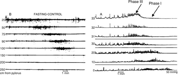
The third suborgan of the stomach, the pacemaker for the MMC, plays a crucial role in the emptying of solids. This pacemaker is located in 80% of patients on the greater curve of the stomach and the remainder in the duodenum (53). Several times daily in the normal fasting state, massive motor waves, dubbed the activity front of the MMC (Fig. 158.2), stimulate emptying of large particles through the pylorus and into the small intestine. Disruption of the pacemaker of the MMC may in part explain the high rate of GI symptoms after partial gastrectomy/bariatric surgery. For example, without a pacemaker and MMC to clear particles greater than 1 mm, bezoars may form; in theory, bezoars may predispose to aspiration.
In addition, these activity fronts of phase III of the MMC are essential in the function and coordination of the stomach and entire small bowel. The activity front of phase III is the “gastrointestinal housekeeper” and propagates all the way to the ileum. In addition to assisting gastric emptying of large particles, these waves clear bacteria from the proximal small bowel to prevent the syndrome of bacterial overgrowth. These waves also clear the entire GI tract of shed epithelial cells. Interestingly, caloric intake of as little as 200 kcal inhibits these important waves. This is the principle why an astute public speaker will eat a candy bar to prevent stomach “growling” on the lecture circuit. Theoretically in the ICU, patients receiving continuous enteral feedings may have less frequent or diminished activity fronts of the MMC. This lack of a GI housekeeper could predispose to tube-feed diarrhea, bloating, abdominal pain, bacterial overgrowth and translocation, poor stomach emptying, and aspiration.
|
|
|
Figure 158.2. Activity front of the MMC seen by antroduodenal manometry. The y axis is the distance from the pylorus at multiple leads, and the x axis is time. Phase III is the Activity Front (AF) of the MMC. MMC, migrating motor complex. (From Braz J Med Biol Res.1998;31(7):889–900, with permission.) |
The best studied method of measuring the activity fronts of the MMC is by antroduodenal (small bowel) manometry. Manometry requires insertion of a pressure-sensitive catheter in a cooperative patient, under fluoroscopy, into the proximal small bowel. Three leads are antral and three are duodenal. In only extraordinary cases, manometry can be accomplished in the ICU. However, in outpatients, the antroduodenal manometry catheter monitors fed and fasting states and responses to various prokinetic agents. Generally, low-dose octreotide—50 µg at bedtime—is the best stimulant of MMC activity fronts in ambulatory patients. However, in gastroparetics, our anecdotal experience with small bowel manometry favors azithromycin liquid, 400 mg each morning (which has virtually no risk of prolonging QT), or erythromycin liquid, 200 mg every 6 hours, 30 min before meals if the Q-Tc is less than 440 msec. The risk of sudden death with erythromycin is highly controversial (see below, Gastroparesis).
The first-mentioned suborgan of the stomach, the fundus, accounts for differences between liquid and solid emptying. Relaxation and enlargement of the fundus are crucial to the digestion of large liquid meals or solid meals that contain liquid. The vagus nerve mediates this relaxation of the fundus (54). Diabetics may have a vagal neuropathy and, together with surgical vagotomy patients, often lack this ability of the stomach to accommodate, leading to rapid liquid emptying, postprandial pain, and diarrhea. The effect of metoclopramide in the symptom relief of gastroparesis may be due to procholinergic stimulation of fundic relaxation and compliance.
The nutritional content of gastric contents also regulates how the stomach processes food. For example, isotonic materials empty quickest, whereas hypertonic ones empty slowest, with hypotonic materials emptying at intermediate speed. Proteins and carbohydrates empty equally well and much better than fats, which stimulate cholecystokinin, thought to perturb gastric emptying in such disorders as chronic pancreatitis (55). Delays in the gastric clearance of fat may have profound importance: common diabetic drinks and enteral formulations are usually low in carbohydrates, but their high fat content in theory may decrease gastric emptying. High-glucose concentrations, at a threshold of 8%, may also empty slower than 1% glucose solutions, which empty quickly, at about the same rate as 0.9% NaCl (56).
The stomach and small bowel are also in close communication via the gastroileal reflex. When high-nutrient contents reach the ileum, normal patients have a feedback reflex that delays release of stomach contents into the duodenum and decreases gastric emptying. In animals, this can be induced by instillation of fat into the ileum. The gastroileal reflex, in theory, may partly explain the paradox of the typical ICU patient: intolerant of tube feeds, with simultaneous diarrhea—due to poor absorption and high nutrient contents reaching the bacteria of the large bowel—and bloating/distention due to gastroparesis.
In the following text, we will investigate disorders of gastric and small intestinal motility.
Gastroparesis
Definitions of gastroparesis vary but, in general, the hallmark of gastroparesis is a delay in the emptying of solids. Patients complain of nausea (93%), abdominal pain (90%), early satiety (86%), and vomiting (68%) (57). Another common complaint is bloating, either perceived or frankly visible; patients may describe changing belt or pant size after meals.
Gastroparesis may contribute to aspiration pneumonia by two mechanisms. The first is by increasing gastroesophageal reflux. For example, up to 15% of ambulatory patients with refractory GERD have gastroparesis (58), leading one to speculate that gastroparesis may predispose to aspiration pneumonia in the ICU (59). In addition to promoting more frequent reflux in the ICU, gastroparesis stimulates colonization of the stomach with Gram-negative enteric and nosocomial flora, putting patients in the ICU with gastroparesis at increased risk for Gram-negative pneumonia (60).
The diagnosis of gastroparesis can be problematic. Barium studies lack caloric content and are liquid based. Therefore, they are a poor reflection of gastric or, for that matter, small bowel motility. Evidence suggests noncaloric liquids empty based only on the pressure they apply to the luminal wall (61). However, gastric scintigraphy with a technitium-99 labeled egg sandwich more accurately measures gastric emptying, especially of solids. Unfortunately, nearly every large medical center in this country seems to have its own protocol, controls, and standards for this test. In addition, a single patient's gastric emptying can vary from day to day due to nicotine intake or to recent narcotic or prokinetic ingestion. Male and female populations have different standards for gastric emptying, but generally a half-time of emptying of greater than 90 minutes implies gastroparesis. More specificity can be gained by extending the amount of time the patient remains in the scanner. For example, the gastric residual at 2 hours is the most common measurement in community hospitals, and although sensitivity may be 100%, specificity is only 20%. The 4-hour measurement is more accurate with 100% sensitivity and 70% specificity (62). However, even the 2-hour measurement is impractical for the ICU setting, and gold standards to compare against scintigraphy are lacking. In addition, clinicians often encounter the quandary of a patient with borderline emptying at 100 minutes. One way to handle such a case is to recollect that, in our hands, most patients with symptomatic gastroparesis have emptying half-times greater than 120 minutes. However, many patients with normal half-times clinically benefit from prokinetics. Therefore, in borderline cases, we move to antroduodenal manometry or treat empirically.
Causes for gastroparesis are similar to almost all motility disorders and are quite diverse, including metabolic disorders: diabetes, hypothyroidism, uremia; drug-induced emptying disorders: narcotics; infiltrative processes: amyloid, scleroderma; and backflow-related disorders such as portal hypertension, right-sided congestive heart failure (CHF), or hypoalbuminemia; nonetheless, many remain idiopathic. Diabetics presenting with gastroparesis often have evidence of neuropathy. Indeed, a vagal neuropathy is believed to be the fundamental problem in diabetic gastroparesis. Due to the rising incidence of type 2 diabetes, the number of gastroparesis patients with type 2 diabetes now equals that with type 1 diabetes. Idiopathic patients are more often female of childbearing age. The hormone progesterone may be responsible for hyperemesis gravidarum and idiopathic gastroparesis.
A hallmark of gastroparesis treatment is medical management. Erythromycin, a motilin agonist, is one of the strongest pharmacologic stimulants of gastric emptying (63,64). Unfortunately, it is short acting, limited by tachyphylaxis, and must be given in intravenous (IV) form immediately before eating, or in PO (oral) form about 30 minutes before each meal. Given the fast-paced environment in today's intensive care units, it may be impractical to dose this medicine at the exact time it is needed. Because gastric emptying to solids is slower than that of liquids, we prefer the suspension over the tablet form, 100 to 200 mg three times daily and before meals. However, IV administration may be more effective, and we have some experience with outpatients who require a PICC (peripherally inserted central catheter) line with chronic four-times-daily IV erythromycin. Another use of erythromycin in the ICU was found in one study to increase the tolerance of enteral nutrition in critically ill patients (66).
Much controversy surrounds the dangers of QTc prolongation in patients taking erythromycin. One methodologically problematic study found several deaths due to dysrhythmias in patients taking chronic erythromycin (67). Although these risks may be overstated, to be cautious, we recommend avoiding P450 inhibitors and other medications that also prolong the QTc while on erythromycin, such as several of the antidepressants and antidysrhythmics, including common ICU drugs such as fluconazole, quinidine, amiodarone, procainamide, and haloperidol. Fortunately, another option is azithromycin which does not prolong the QT interval (65). During antroduodenal manometry, we see pronounced antral contractions from azithromycin at a dose of 400 mg liquid orally 30 min before breakfast (unpublished data). However, like all macrolides, azithromycin can cause significant diarrhea. This side effect can be useful in the patient with adynamic ileus, chronic colonic inertia, or constipation-predominant irritable bowel syndrome, but can be problematic in ICU patients with antibiotic or tube-feed–associated diarrhea. Another drawback of the macrolides is some patients will develop abdominal pain and nausea. Part of this phenomenon can be explained by experiments showing that overdrive pacing the stomach faster than its baseline contraction rate of three waves per minute can sometimes delay gastric emptying (68). Therefore, it is not unreasonable to start low, at 100 mg liquid orally three times daily 30 minutes before meals.
Another agent in the treatment of gastroparesis is metoclopramide, a mixed dopamine antagonist and procholinergic. This drug has fallen out of favor in recent years for several reasons. First, 20% of patients develop Parkinsonism early or sometimes very late into treatment. Usually, this is reversible. However, 3% of patients taking metoclopramide will develop irreversible tardive dyskinesia (69). In addition, many patients experience restlessness and agitation. Metoclopramide is not as potent a stimulant of gastric emptying as the macrolides, and stimulates colon and esophageal motility very poorly. The typical dose is 5 to 10 mg three times daily, 30 min before meals IV or PO.
Because of the difficulties with macrolides and metoclopramide, gastroenterologists have sought other agents. One of those was cisapride, which is a very strong stimulant of all enteric motility. Unfortunately, several deaths due to torsades de pointes occurred, and it is no longer available for use (70). However, on a compassionate basis, it is still available from the manufacturer.
Zelnorm (tegaserod) is often used off-label to supplement other therapies for gastroparesis, but in rare patients can be used as monotherapy (71). It is a partial 5HT4 agonist. We typically crush 6 mg mixed with 2 to 3 ounces of liquid or applesauce three times daily 30 minutes before eating. Side effects are relatively rare but include diarrhea and headaches. As the side effects indicate, it is useful for chronic constipation and FDA approved for women with constipation-predominant irritable bowel syndrome and men and women with chronic constipation. However, it has not been well tested in patients with creatinine (Cr) clearance less than 40 mL/minute. Unfortunately, in February 2007, the FDA took tegaserod off the market due to a very low absolute risk of cardiac events.
Bethanecol is an older drug, formerly used in postsurgical ileus and bladder stasis, but may be useful in the gastroparetic patient who is intolerant of macrolides (72). As bethanecol is a procholinergic agent, one must monitor for potentially life-threatening reactions such as reactive airways disease, bradycardia, prostate obstruction, and diarrhea. The typical dose for gastroparesis is 25 mg orally four times daily.
Serum glucose is a strong inhibitor of gastric emptying (73,74); therefore, tight control of glucose is fundamental to treating gastroparesis. We aim for a glucose less than 200 mg/dL, even postprandially, especially in patients admitted for nausea and vomiting of gastroparesis. In addition, hyperglycemia attenuates the effects of prokinetics (75). Hyperglycemia also reduces the frequency of MMCs and affects small bowel motility (76). Therefore, tight glucose control may benefit other motility disturbances, especially postoperative ileus.
In ambulatory gastroparesis patients, the choice of narcotic may help patients considerably for not only gastroparesis, but all the slow motility disorders. For example, we prefer Ultram and propoxyphene, which have fewer effects on motility than traditional mu-agonists such as morphine, fentanyl, and Dilaudid (77). A promising treatment in gastroparesis—and in all motility, for that matter—is a mu-opiate antagonist that does not cross the blood–brain barrier. A recent study showed benefit in postoperative ileus patients receiving an investigational opiate antagonist agent (78). However, although other investigators have successfully used agents similar to this in dog models of gastroparesis, results in humans have been disappointing (79,80,81).
In addition to changing opiates, discontinuing other antimotility agents such as calcium channel blockers, anticholinergics, and alpha-2 antagonists such as clonidine is important in the management of gastroparesis (82).
Dumping Syndrome
Dumping syndrome is a constellation of postprandial symptoms in patients who have undergone surgical vagotomy and gastric drainage procedures such as pyloroplasty, antrectomy, or gastric resections (83,84). Inadvertent damage to the vagal nerve as in esophageal surgery or from neuropathy can also cause dumping syndrome. Therefore, ICU clinicians may encounter this disorder. In the dumping syndrome, abnormally rapid emptying of gastric contents leads to early delivery of osmotically rich food into the small intestine, resulting in volume shifts and the excessive release of vasoactive peptides and insulin. Symptoms include adrenergic discharge with symptoms such as tachycardia, diaphoresis, agitation or confusion, diarrhea, abdominal cramps, and even hypotension. Measurable hypoglycemia was once thought to be common in dumping syndrome, but further studies have not concurred; perhaps the autonomic nervous system provides sufficient counterregulation to insulin surge in most patients to prevent frank hypoglycemia. A key historical point in the diagnosis of dumping syndrome is that patients are free of these symptoms under fasting conditions. The symptoms are classified as early dumping, within the first 30 to 60 minutes after meal ingestion, and less commonly, late dumping, within 90 to 240 minutes after meals. Liquids and foods rich in carbohydrates are generally not well tolerated, and patients may lose weight due to fear of eating meals (84).
Dietary adjustments are fundamental to the management of the dumping syndrome (85). Patients should divide their caloric intake over at least six meals per day and minimize intake of fluids with solids. Meals rich in carbohydrates, such as Boost or Ensure, commonly induce dumping symptoms. Therefore, dumping patients should eat meals high in protein and fat and low in carbohydrates. Because lactose is absorbed in the jejunum, many patients experience milk intolerance after gastrojejunostomy and respond to restriction of lactose-containing products. The addition of agents like pectin or guar gum that increase the meal viscosity may help patients with dumping symptoms, although these supplements are not universally well tolerated. Dietary fiber may be effective in dumping syndrome (86,87). Acarbose, which delays carbohydrate absorption, may prevent late dumping (88,89). However, diarrhea due to fermentation of unabsorbed carbohydrates limits long-term use.
Mechanical Ventilation/Pressors and Stomach and Small Intestine Motility
Two very common ICU treatments, mechanical ventilation and pressors, cause profound motility disturbances of the stomach and small bowel. Two observational studies measured antroduodenal manometry in mechanically ventilated ICU patients, most on opiate sedation and dopamine. These studies found marked disruptions in the normal fed state and increased phase III activity fronts—which should be off to facilitate mixing and absorption in the fed state—leading to poor tolerance of enteral feeding (90,91). Low-dose dopamine causes a similar effect in healthy volunteers (92) and, in a recent randomized controlled trial, in hemodynamically stable ICU patients (93). Perhaps this effect causes the diarrhea and malabsorption so often seen in critically ill patients on initiation of enteral nutrition. In the latter trial, despite the exclusion of opiate use, the dopamine group also had significantly fewer fasting antral contractions. Thus, dopamine may delay gastric emptying independent of opiate use in the ICU. However, the astute reader may recognize a possible confounder in this study: Most of the patients received IV propofol, which has a high fat content and can delay gastric emptying. However, a second study of critically ill patients also found delayed absorption of acetaminophen, a marker of gastric emptying (94). In the acetaminophen study, significantly higher volumes of gastric contents were aspirated while on dopamine. Dopamine may also prevent the normal fundic relaxation that is essential to prevent aspiration during ingestion of large volumes (95). Perhaps metoclopramide, a dopamine antagonist, which primarily stimulates fundic relaxation and accommodation, could reverse this effect of dopamine (see gastroparesis section for pitfalls of metoclopramide use). Vasopressin may also delay gastric emptying (96). Overall, mechanical ventilation and pressor use may decrease antral motility and reduce fundic relaxation, perhaps accounting for the bloating and high gastric tube outputs sometimes seen in enterally fed ICU patients.
Intestinal Ileus (Acute Intestinal Pseudo-obstruction)
Although many ICU patients have the aberrant, fed-state stimulation of MMCs demonstrated above, a more common scenario is inhibition of small intestinal motility in the ICU, leading to the condition of the small intestinal ileus, or paralytic ileus, an acute delay in caudad passage of intestinal contents in the absence of obstruction.
Patients with ileus present with distention, poorly localized abdominal pain, nausea, vomiting, obstipation, and decreased bowel sounds. Differentiating ileus from mechanical obstruction can be challenging, and an obstruction series abdominal film can be suggestive. However, passage of computed tomography (CT) contrast into the colon within 4 hours virtually excludes mechanical obstruction (97).
Recent surgery or bowel manipulation can decrease bowel motility, leading to ileus. Normally, small intestinal motility returns within 24 hours after surgery, whereas gastric motility returns after 48 hours and that of the colon after 3 to 5 days. Various surgical factors are thought to account for differences in length of postoperative ileus, such as vagotomy, degree of intestinal manipulation, and presence of enterotomy, but these have not been proven (98,99). Several investigators have shown that activity of the left colon is key to recovery from postoperative ileus (100,101).
Causes of ileus are often multifactorial. Laparotomy decreases the amplitude of MMCs; activation of the sympathetic nervous system is likely a major factor. Similarly, inflammation plays an important role and may account for decreases in postoperative ileus in laparoscopic, as compared to open, surgeries (102). Epidural anesthesia may decrease the incidence of ileus compared to conventional narcotic analgesia (103). Sepsis, mesenteric ischemia, myocardial infarction, lower lobe pneumonia, lower rib fractures, chronic mesenteric ischemia, phenothiazines, calcium channel blockers (especially the nondihydropyridines), hypokalemia, hypomagnesemia and hypermagnesemia, hyponatremia, hypercalcemia—please note that because many ICU patients are hypoalbuminemic, an ionized calcium value is crucial here—and thyroid disorders have all been associated with intestinal ileus (104). Hypoalbuminemia may lead to bowel edema, which may account for part of the decreased motility seen in portal hypertension (105).
Most patients can be managed conservatively by identifying and treating/removing contributors to ileus. In patients with marked distention, pain, or respiratory compromise, NG suctioning should be initiated. In our practice, we recommend physical therapy and turning or sitting patients upright every shift. Some have advocated serial rectal exams, a brief trial of a rectal tube, or gentle tap water or 1 to 2 Fleet enemas (although, because these contain sodium phosphate, they should not be used in hypernatremic, hyperphosphatemic patients) to stimulate motility. Oral and stimulant laxatives should be avoided, especially lactulose, which releases hydrogen gas that can add to the problem, especially if mechanical obstruction has not definitively been ruled out. Unfortunately, prokinetic agents have so far been disappointing in the treatment of intestinal ileus. Metoclopramide is more potent in the foregut than hindgut. Erythromycin, a more potent foregut agent, is also active in the hindgut, but has not been shown to be effective in postoperative patients (106). The latter agent and tegaserod may have roles in the medical patient with ileus. Many agents are being tested in this exciting area of motility research.
Chronic Intestinal Pseudo-obstruction
Although it would be quite rare to diagnose chronic intestinal pseudo-obstruction in the ICU, for completeness, we will review the presentation, pathophysiology, causes, and treatments of this family of disorders. These rare motility disorders are characterized by symptoms and signs of chronic nonmechanical intestinal obstruction. Many of these disorders can affect the GI tract in several areas, causing megacolon, megaesophagus, megaduodenum, and so forth. The presence of small bowel diverticula should prompt a search for one of these disorders. Patients can present with steatorrhea from small bowel overgrowth, alternating constipation and diarrhea depending on recent antibiotic use, feculent vomiting and halitosis, gastroparesis, pseudoachalasia, or even GI hemorrhage.
Generally these disorders are characterized as either myopathic or neuropathic, and can be congenital or acquired.
The visceral myopathies involve degeneration and fibrosis of the muscularis propria, which can involve the smooth muscle of the bowel (enlarged esophagus, megaduodenum, redundant colon); iris (mydriasis); face (ptosis, ophthalmoplegia); bladder (megacystitis); and uterus (uterine inertia). They can present at any age, genetically or sporadically (107). Histology may show absence of actin, degenerating myofibrils, and mitochondrial abnormalities (108). Systemic disorders can cause visceral myopathy such as scleroderma and amyloidosis. Barium enema may show lack of haustrations, unlike the neuropathic disorders reviewed below. Antroduodenal manometery may show low-amplitude or absent MMCs (109).
In contrast, the visceral neuropathies are degenerative disorders of the myenteric plexus but can also be familial or sporadic. Histologically, these disorders display degeneration and/or inflammation of axons, dendrites, and absence of silver staining, occasionally with viral inclusions of cytomegalovirus (CMV) (110) or Epstein-Barr virus (EBV) (111). Other systemic contributors to visceral neuropathy include myxedema, Parkinson, narcotic bowel syndrome, late-stage Chagas disease, tumor or stroke of the medulla, acute encephalitis, and paraneoplastic neuronal degeneration, often from occult small cell carcinoma. This association is important to recognize because the time from diagnosis of extraintestinal primary small cell tumor to death is less than 1 year (112). In contrast to the myopathic disorders, the neuropathic disorders show uncoordinated bursts of activity, and abnormal propagation and configuration of MMCs (113). As in the myopathic disorders, suction biopsy can sometimes be diagnostic, but often a full thickness biopsy is required (113).
Treatment of visceral myopathy and neuropathy can be medical with erythromycin (115) or low-dose octreotide, 50 µg at bedtime, especially in scleroderma (114,115), nutritional with addition of B12 and fat-soluble vitamins with or without TPN, or surgical with resection of involved segments (116).
Large Intestine
Diarrhea
Diarrhea is a common ICU problem. Although a comprehensive approach to acute and chronic diarrhea is too expansive for this chapter, several important ICU diagnoses need to be ruled out, including Clostridium difficile toxemia, medication-induced diarrhea, acute mesenteric ischemia, pseudodiarrhea, and malabsorption.
The diagnosis of C. difficile infection is covered in depth elsewhere in this text, but is important to mention because of several common pitfalls. First, C. difficile can rarely be toxin negative, even when the standard three samples are sent for analysis, with colonoscopy serving as the gold standard (117). Therefore, if clinical suspicion for C. difficile colitis or enteritis is high, antibiotic therapy and isolation should be initiated before toxin results come back, and should be continued until at least three samples are toxin negative—potentially longer if GI consultation recommends. Second, care must be taken in interpreting a single positive toxin, especially in the patient at risk for antibiotic-associated non–C. difficile diarrhea. For example, 20% of patients have a false-positive C. difficile toxin. These patients actually have medication-related, antibiotic-associated diarrhea or simple colonization, without signs of serious illness such as fever, abdominal pain, leukocytosis, and acute hypoalbuminemia (118). Removing the offending agent, often a macrolide, is the definitive management of antibiotic-associated diarrhea and is also important in C. difficile diarrhea. Recall that the diarrhea of C. difficile is often hemoccult positive. For patients who must remain on antibiotics, much controversy surrounds the use of probiotic agents such as lactobacillus and Saccharomyces boulardii in the prevention and treatment of antibiotic-associated diarrhea. These agents are generally safe but are not FDA-regulated, and there have been several case reports of S. cerevisiae fungemia and lactobacillus bacteremia in immunocompromised patients due to handling of central catheters after placing the medication in feeding tubes (119).
Other bacterial infectious enteritides are quite rare in the patient who develops diarrhea. However, in the ICU patient with diarrhea admitted from the emergency department (ED) for other reasons, it is reasonable to test for the most common causes of acute infectious bacterial enteritis, such as Campylobacter, salmonella, shigella, and Escherichia coli 0157:H7. Recall that these patients can appear toxic, and any of these enteroinvasive bacteria can present with hemolytic-uremic syndrome or toxic megacolon. Campylobacter jejuni, as its name implies, can present with jejunal thickening on CT resembling mesenteric ischemia. Recall that a classic finding of salmonellosis is fever without tachycardia, one of the rare infections other than mycoplasma that give that presentation.
The diarrhea of acute mesenteric ischemia is important to recognize. It is often heme positive and can appear maroon or melenic. Sloughing of mucosa may be seen and, in addition to sudden distention, may portend infarction. In the patient with pain out of proportion to physical exam early in the course, many signs are absent, and a high clinical suspicion must be maintained. A rancid odor may be appreciated but can be stifled by a rectal tube with balloon, only to be noticed when the bag is changed. Patients may not appear toxic until late in the course when bowel infarction, peritonitis, and lactic acidosis occur. In our experience, patients at highest risk are vascular surgery patients, those undergoing cardiopulmonary bypass, patients with embolic risk whose anticoagulants are held for bleeding, and severely hypotensive patients. Digoxin can decrease mesenteric flow in patients on multiple pressors (120).
Another common cause of diarrhea in ICU patients is iatrogenic diarrhea, which is important to recognize. Oral magnesium, selective serotonin reuptake inhibitors (SSRIs), proton pump inhibitors (PPIs), H2blockers, caffeine/theophylline, antibiotics, nonsteroidal anti-inflammatory agents (NSAIDS), colchicine, gastroparesis therapies, ursodeoxycholic acid (URSO), senna/colace orders from the patient's time on the general ward, and Kayexalate are common offenders. Some common surgical procedures that cause diarrhea are cholecystectomy, partial pancreatectomy, long bowel resections, partial ileal resections, and ileoanal anastomoses. Diarrhea post cholecystectomy and after ileal resections less than 100 cm in length can be treated with cholestyramine, a bile salt binder three times daily (121). This drug can bind and interfere with the absorption of most other medications, so it must be given at least 1 to 2 hours after other drugs. The rare patient with a less than 100-cm ileal resection can develop bile salt depletion and malabsorption if given cholestyramine, although this is common in ileal resections greater than 100 cm. Pancreatic exocrine insufficiency is common after a Whipple procedure because many patients have some degree of chronic pancreatitis prior to the operation. Enteric-coated pancreatic enzyme supplementation, such as with Creon or Ultrase-20, two tabs with meals, is curative.
Another cause of iatrogenic diarrhea is small bowel bacterial overgrowth. In this case, anaerobic bacteria from the colon reflux into the small bowel and prevent absorption of bile salts and nutrients. This is the mechanism behind the diarrhea after ileocecal valve resection. Occasionally, PPIs can cause overgrowth by suppressing the protective effect of stomach acid. Because these bacteria produce folate, blood levels of this B vitamin are often elevated. The D-xylose breath test is the most convenient and sensitive substitute for endoscopic aspiration and culture of duodenal contents (122). However, both tests are inconvenient in ICU patients, and empirical therapy with tetracycline, amoxicillin/clavulanate, trimethoprim/sulfamethoxazole with metronidazole, or quinolones for 1 week is often effective.
Initiation of tube feeds—total enteral nutrition or TEN—can cause diarrhea in the ICU. Patients with low oncotic pressures, with hypotension and/or pressor requirements, and elevated lactic acid are most susceptible. If a patient is at less than average risk for pulmonary aspiration, these feeds can be diluted 50% to improve gastric emptying. In addition, high-glucose containing products such as Ensure or Boost can be exchanged for higher fat content such as Glucerna and vice versa. In the hypoalbuminemic patient, TEN may have to be withheld while total parental nutrition (TPN) temporarily restores oncotic pressure. In the patient with new diarrhea after percutaneous endoscopic gastric tube (PEG) placement, one must be vigilant to rule out inadvertent placement through the colon. If this tube is inserted through the transverse colon, in critically ill patients on broad-spectrum antibiotics, the presence of unaltered tube feeds in the stool may be the only sign. The diagnosis of a colonic perforation during PEG requires a CT scan with PEG-instilled contrast, as a kidney, ureters, and bladder (KUB) will show some free air even in normal post-PEG patients. In the patient with new tube feeds and diarrhea, the ICU clinician may need to rule out jejunal ischemia, as there have been several case reports (123). If, in fact, tube feed diarrhea is idiopathic, a possible treatment is adding soluble partly hydrolyzed guar as a source of fiber. In one randomized controlled trial in mechanically ventilated and septic patients, guar significantly reduced—from 32% to 9%—the percentage of tube feed–induced diarrhea (124). The only enteral formula with fiber is Jevity, but even this may not be sufficient. Elemental or semielemental formulas such as Peptimen may be easier to absorb, especially in ICU patients, but are expensive.
Pseudodiarrhea is important to rule out. Spinal cord patients, diabetic patients, patients with neuropathies/strokes/anticonvulsant therapy, patients with prior episiotomy or prostate surgery, and sedated patients may have baseline low anal sphincter pressure. They may be unable to sense the urge to defecate. If the stool is looser because of tube feeds or antibiotic therapy, the critical care team may start an exhaustive and expensive search for all the causes of diarrhea when a simple rectal exam may be suggestive (125). Treatment is outpatient biofeedback and pelvic floor muscle strengthening.
Hirschsprung Disease
A prototypic, but rare, disorder of constipation is congenital aganglionosis of the colon, also known as congenital megacolon. Although it would be rare to diagnose a patient in the ICU, save for the pediatric ICU (PICU), the physiology of Hirschsprung disease is worthy of a brief discussion and illustrates several broadly applicable motility principles.
Hirschsprung disease results from failure of the neural crest cells to migrate into and from the myenteric plexus in a contiguous region of the left colon. Patients present with severe constipation without palpable stool in the rectal vault, without fecal soiling, but with a narrowed, diseased colon segment and a more proximally dilated, but normal, colonic segment.
Patients suspected of having Hirschsprung disease should undergo anorectal manometry in the gastrointestinal laboratory, an impossibility in most ICU patients. Lack of compensatory rectal relaxation to distention of the rectal balloon is suggestive. These patients should undergo colonic suction biopsy, which can sometimes reach the muscularis propria and rule out visceral neuropathy, myopathy, and amyloidosis. However, lack of myenteric plexus neurons on endoscopic suction biopsy does not entirely prove Hirschsprung disease, and often a laparoscopic full thickness biopsy is required (126).
The lack of nitric oxide–containing neurons, which prevent rectal relaxation, may be the principal mechanism of disease. Chagas disease can even result in a Hirschsprunglike presentation in adults even in the United States (127). Some have found that 70% of adults with acute megacolon may have some histologic features of congenital neurologic disease (128).
Colonic Inertia
Probably the most common cause of refractory constipation in adults is colonic inertia due to generalized decreased colonic motility. A subset of patients may present with obstipation, evidence of colonic dilation, and even respiratory compromise from diaphragmatic impingement. In patients without a prior radiograph, it may be difficult to rule out acute processes such as megacolon. Patients may have a long history of chronic stimulant laxative use. However, much controversy exists over whether this is in fact the cause or the result of colonic inertia.
Less controversial causes include opiates, dehydration/diuretics, calcium channel blockers, clonidine, hypothyroidism, electrolyte imbalance (see ileus section), and immobility. For diagnosis, ambulatory patients can swallow a capsule filled with radio-opaque beads (also known as Sitz markers) and be followed with daily radiographs of the abdomen (KUBs) (129). Markers that are evenly distributed throughout the colon— more than 5 out of 20 at 4 days—are diagnostic of colonic inertia, whereas those that “pile up” can indicate obstruction or pelvic floor dyssynergia.
Ruling out C. difficile in the patient presenting with constipation, bloating, and colonic distention, sometimes with hemoglobin-positive rectal exams as well, is essential. In immunocompromised patients, CMV can present similarly. Although colonic amebiasis would be a rare cause of this presentation (130), the southeastern United States has a higher prevalence than the rest of the nation, especially among individuals at high risk such as travelers to endemic areas and homosexual men. Other causes of infectious colitis can rarely cause toxic megacolon but are in the differential diagnosis.
Treatment is aimed at removing offending agents and gently stimulating bowel movements with tap water or Fleets enemas (see section on ileus). Most acute presentations can be treated with manual disimpaction. Rarely would a patient benefit from emergency colonoscopy, unless manual disimpaction and enemas have failed, and colonic dilation proximal to the impaction exceeds 10 cm in the cecum. Prokinetics and oral or stimulant laxatives should not be used until the impaction has been removed. After relief, patients should be maintained on a chronic, safe bowel regimen to prevent impaction, such as once-daily Miralax, or, in patients with normal renal function, with milk of magnesia or magnesium citrate, 1 to 2 teaspoons daily.
Ogilvie Syndrome
One of the most important motility associated diseases is acute colonic pseudo-obstruction. When this condition coincides with recent orthopedic, trauma, gynecologic, or other surgery, it is classically known as Ogilvie syndrome. This is important to distinguish from simple small bowel ileus or from acute colonic obstruction from stool impaction, and so forth, which require different management.
|
|
|
Figure 158.3. Ogilvie syndrome. This 95-year-old veteran presented with distention, obstipation, and self-elicited tympany. Note extremely dilated loops of colon. Note hip hardware, a common comorbidity. (Courtesy of Veterans Administration Hospital, Gainesville, Florida.) |
Typical Ogilvie patients present on initiation of diet several days after surgery with acute or subacute colonic dilation, distention, often with lack of gas passage, sometimes with nausea, vomiting, or respiratory compromise due to diaphragmatic compression. Generally, not much stool and no transition point is seen (Fig. 158.3). Massive dilation can occur and may lead to perforation, especially when cecal diameter approaches 11 to 12 cm. The cecum is most susceptible to baro-induced ischemia and perforation, as it is the thinnest walled area of the colon. Laplace's law states that transmural pressure is highest across the thinnest portion of a wall. Once pressure inside the cecum exceeds that of the superior mesenteric vein, ischemia can occur (131). Predisposing factors to Ogilvie syndrome include colonic inertia, age, immobility, electrolyte imbalance, or neurologic conditions, such as strokes or parkinsonism with dysautonomia.
If the colon is dangerously dilated, the patient should undergo nasogastric tube suctioning, conversion to nothing-by-mouth status, perhaps with total parenteral nutrition, and gentle stimulation with serial rectal exams and enemas. In some patients, a carefully placed rectal tube can release some of the gas buildup if the dilation extends to the rectum. As in the case of fecal impaction, lactulose, which produces more gas, and other oral laxatives should be avoided. The gold standard in management for many years was an emergency water-soluble contrast enema in the radiology suite. The hyperosmolarity of the enema often induces evacuation and at the same time rules out obstruction. The waning of expertise in this technique, as well as the widespread and emergency availability of colonoscopy, has resulted in a shift in this paradigm. Careful colonoscopy without oral bowel lavage and with minimal insufflation can decompress a severely dilated colon, but is associated with a 1% or higher risk of perforation in this setting. Placement of a decompression tube has a debatable effect on recurrence (132). Therefore, since 1999, some proponents have advocated pharmacologic management with erythromycin (133), cisapride (134)—a mixed procholinergic and 5HT4 agonist no longer available in the United States, and neostigmine, a potent acetylcholinesterase blocker that has many of the side effects, albeit much more temporary, of acute nerve gas poisoning. Therefore, patients receiving neostigmine in our institution must be at the intermediate care unit (IMC) or higher level of care, with atropine instantly available. We do not use neostigmine in actively wheezing patients, those on oxygen for chronic obstructive pulmonary disease (COPD), patients with coronary artery disease, or those at higher-than-average risk of bradycardia and asystole, such as those on several atrioventricular (AV) nodal-blocking agents such as amiodarone or a calcium channel blocker with a beta-blocker. We typically notify cardiology or the critical care team of impending neostigmine use and ask patients to sign a consent form. The standard dose is 2.5 mg IV slow infusion over 1 to 3 minutes. An effect is generally seen within 2 to 20 minutes. The response can be dramatic, with massive evacuation of stool and gas and instant relief. The dose may be repeated 2 to 3 times very carefully. A prospective trial showed a 91% response rate compared to 0% with placebo (135). Nevertheless, many patients may have improved on more aggressive nonneostigmine medical therapy (136).
In the presence of peritonitis, leukocytosis, fever, or a cecal diameter of greater than 12 cm, surgery may be necessary, which, if ischemia is present, usually includes right hemicolectomy, ileostomy, and mucous fistula formation.
Sigmoid Volvulus
Similar principals can be applied to the management of sigmoid volvulus, the “medical” volvulus; many patients are elderly with a history of chronic constipation and other comorbid diseases (137). In one series, up to 13% were in chronic institutions. Most present with distension and obstipation, but about 30% report pain. Significant tenderness on exam may portend ischemia and peritonitis. Radiographically, a sigmoid volvulus may appear as a markedly dilated, ahaustral, sigmoid colon, with paucity of gas in the rectum. The dilated sigmoid loop may extend into the right upper quadrant, assuming a C or bent inner tube shape (Fig. 158.4). In nontoxic patients without signs of peritonitis, emergent sigmoidoscopy is the procedure of choice, using minimal air insufflation, and is successful 60% of the time (139). Once the transition point is passed, a visible untwisting can be seen, associated with massive passage of stool and gas. Our practice is to do a colonoscopy, or at least reach the transverse colon whenever possible, to look for an underlying stricturing lesion. However, because of poor preparation, emergency sigmoidoscopy cannot rule out small areas of ischemic mucosa and may delay definitive surgery in some patients. Careful placement of a rectal tube may decrease recurrence, which is quite high, on the order of 50%. Therefore, after emergent sigmoidoscopy, our practice is to continue to cleanse the bowel with enemas and, if tolerated, with gentle oral lavage to permit more accurate exam of the colon at the time of recurrence. Overall mortality of sigmoid volvulus is 8% (140), mostly due to the 20% of patients with ischemia who have a mortality rate of 80% in an older study (138), but a more recently reported rate is 25% (137).
|
|
|
Figure 158.4. Sigmoid volvulus. KUB showing a dilated loop of sigmoid displaced to the periumbilical region with the appearance of a C or bent inner tube. Note fecal material in descending colon. KUB, kidney, ureters, and bladder. (Courtesy of Veterans Administration Hospital, Gainesville, Florida.) |
Summary
The ICU is home to many motility disorders. Some universal principles of diagnosis include ruling out emergencies such as impending colonic perforation, intestinal ischemia, food impaction, and so forth. In managing these disorders, some basic principles apply: (a) in the “slow disorders” such as gastroparesis, ileus, constipation, Ogilvie's etc., correct electrolytes, rule out occult thyroid disease, keep glucose less than 150 mg/dL, minimize tubes and drains—especially in the upper GI tract, and remove offending agents (Table 158.2) such as calcium channel blockers, central alpha 2 antagonists, and pure mu-opiates; (b) in the “fast disorders,” such as diarrhea, avoid magnesium-containing medications, SSRIs, PPIs, and unnecessary antibiotics. In patients that fail TEN, consider motility as a cause. Consider guar for diarrhea and changing fat and sugar content for distention. Rule out hypoalbuminemia and lactic acidosis. In colonic distention, distinguish among fecal impaction (perhaps from chronic inertia) versus Ogilvie versus sigmoid volvulus. Basic management is the same: (i) use clinical assessment to rule out impending infarction; (ii) rule out cecal diameter greater than 10 cm; (iii) apply gentle enemas and rectal tube if the patient is nontoxic; (iv) consider promotility agents only in Ogilvie (the only case in which neostigmine is used) or decompressed volvulus or disimpacted colonic inertia.
|
Table 158.2 Factors Affecting Motility |
|||||||||||||||
|
|||||||||||||||
Pearls
Motility Disorders of the Esophagus
· Oropharyngeal disorders predispose to aspiration and may be due to stroke, medications, and neurodegenerative or striated muscle disorders. Obtain rehab barium swallow and speech pathology consults early.
· Do not insert a Dobbhoff or NGT into a patient with a distal esophageal hypomotility disorder until epiphrenic diverticula have been ruled out by barium swallow.
· A wide mediastinum may be GI in origin.
· The mucosa of the esophagus is almost never affected by ischemia.
· Be aware of the disorders and drugs that affect LES pressure, as these may contribute to aspiration.
· NGT and Dobbhoff tubes lower LES and predispose to aspiration and esophagitis.
· During cardiac arrest, remember the importance of the LES and maneuvers to decrease gastric ventilation.
· Patients with connective tissue disease, especially those with esophageal dysmotility, are more vulnerable than the average patient to esophageal damage and aspiration.
· A food impaction must be removed immediately by endoscopy. Many are due to motility disturbances.
Motility Disorders of the Stomach
· Remember the stomach is the origin of the MMC in 80% of patients.
· Gastroparesis can predispose to aspiration of nosocomial Gram-negative bacteria and TEN intolerance.
· Secondary causes of gastroparesis, as in most motility disorders, include hyperglycemia, hypothyroidism, hypokalemia, hypercalcemia, uremia, high progesterone states, portal hypertension, connective tissue disease, right-sided CHF, and hypoalbuminemia.
· Treat by keeping blood glucose level less than 150 mg/dL, correcting electrolytes, avoiding precipitants such as opiates, calcium channel blockers, and clonidine.
· If possible, use propoxyphene and/or tramadol for pain control in gastroparetics and other patients with slow motility disorders.
· Erythromycin is the mainstay: start low at 100 mg liquid 30 minutes before meals or IV 10 minutes before meals, titrating to 200 four times daily if QTc is acceptable. Watch for P450 inhibitors and long QTc interactions.
· Some patients may benefit from azithromycin, which does not prolong the QTc, instead of erythromycin.
· Any drug that stimulates gastric emptying can cause diarrhea.
· Gastroparesis is worsened by high salt, sugar, fat, and fiber content. Thus, in outpatients, where aspiration is not a risk, tube feeds can be diluted 50% with water.
· In contrast, patients with dumping benefit from low-carbohydrate, high-fat, high-fiber diets.
Motility Disorders of the Small Bowel
· Recognize why some patients on pressors fail enteral nutrition.
· In the management of ileus:
· Correct electrolytes.
· Increase patient mobility.
· Use gentle enemas but not oral-stimulant laxatives or lactulose.
· Avoid calcium channel blockers, clonidine, anticholinergics, and opiates.
· Consider oral opiate antagonists, prokinetics, and epidural anesthesia in patients at high risk for postop ileus.
· Rule out small cell carcinoma in susceptible patients with new and unexplained constipation, gastroparesis, small or large bowel dysmotility.
Motility Disorders of the Large Intestine
· Common causes of diarrhea include magnesium salts, SSRIs, PPIs, antibiotics, TEN.
· In TEN diarrhea, consider changing or diluting formula if aspiration is not likely. Also rule out jejunal ischemia and misplaced PEG, and consider adding guar-based fiber.
· Be able to distinguish false-positive C. difficile toxin in antibiotic-associated diarrhea from patients with C. difficile disease and false-negative toxin.
· Distinguish Ogilvie syndrome from sigmoid volvulus and from fecal impaction with megacolon.
· The initial approach to nontoxic Ogilvie and sigmoid volvulus patients is rectal tube placement, gentle enemas, correction of electrolytes, serial KUBs, and removal of antimotility agents such as narcotics, anticholinergics, and calcium channel blockers.
· Ogilvie patients can also be started on erythromycin if QTc interval is acceptable.
· If the patient fails to improve or if cecal diameter rises to about 10 cm or enough to cause respiratory insufficiency, urgent sigmoid or colonoscopy is required.
· Toxic patients should go the operating room (OR) or undergo cecostomy in the case of Ogilvie. Gastrografin enema in the radiology suite and IV neostigmine are other options, the cecostomy, only if the patient is not wheezing, bradycardic, and does not have coronary artery disease, or is in an IMC or higher level of care with atropine and resuscitation equipment for asystole easily available.
References
1. Weisbrodt NW, Christianson J. Gradients of contractions in the opossum esophagus. Gastroenterology. 1972;62:1159–1166.
2. Yamato S, Saha JK, Goyal RK. Role of nitric oxide in lower esophageal sphincter relaxation to swallowing. Life Sci. 1992;50:1263–1272.
3. Diamant NE, El Sharkawy TY. Neural control of esophageal peristalsis. A conceptual analysis. Gastroenterology. 1977;72:546–556.
4. Dodds WJ, Christianson J, Dent J, et al. Pharmacologic investigation of primary peristalsis in smooth muscle portion of opossum esophagus. Am J Physiol. 1979;237:E561–E566.
5. Gidda JS, Boyinski JP. Swallow evoked peristalsis in opossum esophagus: role of cholinergic mechanisms. Am J Physiol. 1986;251:G779–G781.
6. Richter JE, Wu WC, Johns DN, et al. Esophageal manometry in 95 healthy volunteers. Variability of pressures with age and frequency of abnormal contractions. Dig Dis Sci. 1987;32:583–592.
7. Paterson WG, Rattan S, Goyal RK. Esophageal responses to transient and sustained esophageal distension. Am J Physiol. 1988;255:G587–G595.
8. Katz PO, Richter JE, Cowan R, et al. Apparent complete lower esophageal sphincter relaxation in achalasia. Gastroenterology. 1986;90:978–983.
9. Kahrilas PJ, Dodds WJ, Dent J, et al. Upper esophageal sphincter function during deglutition. Gastroenterology. 1988;95:52–62.
10. Horner J, Massey EW, Riski JE, et al. Aspiration following stroke: clinical correlates and outcome. Neurology. 1988;38:1359–1362.
11. Logeman JA, Blonsky ER, Boshes B. Dysphagia in parkinsonism. JAMA. 1975;231(1):69–70.
12. Siebens H, Trupe E, Siebens A, et al. Correlates and consequences of eating dependency in the institutionalized elderly. J Am Geriatr Soc. 1986;34:192–198.
13. Goher ME. The prevalence of swallowing disorders in two teaching hospitals. Dysphagia. 1986;1:3–6.
14. Smithard DG, O'Neill PA, Park C, et al. Complications and outcome after acute stroke: does dysphagia matter? Stroke. 1996;27:1200–1204.
15. Splaingard ML, Hutchins B, Sulton LD, et al. Aspiration in rehabilitation patients: videofluoroscopy vs bedside clinical assessment. Ach Phys Med Rehabil. 1988;69:637–640.
16. Logemann JA. The role of the modified barium swallow in management of patients with dysphagia. Otolaryngol Head Neck Surg. 1997;116(3):335–338.
17. Cook IJ. Chapter 10. Disorders causing oropharyngeal dysphagia In: Castell DO, Richter J, eds. The Esophagus. 4rd ed. Philadelphia, PA: Lippincott Williams & Wilkins; 2003:197–198.
18. Norton B, Homer WM, Donnelly MT, et al. A randomized, prospective comparison of percutaneous endoscopic gastrostomy and nasogastric tube feeding after acute dysphagic stroke. BMJ. 1996;312(7022):13–16.
19. Murray JA, Ledlow A, Launspach J, et al. The effects of recombinant human hemoglobin on esophageal motor function in humans. Gastroenterology. 1995;109:1241–1248.
20. Konturek JW, Gillessen A, Domschke W. Diffuse esophageal spasm: a malfunction that involves nitric oxide? Scand J Gastroenterol. 1995;30:1041–1045.
21. Richter JE, Hackshaw BT, Wu WC, et al. Edrophonium: a useful provocative test for esophageal chest pain. Ann Int Med. 1985;103:14–21.
22. Orlando RC, Bozymski EM. Clinical and manometric effects of nitroglycerin in diffuse esophageal spasm. N Engl J Med. 1973;289:23–25.
23. Hongo M, Traube M, McCallum RW. Comparison of effects of nifedipine, propantheline bromide, and the combination on esophageal motor function in normal volunteers. Dig Dis Sci. 1984;29:300–304.
24. Davies HA, Lewis MJ, Rhoads J, et al. Trial of nifedipine for prevention of esophageal spasm. Digestion. 1987;36:81–83.
25. Cannon RO 3rd, Quyyumi AA, Mincemoyer R, et al. Imipramine in patients with chest pain despite normal coronary angiograms. N Engl J Med. 1994;330:1411–1417.
26. Richter JE, Dalton C, Bradley L, et al. Oral nifedipine in the treatment of noncardiac chest pain in patients with the nutcracker esophagus. Gastroenterology. 1987;93:21.
27. Ali GN, Hunt DR, Jorgensen JE, et al. Esophageal achalasia and coexistent upper esophageal sphincter relaxation disorder presenting with airway obstruction. Gastroenterology. 1995;109:1328–1332.
28. Cohen S, Lipschutz W. Lower esophageal sphincter dysfunction in patients with achalasia. Gastroenterology. 1971;61:814–820.
29. Mayberry JF. Epidemiology and demographics of achalasia. Gastrointest Endosc Clin N Am. 2001;11:235–247.
30. Niwamoto H, Okamoto E, Fujimoto J, et al. Are human herpes viruses or measles virus associated with esophageal achalasia? Dig Dis Sci. 1995;859–864.
31. Cassella RR, Ellis FH Jr, Brown AL Jr. Fine structure changes in achalasia of the esophagus, I: vagus nerves. Am J Pathol. 1965;279:46–54.
32. Goldblum JR, Rice TW, Richter JE. Histopathologic features in esophagomyotomy specimens from patients with achalasia. Gastroenterology. 1996;111:648–654.
33. Verne GN, Hahn AB, Pineau BC, et al. Association of LSA-DR and -DQ alleles with idiopathic achalasia. Gastroenterology. 1999;117:26–31.
34. Wagh MS, Matloff DS, Carr-Locke DL. Life-threatening acute airway obstruction in achalasia. Med Gen Med. 2004;6(3):12.
35. Arcos E, Medina C, Mearin F, et al. Achalasia presenting as acute airway obstruction. Dig Dis Sci. 2000;45(10):2079–2083.
36. Vaezi MJ, Richter JE, Wilcox CM. Botulinum toxin versus balloon dilation in the treatment of achalasia: a randomized trial. Gut. 1999;44;231–239.
37. Gelfand M, Rozen P, Gilat T. Isosorbide dinitrate and nifedipine treatment of achalasia. Ann Intern Med. 1982;83:963–969.
38. Eherer AJ, Schwetz I, Hammer HF, et al. Effect of sildenafil on esophageal motor function in healthy subjects and in patients with esophageal motility disorders. Gut. 2002;50:758–764.
39. Becker BS, Burakoff R. The effect of verapamil on the lower esophageal sphincter in normal subjects and in achalasia. Am J Gastroenterol. 1983;78:773–776.
40. Bowman FP, Mengazzi JJ, Check BD, et al. The lower esophageal sphincter pressure during prolonged cardiac arrest and resuscitation. Ann Emerg Med. 1995;26:216–219.
41. Wenzel V, Idris AH, Banner MJ, et al. Influence of volume on the distribution of gas between the lungs and stomach in the unintubated patient receiving positive pressure ventilation. Crit Care Med. 1998;26:264–268.
42. Wenzel V, Idris AH, Banner MJ, et al. Respiratory system compliance decreases after cardiopulmonary resuscitation and stomach inflation: impact of large and small tidal volumes on calculated peak airway pressure. Resuscitation. 1998;38:113–118.
43. Lawes EG, Baskett PJF. Pulmonary aspiration during unsuccessful cardiopulmonary resuscitation. Intensive Care Med 1987;13:379–382.
44. Gabrielli A, Wenzel V, Layon J, et al. Lower esophageal sphincter pressure measurement during cardiac arrest in humans: potential implications for ventilation of the unprotected airway. Anesthesiology. 205;103:897–899.
45. Dorges V, Ocker H, Hagelberg S, et al. Smaller tidal volumes with room air are not sufficient to ensure adequate oxygenation during basic life support. Resuscitation. 2000;44:37–41.
46. Wagner-Berger HG, Wenzel V, Stallinger A, et al. Decreasing peak flow rate with a new bag-valve mask device: effects of respiratory mechanics and gas distribution in a bench model of the unprotected airway. Resuscitation. 2003;57:193–197.
47. Baskett PJ, Baskett TF. Resuscitation great. Brian Sellick, cricoid pressure and the Sellick maneuver. Resuscitation. 2004;61:5–7.
48. Cohen S, Lauffer I, Snape WJ et al. The gastrointestinal manifestations of scleroderma: pathogenesis and management. Gastroenterology. 1980;79:155.
49. Lieb JG II, Katzka D. Motility disorders of the esophagus. In: Parkman HP, Fisher RS, eds. Lichtenstein G, series ed. A Clinician's Guide to Acid Peptic and Motility Disorders. Thorofare, NJ: Slack Publishers; 2006.
50. Desai TK, Stecevic V, Chang CH, et al. Association of eosinophilic inflammation with esophageal food impaction in adults. Gastrointest Endosc. 2005;61(7):795–801.
51. Camilleri M, Malagelada JR, Brown ML, et al. Relation between antral motility and gastric emptying of solids and liquids in humans. Am J Physiol. 1985;249(5 Pt 1):G580–585.
52. Meyer JH, Thomson JB, Cohen MB, et al. Sieving of solid food by the canine stomach and sieving after gastric surgery. Gastroenterology. 1979;76(4):804–813.
53. Tanaka M, Sarr MG, Van Lier Ribbink JA Gastrointestinal motor patterns: motilin as a coordinating factor. J Surg Res. 1989;47(4):325–331.
54. Fich A, Neri M, Camilleri M, et al. Stasis syndromes following gastric surgery: clinical and motility features of 60 symptomatic patients. J Clin Gastroenterol. 1990;12(5):505–512.
55. Chowdhury RS, Forsmark CE, Davis RH, et al. Prevalence of gastroparesis in patients with small duct chronic pancreatitis. Pancreas. 2003;26:235–238.
56. McHugh PR, Moran TH: Calories and gastric emptying: a regulatory capacity with implications for feeding. Am J Physiol. 1979;236:R254–R260.
57. Hoogerwerf WA, Pasricha PJ, Kalloo AN, et al. Pain: the overlooked symptom in gastroparesis. Am J Gastroenterol. 1999;94(4):1029–1033.
58. Maddern J, Jamieson G, Myers J. Effect of cisapride on delayed gastric emptying in gastroesophageal reflux disease. Gut. 1991;32:470–474.
59. Drakulovic MB, Torres A, Bauer TT, et al. Supine body position as a risk factor for nosocomial pneumonia in mechanically ventilated patients: a randomised trial. Lancet. 1999;354:1851–1858.
60. Heyland D, Mandell L. Gastric colonization by Gram negative bacilli and nosocomial pneumonia in the intensive care unit patient: evidence of causation. Chest. 1992;101(1):187–193.
61. Kelly KA. Gastric emptying of liquids and solids: roles of proximal and distal stomach. Am J Physiol. 1980;239:G71–G76.
62. Thomforde GM, Camilleri M, Phillips SF, et al. Evaluation of an inexpensive screening scintigraphic test of gastric emptying. J Nucl Med. 1995;36(1):93–96.
63. Urbain JLC, Vantrappen G, Janssens J, et al. Intravenous erythromycin dramatically accelerates gastric emptying in gastroparesis diabeticorum and normals and abolishes the emptying discrimination between solids and liquids. J Nucl Med. 1990;31:1490–1493.
64. Fraser R, Shearer T, Fuller J, et al. Intravenous erythromycin overcomes small intestinal feedback on antral, pyloric, and duodenal motility. Gastroentrerology. 1992;103:114–119.
65. Iannini PB. Cardiotoxicity of macrolides, ketolides, and fluoroquinolones that prolong the QTc. Expert Opin Drug Saf. 2002;1(2):121–128.
66. Booth CM, Heyland DK, Paterson WG. Gastrointestinal promotility drugs in the critical care setting: a systematic review of the evidence. Crit Care Med. 2002;30:1429–1435.
67. Ray WA, Murray KT, Meredith S, et al. Oral erythromycin and the risk of sudden death from cardiac causes. N Engl J Med. 2004;351(11):1089–1096.
68. Ouyang H, Xing J, Chen JD. Tachygastria induced by gastric electrical stimulation is mediated via alpha- and beta-adrenergic pathway and inhibits antral motility in dogs. Neurogastroenterol Motil. 2005;17(6):846–853.
69. Ganzini L, Casey DE, Hoffman WF, et al. The prevalence of metoclopramide-induced tardive dyskinesia and acute extrapyramidal movement disorders. Arch Intern Med. 1993;153:1469–475.
70. Wysowski DK, Corken A, Gallo-Torres H, et al. Postmarketing reports of QT prolongation and ventricular arrhythmia in association with cisapride and Food and Drug Administration regulatory actions. Am J Gastroenterol. 2001;96:1698–1703.
71. Beglinger C. Tegaserod: a novel, selective 5-HT4 receptor partial agonist for irritable bowel syndrome. Int J Clin Pract. 2002;56:47–51.
72. Regional gastric contractility alterations in a diabetic gastroparesis mouse model: effects of cholinergic and serotonergic stimulation. Am J Physiol Gastrointest Liver Physiol. 2004;287(3):G612–619.
73. Fraser RJ, Horowitz M, Maddox AF, et al. Hyperglycaemia slows gastric emptying in type 1 (insulin-dependent) diabetes mellitus. Diabetologia. 1990;33(11):675–680.
74. Schvarcz E, Palmer M, Aman J, et al. Physiological hyperglycemia slows gastric emptying in normal subjects and patients with insulin-dependent diabetes mellitus. Gastroenterology. 1997;113(1):60–66.
75. Petrakis IE, Kogerakis N, Vrachassotakis N, et al. Hyperglycemia attenuates erythromycin-induced acceleration of solid-phase gastric emptying in healthy subjects. Abdom Imaging. 2002;27(3):309–314.
76. Bjomsson ES, Urbanavicius V, Eliasson B, et al. Effects of hyperglycemia on interdigestive gastrointestinal motility in humans. Scand J Gastroenterol. 1994;29:1096–1104.
77. A comparison of the abuse liability of tramadol, NSAIDs, and hydrocodone in patients with chronic pain. J Pain Symptom Manage. 2006;31(5):465–476.
78. Taguchi A, Sharma N, Saleem R, et al. Selective postoperative inhibition of gastrointestinal opioid receptors. N Engl J Med. 2001;345:13.
79. Yuan CS, Foss JE. Oral methylnaltrexone for opioid-induced constipation. JAMA. 2000;284:1383–1384.
80. Foss JF, Yuan CS, Roizen MF, et al. Prevention of apomorphine- or cisplatin-induced emesis in the dog by a combination of methylnaltrexone and morphine. Cancer Chemother Pharmacol. 1998;42:287–291.
81. Moerman I, Franck P, Camu F. Evaluation of methylnaltrexone for the reduction of postoperative vomiting and nausea incidences. Acta Anaesthesiol Belg. 1995;46:127–132.
82. Jones KL, Russo A, Stevens JE, et al. Predictors of delayed gastric emptying in diabetes. Diabetes Care. 2001;24:1264–1269.
83. Carvajal SH, Mulvihill SJ. Postgastrectomy syndromes/dumping and diarrhea. Gastroenterol Clin North Am. 1994;23:61–79.
84. Vecht J, Masclee AA, Lamers CB. The dumping syndrome. Current insights into pathophysiology, diagnosis and treatment. Scand J Gastroenterol Suppl. 1997;223:21–27.
85. Cuschieri A. Surgical management of severe intractable post-vagotomy diarrhoea. Br J Surg. 1986;73:981–984.
86. Bouras EP, Scolapio JS. Gastric motility disorders. Management that optimizes nutritional status. J Clin Gastroenterol. 2004;38:549–557.
87. Kneepkens CM, Fernandes J, Vonk RJ. Dumping syndrome in children. Diagnosis and effect of glucomannan on glucose tolerance and absorption. Acta Paediatr Scand. 1988;7:279–286.
88. Speth PAJ, Jansen JB, Lamers CB. Effect of acarbose, pectin, a combination of acarbose with pectin, and placebo on postprandial reactive hypoglycemia after gastric surgery. Gut. 1983;24:798–802.
89. Lyons TJ, McLoughlin JC, Shaw C, et al. Effect of acarbose on biochemical responses and clinical symptoms in dumping syndrome. Digestion. 1985;31:89–96.
90. Dive A, Moulart M, Mahieu P. Gastroduodenal motility in mechanically ventilated critically ill patients. A manometric study. Crit Care Med. 1994;22:441–447.
91. Dive A, Miesse C, Jamart J. Duodenal motor response to continuous enteral feeding is impaired in mechanically ventilated patients. Clin Nutr. 1994;13:302–306.
92. Marzio L, Neri M, Cuccurullo F, et al. Dopamine interrupts gastrointestinal fed motility pattern in humans. Effect on motilin and somatostatin blood levels. Dig Dis Sci. 1990;35:327–332.
93. Dive A, Foret F, Jamart J, et al. Effect of dopamine on gastrointestinal motility during critical illness. Intens Car Med. 2000;26:901–907.
94. Tarling M, Toner C, Withington P, et al. A model of gastric emptying using paracetamol absorption in intensive care patients. Intensive Care Med. 1997;23:256–260.
95. Hartley M, Sarginson R, Green C. Gastric pressure response to low dose dopamine infusion in normal man. Clin Nutr. 1992;11:23–29.
96. Xing J, Qian L, Chen J. Experimental gastric dysrhythmias and its correlation with in vivo gastric muscle contractions. World J Gastroenterol. 2006;12(25):3994–3998.
97. Peck J, Milleson T, Phelan J. The role of CT with contrast and SBFT in the management of small bowel obstruction. Am J Surg. 1999;177:375.
98. Graber J, Schulte W, Condon R, et al. Relationship of the duration of postoperative ileus to extent and site of operative dissection. Surgery. 1982;92:87.
99. Condon R, Cowles V, Schulte W, et al. Resolution of postoperative ileus in humans. Gut. 1986;203:574.
100. Waldhausen J, Shaffrey M, Skenderis B, et al. Gastrointestinal, myoelectric, and clinical patterns of recovery after laparotomy. Ann Surg. 1990;211:777.
101. Cordon R, Cowels V, Ferraz A, et al. Human colonic smooth muscle electrical activity during and after recovery from postoperative ileus. Am J Physiol. 1995;269:G408.
102. Kehlet H. Postoperative ileus. Gut. 2000;47:iv85.
103. Holte K, Kehlet H. Postoperative ileus, a preventable event. Br J Surg. 2000;87:1480.
104. Turnage R, Bergen C. Intestinal obstruction and ileus. In: Sleisinger and Fordtran's Gastrointestinal and Liver Disease. 7th ed. Philadelphia, PA: WB Saunders; 2003.
105. Reilly JA Jr, Forst CF, Quigley EM, Rikkers LF. Gastric emptying of liquids and solids in the portal hypertensive rat. Dig Dis Sci. 1990;35(6):781–786.
106. Brungard T, Kale-Pradhan P. Prokinetic agents for the treatment of postoperative ileus in adults: a review of the literature. Pharmacotherapy. 1999;19:419.
107. Schuffler M, Lowe M, Bill A. Studies of idiopathic chronic intestinal pseudoobstruction, I: hereditary hollow visceral myopathy: clinical and pathological studies. Gastroenterology. 1979;77:664.
108. Smith V, Lake B, Kamm M, et al. Intestinal pseudoobstruction with deficient smooth muscle a-actin. Histopathology. 1992;21:535.
109. Verne GN, Sninski C. Chronic intestinal pseudoobstruction. Dig Dis Sci. 1995;13:163.
110. Krisnamurthy S, Schuffler M. pathology of the neuromuscular disorders of the small intestine and colon. Gastroenterology. 1987;93:610.
111. Besnard M, Faure C, Fromont-Hankard G, et al. Intestinal pseudoobstruction and acute pandysautonomia associated with Epstein-Barr infection. Am J Gastroenterol. 2000;95:280.
112. Chinn J, Schuffler M. Paraneoplastic visceral myopathy as a cause of severe gastrointestinal motor dysfunction. Gastroenterology. 1988;95:1279.
113. Barenet J, McDonnell W, Appleman H, et al. Familial visceral neuropathy with neuronal intranuclear inclusions: diagnosis by rectal biopsy. Gastroenterology. 1992;102:684.
114. Soudah H, Hasler W, Owyang C. Effect of octreotide on intestinal motility and bacterial overgrowth in scleroderma. N Engl J Med. 1991;325:1461.
115. Verne GN, Eaker E, Hardy E, et al. Effect of octreotide and erythromycin on idiopathic and scleroderma-associated intestinal pseudo-obstruction. Dig Dis. Sci. 1995;40:1892.
116. Schuffler M, Leon S, Krishnamurthy S. Intestinal pseudoobstruction caused by a new form of visceral neuropathy. Palliation by radical small bowel resection. Gastroenterology. 1985;89:1152.
117. Borriello S, Larson H, Welch A. Enterotoxigenic Clostridium perfringens: a possible cause of antibiotic associated diarrhea. Lancet. 1984;(1):305.
118. Bartlett J. Clostridium difficile. Clinical considerations. Rev Infect Dis. 1990;12:S244.
119. Burkhardt O, Köhnlein T, Pletz M, et al. Saccharomyces boulardii induced sepsis: successful therapy with voriconazole after treatment failure with fluconazole. Scand J Infect Dis. 2005;37(1):69–72.
120. Kurland B, Brandt L, Delany H. Diagnostic tests for intestinal ischemia. Surg Clin N Am. 1992;72:85.
121. Arrambride K, Santa Ana C, Schiller L, et al. Loss of absorptive capacity for sodium chloride as a cause of diarrhea following partial ilial and right colon resection. Dig Dis Sci. 1989;34:193.
122. King C, Toskes P, Guilarte T, et al. Detection of small bowel bacterial overgrowth by means of a C14 -D-xylose breath test. Gastroenterology. 1979;77:75.
123. Schunn CD, Daly JM. Small bowel necrosis associated with postoperative jejunal tube feeding. J Am Coll Surg. 1995;180:410–416.
124. Spapen H, Diltoer M, Van Malderen C, et al. Soluble fiber reduces the incidence of diarrhea in septic patients receiving total enteral nutrition: a prospective, double-blind, randomized, and controlled trial. Clin Nutr. 2001;20:301–305.
125. Tatar EL.Trivedi C. Pseudodiarrhea caused by vaginal pessary in an elderly patient. J Am Geriatr Soc. 2005;53:1083.
126. Preston D, Lennard-Jones J, Thomas B. Towards a radiographic definition of idiopathic megacolon. Gastrointest Radiol. 1985;10:167.
127. Holbert R, Magiris E, Hirsch C. Chagas' disease: a case in south Mississippi. J Miss State Med Assoc. 1995;36:1.
128. Basilova T, Vorob'ev G, Nasyrina T. Changes in the intramural nervous system in idiopathic megacolon in adults [in Russian]. Arkh Patol. 1995;57:28.
129. Metcalf A, Phillips S, Zinsmeister A, et al. A simplified assessment of segmental colonic transit. Gastroenterology. 1987;92:40.
130. Suarez Artacho G, Olano Acosta MC, Vazquez Monchul J, et al. Acute fulminant colitis caused by intestinal amebiasis. Rev Esp Enferm Dig. 2006;98(7):559–560.
131. Sleisenger, page 2126.
132. Stephenson K, Rodrigues-Bigas M. Decompression of the large intestine in Oglivie's syndrome by a colonoscopically placed long intestinal tube. Surg Endosc. 1994;8:116.
133. Armstrong D, Ballantyne G, Modlin I. Erythromycin for reflex ileus in Ogilvie's syndrome. Lancet. 1991;337:378.
134. MacColl C, MacConnell K, Baylis B, et al. Treatment of acute colonic pseudoobstruction (Ogilvie's syndrome) with cisapride. Gastroenterology. 1990;98:773.
135. Pronec R, Saunders M, Kimmey M. Neostigmine for the treatment of acute colonic pseudoobstruction. N Engl J Med. 1999;341:137.
136. Sloyer A, Panella V, Demas B, et al. Ogilvie's syndrome. Successful management without colonoscopy. Dig Dis Sci. 1988;33:1391.
137. Grossman E, Longo W, Stratton M, et al. Sigmoid volvulus in the department of veterans affairs medical centers. Dis Colon Rectum. 2000;43:414.
138. Arnold G, Nance F: Volvulus of the sigmoid colon. Ann Surg. 1973;177:527.
139. Brothers T, Strodel W, Eckhauser F. Endoscopy in colonic volvulus. Ann Surg. 1987;206:1.
140. Ballantyne G. Review of sigmoid volvulus. History and results of treatment. Dis Colon Rectum. 1982;25;494.
141. McNally P. GERD. In: GI/Liver Secrets. 2nd ed. Philadelphia, PA: Hanley & Belfus; 2001:9.
142. van der Hoeven CW, Attia A, Deen L, et al. The influence of anaesthetic drugs on the lower oesophageal sphincter in propofol/nitrous oxide anaesthetized dogs. Pressure profilometry in an animal model. Acta Anaesthesiol Scand. 1995;39(6):822–826.