Thomas S. Huber
Mesenteric ischemia is a generic term that implies inadequate blood flow to the intestines. It is relevant from a clinical standpoint as two separate disease processes: acute mesenteric ischemia (AMI) and chronic mesenteric ischemia (CMI). Although the underlying process is similar (i.e., inadequate intestinal blood flow), the clinical presentation, diagnostic concerns, and treatment algorithms are different. CMI is almost exclusively related to visceral artery occlusive disease from atherosclerosis and is relevant to intensive care physicians primarily because of the multiple organ dysfunction that occurs after revascularization. AMI results from a broad spectrum of underlying conditions including arterial emboli, in situ thrombosis in the setting of visceral arterial occlusive disease, mesenteric venous thrombosis, nonocclusive mesenteric ischemia (NOMI), and acute aortic dissections. AMI is relevant to intensivists not only because of the early postoperative concerns, but also because it can occur in critically ill patients with other active problems (e.g., post coronary artery bypass, acute pancreatitis). A thorough understanding of both clinical entities is essential for all intensive care physicians. The underlying pathophysiology and treatment of CMI will be discussed first to provide a foundation for addressing AMI. Although not traditionally considered AMI, isolated colon ischemia will also be discussed for completeness.
Chronic Mesenteric Ischemia
Pathophysiology
The underlying pathophysiology of CMI is the inability to achieve postprandial hyperemic intestinal blood flow. Intestinal blood flow in the normal fasting state is fairly modest but increases markedly postprandially with the magnitude contingent on the size and composition of the meal. Most of the hyperemic changes occur in the pancreas and small bowel and peak within 30 to 60 minutes after eating (1,2). In the presence of significant arterial occlusive disease, this postprandial hyperemia is not possible and patients develop ischemic pain similar to angina pectoris appropriately termed mesenteric angina.
|
|
|
Figure 159.1. A diagram of the collateral pathways for the mesenteric vessels is shown. The celiac axis and superior mesenteric arteries communicate through the superior and inferior pancreaticoduodenal arteries, respectively. The superior and inferior mesenteric arteries communicate through the meandering artery and the marginal artery of Drummond with the meandering artery serving as the dominant collateral. The inferior mesenteric artery communicates with the internal iliac artery through the hemorrhoidal vessels. (From Zelenock GB. Visceral occlusive disease. In Greenfield LJ, ed. Surgery: Scientific Principles and Practice. Philadelphia, PA: Lippincott Williams & Wilkins; 2001:1691, with permission.) |
The normal visceral circulation is fairly redundant and has a rich collateral network (Fig. 159.1). The celiac axis communicates with the superior mesenteric artery through the superior and inferior pancreaticoduodenal arteries. The superior mesenteric artery communicates with the inferior mesenteric artery via the meandering mesenteric artery that runs within the proximal mesentery. This collateral has multiple eponyms, but should be differentiated from the less significant collateral known, the marginal artery of Drummond, located in the distal mesentery. Last, the inferior mesenteric artery communicates with the internal iliac artery via the superior and middle hemorrhoidal arteries. The symptoms of mesenteric ischemia usually do not occur unless two of the three visceral vessels (i.e., celiac axis, superior mesenteric artery, inferior mesenteric artery) are significantly diseased because of this rich collateral network. However, symptoms may occur with isolated disease in the superior mesenteric artery in the absence of adequate collaterals (3).
The overwhelming majority of visceral artery stenoses are due to atherosclerosis. Various other causes have been reported including neurofibromatosis, fibromuscular disease, rheumatoid disorders, aortic dissection, radiation, Buerger disease, and certain drugs, although these are collectively less common. Symptomatic visceral artery occlusive disease (i.e., CMI) is relatively rare despite the prevalence of visceral artery stenoses. Indeed, a hemodynamically significant visceral artery stenosis (>50% diameter reduction) has been reported in up to 27% of patients with peripheral arterial occlusive disease undergoing arteriography (4) and in up to 5 to 10% of unselected patients at autopsy (5). The atherosclerotic process usually involves just the origin of the visceral vessels. Patients with occlusive disease in the celiac axis and superior mesenteric artery frequently have renal artery lesions (and vice versa). The risk factors for visceral artery atherosclerosis are the same as those for the other vascular beds.
Clinical Presentation and Diagnosis
The appearance and presentation of patients with CMI is fairly characteristic. Most patients are cachectic, elderly women with strong smoking histories. Indeed, CMI is one of the few cardiovascular problems more common in women with a reported ratio of approximately 3:1 (6). The mean age (±SD) of patients undergoing repair in a recent national series was 66 ± 11 although the disease process is not exclusive to the elderly and is frequently found in patients in their fourth and fifth decades of life (6).
Abdominal pain is usually the presenting symptom. It initially occurs postprandially but may progress to a persistent nature in the latter stages of the disease process. Unfortunately, the pain has no specific characteristics. As a result of the pain, patients develop a fear of food and avoid eating. The net result is a predictable weight loss with a mean of 20 to 30 lb in several recent clinical series (7,8,9). It should be noted that the cause of the weight loss is poor nutrition rather than an abnormality of intestinal absorption. Patients may develop nausea/vomiting, constipation, or diarrhea. Indeed, AMI is a fairly strong cathartic, and motility symptoms rather than pain may be the predominate symptom. Patients with CMI frequently have evidence of systemic vascular disease although there are no characteristic physical findings.
The diagnosis of CMI requires the appropriate clinical history and the presence of significant visceral artery occlusive disease. The diagnostic approach is a stepwise one with consideration of the more common problems first. Although the appearance and clinical presentation of patients with CMI is fairly characteristic, the differential diagnosis of patients with abdominal pain and weight loss is extensive and includes gastrointestinal malignancy first and foremost. Indeed, CMI is usually not even considered by most primary care providers, and this is reflected by the fact that the mean duration from presentation to diagnosis exceeds a calendar year and 2.8 diagnostic tests (10). The initial diagnostic workup for patients with abdominal pain and weight loss should include esophagogastroduodenoscopy, colonoscopy, abdominal ultrasound, and an abdominal/pelvic computed tomography (CT) scan. Notably, gastric ulcers are relatively common in patients with CMI and likely result from ischemia (11). A surprising number of patients are subjected to cholecystectomy as part of their workup before the definitive diagnosis of CMI is made.
Mesenteric duplex scan is an excellent screening tool for visceral artery stenosis with reported sensitivity and specificity rates of approximately 80% (12). However, it is technically challenging, operator dependent, and not available in all centers. Various criteria have been proposed to grade the severity of stenoses based on the systolic/diastolic velocities or frequency shifts (12,13). The various criteria are probably equivalent as far as their accuracy in grading the degree of stenosis. However, it is imperative that they be validated at each institution by comparison with the institutional gold standard (i.e., CT arteriogram, contrast arteriogram). Various provocative tests have been proposed to unmask clinically significant visceral artery stenoses, although their utility is unclear and they have not achieved widespread use (14,15).
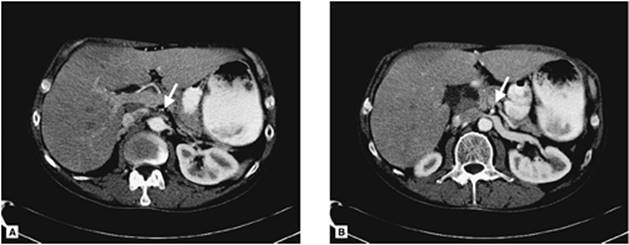
CT arteriography has largely replaced catheter-based contrast arteriography as the diagnostic study of choice for patients with CMI (Fig. 159.2) and is usually sufficient to plan open surgical revascularization (16,17,18). CT arteriography is safe, noninvasive, and almost universally available. It is very good for identifying occlusive disease in the superior mesenteric artery and celiac axis. Furthermore, it is useful for identifying the presence of collateral vessels between the visceral vessels (i.e., celiac axis, superior mesenteric, inferior mesenteric) and/or the internal iliac arteries that suggest hemodynamically significant stenoses. Additionally, it is very good for evaluating other intra-abdominal processes.
Catheter-based contrast arteriography is very useful for evaluating the visceral circulation and has traditionally been used as the definitive diagnostic test for CMI (Fig. 159.3A). The major advantage of contrast arteriography over CT arteriography is that therapeutic interventions can be performed at the time of the diagnostic procedure. However, it is an invasive procedure with a small but finite complication rate. A lateral arteriogram is mandatory as part of the examination to accurately assess the origins of the celiac axis and superior mesenteric artery due to their anterior/posterior orientation. The significant findings on arteriogram include ostial stenoses of the celiac axis and superior mesenteric artery, the presence of visceral collaterals, and the presence of central aortic atherosclerotic disease. A small percentage of patients with CMI may have visceral artery aneurysms, presumably from increased flow through the collateral vessels.
Magnetic resonance (MR) arteriography has been used as a diagnostic study for patients with CMI and offers many of the advantages of CT arteriography (19,20). However, it is not as universally available as CT arteriography. Furthermore, it is not practical and may be contraindicated for many patients including those in the intensive care unit. Last, MR arteriography tends to overestimate the degree of stenosis in the visceral vessels.
Treatment Strategies
All patients with CMI require treatment. The natural history of the untreated disease process is death either from inanition or bowel infarction. The theoretical treatment options include medical management with total parenteral nutrition or revascularization by either endovascular or open surgical techniques. The role of long-term parenteral nutrition is very limited given its complexity, expense, and complications, particularly those associated with the infusion catheters. Additionally, patients with CMI may not be able to metabolize the parenteral nutrition. Endovascular treatment (angioplasty with or without stenting) has emerged as the initial revascularization option for most patients with CMI but should be viewed as an alternative to the open surgical approach (21,22,23,24,25,26,27). Endovascular treatment has consistently been shown to have a lower mortality rate, a lower complication rate, and shorter hospital length of stay when compared to open surgical revascularization. However, the patency rates for the endovascular approach are lower. Notably, this has not been associated with a decrease in survival, and vessel thrombosis and/or recurrent stenosis has not necessarily resulted in AMI. The endovascular approach can serve as an excellent bridge to open surgical revascularization for debilitated patients who are poor initial candidates for a major surgical procedure. The recent development of lower-profile (i.e., smaller-diameter) angioplasty balloon/stent systems has further extended the applications of the endovascular approach (25), and reasonable short-term results have been reported for patients with even occluded (versus stenotic) vessels (23).
|
|
|
Figure 159.2. Two CT arteriograms are shown in a patient with chronic mesenteric ischemia (CMI) and an occluded celiac axis and superior mesenteric artery. A: The origin of the superior mesenteric artery is shown with the arrow. There is no contrast within the lumen of the vessel at this cross section. B: The superior mesenteric artery is shown with the arrow in this cross section that is 10 mm caudal to the first image. The artery is patent at this level as reflected by the contrast within the lumen. |
|
|
|
Figure 159.3. A lateral aortogram of a patient with chronic mesenteric ischemia (CMI) is shown. A: There is a moderate stenosis in the proximal superior mesenteric artery as shown with the arrow. B: No residual stenosis is seen in the superior mesenteric artery after placement of an intraluminal stent. |
Endovascular Revascularization
The preoperative evaluation prior to endovascular treatment is essentially the same for all catheter-based contrast arteriography. Patients with a contrast allergy should be treated with an appropriate steroid preparation. Patients with elevated serum creatinine levels (serum creatinine 1.5–2.0 mg/dL) should receive gentle hydration and acetylcysteine, although admittedly their benefits are somewhat unsubstantiated.
Percutaneous access can be obtained through either the femoral or brachial arteries although the latter is favored for therapeutic procedures given the vector forces associated with the catheters/sheaths. A flush aortogram is performed in both the anteroposterior and lateral projections. Since most lesions in the superior mesenteric artery and celiac axis are orificial and located in the proximal 2 cm, selective catheterization is not usually necessary unless a distal lesion is suspected or the extent of the lesion cannot be determined. A >50% diameter reduction of the superior mesenteric artery is usually considered clinically significant regardless of whether or not the celiac axis is involved. In contrast, the diagnosis of CMI should be questioned in the presence of an isolated celiac axis stenosis. Symptomatic stenoses can be treated at the time of the diagnostic arteriogram (Fig. 159.3B). The orificial stenoses in the mesenteric vessels are refractory to angioplasty alone, and primary stenting is recommended. Balloon angioplasty with selective stenting is reserved for midsegment lesions. Balloon-expandable stents (vs. self-expanding stents) are preferred for the orificial stenoses due to their superior radial forces and controlled deployment mechanism.
The postoperative care after mesenteric angioplasty/stenting is comparable to that for other peripheral endovascular procedures. Patients are admitted to the general care hospital ward for overnight observation and started on clopidogrel. Most patients notice a marked improvement of their postprandial symptoms shortly after the procedure. A fasting mesenteric duplex ultrasound scan is obtained on the morning after the procedure to serve as a baseline. Elevated velocities are occasionally noted in the duplex scan despite a technically satisfactory arteriographic result and complete resolution of the preoperative symptoms. The explanation for these abnormal duplex findings is unclear although we have elected to follow the patient's clinical course in this setting and only repeat the arteriogram and/or intervention if there is a significant change. A repeat duplex examination is performed at 1 month and aspirin (325 mg/day) is substituted for the clopidogrel at that time. The subsequent follow-up with serial duplex examination is comparable to that outlined for open revascularization.
Open Surgical Revascularization
The preoperative workup for patients undergoing open mesenteric revascularization is comparable to that for other major vascular surgical procedures and includes optimization of all organ systems. Multiple algorithms have been developed to reduce the cardiac risk for vascular surgical patients undergoing noncardiac procedures (28) although their utility is unclear given the results of several recent publications (29,30). Regardless of the specific preoperative cardiac evaluation, all patients should likely be on aspirin, a beta-blocker (if not contraindicated), and a cholesterol-lowering agent (preferably a statin/HMG Co-A reductase inhibitor) (31). A CT or contrast arteriogram is mandatory to both confirm the diagnosis and plan the operative procedure. There is no clear-cut role for extended preoperative parenteral alimentation to replete the nutritional stores, and, indeed, it may actually be detrimental.
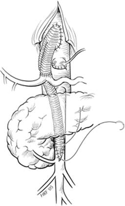
Various open surgical procedures have been reported although the antegrade aortoceliac/superior mesenteric artery bypass (Fig. 159.4) and the retrograde aortosuperior mesenteric artery bypass (Fig. 159.5) are the most common. Unfortunately, the relative infrequency of the problem has prevented the requisite randomized controlled trials. The advantages of the antegrade aortoceliac/superior mesenteric bypass are that both visceral vessels are revascularized and that the supraceliac aorta (the origin of the bypass) is usually free of atherosclerotic occlusive disease. The major disadvantage is the complexity of the procedure. In contrast, retrograde aortosuperior mesenteric artery bypass is relatively straightforward, although only a single vessel is revascularized and the graft is prone to kinking given its obligatory retrograde course that traverses both caudal to cephalad and posterior to anterior. The optimal choice for a specific patient is contingent on his or her comorbidities with the retrograde bypass generally reserved for patients who will not tolerate the more complex antegrade procedure.
|
|
|
Figure 159.4. The completed antegrade bypass from the supraceliac aorta to both the celiac axis and the superior mesenteric artery is shown. (From Huber TS, Lee WA. Revascularization for chronic mesenteric ischemia. In: Zelenock GB, Huber TS, Messina LM, et al., eds. Mastery of Vascular and Endovascular Surgery. Philadelphia, PA: Lippincott Williams & Wilkins; 2006:301, with permission.) |
The immediate postoperative care for patients undergoing revascularization for CMI is frequently complicated by the development of multiple organ dysfunction and is distinctly different from that associated with most other abdominal vascular surgical procedures such as aortobifemoral bypass for aortoiliac occlusive disease. This propensity to develop multiple organ dysfunction likely accounts for the prolonged intensive care and total hospital length of stays and is one of the leading causes of death in the postoperative period (32). The responsible mechanism for this multiple organ dysfunction is likely the visceral ischemia and reperfusion phenomenon inherent to the revascularization. This process has been reported to induce a complex response involving several interrelated inflammatory mediators that have the potential to cause both local and distant organ injury (33). In a detailed study, Harward et al. (32) characterized the individual organ system dysfunction after revascularization for both AMI and CMI. They reported that the serum hepatic transaminases (i.e., serum glutamic-oxaloacetic transaminase [SGOT], serum glutamic-pyruvic transaminase [SGPT]) increased 90- to 100-fold immediately postoperatively and did not normalize for 7 to 10 days, the platelet counts fell below 40,000 per microliter within 12 to 24 hours and remained abnormal for the first 3 to 6 days, and the prothrombin (PT) and partial thromboplastin (PTT) times became elevated and stayed elevated also for 3 to 6 days. Perhaps most notably, they reported that the overwhelming majority of patients developed a significant pulmonary injury characterized by an elevated mean shunt fraction and a radiographic picture of the acute respiratory distress syndrome that manifested between 1 to 3 days and persisted for 5 to 8 days. Jimenez et al. (7) documented a 64% incidence of multiple organ dysfunction and a 53% incidence of prolonged mechanical ventilation after antegrade revascularization for CMI and further corroborated the findings by Harward et al. (32).
|
|
|
Figure 159.5. The completed retrograde bypass from the terminal aorta/proximal right common iliac artery to the superior mesenteric artery bypass is shown. (From Huber TS, Lee WA. Revascularization for chronic mesenteric ischemia. In Zelenock GB, Huber TS, Messina LM, et al., eds. Mastery of Vascular and Endovascular Surgery. Philadelphia, PA: Lippincott Williams & Wilkins; 2006:301, with permission.) |
The optimal management strategy for patients in the early postoperative period after mesenteric revascularization is to simply support the individual organ systems until the dysfunction resolves. Admittedly, not all patients develop organ dysfunction, but the incidence is quite high and somewhat unpredictable. The optimal ventilator management remains unresolved. We have usually extubated patients in the early postoperative period when they satisfy the various weaning criteria and have been reluctant to maintain them on mechanical ventilation in anticipation that they may develop a lung injury. However, it is not infrequent that they need to be reintubated and started back on mechanical ventilation. The thrombocytopenia and coagulopathy are usually managed expectantly with platelet and/or plasma transfusions reserved for severely depressed platelet counts and/or any clinical evidence of bleeding. Notably, the report by Harward et al. (32) suggested that the inherent coagulopathy after mesenteric revascularization was not responsive to vitamin K. Patients should be maintained on total parenteral nutrition throughout the postoperative period until their bowel function returns. This is particularly important given the fact that most patients are severely compromised from a nutritional standpoint. Unfortunately, patients may have a prolonged ileus after revascularization and parenteral nutrition is required for some time. The bypass should be interrogated prior to discharge to confirm the technical adequacy of the reconstruction. Essentially the same imaging studies used to make the diagnosis are options. Our preference is mesenteric duplex although examination in the early postoperative period is frequently compromised by the persistent ileus and the presence of bowel gas. Patients with acute changes in their clinical status should also undergo visceral imaging to confirm that their bypass is patent. It can be difficult to differentiate multiple organ dysfunction that is a sequelae of the ischemia and reperfusion injury from AMI secondary to graft thrombosis. Serum lactate levels may be helpful in this setting.
All patients who undergo revascularization for CMI require long-term follow-up. Patients are usually seen frequently in the early postoperative period until all their active issues resolve and then every 6 months thereafter with a mesenteric duplex to confirm graft patency and to identify any graft or anastomotic related problems. Objective assessment of graft patency is critical and significantly better than the return of symptoms that has been used as a surrogate marker (34). All abnormalities on duplex imaging merit further investigation with additional imaging and/or intervention.
Diarrhea is a common complaint after revascularization for CMI and can persist for several months. It is more common in patients with preoperative diarrhea and can be so severe that it necessitates total parenteral nutrition. Notably, Jimenez et al. (7) reported that 33% of the patients in their series experienced significant postoperative diarrhea and that it persisted greater than 6 months in 24%. Furthermore, Kihara et al. (35) reported that patients had almost two stools/day (1.9 ± 0.4) after revascularization for CMI. The cause of the diarrhea is unclear but may be related to intestinal atrophy, bacterial overgrowth, or disruption of the mesenteric neuroplexus.
Outcome
The outcome after mesenteric revascularization for CMI is quite good. The perioperative mortality rate after open surgical revascularization is <15% (6,7,8,10,34,35,36,37,38,39) whereas that for the endovascular treatment is <5% (9,21,22,23,24,26,27,40,41,42,43,44,45,46,47,48,49). The corresponding complication rates are approximately 30% and 15% for the open and endovascular treatments, respectively. The initial technical success rate for the endovascular treatment is approximately 90%. The objectively documented 5-year patency rates after open revascularization are approximately 75%; patency rates after endovascular treatment are not as well described, but likely fall short of those reported for the open treatment. The 5-year survival after either treatment is approximately 75%, and most patients return to their presymptoms weight.
Pearls
· The underlying pathophysiology of CMI is the inability to achieve postprandial hyperemic intestinal blood flow.
· The symptoms of mesenteric ischemia usually do not occur unless two of the three visceral vessels are significantly diseased because of the rich collateral network.
· Patients presenting with abdominal pain and weight loss should initially undergo an evaluation to rule out a gastrointestinal malignancy.
· CT arteriography has largely replaced catheter-based contrast arteriography as the diagnostic study of choice for patients with CMI.
· Endovascular treatment has emerged as the initial revascularization option for most patients with CMI, but should be viewed as an alternative to open surgical revascularization.
· The immediate postoperative care for patients undergoing revascularization for CMI is frequently complicated by the development of multiple organ dysfunction.
· The optimal management strategy for patients in the early postoperative period after mesenteric revascularization is to support the individual organ systems until the dysfunction resolves.
Acute Mesenteric Ischemia
Acute mesenteric ischemia (AMI) is the end point for several distinct disease processes. Mesenteric emboli and in situ thrombosis are the most common among these and account for approximately 50% and 25% of the cases, respectively (50,51). Nonocclusive mesenteric ischemia (NOMI, 20%), mesenteric venous thrombosis (5%), and aortic dissections account for the balance (50,51). The underlying pathophysiology of AMI is that the impaired intestinal perfusion leads to mucosal compromise. This results in the release of the intracellular contents and the influx of substances (including bacteria) from the lumen of the bowel. This can lead to the activation of the systemic inflammatory response, resulting in both local and distant organ dysfunction (e.g., lung injury). If the impaired perfusion persists, bowel infarction with perforation and peritonitis ensues. The immediate clinical concerns for patients with AMI are to reverse the underlying clinical condition and prevent bowel infarction. The clinical presentation, diagnostic approach, and treatment for the various causes of AMI are similar, but there are distinct differences that mandate individual consideration. AMI from an embolus will be discussed in depth given the fact that it is the most common cause, and the respective differences will be highlighted for the other causes. Fortunately, the underlying cause can usually be determined from the history and clinical setting. Not surprisingly, the morbidity and mortality associated with AMI are significant. The optimal therapy requires prompt diagnosis and definitive treatment although this is often difficult given the susceptible patient population and common clinical scenarios. Indeed, AMI may be either the cause or the effect of the patient's critical illness.
Embolus
Pathophysiology
The emboli responsible for AMI usually lodge in the superior mesenteric artery and originate from the heart. The intracardiac thrombus is related to either atrial fibrillation, an acute myocardial infarction, or a ventricular aneurysm. Patients frequently have prior embolic events from the same source although they are not necessarily limited to the superior mesenteric artery (e.g., common femoral artery bifurcation presenting with acute lower extremity ischemia). Notably, the material that constitutes these macroemboli is actually quite large as might be predicted from the size of the arteries involved. This is in contradistinction to the micron-sized atheroembolic particles that commonly cause blue toes after invasive arteriographic procedures (i.e., artery–artery emboli). The extent of bowel ischemia and/or infarction after an embolus to the superior mesenteric artery is contingent on the extent of the collateral circulation, the pattern of the arterial occlusion, and the duration of the ischemia. In this setting, the bowel progresses from ischemia to infarction in a time-dependent fashion although it may remain viable for 6–12 hours. Acute embolic occlusion of the superior mesenteric usually results in ischemia/infarction of the bowel from the proximal jejunum to the transverse colon. The duodenum and descending colon are usually spared because they are supplied by branches of the celiac axis and inferior mesenteric artery, respectively.
Clinical Presentation and Diagnosis
The diagnosis of AMI from an embolus (or other causes of AMI) may be difficult. The differential list of diagnoses includes all the more common causes of acute abdominal pain. The diagnosis is confounded by the fact that the patients are often critically ill, and thus, their history/physical examination may not be reliable. Diagnosis requires a high index of suspicion and an aggressive approach since delays in diagnosis adversely affect outcome. This requires an appreciation of the types of patients and clinical scenarios in which AMI occurs including post myocardial infarction, end-stage renal disease (52), and post coronary artery bypass grafting (53,54,55).
Patients with AMI from an embolus usually present with diffuse abdominal pain. The classic description of the pain secondary to AMI is pain out of proportion to the physical findings although this scenario is not always present. Unfortunately, the pain is neither specific nor localized to a particular abdominal quadrant. Peritoneal signs can be present, but they usually occur late in the process and suggest bowel perforation. Patients often experience nausea/vomiting and/or diarrhea, but again these are fairly nonspecific complaints. Notably, AMI is a potent cathartic. Similar to the physical examination, the routine chemistry and hematologic laboratory studies are usually nonspecific and insensitive. Patients frequently have mild abnormalities of their laboratory values including an elevated white blood count, a decreased platelet count, an elevated hematocrit, and a mildly elevated amylase level. The hemodynamic status of the patients at the time of presentation ranges from normovolemia to profound hypovolemic shock with acidosis and is contingent on the status of the bowel and the duration of symptoms.
Various diagnostic studies are available to help make the diagnosis of AMI secondary to an embolus. Plain abdominal radiographs have been used traditionally for patients with acute abdominal pain and can be helpful to demonstrate free air from an intestinal perforation or identify other causes of abdominal pain. However, patients with AMI frequently have either normal plain radiographs or demonstrate nonspecific findings (e.g., ileus). CT arteriography with 1- to 2-mm cuts has emerged as the diagnostic study of choice for patients with AMI secondary to an embolus (56,57,58). Notably, the study is performed using only intravenous contrast since both oral and rectal contrast potentially interfere with the arteriogram itself. Both stenoses and occlusions in the visceral vessels are well demonstrated on CT arteriography. In patients with AMI secondary to an embolus, a meniscus sign can often be seen in the mid/distal superior mesenteric artery. The images obtained using the CT arteriogram protocol are also excellent for the nonvascular structures. The significant nonvascular findings of AMI include bowel wall thickening, bowel wall gas, bowel/solid organ infarction, and hepatic/portal venous gas. Indeed, Paran et al. (57) reported that most patients with portal/ mesenteric venous gas had mesenteric ischemia and that the associated mortality rate was 86%. Mesenteric duplex, although an excellent screening test for CMI, is not usually helpful in patients with AMI because abdominal distention/gas precludes the accurate interrogation of the visceral vessels.
Standard contrast arteriography can be used as an alternative to CT arteriography, and indeed, has been the traditional imaging study for the visceral vessels in the setting of AMI from an embolus or in situthrombosis. Similar to studies in patients with CMI, it can potentially serve as both a diagnostic test and therapeutic modality since an intervention can be performed at the same time. The major disadvantages are the obligatory time required to obtain the procedure and the small, but finite, complications associated with the contrast agent and vessel cannulation. Given the new treatment algorithms with vascular surgeons assuming the traditional roles of the interventional radiologists, it is conceivable that the diagnostic contrast arteriogram could be performed in an operating room with a fixed imaging unit. The definitive operative procedure could be performed at the same setting if the arteriogram confirmed the diagnosis.
Laparoscopy offers an additional diagnostic modality for patients with AMI (59,60). It can be used to assess the viability of the bowel and confirm the diagnosis. Furthermore, it can be performed in the intensive care unit with sedation and, therefore, is feasible for unstable patients with a suspected intra-abdominal process. Notably, the authors of the evidence-based guidelines from the European Association for Endoscopic Surgery concluded that laparoscopy had an unclear or limited role in the setting of AMI (61). However, they did state that it may be indicated if the routine diagnostic studies are inconclusive. Laparotomy remains the definitive diagnostic test for patients with AMI. However, the diagnostic studies outlined above are usually sufficient.
Treatment Strategies
Patients should be taken emergently to the operating room for definitive treatment once the diagnosis of AMI from an embolus is made. An extensive preoperative evaluation is unnecessary and potentially harmful in light of the narrow window for salvaging the bowel. There is essentially no role for medical management alone in this setting. Patients should be systemically anticoagulated with heparin to prevent further clot development and started on broad-spectrum antibiotics against enteric organisms. Importantly, patients with AMI are frequently hypovolemic and should be volume resuscitated prior to the induction of anesthesia. This can be performed fairly expeditiously and should not delay transfer to the operating room.
Both midline and transverse abdominal incisions provide adequate exposure to the visceral vessels, and the choice is contingent on surgeon preference. The diagnosis of AMI from an embolus is usually confirmed by the distribution of the ischemic/infarcted bowel that extends from the proximal jejunum to the transverse colon. However, the diagnosis should be further substantiated by interrogating the visceral vessels with continuous wave Doppler. The embolus may be extracted from the superior mesenteric artery using a Fogarty thromboembolectomy catheter. Although there are several approaches to the superior mesenteric artery, the easiest approach is to incise the base of the transverse mesocolon after retracting the transverse colon itself cephalad. The arteriotomy in the superior mesenteric artery may be performed either longitudinally or horizontally. Although the longitudinal arteriotomy needs to be closed with a vein patch to prevent narrowing its lumen, it is the preferred approach because it affords greater flexibility in case a bypass procedure is necessary.
The management of the bowel for patients with AMI merits further comment. All of the bowel that is obviously dead should be resected, and intestinal anastomoses should be avoided in favor of proximal and distal stomas. This mandates a second procedure to restore bowel continuity, but it allows the bowel (i.e., mucosa of the stoma) to be examined at the bedside during the postoperative period. Furthermore, it avoids using ischemic or borderline ischemic tissues for the anastomosis. Bowel that is ischemic though not frankly necrotic should be revascularized and then re-examined before any final decision about resection. A conservative approach is justified in this setting because many of the borderline areas will remain viable after revascularization. Admittedly, the differentiation between viable and nonviable bowel is difficult. Various complicated modalities have been described to help differentiate viable from nonviable bowel in this setting although they have not been universally adopted. Simple adjuncts include visual inspection for peristalsis, use of continuous wave Doppler to detect arterial signals within the mesentery, and intravenous fluorescein in combination with a Wood lamp. Notably, approximately 100 to 150 cm of small bowel is necessary for nutritional absorption.
A decision to perform a second-look operation to reassess the viability of the bowel should be made at the time of the initial procedure. This is routinely performed 24 to 48 hours after the first procedure, a time usually sufficient for the marginal bowel to declare itself. A recent retrospective review has questioned the role of the second-look operation and reported that survival was actually greater in those patients in whom it was not performed (62). Admittedly, there was a tremendous selection bias in this review and the authors conceded that the experience of the surgeon is likely the key factor regarding the decision to perform a second look. A “damage control” operation may be justified in a small subset of unstable patients with AMI as suggested by Freeman and Graham (63). This includes emergent laparotomy, resection of obviously dead bowel, and creation of proximal/distal stomas, leaving the abdomen open and deferring the definitive vascular/gastrointestinal procedure until later.
Despite its definitive role for patients with CMI, there is likely little role for endovascular therapies in patients with AMI secondary to an embolus. The obligatory time for endovascular treatment, including chemical lysis, is too long given the threatened bowel and the potential to progress from ischemic bowel to infarcted bowel. Furthermore, it does not allow direct assessment of the bowel, and the chemical lysis may cause intestinal bleeding from mucosal sloughing. A recent systematic review of the literature examining the role of thrombolysis for acute superior mesenteric artery occlusion found insufficient evidence to support the practice despite a few case reports (64,65,66,67,68,69,70).
The postoperative course after embolectomy for AMI is similar to that after revascularization for CMI although the incidence of postoperative complications and multiple organ dysfunction are greater. As noted above, revascularization may cause an ischemia/reperfusion injury that affects both local and distant organ systems. Accordingly, patients are at risk for developing an abdominal compartment syndrome. Bladder pressures can be measured, and the abdomen closure can be dissembled as necessary. Patients should be continued on broad-spectrum antibiotics throughout the early postoperative period. Furthermore, they need to be anticoagulated long term due to the potential for recurrent emboli.
Outcome
The mortality rate for patients with AMI is approximately 70%, and this rate has changed very little over the past several decades (51,68,71,72). Unfortunately, most of the case series tend to encompass all of the causes rather than a specific one (e.g., embolus). A recent systematic review by Schoots et al. (68) reported that the mortality rates for AMI from mesenteric venous thrombosis were better than those for arterial problems and that the mortality rates for mesenteric emboli were better than those for in situ thromboses. The aggregate mortality rates in their study by cause are listed: mesenteric venous thrombosis, 32%; embolus, 54%; NOMI, 74%; in situ thrombosis, 77%. Several predictable factors have been associated with mortality in the various case series including patient age, time to definitive surgery, shock, acidosis, leukocytosis, cardiac status, and coagulopathy (71,73,74). Most deaths in a recent report were due to multiple organ failure (75).
In Situ Thrombosis
Patients with visceral artery occlusive disease may also present with AMI secondary to in situ thrombosis. The presentation is superimposed on the symptoms of CMI in more than 50% of the patients (76) and can usually be differentiated from the other causes of AMI by the history and clinical setting. However, it is important to emphasize that patients may present with AMI as the initial symptom of their visceral occlusive disease. The clinical presentation, diagnostic approach, and immediate postoperative care of patients with AMI secondary to in situ thrombosis is similar to that outlined above for emboli although the operative approach is somewhat different.
Patients with AMI secondary to in situ thrombosis require a mesenteric bypass. Although antegrade aortoceliac/superior mesenteric artery bypass is probably the optimal bypass for CMI, retrograde bypass from the infrarenal aorta or common iliac artery is likely the optimal procedure for AMI (Fig. 159.5). The objectives and treatment are somewhat different in the acute setting (i.e., AMI vs. CMI). The main objective is to restore blood flow to the ischemic vascular bed as safely and expeditiously as possible. This usually requires only bypass to the superior mesenteric artery. Patients with isolated celiac axis stenosis rarely develop AMI because the collateral blood flow to the foregut is so good and the liver may be sustained on portal blood flow alone. Prosthetic conduits are relatively contraindicated in the setting of bowel infarction and/or perforation due to the potential for postoperative graft infection. Autogenous conduits with either saphenous or superficial femoral vein are suitable although the latter may be more durable.
The role of endovascular treatment for patients with AMI secondary to in situ thrombosis is likely limited for the reasons noted above for AMI secondary to an embolus. Wyers et al. (77) have reported a small case series using novel hybrid open/endovascular approach in which a stent is placed retrograde through the superior mesenteric artery at the time of laparotomy. This approach allows assessment of the bowel and is potentially less morbid than the more traditional open revascularization.
Occasionally patients will undergo bowel resection for infarction by a nonvascular surgeon, and the diagnosis of mesenteric ischemia will be missed both preoperatively and intraoperatively. It is important to emphasize that infarction of the bowel is not a spontaneous event, but rather an end-stage complication of another disease process; it is imperative that the cause of all bowel infarctions be established in an attempt to prevent recurrences. An appropriate imaging study (i.e., CT arteriogram, contrast arteriogram) should be obtained in the early postoperative period, and the necessary treatment including anticoagulation and revascularization implemented in a timely fashion.
Nonocclusive Mesenteric Ischemia
Pathophysiology
Nonocclusive mesenteric ischemia (NOMI) represents an abnormal or paradoxical mesenteric vasoconstriction characterized by the loss of autoregulation. Shock or circulatory stress normally causes mesenteric vasoconstriction in an attempt to maintain cerebral and/or cardiac perfusion. The mesenteric vasoconstriction ordinarily resolves when the underlying circulatory disorders are corrected; persistent vasoconstriction results in NOMI. There are multiple potential causes for NOMI including cardiogenic shock, sepsis, burn injury, trauma, pancreatitis, digitals, vasopressors, and renal failure (78,79,80,81,82,83). Indeed, almost any underlying condition that can precipitate shock or circulatory stress may precipitate NOMI.
Clinical Presentation and Diagnosis
Similar to the other causes of AMI, the diagnosis of NOMI requires a high index of suspicion and the proper clinical setting. Patients may develop abdominal pain, although the physical examination is frequently unreliable due to the other active medical issues and altered sensorium. Laboratory abnormalities are common including acidosis, leukocytosis, elevated lactate levels, and hyperamylasemia, but these are all relatively nonspecific markers of the underlying shock state. Contrast arteriography has traditionally been the diagnostic study of choice and is also potentially therapeutic. The significant findings on arteriogram for patients with NOMI include segmental stenosis/narrowing of the superior mesenteric artery in a string-of-beads appearance. Furthermore, there is narrowing of the branches of the superior mesenteric artery at their origins, spasm of the mesenteric arcades, and impaired filling of the intramural branches. The contrast arteriogram can also be helpful to rule out the other potential causes of AMI. The published experience with CT arteriography in the setting of NOMI is limited, although it likely affords the same advantages as contrast arteriography with fewer risks.
Treatment Strategies
The initial treatment of patients with NOMI is nonoperative and directed at correcting the underlying condition that precipitated the circulatory stress. Specifically, patients should be resuscitated in an attempt to improve their cardiac output and systemic perfusion. All vasoactive drugs should be stopped (if possible), and patients should be started on broad-spectrum antibiotics directed against enteric organisms. Furthermore, patients should be systemically anticoagulated unless contraindicated. Despite these efforts, the characteristic mesenteric vasoconstriction may persist. Continuous intra-arterial papaverine, administered through an infusion catheter placed into the superior mesenteric artery, may reverse the vasoconstriction (84,85). A 45-mg test dose of papaverine (i.e., short-acting calcium channel blocker) should be given over 15 minutes, and a continuous infusion of 30 to 60 mg/hour should be started if no adverse reactions are encountered. Serial mesenteric arteriograms should be performed to monitor the response to the papaverine with the first performed 1 hour after initiating therapy. The intra-arterial infusion may be continued up to 24 hours. It should be noted that the infusion will reverse the mesenteric vasoconstriction only if the underlying hemodynamic instability is corrected. Operative treatment of NOMI should be reserved only for the clinical scenario when bowel infarction is suspected.
Mesenteric Venous Thrombosis
Pathophysiology
Mesenteric venous thrombosis may also result in AMI and is considered in this section for completeness. However, the associated degree of bowel ischemia is usually less than with arterial occlusion from either an embolus or in situ thrombosis. The pathophysiology is similar to venous thrombosis in other vascular beds and may be explained in terms of the Virchow classic triad of stasis, intimal injury, and hypercoagulable states. Mesenteric venous thrombosis results in edema in the bowel and mesentery with significant third-space fluid losses. This may result in bloody ascites and, indeed, a bloody tap at the time of paracentesis may be diagnostic. Progression to bowel infarction is contingent on the magnitude of the clot load and its distribution. Clot localized to the portal or superior mesenteric vein does not usually lead to bowel infarction because of the collateral channels whereas clot in the peripheral mesenteric veins is more likely to do so. The natural history of untreated mesenteric venous thrombosis is poor and almost universally progresses from bowel infarction to perforation and death.
Mesenteric venous thrombosis may result from abnormalities in any of the components of the Virchow triad (i.e., stasis, intimal injury, hypercoagulable state). Stasis may result from congestive heart failure or portal hypertension whereas intimal injury may result from general anesthesia or any number of intra-abdominal infectious processes. A hypercoagulable state is perhaps the strongest of the contributory factors and has been identified in up to 90% of patients with mesenteric venous thrombosis (86).
Clinical Presentation and Diagnosis
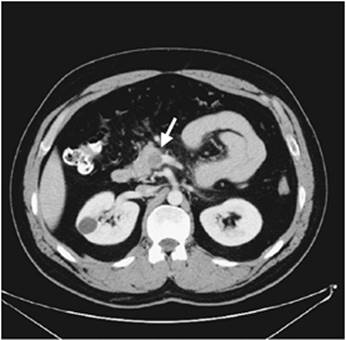
Patients with mesenteric venous thrombosis usually present with vague, mild abdominal pain. The pain is usually insidious in onset and frequently present for some time before the patients seek medical attention. Furthermore, the pain is not usually localized to any specific quadrant. Physical examination is notable only for mild, diffuse abdominal pain. Peritoneal signs suggest bowel infarction, but are found only later in the disease process. An abdominal CT scan is the diagnostic study of choice (87,88). The significant findings include bowel edema and thrombus within the mesenteric veins with inflammation of the vessel wall (Fig. 159.6). Plain abdominal radiographs may suggest abdominal wall edema and are helpful to rule out other causes of the abdominal pain. Standard catheter-based contrast arteriography may be helpful but is inferior to CT. The arteriographic findings that suggest mesenteric venous thrombosis include arterial spasm with a prolonged arterial phase, opacification of the bowel wall, extravasations of the contrast into the bowel lumen, and visualization of the venous thrombus.
Treatment Strategies
The primary treatment of patients with mesenteric venous thrombosis is anticoagulation. Patients should be aggressively anticoagulated with heparin when the diagnosis is made and should be maintained on long-term oral anticoagulation. Similar to patients with massive iliofemoral deep venous thrombosis, it may be difficult to achieve effective anticoagulation initially using the standard dosing schedules for heparin (i.e., 80 units/kg bolus, 18 units/kg per hour drip), presumably secondary to the clot burden. Larger doses of heparin may be required, and the adequacy of heparinization (e.g., partial thromboplastin time) may need to be monitored more frequently. Although the required dosage of heparin may be unsettling, the potential clot propagation and its associated complications likely exceed any increased risk from bleeding. A hypercoagulable workup for the standard hematologic abnormalities should be performed prior to initiation of anticoagulation. However, long-term anticoagulation should be continued even in the absence of an identifiable hypercoagulable state since it is likely that many of these patients have some type of hypercoagulable disorder even though it may not be characterized on initial screening. Additionally, patients frequently require fluid resuscitation at the time of diagnosis due to the significant third-space losses from the bowel edema.
|
|
|
Figure 159.6. A CT scan of a patient with mesenteric venous thrombosis is shown. Note the thrombus in the superior mesenteric vein at its confluence with the splenic vein as shown with the arrow. |
Exploratory laparotomy should be reserved for cases in which bowel infarction is suspected. The intraoperative findings include edematous/rubbery bowel, bloody ascites, and thrombus within the mesentery. A wide resection of the bowel should be performed in the presence of infarction. Primary enteric anastomosis is probably safe if the margins of resection are free of thrombus within the mesentery. Proximal and distal stomas are advisable if the viability of the bowel at the margins of resection is questionable. Patients are at risk for additional or ongoing thrombosis with the most common site being the margins of the resection and/or the anastomosis. Mechanical thrombectomy is not advocated at the time of laparotomy because of the extensive clot burden.
Endovascular treatment including chemical lysis likely has a limited role for patients with mesenteric venous thrombosis despite several case reports (89,90,91). Similar to the concerns with mechanical thrombectomy, the clot burden is very significant and usually extends from the small peripheral collaterals in the mesentery to the portal and mesenteric veins.
Outcome
As noted above, the mortality rate (approximately 30%) for mesenteric venous thrombosis is significantly less than for the arterial causes of AMI (68). Death has been associated with portal vein thrombosis, systemic venous thromboembolism, and obesity. The increased mortality rate associated with venous thromboembolism underscores the importance of early, adequate anticoagulation.
Aortic Dissection
Patients with acute aortic dissection can present with visceral malperfusion and AMI. The management of acute aortic dissections is beyond the scope of this chapter, but the topic is included to emphasize the importance of evaluating the status of the visceral vessels in all patients presenting with acute dissections. The presence of visceral malperfusion significantly increases the mortality rate (92,93). Various endovascular approaches have been used to treat the visceral malperfusion in this setting (94,95). Endovascular revascularization is probably superior to the open approach provided the patients are candidates from an anatomic and technical standpoint.
Protocols and Algorithms
See Fig. 159.7.
Pearls
· The impaired intestinal perfusion associated with AMI leads to mucosal compromise that can lead to the activation of the systemic inflammatory and bowel infarction with perforation.
· The immediate clinical concerns for patients with AMI are to reverse the underlying clinical condition and prevent bowel infarction.
· The cause of AMI (i.e., embolus, in situ thrombosis, NOMI, mesenteric venous thrombosis, dissection) can usually be determined from the history and clinical setting.
· The emboli responsible for AMI usually lodge in the superior mesenteric artery and originate from the heart as a result of atrial fibrillation, an acute myocardial infarction, or a ventricular aneurysm.
· In patients with in situ thrombosis, the presentation of AMI is superimposed on the symptoms of CMI in more than 50% of the patients.
· NOMI represents an abnormal or paradoxical mesenteric vasoconstriction characterized by the loss of autoregulation.
· Mesenteric venous thrombosis is associated with a high incidence of hypercoagulable states and merits long-term anticoagulation even in the absence of an identifiable condition.
· CT arteriography with 1- to 2-mm cuts has emerged as the diagnostic study of choice for patients with AMI.
· Patients with AMI from an embolus or in situ thrombosis require emergent operative treatment.
· Operative treatment for patients with NOMI and mesenteric venous thrombosis is reserved for cases in which bowel infarction is suspected.
Colon Ischemia
Isolated colon ischemia can occur after both open and endovascular aneurysm repair. Furthermore, it can develop as a complication of hemodynamic shock, similar to NOMI. Ischemic colitis has been reported to occur in approximately 2% to 13% of open aneurysm repairs (96). The reported incidence depends on the diagnostic algorithm and modality (routine sigmoidoscopy vs. selective sigmoidoscopy) and is dramatically increased after ruptured aneurysm repair. Indeed, the incidence of colonic ischemia after ruptured aneurysm repair in patients undergoing routine colonoscopy is approximately 25 to 40% (96,97). The sigmoid colon is affected most frequently, although all the sections of the colon may be involved. The ischemia may result from inadequate resuscitation, disruption of collaterals, and/or failure to revascularize a hemodynamically significant inferior mesenteric artery. Interestingly, routine reimplantation of the inferior mesenteric artery at the time of aortic reconstruction does not prevent colon ischemia (98). The ischemic colitis associated with endovascular aneurysm repair is more commonly related to atheroembolism than acute internal iliac artery occlusion as might be suspected. The prognosis for ischemic colitis after endovascular aneurysm repair is worse than that for open repair (99).
|
|
|
Figure 159.7. A flow diagram for the evaluation of patients with acute mesenteric ischemia (AMI) is shown. Although patients frequently present with abdominal pain, their sensorium may be altered. The diagnosis should be considered in the critical care setting when patients decompensate acutely. The differential diagnosis should be framed within the appropriate clinical setting. A computed tomography (CT) arteriogram is the diagnostic test of choice although a contrast arteriogram can be used. The CT findings are fairly characteristic for each of the diagnoses. Emergent, open revascularization is required in the setting of an embolus and in situ thrombosis; the bowel should be resected as necessary for ischemia/infarction. Preoperative evaluation should include broad-spectrum antibiotics, anticoagulation, and resuscitation. Medical management alone is usually adequate for patients with nonocclusive mesenteric ischemia (NOMI) and mesenteric venous thrombosis. Intra-arterial vasodilation may be required for persistent vasoconstriction in patients with NOMI. Lifetime anticoagulation is required for mesenteric venous thrombosis. Emergent endovascular revascularization is required for patients with acute aortic dissections and visceral malperfusion. Exploratory laparotomy and bowel resection should be reserved in patients with NOMI, mesenteric venous thrombosis, and aortic dissections for presumed bowel infarction. CMI, chronic mesenteric ischemia. |
Patients with ischemic colitis usually present with bloody diarrhea in contrast to patients with AMI who usually present with abdominal pain. In the most common scenario, patients develop bloody diarrhea on the first or second postoperative day after aortic reconstruction. However, the diagnosis should be considered after aortic reconstruction in the absence of bloody diarrhea in patients with thrombocytopenia, multiple-organ dysfunction, increasing abdominal pain/peritonitis, and generalized failure to thrive. The diagnosis may be confirmed by endoscopy. Although sigmoidoscopy is used most frequently, a complete colonoscopy is likely optimal due to the potential involvement of the other colon segments.
Treatment depends on the endoscopic findings and clinical setting. The endoscopic findings range from mucosal ischemia to transmural necrosis. Unfortunately, it is often difficult to differentiate diffuse mucosal ischemia from transmural necrosis. Patients with mucosal ischemia alone should be treated with bowel rest, broad-spectrum antibiotics, total parenteral nutrition, and serial endoscopic examinations. Many of these lesions resolve spontaneously without long-term sequelae although colonic strictures may develop in a small subset of patients. Patients with transmural colonic necrosis should undergo laparotomy with resection of the involved segment, a proximal diverting colostomy, and a distal Hartmann pouch.
The reported mortality rate in patients with transmural colon necrosis after aortic reconstruction is approximately 85% (96). Maintaining antegrade flow through the internal iliac vessels, routinely implanting the inferior mesenteric artery, and preserving the colonic collateral circulation may reduce or prevent this adverse outcome.
References
1. Fara JW. Postprandial mesenteric hyperemia. In: Shepard AP, Granger DN, eds. Physiology of the Intestinal Circulation. New York, NY: Raven Press; 1984:99.
2. Moneta GL, Taylor DC, Helton WS, et al. Duplex ultrasound measurement of postprandial intestinal blood flow: effect of meal composition. Gastroenterology. 1988;95(5):1294–1301.
3. Carrick RP, Borge MA, Labropolous N, et al. Chronic mesenteric ischemia resulting from isolated lesions of the superior mesenteric artery—a case report. Angiology. 2005;56(6):785–788.
4. Valentine RJ, Martin JD, Myers SI, et al. Asymptomatic celiac and superior mesenteric artery stenoses are more prevalent among patients with unsuspected renal artery stenoses. J Vasc Surg. 1991;14(2):195–199.
5. Croft RJ, Menon GP, Marston A. Does ‘intestinal angina’ exist? A critical study of obstructed visceral arteries. Br J Surg. 1981;68(5):316–318.
6. Derrow AE, Seeger JM, Dame DA, et al. The outcome in the United States after thoracoabdominal aortic aneurysm repair, renal artery bypass, and mesenteric revascularization. J Vasc Surg. 2001;34(1):54–61.
7. Jimenez JG, Huber TS, Ozaki CK, et al. Durability of antegrade synthetic aortomesenteric bypass for chronic mesenteric ischemia. J Vasc Surg. 2002;35(6):1078–1084.
8. Johnston KW, Lindsay TF, Walker PM, et al. Mesenteric arterial bypass grafts: early and late results and suggested surgical approach for chronic and acute mesenteric ischemia. Surgery. 1995;118(1):1–7.
9. Matsumoto AH, Angle JF, Spinosa DJ, et al. Percutaneous transluminal angioplasty and stenting in the treatment of chronic mesenteric ischemia: results and longterm followup. J Am Coll Surg. 2002;194(1 Suppl):S22–S31.
10. Mateo RB, O'Hara PJ, Hertzer NR, et al. Elective surgical treatment of symptomatic chronic mesenteric occlusive disease: early results and late outcomes. J Vasc Surg. 1999;29(5):821–31.
11. Van Damme H, Jacquet N, Belaiche J, et al. Chronic ischaemic gastritis: an unusual form of splanchnic vascular insufficiency. J Cardiovasc Surg (Torino). 1992;33(4):451–453.
12. Moneta GL, Lee RW, Yeager RA, et al. Mesenteric duplex scanning: a blinded prospective study. J Vasc Surg. 1993;17(1):79–84.
13. Zwolak RM, Fillinger MF, Walsh DB, et al. Mesenteric and celiac duplex scanning: a validation study. J Vasc Surg. 1998;27(6):1078–1087.
14. Boley SJ, Brandt LJ, Veith FJ, et al. A new provocative test for chronic mesenteric ischemia. Am J Gastroenterol. 1991;86(7):888–891.
15. Gentile AT, Moneta GL, Lee RW, et al. Usefulness of fasting and postprandial duplex ultrasound examinations for predicting high-grade superior mesenteric artery stenosis. Am J Surg. 1995;169(5):476–479.
16. Cademartiri F, Raaijmakers RH, Kuiper JW, et al. Multi-detector row CT angiography in patients with abdominal angina. Radiographics. 2004;24(4):969–984.
17. Wildermuth S, Leschka S, Alkadhi H, et al. Multislice CT in the pre- and postinterventional evaluation of mesenteric perfusion. Eur Radiol. 2005;15(6):1203–1210.
18. Zandrino F, Musante F, Gallesio I, et al. Assessment of patients with acute mesenteric ischemia: multislice computed tomography signs and clinical performance in a group of patients with surgical correlation. Minerva Gastroenterol Dietol. 2006;52(3):317–325.
19. Hagspiel KD, Leung DA, Angle JF, et al. MR angiography of the mesenteric vasculature. Radiol Clin North Am. 2002;40(4):867–886.
20. Laissy JP, Trillaud H, Douek P. MR angiography: noninvasive vascular imaging of the abdomen. Abdom Imaging. 2002;27(5):488–506.
21. Brown DJ, Schermerhorn ML, Powell RJ, et al. Mesenteric stenting for chronic mesenteric ischemia. J Vasc Surg. 2005;42(2):268–274.
22. Landis MS, Rajan DK, Simons ME, et al. Percutaneous management of chronic mesenteric ischemia: outcomes after intervention. J Vasc Interv Radiol. 2005;16(10):1319–1325.
23. Resch T, Lindh M, Dias N, et al. Endovascular recanalisation in occlusive mesenteric ischemia—feasibility and early results. Eur J Vasc Endovasc Surg. 2005;29(2):199–203.
24. Schaefer PJ, Schaefer FK, Mueller-Huelsbeck S, et al. Chronic mesenteric ischemia: stenting of mesenteric arteries. Abdom Imaging. 2007;32(3):304–309. Epub 2006 Sep 6.
25. Schaefer PJ, Schaefer FK, Hinrichsen H, et al. Stent placement with the monorail technique for treatment of mesenteric artery stenosis. J Vasc Interv Radiol. 2006;17(4):637–643.
26. Silva JA, White CJ, Collins TJ, et al. Endovascular therapy for chronic mesenteric ischemia. J Am Coll Cardiol. 2006;47(5):944–950.
27. Sivamurthy N, Rhodes JM, Lee D, et al. Endovascular versus open mesenteric revascularization: immediate benefits do not equate with short-term functional outcomes. J Am Coll Surg. 2006;202(6):859–867.
28. Eagle KA, Berger PB, Calkins H, et al. ACC/AHA guideline update for perioperative cardiovascular evaluation for noncardiac surgery—executive summary a report of the American College of Cardiology/American Heart Association Task Force on Practice Guidelines (Committee to Update the 1996 Guidelines on Perioperative Cardiovascular Evaluation for Noncardiac Surgery). Circulation. 2002;105(10):1257–1267.
29. McFalls EO, Ward HB, Moritz TE, et al. Coronary-artery revascularization before elective major vascular surgery. N Engl J Med. 2004;351(27):2795–2804.
30. Poldermans D, Bax JJ, Schouten O, et al. Should major vascular surgery be delayed because of preoperative cardiac testing in intermediate-risk patients receiving beta-blocker therapy with tight heart rate control? J Am Coll Cardiol. 2006;48(5):964–969.
31. Smith SC Jr, Blair SN, Bonow RO, et al. AHA/ACC Guidelines for Preventing Heart Attack and Death in Patients with Atherosclerotic Cardiovascular Disease: 2001 update. A statement for healthcare professionals from the American Heart Association and the American College of Cardiology. J Am Coll Cardiol. 2001 Nov 1;38(5):1581–1583.
32. Harward TR, Brooks DL, Flynn TC, et al. Multiple organ dysfunction after mesenteric artery revascularization. J Vasc Surg. 1993;18(3):459–467.
33. Kagan SA, Myers SI. Acute embolic and thrombotic mesenteric ischemia. In: Ernst CB, Stanley JC, eds. Current Therapy in Vascular Surgery. 4th ed. St. Louis, MO: Mosby; 2001:675.
34. McMillan WD, McCarthy WJ, Bresticker MR, et al. Mesenteric artery bypass: objective patency determination. J Vasc Surg. 1995;21(5):729–740.
35. Kihara TK, Blebea J, Anderson KM, et al. Risk factors and outcomes following revascularization for chronic mesenteric ischemia. Ann Vasc Surg. 1999;13(1):37–44.
36. Cho JS, Carr JA, Jacobsen G, et al. Long-term outcome after mesenteric artery reconstruction: a 37-year experience. J Vasc Surg. 2002;35(3):453–460.
37. Foley MI, Moneta GL, Abou-Zamzam AM Jr, et al. Revascularization of the superior mesenteric artery alone for treatment of intestinal ischemia. J Vasc Surg. 2000;32(1):37–47.
38. Moawad J, McKinsey JF, Wyble CW, et al. Current results of surgical therapy for chronic mesenteric ischemia. Arch Surg. 1997;132(6):613–618.
39. Park WM, Cherry KJ Jr, Chua HK, et al. Current results of open revascularization for chronic mesenteric ischemia: a standard for comparison. J Vasc Surg. 2002;35(5):853–859.
40. Allen RC, Martin GH, Rees CR, et al. Mesenteric angioplasty in the treatment of chronic intestinal ischemia. J Vasc Surg. 1996;24(3):415–421.
41. Cognet F, Ben Salem D, Dranssart M, et al. Chronic mesenteric ischemia: imaging and percutaneous treatment. Radiographics. 2002;22(4):863–879.
42. Hallisey MJ, Deschaine J, Illescas FF, et al. Angioplasty for the treatment of visceral ischemia. J Vasc Interv Radiol. 1995;6(5):785–791.
43. Kasirajan K, O'Hara PJ, Gray BH, et al. Chronic mesenteric ischemia: open surgery versus percutaneous angioplasty and stenting. J Vasc Surg. 2001;33(1):63–71.
44. Maspes F, Mazzetti dP, Gandini R, et al. Percutaneous transluminal angioplasty in the treatment of chronic mesenteric ischemia: results and 3 years of follow-up in 23 patients. Abdom Imaging. 1998;23(4):358–363.
45. Nyman U, Ivancev K, Lindh M, et al. Endovascular treatment of chronic mesenteric ischemia: report of five cases. Cardiovasc Intervent Radiol. 1998;21(4):305–313.
46. Pietura R, Szymanska A, El Furah M, et al. Chronic mesenteric ischemia: diagnosis and treatment with balloon angioplasty and stenting. Med Sci Monit. 2002;8(1):R8–R12.
47. Sharafuddin MJ, Olson CH, Sun S, et al. Endovascular treatment of celiac and mesenteric arteries stenoses: applications and results. J Vasc Surg. 2003;38(4):692–698.
48. Sheeran SR, Murphy TP, Khwaja A, et al. Stent placement for treatment of mesenteric artery stenoses or occlusions. J Vasc Interv Radiol. 1999;10(7):861–867.
49. Steinmetz E, Tatou E, Favier-Blavoux C, et al. Endovascular treatment as first choice in chronic intestinal ischemia. Ann Vasc Surg. 2002;16(6):693–699.
50. Lock G. Acute mesenteric ischemia: classification, evaluation and therapy. Acta Gastroenterol Belg. 2002;65(4):220–225.
51. Safioleas MC, Moulakakis KG, Papavassiliou VG, et al. Acute mesenteric ischaemia, a highly lethal disease with a devastating outcome. Vasa. 2006;35(2):106–111.
52. Bassilios N, Menoyo V, Berger A, et al. Mesenteric ischaemia in haemodialysis patients: a case/control study. Nephrol Dial Transplant. 2003;18(5):911–917.
53. Bolcal C, Iyem H, Sargin M, et al. Gastrointestinal complications after cardiopulmonary bypass: sixteen years of experience. Can J Gastroenterol. 2005;19(10):613–617.
54. Edwards MS, Cherr GS, Craven TE, et al. Acute occlusive mesenteric ischemia: surgical management and outcomes. Ann Vasc Surg. 2003;17(1):72–79.
55. Ghosh S, Roberts N, Firmin RK, et al. Risk factors for intestinal ischaemia in cardiac surgical patients. Eur J Cardiothorac Surg. 2002;21(3):411–416.
56. Lee R, Tung HK, Tung PH, et al. CT in acute mesenteric ischaemia. Clin Radiol. 2003;58(4):279–287.
57. Paran H, Epstein T, Gutman M, et al. Mesenteric and portal vein gas: computerized tomography findings and clinical significance. Dig Surg. 2003;20(2):127–132.
58. Schindera ST, Triller J, Vock P, et al. Detection of hepatic portal venous gas: its clinical impact and outcome. Emerg Radiol. 2006;12(4):164–170.
59. Gagne DJ, Malay MB, Hogle NJ, et al. Bedside diagnostic minilaparoscopy in the intensive care patient. Surgery. 2002;131(5):491–496.
60. Jaramillo EJ, Trevino JM, Berghoff KR, et al. Bedside diagnostic laparoscopy in the intensive care unit: a 13-year experience. JSLS. 2006;10(2):155–149.
61. Sauerland S, Agresta F, Bergamaschi R, et al. Laparoscopy for abdominal emergencies: evidence-based guidelines of the European Association for Endoscopic Surgery. Surg Endosc. 2006;20(1):14–29.
62. Kaminsky O, Yampolski I, Aranovich D, et al. Does a second-look operation improve survival in patients with peritonitis due to acute mesenteric ischemia? A five-year retrospective experience. World J Surg. 2005;29(5):645–648.
63. Freeman AJ, Graham JC. Damage control surgery and angiography in cases of acute mesenteric ischaemia. ANZ J Surg. 2005;75(5):308–314.
64. Calin GA, Calin S, Ionescu R, et al. Successful local fibrinolytic treatment and balloon angioplasty in superior mesenteric arterial embolism: a case report and literature review. Hepatogastroenterology. 2003;50(51):732–734.
65. Lim RP, Dowling RJ, Mitchell PJ, et al. Endovascular treatment of arterial mesenteric ischaemia: a retrospective review. Australas Radiol. 2005;49(6):467–475.
66. Milner R, Woo EY, Carpenter JP. Superior mesenteric artery angioplasty and stenting via a retrograde approach in a patient with bowel ischemia—a case report. Vasc Endovascular Surg. 2004;38(1):89–91.
67. Russo MJ, Chaer RA, Lin SC, et al. Percutaneous endovascular treatment of acute sequential systemic emboli. J Vasc Surg. 2006;43(2):388–392.
68. Schoots IG, Koffeman GI, Legemate DA, et al. Systematic review of survival after acute mesenteric ischaemia according to disease aetiology. Br J Surg. 2004;91(1):17–27.
69. Schoots IG, Levi MM, Reekers JA, et al. Thrombolytic therapy for acute superior mesenteric artery occlusion. J Vasc Interv Radiol. 2005;16(3):317–329.
70. Wakabayashi H, Shiode T, Kurose M, et al. Emergent treatment of acute embolic superior mesenteric ischemia with combination of thrombolysis and angioplasty: report of two cases. Cardiovasc Intervent Radiol. 2004;27(4):389–393.
71. Ritz JP, Germer CT, Buhr HJ. Prognostic factors for mesenteric infarction: multivariate analysis of 187 patients with regard to patient age. Ann Vasc Surg. 2005;19(3):328–334.
72. Moore EM, Endean EC. Treatment of acute intestinal ischemia caused by arterial occlusions. In: Rutherford RB, ed. Vascular Surgery. 6th ed. Philadelphia, PA: Elsevier Saunders; 2005:1718–1727.
73. Yasuhara H, Niwa H, Takenoue T, et al. Factors influencing mortality of acute intestinal infarction associated with SIRS. Hepatogastroenterology. 2005;52(65):1474–1478.
74. Costa-Merida MA, Marchena-Gomez J, Hemmersbach-Miller M, et al. Identification of risk factors for perioperative mortality in acute mesenteric ischemia. World J Surg. 2006;30(8):1579–1585.
75. Park WM, Gloviczki P, Cherry KJ Jr, et al. Contemporary management of acute mesenteric ischemia: factors associated with survival. J Vasc Surg. 2002;35(3):445–452.
76. Kaleya RN, Sammartano RJ, Boley SJ. Aggressive approach to acute mesenteric ischemia. Surg Clin North Am. 1992;72(1):157–182.
77. Wyers MC, Powell RJ, Nolan BW, et al. Retrograde mesenteric stenting during laparotomy for acute occlusive mesenteric ischemia. J Vasc Surg. 2007;45(2):269–275.
78. Ceppa EP, Fuh KC, Bulkley GB. Mesenteric hemodynamic response to circulatory shock. Curr Opin Crit Care. 2003;9(2):127–132.
79. Hirota M, Inoue K, Kimura Y, et al. Non-occlusive mesenteric ischemia and its associated intestinal gangrene in acute pancreatitis. Pancreatology. 2003;3(4):316–322.
80. Imanaka K, Kyo S, Ban S. Possible close relationship between non-occlusive mesenteric ischemia and cholesterol crystal embolism after cardiovascular surgery. Eur J Cardiothorac Surg. 2002;22(6):1032–1034.
81. Katz MG, Schachner A, Ezri T, et al. Nonocclusive mesenteric ischemia after off-pump coronary artery bypass surgery: a word of caution. Am Surg. 2006;72(3):228–231.
82. Ori Y, Chagnac A, Schwartz A, et al. Non-occlusive mesenteric ischemia in chronically dialyzed patients: a disease with multiple risk factors. Nephron Clin Pract. 2005;101(2):c87–c93.
83. Weil J, Sen GR, Herfarth H. Nonocclusive mesenteric ischemia induced by digitalis. Int J Colorectal Dis. 2004;19(3):277–280.
84. Klotz S, Vestring T, Rotker J, et al. Diagnosis and treatment of nonocclusive mesenteric ischemia after open heart surgery. Ann Thorac Surg. 2001;72(5):1583–1586.
85. Luckner G, Jochberger S, Mayr VD, et al. Vasopressin as adjunct vasopressor for vasodilatory shock due to non-occlusive mesenteric ischemia. Anaesthesist. 2006;55(3):283–286.
86. Harward TR, Green D, Bergan JJ, et al. Mesenteric venous thrombosis. J Vasc Surg. 1989;9(2):328–333.
87. Bradbury MS, Kavanagh PV, Chen MY, et al. Noninvasive assessment of portomesenteric venous thrombosis: current concepts and imaging strategies. J Comput Assist Tomogr. 2002;26(3):392–404.
88. Bradbury MS, Kavanagh PV, Bechtold RE, et al. Mesenteric venous thrombosis: diagnosis and noninvasive imaging. Radiographics. 2002;22(3):527–541.
89. Zhou W, Choi L, Lin PH, et al. Percutaneous transhepatic thrombectomy and pharmacologic thrombolysis of mesenteric venous thrombosis. Vascular. 2007;15(1):41–45.
90. Lopera JE, Correa G, Brazzini A, et al. Percutaneous transhepatic treatment of symptomatic mesenteric venous thrombosis. J Vasc Surg. 2002;36(5):1058–1061.
91. Stein M, Link DP. Symptomatic spleno-mesenteric-portal venous thrombosis: recanalization and reconstruction with endovascular stents. J Vasc Interv Radiol. 1999;10(3):363–371.
92. Sandridge L, Kern JA. Acute descending aortic dissections: management of visceral, spinal cord, and extremity malperfusion. Semin Thorac Cardiovasc Surg. 2005;17(3):256–261.
93. Yagdi T, Atay Y, Engin C, et al. Impact of organ malperfusion on mortality and morbidity in acute type a aortic dissections. J Card Surg. 2006;21(4):363–369.
94. Vedantham S, Picus D, Sanchez LA, et al. Percutaneous management of ischemic complications in patients with type-B aortic dissection. J Vasc Interv Radiol. 2003;14(2 Pt 1):181–194.
95. Leprince P, Cluzel P, Bonnet N, et al. An endovascular stent relieves celiac and mesenteric ischemia in acute aortic dissection. Ann Thorac Surg. 2004;78(1):e3–e5.
96. Tollefson DF, Ernst CB. Colon Ischemia following Aortic Reconstruction. Basic Data Underlying Clinical Decision Making. Vascular Surgery. St. Louis, MO: Quality Medical; 1994:111–115.
97. Champagne BJ, Darling RC III, Daneshmand M, et al. Outcome of aggressive surveillance colonoscopy in ruptured abdominal aortic aneurysm. J Vasc Surg. 2004;39(4):792–796.
98. Mitchell KM, Valentine RJ. Inferior mesenteric artery reimplantation does not guarantee colon viability in aortic surgery. J Am Coll Surg. 2002;194(2):151–155.
99. Geraghty PJ, Sanchez LA, Rubin BG, et al. Overt ischemic colitis after endovascular repair of aortoiliac aneurysms. J Vasc Surg. 2004;40(3):413–418.