Lawrence J. Caruso
Larry C. Martin
Immediate Concerns
Major Problems
For critically ill patients, enteral nutrition is preferable to parenteral whenever feasible. However, enteral feeding can be accompanied by multiple challenges. In addition to feeding intolerance and metabolic alterations, enteral support may be hindered by difficulties inserting and maintaining the feeding tube itself. Problems include inability to place a feeding tube into the stomach or small intestine, occlusion of the tube by inspissated feeds or medications, and tube dislodgment.
Mechanical complications of feeding tube placement can be life-threatening. In deeply sedated or comatose patients, inadvertent placement of a nasoenteral tube into the bronchial tree is not uncommon. Percutaneous tube placement can be complicated by bowel perforation or insertion site infection. Thoughtful consideration of the appropriate tube placement method as well as careful technique will minimize complications.
Stress Points
1. The initial consideration for determining the choice of an enteral access system is the anticipated duration of enteral feeding.
· Short term: Patients predicted to need enteral feeding for 2 weeks or less are said to have short-term requirements. These patients are best served with bedside placement of a nasoenteric tube.
· Long term: Patients thought to need enteral feeding for greater than 6 to 8 weeks are described as having long-term requirements. Percutaneous or surgically placed tubes are most appropriate for this subgroup of patients.
· Intermediate: Patients with the anticipated need for enteral feeding for greater than 2 weeks but less than 6 to 8 weeks are said to have an intermediate requirement. These patients are well served by nasally, percutaneously, or surgically placed feeding tubes.
2. Roughly half of critically ill patients have gastric emptying dysfunction. For these patients, enteral tubes may need to be placed beyond the pylorus.
3. Several bedside techniques have been described to aid in achieving postpyloric placement of tubes inserted through the nose. These techniques are highly successful in experienced hands.
4. Fluoroscopic or endoscopic support may be necessary for passing the tip of the nasoenteric tube beyond the pylorus. Even with the assistance of these modalities, success is not guaranteed.
5. Percutaneous gastrostomy has been shown to be less costly and less time consuming, and has fewer complications than surgical gastrostomy.
6. While bolus gastric feeding may occasionally be used, continuous infusion of enteral feeds is better tolerated than bolus feeding (1).
7. Surgically placed jejunostomy tubes are excellent for long-term infusion feeding, but expose the critically ill patient to the risks and complications of an operative procedure.
Essential Diagnostic Tests and Procedures
1. Auscultation of air instilled into the nasogastric or nasoenteric tube is useful for determining proper tube placement. It is important to confirm that the tube was not inadvertently placed into the bronchial tree.
2. After placement of a nasogastric or nasoenteric feeding tube, an abdominal radiograph should be obtained to confirm proper tube location before infusing the enteral formula.
3. If there is concern that a nasoenteric tube has migrated back into the stomach, its position must be clarified with a radiograph.
4. Dislodged percutaneous tubes should be replaced as soon as possible to prevent closure of the tube tract.
Initial Therapy
1. Determine whether the patient can tolerate enteral nutrition.
2. Based on the patient's overall condition, associated medical conditions, the functional status of the gastrointestinal tract, and available hospital resources, the most appropriate type of feeding tube and placement technique can be chosen.
3. If the attempted placement technique is unsuccessful, reconsider the above and choose another enteral access option.
Overview
Critically ill patients will almost invariably require nutritional intervention. Current guidelines support the preferential use of enteral nutrition over parenteral nutrition (1). While parenteral nutrition can effectively deliver protein and calories, it does not prevent intestinal mucosal atrophy (2,3). It is now well recognized that the healthy gut mucosal layer provides a barrier to pathogen invasion (4,5). Microbial invasion through the gut into the systemic circulation is thought to represent a major cause of the nosocomial sepsis, and possibly the multiple organ dysfunction, seen with critical illness (6,7,8,9).
Enteral nutrition is preferable to parenteral in terms of cost, complications, gut mucosal maintenance, and metabolic and immune function (10,11,12,13). However, feeding the patient via the gastrointestinal tract is more difficult than through a central vein. Enteral feeding is commonly frustrated by gastric dysmotility, aspiration, diarrhea, and occasionally intestinal ileus (14). Feeding intolerance, fluid restrictions, and metabolic derangements further limit attempts to establish total enteral support. Appropriate gastrointestinal access can also be challenging to obtain and maintain, and may require surgical placement. Often, after successful access placement, feeding tubes occlude, dislodge, or are inadvertently removed. Despite the obvious obstacles listed in the first paragraph, it is clear that the benefits of enteral nutrition far outweigh the concerns.
Fortunately, there are a multitude of commercially available tube designs and a variety of placement techniques to meet most needs. The choice of tube type, access site, and placement technique will depend on a number of considerations including each patient's unique requirements, clinician preference, and even available resources. This chapter will review the assortment of tube types, the differences between gastrointestinal placement sites, and the variety of access options available. In addition, it will describe the decision-making process required for choosing the best combination of tube, site, and method of placement.
Determining the Most Appropriate Tube Type, Site, and Placement Technique
When considering the initiation of enteral feeding, the clinician must first decide on the most appropriate type of tube to employ, the region in the gastrointestinal tract in which to place the tube, and the technique to access the lumen. Several factors influence the ultimate decision (15):
1. Duration of enteral feeding
2. Current status of the patient
3. Coexisting medical conditions
4. Functional status of the gastrointestinal tract
5. Previous abdominal surgeries
6. Tube brands in stock
7. Availability of radiologic, gastroenterologic, and surgical support
Duration of Enteral Feeding
The initial consideration is the anticipated duration of enteral feeding. Based on the patient's current status and comorbidity, it must be estimated whether the access requirement is to be short, intermediate, or long term. The distinction between the three classifications is somewhat artificial and there are no absolute time separations.
Short Term
Short term usually implies the need for enteral feeding for up to 2 weeks in duration. Short-term enteral feeding is commonly used for patients who tend to be free of significant underlying medical conditions, such as trauma victims and younger patients who undergo uncomplicated major surgery. In most of these patients, access requirements can be satisfied by placing the tube into the gastrointestinal tract using the nasal approach. These tubes are considered to be temporary in that they can be easily removed when they are no longer required. Placement techniques tend to be relatively inexpensive and minimally invasive, and carry a low complication rate. Although these tubes easily dislodge, replacement is usually straightforward.
Long Term
Long-term enteral access can be defined as the need to administer enteral nutritional support for an extended period of time or even permanently. Some authors describe long-term support as that requiring access for more than 4 to 6 weeks (16,17). Regardless of the time, the important consideration for these patients is that the tube needs to be more permanently secured. In most cases, nasally placed tubes are not adequate. For this subgroup of patients, tube placement tends to be more invasive and more expensive and has greater potential for complications. It also usually requires surgical or percutaneous procedures. Patients requiring long-term enteral nutrition are those whose illness is such that recovery will be delayed or permanently incomplete. These patients typically are older, have significant comorbid conditions or end-stage organ dysfunction, and have had a serious acute event.
Intermediate Duration
Intermediate duration is also a relative term but encompasses all patients with requirements for enteral feeding access for greater than 2 weeks but less than 6 to 8 weeks. These patients can be served by any of the available access routes. Though nasally placed tubes are less secure, the ease of replacement can often extend their use for the duration of their necessity. Tubes placed percutaneously or surgically tend to be considered permanent but can be removed without much difficulty should feeding access no longer be necessary. In most cases, the decision can be based on factors such as previous abdominal surgery and the availability of resources.
Site of Tube Placement and Technique
Although the patient's current medical status and comorbid conditions indirectly influence the choice of tube by their effects on the duration of tube utilization, these factors also directly impact the choice of placement site and technique. The more critically ill patients are less likely to tolerate a surgical or endoscopic procedure. In these patients, if enteral feeding is desired, less invasive techniques such as nasal access can be used temporarily until conditions are more conducive to long-term access placement. Patients with complications such as intestinal fistulae, laparotomy wound infections, open abdomens, or sepsis from other sources also may be inappropriate for surgically or percutaneously placed tubes. Other groups at increased risk for complications with surgically or percutaneously placed tubes include obese patients and those with diabetes, cancer, malnutrition, immunocompromised conditions, end-stage liver disease, cirrhosis, ascites, renal failure, uremia, thrombocytopenia, coagulopathy states, and significant heart disease.
Inadequate gastric emptying is common in critically ill patients, occurring in roughly half of general intensive care unit (ICU) patients and up to 80% of patients with traumatic brain injury (18,19,20). Interleukin-1, a cytokine released during stressed states, has been shown to delay gastric emptying when administered to rats (21). Many underlying conditions such as diabetes mellitus, medication use, gastritis, sepsis, and electrolyte abnormalities also affect gastric emptying (22). Due to concerns about inadequate gastric emptying and potential aspiration, many physicians feel compelled to place the tip of the feeding tube in the small bowel. However, while postpyloric feeding has a lower incidence of gastroesophageal regurgitation and a trend toward less aspiration compared to gastric feeding (23), evidence that postpyloric feeding improves outcomes is somewhat lacking.
The Canadian Critical Care Nutrition Guidelines recommended feeding in the small bowel whenever feasible (1). This recommendation is based largely on a meta-analysis of a number of prospective, randomized studies. However, the analysis relied heavily on one particular study (24). Unfortunately, the majority of patients in the postpyloric group were fed into the stomach, calling into question whether the study should be included in the meta-analysis. Two other meta-analyses found no significant differences in the incidence of pneumonia or mortality between patients fed into the stomach or postpyloric (25,26). It should be noted that all of the studies analyzed were fairly small, with the largest study including 101 patients. As a result, the individual studies were underpowered to detect a small but clinically meaningful difference in outcomes. For example, in order to detect a 20% reduction in the incidence of aspiration pneumonia with a baseline rate of 22%, over 2,600 patients would be required (26). Given that the studies were underpowered to show small but meaningful differences in the rates of pneumonia, along with the documented increases in gastroesophageal reflux and microaspiration with gastric compared to postpyloric feeding, it seems appropriate to place the feeding tube postpyloric whenever feasible. However, attempts at postpyloric placement should not unduly delay the start of enteral nutrition, as gastric feedings are often well tolerated.
The extra workload and cost to place the feeding beyond the pylorus is not trivial. While nasogastric tubes can typically be placed fairly easily, placement of postpyloric tubes often involves multiple attempts with as many radiographs. If the tube cannot be properly positioned with a blind technique, endoscopy or fluoroscopy may be required, the latter requiring a trip out of the unit with the associated costs and personnel requirements. Bedside techniques to improve the success of tube placement are discussed below.
Bedside Techniques
Motility Agents
If gastric feeding is used, the addition of a promotility agent may be helpful. Metoclopramide increases upper gastrointestinal motility by blocking dopamine receptors. It has been shown to improve gastric emptying and decrease gastric residual volumes in ICU patients (27,28,29). Erythromycin, a macrolide antibiotic that is also a motilin agonist, increases gastric emptying and tolerance to gastric feeding (29,30,31). The commonly used dose is 200 mg, but 70 mg may be just as effective (32). Unfortunately, patients often develop tachyphylaxis to the drugs after several days, requiring close monitoring of gastric volumes and feeding tolerance (29). Safety concerns should be taken into account when using motility agents. Metoclopramide is associated with movement disorders, which may be irreversible. Erythromycin can prolong the Q-T interval and has many drug interactions. In addition, the long-term use of any antibiotic raises concerns about the development of resistance.
Functional Status of Gastrointestinal Tract
The functional status of the gastrointestinal tract also may influence the technique of tube placement in other ways. Gastroesophageal disease or obstruction may preclude the ability to pass tubes nasally and may also make percutaneous tube placement using the endoscopic approach difficult or impossible. Adhesions from previous upper abdominal surgeries or the anatomic alterations seen after procedures such as gastrectomy or pancreaticoduodenectomy diminish the likelihood of endoscopic and radiologic placement and may make a surgical placement more complicated.
Hospital Resources
Hospital resources—both supplies and personnel—also influence the decision-making process. Hospitals may have a limited selection of tube products, as the cost has become a significant determinant of hospital purchasing. Because many tube designs are similar, substitutions can be successful, and the inability to obtain a specific tube should not preclude the use of enteral nutrition. Human resources also vary from facility to facility. Not all hospitals offer equal expertise in each of the support services required. The choice between radiologic, gastroenterologic, or surgical support may be based to some degree on the strength and weaknesses of these departments within the hospital.
Summary
The selection of the most appropriate tube type, access site, and placement technique for the critically ill is based on the consideration of these interrelated factors. Therefore, the final decision varies from patient to patient. Success is never ensured but occurs most often when all of the dependent variables are considered and the approach is flexible to allow for change when the situation warrants creativity. To aid in the decision-making process, a useful algorithm is provided (Fig. 36.1).
Tube Options and Designs
Currently, a large variety of tube designs are commercially available. For nasal placement, tubes tend to be thin, soft, and flexible. Most are polyvinyl chloride, silicone, or polyurethane. Many are weighted at the tip with mercury, whereas others are unweighted. Most contain a thin metal stylet to assist in placement. All are radiopaque to enable confirmation of position by radiographic study. Their soft, flexible construction and narrow diameter enables these tubes to be well tolerated by the patient. However, these tubes are prone to occlusion, particularly if feeding is interrupted without irrigating the tube free of formula and when the tube is used for the delivery of crushed or dissolved medications.
|
|
|
Figure 36.1. Algorithm for determining appropriate enteral access for the critically ill. |
Percutaneous
Percutaneously placed tubes also come in a variety of commercially prepared designs. These tubes tend to be wider in diameter than those used for nasal placement, which decreases but does not completely prevent the likelihood of occlusion. Placement requires fluoroscopic or endoscopic assistance to direct the tube into the appropriate lumen after skin puncture. These tubes are less likely to dislodge but can leak, cause pain at the insertion site, and induce a local soft tissue infection. With long-term use, they also may begin to deteriorate. Some models have separate ports for simultaneous jejunal feeding and gastric decompression. Most are secured from within the lumen of the viscus by a small flange or balloon contained within the tip; removal may require endoscopy.
Surgical
The choices for surgical access are also extensive. Balloon- and mushroom-tipped soft rubber tubes are commonly used for gastrostomy tubes. These have a wide bore and are flexible. Like their percutaneously placed counterparts, they may leak, cause pain at the insertion site, and induce a local soft tissue infection. The rubber tubes also have been known to deteriorate with time. Some models on the market have multiple lumens and ports to allow for gastric decompression and jejunal nutrient infusion.
Jejunostomy Tubes
Feeding tubes used for jejunal access tend to be smaller in diameter than surgically placed gastrostomy tubes but wider than the nasally inserted models. In the early 1970s, the needle catheter jejunostomy was introduced (33). This consisted of a tube similar in design to those used for nasal insertion in that it was extremely narrow in diameter. Although easy to insert, the needle catheter jejunostomy has fallen from favor because it is a poor conduit for most enteral formulas. The small tube size employed was prone to kinking and frequently was too narrow to allow rapid flow of the more viscous feeding solutions (15,34), and the small diameter did not allow for medication administration through the tube. Other acceptable options for jejunostomy placement include soft rubber tubes and biliary T tubes. Their wider internal diameter improves infusion and decreases the likelihood of occlusion. Red rubber or Robinson catheters are excellent choices. Unfortunately, these tubes are prone to dislodgement and need to be secured diligently. Balloon- and mushroom-tipped tubes are less susceptible to inadvertent removal because their tips are wider than their shafts and anchor them in place. Although popular with some, these tubes are disdained by many. The widened tip can potentially obstruct the narrow lumen or erode the wall of the jejunum. The biliary T tube provides some of the security of the balloon- and mushroom-tipped tubes with less risk of intestinal obstruction or erosion, but must be fashioned for easy removal. As with the gastric tubes, tube deterioration, leakage, and site infection may occur.
Placement Technique Options
The myriad techniques available for feeding tube placement ensure the potential for obtaining enteral access in nearly all critically ill patients. As stated earlier, the many factors involved in choosing the most appropriate tube type and access site also determine the technique for placement.
Nasogastric
The least complicated and quickest feeding access is the nasogastric tube. Although the stiffer, wider tubes used for gastric decompression also can be used for feeding, it may be preferable to replace these tubes with the thin, softer, more flexible tubes. Placement is similar to that of the drainage sump tubes. The tube is lubricated and then passed blindly through a nostril. It travels down the posterior pharynx into the esophagus and then into the stomach. Placement is aided by wire stylets or weighted tips. Complications are similar to those of the sump tube, such as epistaxis, rhinitis, esophageal perforation or hemorrhage, pneumothorax, and inadvertent placement into the trachea or bronchus.
Nasoduodenal and Nasojejunal
Zaloga (35) found that only 5% of weighted small-bore feeding tubes pass spontaneously through the pylorus. Manually passing the soft, thin, flexible tube across the length of the stomach and beyond the pylorus is often challenging. Often, the tube coils back into the fundus or cannot easily be negotiated through the antrum and pylorus. In most cases, the difficulty in tube advancement into the duodenum is related to the blind nature of the passage and to the inability to steer or guide the tube (Fig. 36.2). Success varies from 49% to 92% (35,36).
Several techniques have been described to aid in passage. Some authors have reported improved success with simple maneuvers such as placing the patient in the right lateral decubitus position, giving the tube a gentle clockwise twist as it is being passed, or bending the tip of the stylet (35,37). Using a dedicated feeding tube placement service, Zaloga reported a success rate of 92% with such a technique. House officers who learned the technique were able to achieve placement rates of 70% to 80%.
The use of ultrasound (38), electrocardiogram (39), or an electromagnetic transmitter (40) for location of the tip of the tube during insertion have all been described and may decrease the time of insertion and the number of confirmatory radiographs. However, these techniques do not actually guide the tube in its course. A technique using an industrial magnet to guide a magnet-tipped tube into the duodenum has also been described (41). All these techniques have reported success rates above 75%, but large trials comparing them to other techniques are lacking.
|
|
|
Figure 36.2. Nasoenteric feeding tube. |
Another approach to postpyloric feeding tube placement involves the administration of a promotility agent just prior to tube insertion. Metoclopramide and erythromycin have been commonly used to increase upper gastrointestinal motility. In one study of 10 patients, metoclopramide, 20 mg IV, given 10 minutes before insertion increased the success rate of transpyloric feeding tube placement compared to placebo (42). However, a larger study using 10 mg of metoclopramide showed no benefit (43). Kittinger et al. (44) found that when metoclopramide was given after tube insertion into the stomach, it improved tube passage only in diabetic patients. The data for erythromycin are limited but more consistent. Administration of erythromycin 200 mg or 500 mg IV prior to feeding tube insertion has been shown to increse the success of postpyloric placement and to decrease the time needed for insertion (45,46).
Continuous pH monitoring using a specially designed tube that has a built-in pH sensor in the tip has been described to facilitate passage by enabling recognition of the tube tip location (43,47). Generally, lower pH values are found in the stomach compared with the duodenum, even in patients receiving H2-receptor antagonists (35). This device may obviate the need for radiographic confirmation of tube location. Of course, where the need for fewer radiographs may save money, the requirement for specialized tubes and a pH monitor may offset the savings. These tubes are generally two to three times more expensive than traditional tubes, and most pH monitors cost a few hundred dollars.
Serial or continuous pH monitoring also can be used to indicate if the tube has migrated back into the stomach. Strong et al. (47) reported a 100% correlation between the radiographic documentation of tube location with the measured pH in eight patients. In that study, all changes in tube tip location as interpreted by pH were confirmed by radiography. However, the ability to use pH values to determine location may be less accurate in the setting of H2-blocker administration or with achlorhydria, where the stomach has less acid production and, hence, higher than normal pH readings. Despite the advantages of knowing tube tip location, Heiselman et al. (43) were still only 79% successful in getting the tip of the tube beyond the pylorus.
Fluoroscopic guidance is an excellent method for placing the tip of the feeding tube beyond the pylorus (48). Because the tubes are radiopaque, they are easily seen under fluoroscopy. Tube passage can then be guided by “direct vision.” Though this technique increases the likelihood of success, it has several drawbacks: the critically ill patient must be transported to the radiology department or the radiography equipment must be brought to the ICU. The technique also exposes the patient to radiation. Endoscopy provides an alternate means of delivering the tube past the pylorus, but it is also the most invasive. After positioning the tube into the stomach, an esophagogastroscopy is performed. The tip of the tube, or a suture secured to the tip, can then be grasped with a biopsy forceps that was passed through the endoscope and dragged into the duodenum. Although this technique is effective, the tube may get pulled back into the stomach by the withdrawal of the endoscope, and the procedure places the patient at risk for all of the complications associated with endoscopy, including injury to the esophagus, stomach, or duodenum; perforation; bleeding; and aspiration. Nasoenteric tubes also can be placed at the time of laparotomy for patients requiring temporary jejunal access.
There is no consensus in the literature as to whether weighted or unweighted tubes pass into the duodenum more often. Levenson et al. (49) demonstrated that there was no apparent difference in the likelihood of tube passage between weighted and unweighted tubes. Lord et al. (50) found that with the addition of preinsertion metoclopramide, unweighted tubes were significantly more likely to pass than weighted (84% vs. 36%, respectively). In contrast, Whatley et al. (42) found that 80% of weighted tubes passed when metoclopramide was given before insertion.
Even after the successful nasoenteric placement, tubes easily can be inadvertently removed or pulled back into the stomach. No method for tube immobilization (i.e., sutures, bridles, or taping) is completely secure. Occasionally, replacement requires considerable effort.
Percutaneous Gastrostomy
Although surgically placed gastrostomy tubes have long been heralded as the gold standard for obtaining stable, long-term gastric luminal access, numerous published reports demonstrate that percutaneous placement offers all of the same advantages but with a significantly lower complication rate. This technique involves the placement of an access tube into the gastric lumen using a direct puncture of the skin and abdominal and gastric walls. Endoscopic or radiographic assistance is essential. Percutaneous endoscopic gastrostomy (PEG) tube placement generally requires one of two techniques, termed “push” and “pull.” There are numerous commercially available insertion kits that contain the tube and the equipment necessary for placement. A formal esophagogastroduodenoscopy is performed first to rule out upper gastrointestinal disease that precludes tube placement. The endoscope is then steered against the anterior gastric wall to transilluminate the stomach's position through the abdominal wall and skin. If successful, a small needle is then passed through a skin puncture into the gastric lumen. With the pull technique, a long nylon guidewire is inserted through the needle and grabbed with a snare that was fed through the endoscope. The guidewire is then dragged out of the patient's mouth and used to guide the tube down the esophagus, into the stomach, and out through the skin. With the push method, the catheter is passed directly over the guidewire with a peel-away sheath.
Complication Rates
The incidence of major complications with this technique is generally described as between 1% and 3%, and the reported mortality rate is about 0.5% (51,52). Complications include colonic injury, gastric perforation, hemorrhage, leakage with peritonitis, necrotizing fasciitis, and skin infection (53,54). In addition to having a lower complication rate than the open technique, the PEG procedure has been shown to require less time (10–30 minutes) to perform and has a lower cost overall (52,55,56). It also can usually be performed under local or intravenous sedation. Conditions that preclude the ability to perform upper endoscopy such as obstruction, varices, and severe Candida esophagitis are contraindications to this procedure. Relative contraindications include ascites—with its attendant risk of peristomal leakage as well as peritonitis from gastric leakage—and previous upper abdominal surgery, because the adhesions that form after surgery may prevent the stomach from being manipulated up to the abdominal wall. If transillumination cannot be achieved, the procedure should be aborted.
An alternative method for percutaneous gastrostomy is the radiographic approach. The stomach is distended with air using a nasogastric tube. Radiopaque contrast is then instilled in the stomach to enable it to be seen with fluoroscopy. The abdominal wall then can be pressed down onto the stomach. A needle is passed into the lumen and a guidewire inserted through the needle. The tract is dilated to the appropriate diameter and the tube is pushed into the lumen. This technique eliminates the need for endoscopy; however, many of the same contraindications and complications apply. In addition, this method does not allow inspection of the mucosa before tube placement or direct visualization of tube position after it is in place.
Percutaneous Jejunostomy
With the success of the percutaneous gastrostomy, an interest arose in applying the same technology to jejunal access for patients with a significant risk of aspiration or abnormal gastric motility. Specialized two-lumen tubes are available, with a gastric port for decompression and an extended jejunal limb for feeding. These tubes can be inserted either by the endoscopic technique or with radiographic assistance (57).
Complication Rates
Despite the similarity of the percutaneous endoscopic jejunostomy (PEJ) technique with percutaneous gastrostomy placement, the procedure has been found to have a much higher complication rate. The published rate of tube dysfunction ranges from 30% to 85% (58,59,60). Ironically, there seemed to be no decrease in aspiration rate with these jejunal tubes for patients with an increased aspiration risk. This may not be indicative of a failure of the tube in preventing the reflux of feeding formula. In fact, formula is rarely recovered from the tracheal aspirate. In all likelihood, the lack of improvement in aspiration suggests that the aspiration is mainly oral and pharyngeal secretions. In addition, like nasojejunal access, passage of these tubes through the pylorus is not always successful, and they have been known to migrate back into the stomach.
In one study of PEJ tubes, DiSario et al. (59) described a 95% serious complication rate, a 50% mortality rate, and a 70% incidence rate of tube failure. The alarmingly high mortality rate resulted predominantly from aspiration. However, the investigators did not make the distinction as to whether patients aspirated feeds or oropharyngeal secretions. In addition, they did not radiographically check tube position after aspiration to see if the tip had migrated back into the stomach. Tube failure was also a significant problem in this study. Occlusion represented greater than half of the tube complications. This was attributed to using the tube to deliver crushed tablets and inspissated feeding solutions. Proper use of the catheters should minimize these problems. Wolfsen et al. (58) also reported a higher incidence of complications with PEJ catheters compared with PEG tubes. However, in contrast to the DiSario study, they reported a 36% incidence of tube dysfunction and a 17% incidence of aspiration. Although complications were more likely to occur in patients with PEJ versus PEG tubes, the study was not randomized, and the differences may be related more to the underlying diseases of the patients than to the tubes themselves. The significant complication and tube failure rates reported in the literature suggest that, currently, the percutaneous endoscopically placed jejunostomy tube may not be the best option for long-term feeding (60).
Surgical Gastrostomy
Before the development of safe and effective methods for percutaneous tube placement, surgical techniques were the most commonly employed. After entering the abdominal cavity, a large-bore tube is placed directly into the gastric lumen and then pulled through the abdominal wall and out through the skin. The tubes used are usually balloon or mushroom tipped to prevent dislodgement. To minimize the risk of leakage and peritonitis, the tube is secured with two or three concentric pursestring sutures, and the anterior wall of the stomach is tacked to the undersurface of the abdominal wall to obliterate any potential space around the tube. This technique is generally referred to as the Stamm gastrostomy.
Complication Rates
The overall complication rate varies from 2.5% to 24% in the literature, with a major complication rate of about 10% (51,52). However, the wide range may be more indicative of the severity of illness of the patient populations than from the procedure itself. In addition to the complications associated with any gastric tube, surgical placement adds the increased risks associated with surgery and anesthesia. For patients requiring laparotomy for other reasons, the gastrostomy tube can be inserted at that time with minimal additional time or morbidity. However, for patients not requiring abdominal surgery, this technique requires a laparotomy and an anesthetic. In patients without previous abdominal surgery, the procedure can be brief and limited. The incision type can be either limited vertical midline or small left upper quadrant transverse one. Some surgeons can perform this procedure with local anesthesia, which avoids the risk associated with general anesthetics. Patients who have had previous abdominal surgery may pose a greater risk for complications because the procedure may be more involved. Adhesions—scar tissue that forms in the abdominal cavity as a result of surgery—may extend the incision size and the length of time necessary for tube placement. In addition, it increases the likelihood of injury to the stomach or intestines.
Recently, new minimally invasive techniques have been described using laparoscopy (61). These procedures have a decreased morbidity compared with the traditional open techniques because they do not require the laparotomy incision. However, they require a surgeon with experience in laparoscopic surgery and may be impossible in patients with previous abdominal surgery.
Surgical Jejunostomy
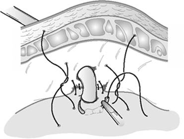
Like the surgically placed gastrostomy, the technique for open jejunostomy tube placement is generally safe; however, it can be more risky in patients who have had abdominal surgery, and typically requires general anesthesia. The most commonly employed techniques minimize the risks of leak from the bowel around the tube by either placing a pursestring suture around the tube (Stamm) or creating a serosal tunnel from the bowel wall overlying a portion of the tube (Witzel). After tube insertion into the jejunal lumen, the bowel is plicated to the undersurface of the anterior abdominal wall with three to four silk sutures placed circumferentially around the tube to further minimize the risk of leak and peritonitis (Fig. 36.3). The bowel is also carefully positioned for plication to prevent kinking or tube erosion.
To facilitate tube placement and minimize the risk of leak, the needle catheter jejunostomy technique was developed. It was first described by Delaney et al. (33) in 1973 as a safe and simple alternative to standard jejunostomy tube placement.
This method employs a narrow tube that is inserted by needle puncture through the antimesenteric border of the jejunal wall obliquely to create a subserosal tunnel. The tube is secured with a pursestring suture and then passed through the abdominal wall and skin. Few major complications were reported with this technique. Page et al. (62) had a 1% major and 1.5% minor complication rate in 199 patients. As stated earlier, these tubes tend to be unreliable and therefore are unpopular with many surgeons.
|
|
|
Figure 36.3. Plication of the jejunum to the anterior abdominal wall. By suturing the jejunum to the abdominal wall circumferentially around the tube, the risk of leakage is decreased. |
|
|
|
Figure 36.4. A surgically placed jejunostomy tube using a biliary T tube. |
More commonly used is a large-bore, soft flexible catheter with additional holes placed into the jejunal lumen with a single pursestring suture. A biliary T tube can also be used for jejunal feeding, and is less likely to dislodge than the straight tube (Fig. 36.4). The limbs are each cut to approximately 3 cm in length and the back of the T limb is slit lengthwise to facilitate removal. After tube insertion into the jejunal lumen, the bowel is plicated as described earlier.
As with gastrostomy tube placement, laparoscopic insertion techniques also have been developed. One such procedure attaches the jejunum to the anterior abdominal wall with four specially designed T fasteners. After this is completed, a large-bore needle is inserted into the jejunal lumen. A guidewire is then threaded through the needle and the catheter passed over a guidewire with the use of a peel-away introducer (63).
Jejunostomy tubes often become occluded, leak, or are removed either deliberately or inadvertently. Replacement is most easily performed with fluoroscopic assistance and should be attempted as soon as dislodgement is noted (57). This technique may even be successful for reinserting tubes that have been removed in the distant past. Because the jejunal limb used for the jejunostomy is routinely fixed to the undersurface of the abdominal wall, luminal access can be achieved by passing a needle catheter through the skin site or scar. Once the tip of the needle is in the lumen, a long guidewire is then directed into the lumen and used to direct the tube.
Novel Approaches to Enteral Access
A few novel approaches to enteral access are included for completeness. These techniques were performed in patients unable to have access established with more conventional methods. In two case reports, the duodenal lumen was safely cannulated by fluoroscopy- or computed tomography–guided puncture using a lumbar approach (64,65).
Summary
Enteral nutrition is fully recognized as an important addition to the comprehensive care of the critically ill and the preferred means of nutritional support. However, the successful establishment of secure and dependable enteral feeding access may be challenging. Several factors, including the patient's current condition, past medical and surgical history, status of the gastrointestinal tract, and hospital resources available to the clinician determine the best tube type, access choice, and placement method. Fortunately, several options are currently available so that there should be few reasons—short of gastrointestinal tract dysfunction—that preclude the use of enteral nutrition.
No single enteral access combination is superior to the others. None guarantees success in all instances. Using a flexible strategy based on the unique characteristics of the patient and the patient care environment will maximize the likelihood of success. Persistence also pays dividends. Failure of one attempt should not be a cause to abandon enteral feeding. A fresh new approach, possibly using another access option, may ultimately succeed. Only with patience, determination, and an understanding of the available access options will the greatest number of critically ill patients reap the benefits of enteral nutrition.
References
1. Clinical Practice Guideline for Nutrition Support in the Mechanically Ventilated, Critically Ill Adult Patient. http://www.criticalcarenutrition.com/index.php?option=com_content&task=view&id=34&Itemid=59. Accessed February 20, 2008.
2. Moore FA, Moore EE, Jones TN, et al. TEN versus TPN following major abdominal trauma-reduced septic morbidity. J Trauma. 1989;29:916.
3. Kudsk KA, Croce MA, Fabian TC, et al. Enteral versus parenteral feeding: effects on septic morbidity after blunt and penetrating abdominal trauma. Ann Surg. 1992;215:503.
4. Gianotti L, Alexander JW, Nelson JL, et al. Role of early enteral feeding and acute starvation on postburn bacterial translocation and host defense: prospective, randomized trials. Crit Care Med. 1994;22:265.
5. Heyland DK, Cook DJ, Guyatt GH. Enteral nutrition in the critically ill patient: a critical review of the literature. Intensive Care Med. 1993;19:435.
6. Babineau TJ, Blackburn GL. Time to consider early gut feeding. Crit Care Med. 1994;22:191.
7. Grant JP, Snyder BA. Use of L-glutamine in total parenteral nutrition. J Surg Res. 1988;44:506.
8. Johnson LR, Copeland EM, Dudrick JT, et al. Structural and hormonal alterations in the gastrointestinal tract of parenterally fed rats. Gastroenterology. 1975;68:1170.
9. Wilmore DW, Smith RJ, O'Dwyer ST, et al. The gut: a central organ after surgical stress. Surgery. 1988;104:917.
10. Page CP. The surgeon and gut maintenance. Am J Surg. 1989;158:485.
11. Deitch EA. Multiple organ failure: pathophysiology and potential future therapy. Ann Surg. 1992;216:117.
12. Carrico J, Meakin JL. Multiple organ failure syndrome. Arch Surg. 1986;121:196.
13. Border JR, Hassett J, LaDuca J, et al. The gut origin septic states in blunt multiple trauma (ISS = 40) in the ICU. Ann Surg. 1987;206:427.
14. Marshall JC, Christou NV, Meakins JL. The gastrointestinal tract: the “undrained abscess” of multiple organ failure. Ann Surg. 1993;218:111.
15. Thibault A: Care of feeding tubes. In: Borlase BC, Bell SJ, Blackburn GL, et al., eds. Enteral Nutrition. New York: Chapman & Hall; 1994:197.
16. Koruda MJ, Guenter P, Rombeau JL. Enteral nutrition in the critically ill. Crit Care Clin. 1987;3:133.
17. Minard G. Enteral access. Nutr Clin Pract. 1994;9:172.
18. Ritz MA, Fraser R, Edwards N, et al. Delayed gastric emptying in ventilated critically ill patients: Measurement by 13C-octanoic acid breath test. Crit Care Med. 2001;29(9):1744.
19. Tarling MM, Toner CC, Withington PS. A model of gastric emptying using paracetamol absorption in intensive care patients. Intensive Care Med. 1997;23:256.
20. Kao CH, ChangLai SP, Chieng PU, et al. Gastric emptying in head-injured patients. Am J Gastroenterol. 1998;93(7):1108.
21. Suto G, Kiraly A, Tache Y. Interleukin 1 beta inhibits gastric emptying in rats: mediation through prostaglandin and corticotrophin-releasing factor. Gastroenterology. 1994;106(6):1568.
22. McClave SA, Snider HL, Lowen CC, et al. Use of residual volume as a marker for enteral feeding intolerance: prospective blinded comparison with physical examination and radiographic findings. JPEN J Parenter Enter Nutr. 1992;16:99.
23. Heyland DK, Drover JW, MacDonald S, et al. Effect of postpyloric feeding on gastroesophageal regurgitation and pulmonary microaspiration: results of a randomized controlled trial. Crit Care Med. 2001;29:1495.
24. Taylor SJ, Fettes SB, Jewkes C, et al. Prospective, randomized, controlled trial to determine the effect of early enhanced enteral nutrition on clinical outcome in mechanically ventilated patients suffering head injury. Crit Care Med. 1999;27(11):2525.
25. Marik PE, Zaloga GP. Gastric versus post-pyloric feeding: a systematic review. Crit Care. 2003;7(3):R46.
26. Ho KM, Dobb GJ, Webb SAR. A comparison of early gastric and post-pyloric feeding in critically ill patients: a meta-analysis. Intensive Care Med. 2006;32:639.
27. Jooste CA, Mustoe J, Collee G. Metoclopramide improves gastric motility in critically ill patients. Intensive Care Med. 1999;25:464.
28. Calcroft RM, Joynt G, Hung V. Gastric emptying in critically ill patients: a randomized, blinded, prospective comparison of metoclopramide with placebo. Intensive Care Med. 1997;23(Suppl):S138.
29. Nguyen NQ, Chapman MJ, Fraser RJ, et al. Erythromycin is more effective than metoclopramide in the treatment of feed intolerance in critical illness. Crit Care Med. 2007;35:483.
30. Dive A, Miesse C, Galanti L, et al. Effect of erythromycin on gastric motility in mechanically ventilated critically ill patients: a double-blind, randomized, placebo-controlled study. Crit Care Med. 1995;23:1356.
31. Chapman MJ, Fraser RJ, Kluger MT, et al. Erythromycin improves gastric emptying in critically ill patients intolerant of nasogastric feeding. Crit Care Med. 2000;28:2334.
32. Ritz MA, Chapman MJ, Fraser RJ, et al. Erythromycin dose of 70 mg accelerates gastric emptying as effectively as 200 mg in the critically ill. Intensive Care Med. 2005;31:949.
33. Delaney HM, Carnevale NH, Garvey JW. Jejunostomy by a needle catheter technique. Surgery. 1973;73:786.
34. Jones TN, Moore EE, Moore FA. Early postoperative feeding. In: Borlase BC, Bell SJ, Blackburn GL, et al. eds. Enteral Nutrition. New York: Chapman & Hall; 1994:78.
35. Zaloga GP. Bedside method for placing small bowel feeding tubes in critically ill patients: a prospective study. Chest. 1991;100:1643.
36. Welch SK, Hanlon MD, Waits M, et al. Comparison of four bedside indicators used to predict duodenal feeding tube placement with radiography. JPEN J Parenter Enter Nutr. 1994;18:525.
37. Thurlow PM. Bedside enteral feeding tube placement into duodenum and jejunum. JPEN J Parenter Enter Nutr. 1986;10:104.
38. Hernandez-Socorro CR, Marin J, Ruiz-Santana S, et al. Bedside sonographic-guided versus blind nasoenteric feeding tube placement in critically ill patients. Crit Care Med. 1996;24(10):1690.
39. Keidan I, Gallagher TJ. Electrocardiogram-guided placement of enteral feeding tubes. Crit Care Med. 2000;28:2631.
40. Gray R, Tynan C, Reed L, et al. Bedside electromagnetic-guided feeding tube placement: an improvement over traditional placement technique? Nutr Clin Pract. 2007;22(4):436.
41. Gabriel SA, Ackermann RJ, Castresana M. A new technique for placement of nasoenteral feeding tubes using external magnetic guidance. Crit Care Med. 1997;25(4):641.
42. Whatley K, Turner WW, Dey M, et al. When does metoclopramide facilitate transpyloric intubation? JPEN J Parenter Enter Nutr. 1984;8:679.
43. Heiselman DE, Vidovich RR, Milkovich G, et al. Nasointestinal tube placement with a pH sensor feeding tube. JPEN J Parenter Enter Nutr. 1993;17:562.
44. Kittinger JW, Sandler RS, Heizer WD. Efficacy of metoclopramide as an adjunct to duodenal placement of small-bore feeding tubes: a randomized, placebo-controlled, double-blind study. JPEN J Parenter Enter Nutr. 1987;11:33.
45. Kalliafas S, Choban PS, Ziegler D, et al. Erythromycin facilitates postpyloric placement of nasoduodenal feeding tubes in intensive care unit patients: randomized, double-blinded, placebo-controlled trial. JPEN J Parenter Enteral Nutr. 1996;20(6):385.
46. Griffith DP, McNally T, Battey CH, et al. Intravenous erythromycin facilitates bedside placement of postpyloric feeding tubes in critically ill adults: a double-blind, randomized, placebo-controlled study. Crit Care Med. 2003;31:39.
47. Strong RM, Gribbon R, Durling S, et al. Enteral tube feedings utilizing a pH sensor enteral feeding tube. Nutr Suppl Sero. 1988;8:11.
48. Grant JP, Curtas MS, Kelvin FM. Fluoroscopic placement of nasojejunal feeding tubes with immediate feeding using a nonelemental diet. JPEN J Parenter Enter Nutr. 1983;7:299.
49. Levenson R, Turner WW, Dyson A, et al. Do weighted nasogastric feeding tubes facilitate duodenal intubations? JPEN J Parenter Enter Nutr. 1988;12:135.
50. Lord LM, Weser-Maimone A, Pulhamus M, et al. Comparison of weighted vs unweighted enteral feeding tubes for efficacy of transpyloric intubation. JPEN J Parenter Enter Nutr. 1993;17:271.
51. Larson DE, Burton DD, Schroeder KW, et al. Percutaneous endoscopic gastrostomy: indications, success, complications, and mortality in 314 consecutive patients. Gastroenterology. 1987;93:48.
52. Grant JP. Comparison of percutaneous endoscopic gastrostomy with Stamm gastrostomy. Ann Surg. 1988;270:598.
53. Peasarini AC, Dittler HJ. Feeding tube perforation as a complication of percutaneous endoscopic gastrostomy. Endoscopy. 1992;24:235.
54. Saltzberg DM, Anand K, Juvan P, et al. Colocutaneous fistula: an unusual complication of percutaneous endoscopic gastrostomy. JPEN J Parenter Enter Nutr. 1987;11:86.
55. Dwyer KM, Watts DD, Thurber JS, et al. Percutaneous endoscopic gastrostomy: the preferred method of elective feeding tube placement in trauma patients. J Trauma. 2002;52:26.
56. Kirby DF, Craig RM, Tsang T-K, et al. Percutaneous endoscopic gastrostomies: a prospective evaluation and review of the literature. JPEN J Parenter Enter Nutr. 1986;10:155.
57. Lambiase RE, Dorfman GS, Cronan JJ, et al. Percutaneous alternatives in nutritional support: a radiologic perspective. JPEN J Parenter Enter Nutr. 1988;12:513.
58. Wolfsen HC, Kozarek RA, Ball TJ, et al. Tube dysfunction following percutaneous gastrostomy and jejunostomy. Gastrointest Endosc. 1990;36:261.
59. DiSario JA, Foutch PG, Sanowski RA. Poor results with percutaneous endoscopic jejunostomy. Gastrointest Endosc. 1990;36:257.
60. Henderson JM, Strudel WE, Gilinsky NH. Limitations of percutaneous endoscopic jejunostomy. JPEN J Parenter Enter Nutr. 1993;17:546.
61. Reiner DS, Leitman IM, Ward RJ. Laparoscopic Stamm gastrostomy with gastropexy. Surg Laparosc Endosc. 1991;1:189.
62. Page CP, Carlton PK, Andrassy RJ, et al. Safe, cost-effective postoperative nutrition: defined formula diet via needle catheter jejunostomy. Am J Surg. 1979;138:939.
63. Duh Q-Y, Way LW. Laparoscopic jejunostomy using T-fasteners as retractors and anchors. Arch Surg. 1993;128:105.
64. Cwikiel W. Percutaneous duodenostomy-alternative route for enteral nutrition. Acta Radiol. 1991;32:153.
65. Koolpe HA, Dorfman D, Kramer M. Translumbar duodenostomy for enteral feeding. AJR Am J Roentgenol. 1989;153:299.