Richard E. Moon
John Paul M. Longphre
The first recorded attempt to use hyperbaric therapy was in 1662, when Henshaw in Britain used an organ bellows to manipulate the pressure within an enclosed chamber designed to seat a patient. He recommended high pressure for acute diseases and low pressure for chronic diseases (1). The pressure fluctuations in either direction were probably quite small. Widespread use of hyperbaric therapy began in the 19th century. At that time, powerful pneumatic pumps were designed, which could be used to compress chambers with air. Physicians in France and Britain used compressed air treatment for miscellaneous conditions. Junod used pressures of 1.5 atmospheres absolute (ATA) to treat patients, but did experiments up to 4 ATA (2). Simpson, using pressures in the range of 1.3 to 1.5 ATA, reported treating a variety of complaints, including dysphonia, asthma, tuberculosis, menorrhagia, and deafness (1), although without any physiologic basis.
Compressed air construction work was also developed in the 1800s, in which men were exposed to elevated ambient pressure within compartments for the purpose of excavating tunnels or bridge piers in muddy soil that was otherwise subject to flooding. Upon decompression at the end of a work shift, workers often developed joint pains or neurologic manifestations (caisson disease, the bends, or decompression sickness). Although the pathophysiology (nitrogen bubble formation in tissues; see below) was not understood, it was observed that recompression of these individuals could relieve the symptoms. Administration of recompression therapy became routine during construction of the Hudson River tunnel in the 1890s (3). All of these treatments used compressed air. Although oxygen breathing under pressure had been suggested for the treatment of decompression sickness as early as 1897 (4) and was used intermittently over the next 30 years, systematic study and use of hyperbaric oxygen would not occur until much later.
Oxygen administration during recompression therapy for decompression sickness increased the efficacy of the treatment (5,6) and is now routinely used for both decompression sickness and gas embolism. The administration of oxygen at increased ambient pressure became known as hyperbaric oxygen (HBO) therapy. In the 1950s, pilot investigations were performed of HBO as a therapy for diseases other than those related to gas bubbles, including carbon monoxide poisoning, clostridial myonecrosis (gas gangrene), and later, selected chronic wounds.
For many years, the Undersea and Hyperbaric Medical Society has regularly reviewed and published information regarding the use of HBO in selected diseases (7), and its recommendations have been widely accepted. The list of accepted indications (7) contains a heterogeneous group of conditions (Table 39.1), suggesting that more than one mechanism mediates the clinical effects of HBO, including the increase in ambient pressure (partly responsible for its efficacy in conditions caused by gas bubble disease) and pharmacologic effects of supraphysiologic increases in blood and tissue PO2 as discussed below.
Effects of Hyperoxia
Blood Gas Values
Under normal clinical HBO therapy conditions (2–3 ATA), breathing 100% oxygen can lead to arterial PO2 (PaO2) values that are 10 to 17 times higher than normal (8,9). PaO2 levels can rise from the normal of 90 to 100 mm Hg (breathing air at sea level, i.e., 1 ATA or normobaria) to 1,000 to 1,700 mm Hg in healthy subjects breathing 100% oxygen at 2 to 3 ATA (see Table 39.2).
|
Table 39.2 Blood gas and hemodynamic values in 14 healthy adults breathing spontaneously (mean ± standard deviation) |
||||||||||||||||||||||||||||||||||||||||||||||||||||||||||||||||||||||
|
||||||||||||||||||||||||||||||||||||||||||||||||||||||||||||||||||||||
One effect is an increase in blood oxygen content:

where Hb is hemoglobin concentration (g/dL), SaO2 is arterial Hb-O2 saturation, and PaO2 is arterial oxygen tension.
The second term of Eq. 1 represents the dissolved oxygen proportion, which under normal circumstances represents a small fraction of total arterial oxygen content, and is therefore often disregarded. However, during HBO, this dissolved fraction is substantially increased (see Table 39.2). In fact, mixed venous Hb-O2 saturation is 100% under resting conditions while breathing 100% oxygen at 3 ATA. Thus, oxygen delivery can be maintained under these circumstances without hemoglobin. This was shown by Boerema et al. in a swine model (10).
PaCO2 is not significantly affected by the increased pressure (8,9,11), although the venoarterial PCO2 difference is slightly increased, mostly because of a reduction in cardiac output.
|
Table 39.1 Conditions amenable to treatment with hyperbaric oxygen therapy |
||
|
Vasoconstriction
Hyperoxia causes peripheral vasoconstriction (8,9,12), regardless of atmospheric pressure (13). At a mere 2 ATA, systemic vascular resistance can increase by 30% in dogs (14). The mechanisms for this include scavenging of nitric oxide (NO) by superoxide anion (O2-) (15) and increased binding of NO at high PO2 to hemoglobin, forming S-nitrosohemoglobin (9). Vasoconstriction has the positive effect of reducing edema in injured tissues and surgical flaps (discussed later). During HBO, the arterial blood O2 content is sufficiently high that despite vasoconstriction and reduced blood flow, oxygen delivery is increased (16) (see also Table 39.2). Although peripheral vasoconstriction occurs in normal skin during hyperbaric oxygen exposure, repetitive intermittent HBO appears to increase the microvascular blood flow of healing wounds (17).
Hemodynamics
Heart rate and cardiac output both decrease by 13% to 35% under hyperbaric conditions (Table 39.2) (8,9,14,18,19). Small changes may occur in systemic and pulmonary artery pressure, with an increase in systemic vascular resistance (SVR) and a decrease in pulmonary vascular resistance (PVR) (9). Despite the reduced cardiac output, oxygen delivery is increased (Fig. 39.1).
Organ Blood Flow
Studies in large animals indicate that the decrease in peripheral blood flow is limited primarily to the cerebral and peripheral vascular beds, with other organs unaffected (14). In rats, HBO has been shown to decrease organ blood flow, including the myocardium, kidney, brain, ocular globe, and gut (15,20,21,22). In autonomically blocked conscious dogs at 3 ATA, coronary blood flow is decreased (23). Another dog study at 2 ATA revealed no change in coronary, hepatic, renal, or mesenteric blood flow (14).
Cellular and Tissue Effects
In a myocutaneous flap model during reperfusion following 4 hours of ischemia, Zamboni et al. described a delayed decrease in blood flow (24). This flow reduction appears to be associated with adherence of leukocytes to the endothelium of the small vessels, an effect that is significantly inhibited by HBO. A delayed reduction in cerebral blood flow has also been observed after arterial gas embolism in the brain (25), which has similarly been attributed to leukocyte accumulation in the capillaries (26). HBO reduces cerebral infarct volume and myeloperoxidase activity, a marker of neutrophil recruitment (27). In other studies using animal models, it has been observed that HBO pretreatment reduces ischemia/reperfusion injury to the liver (28). HBO reduces ischemia/reperfusion injury to the intestine (29,30) and muscle (31), as well as reducing ischemia-induced necrosis in muscle (32,33,34,35,36,37), brain (38,39), and kidney (40). One mechanism for this effect of HBO appears to be the inhibition of leukocyte β2-integrin function (41,42,43). Part of the beneficial effect of HBO in these settings is speculated to be due to the prevention of endothelial leukocyte adherence. After focal ischemia, HBO also reduces postischemic blood–brain barrier damage and edema (44) and has an antiapoptotic effect (45).
Antibacterial Effects
The increase in PO2 during HBO can be toxic to anaerobic bacteria, which lack antioxidant defense mechanisms. In addition, HBO has effects on aerobic organisms via neutrophil mechanisms. Killing of aerobic bacteria by leukocytes is related to the O2-dependent generation of reactive oxygen species within the lysosomes. In vitro studies have demonstrated that phagocytic killing of Staphylococcus aureus by polymorphonuclear leukocytes becomes less effective as ambient PO2 is decreased. This mechanism appears to be important in vivo when tissue PO2 is low (e.g., in osteomyelitis) (46). In an animal model of osteomyelitis, the cidal effect of tobramycin against Pseudomonas was increased when tissue PO2 was raised by the administration of 100% O2 at increased ambient pressure (47). Published evidence also supports an augmentation of penicillin by HBO in the treatment of soft tissue streptococcal infections (48).
|
|
|
Figure 39.1. Arterial O2 content and delivery while breathing air at 1 atmosphere absolute (ATA) or 100% oxygen at 3 ATA. Measurements are shown in a group of normal volunteers. (Data from McMahon TJ, Moon RE, Luschinger BP, et al. Nitric oxide in the human respiratory cycle. Nat Med. 2002;8:711–717.) |
Oxygen Toxicity
Pharmacology
Exposure of an animal to increased partial pressure of oxygen results in higher rates of endogenous production of reactive oxygen species, including superoxide anion (O2-), hydroxyl radical (OH•), hydrogen peroxide (H2O2), and singlet oxygen, which are responsible for tissue oxygen toxicity (49,50,51). Tissue O2 toxicity includes the following: Lipid peroxidation, sulfhydryl group inactivation, oxidation of pyridine nucleotides, inactivation of Na+–K+–ATPase and inhibition of DNA, and protein synthesis. Toxic effects of these species depend upon both dose and duration of O2 exposure. In the central nervous system, HBO initially reduces NO availability and causes vasoconstriction. HBO stimulates neuronal nitric oxide production and causes the accumulation of peroxynitrite. Prior to onset of a seizure, NO levels and blood flow both increase above control levels (52,53). This, in turn, decreases brain γ-aminobutyric acid (GABA) levels, creating an imbalance between glutamatergic and GABAergic synaptic function, which is believed to be partly responsible for central nervous system (CNS) O2 toxicity (54).
Clinical Effects
At sufficiently high PO2, any organ can be susceptible to oxygen toxicity. However, within the clinical range of inspired PO2 (1–3 ATA), the most susceptible tissues are the lung, brain, retina, lens, and peripheral nerve.
Brain
Oxygen toxicity of the central nervous system produces a wide variety of manifestations (55). The most common mild symptom is nausea; the most dramatic is generalized nonfocal convulsions. These are usually self-limited, even without pharmacologic treatment, and have no long-term effects. The occurrence of a hyperoxic seizure does not imply the development of a convulsive disorder. Factors that increase the risk of CNS oxygen toxicity include hypercapnia and probably fever.
CNS O2 toxicity is uncommon when inspired PO2 is less than 3 ATA. While in-water convulsions in divers have been recorded at an inspired PO2 of 1.3 ATA, convulsions during clinical hyperbaric oxygen therapy occur in only a small fraction of treatments. Approximately 0.02% of treatments at an inspired PO2 of 2 ATA and 4% at 3 ATA. At an inspired PO2 less than 3 ATA, the risk of convulsions increases markedly, particularly in patients with sepsis. While anecdotal reports suggest that HBO may precipitate seizures in patients who have an underlying predisposition (56), there are no epidemiologic data to confirm this. When indicated, HBO should not be withheld on the basis of an underlying seizure disorder.
Both CNS and pulmonary toxicity can be delayed by the use of air breaks (a period of a few minutes where air is administered in lieu of 100% oxygen) (57,58,59,60). Oftentimes, the aura of a hyperoxic convulsion occurs in the form of nausea or facial paresthesias. The patient can be given an air break to avert such a convulsion. Once the symptoms have resolved (usually within a few minutes), the oxygen can be restarted without recurrence. During the tonic-clonic phase of a seizure, the airway may be obstructed. Therefore, it is imperative that chamber pressure not be reduced during this time in order to avoid pulmonary barotrauma and the possibility of arterial gas embolism. After a convulsion, some practitioners recommend administering prophylactic medication for the duration of HBO.
Prophylactic anticonvulsants such as phenytoin, phenobarbital, or benzodiazepines can reduce the chance of convulsions when utilizing clinical treatment schedules with a significant risk of CNS O2 toxicity (e.g., treatment pressure >3 ATA). The authors' practice is to load septic patients intravenously with phenobarbital as tolerated, up to 12 mg/kg, prior to hyperbaric oxygen treatment at 3 ATA, with doses every 8 hours to maintain a serum concentration in the therapeutic anticonvulsant range. When using inspired PO2 ≤2.8 ATA, the risk of CNS toxicity is sufficiently low that prophylactic anticonvulsant therapy is not required.
Hyperoxic seizures and other CNS manifestations in diabetics can be caused by HBO-induced reduction in blood glucose. Therefore, the occurrence of CNS O2 toxicity in a patient with diabetes during HBO treatment should prompt the immediate measurement of plasma glucose. When blood PO2 is extremely high, bedside glucose measurement devices, particularly those dependent upon a glucose oxidase reaction, can be inaccurate, producing measurements that significantly underestimate the true value (61). Laboratory-based glucose measurement is usually accurate.
Lungs
Pulmonary oxygen toxicity during hyperbaric oxygen therapy is also PO2 and time dependent. Clinical HBO protocols have been empirically developed to minimize the risk of pulmonary O2 toxicity, which almost never occurs during routine daily or twice-daily clinical treatments. However, it can occur during extended treatments that are used for treating gas embolism or decompression sickness, in which inspired PO2 is as high as 2.8 ATA. The initial manifestation is usually a burning substernal chest pain and cough (62), which is most likely due to tracheobronchitis. Continued exposure to oxygen can produce more severe manifestations such as dyspnea and acute respiratory distress syndrome (ARDS). Measurable abnormalities include reduced forced vital capacity and carbon monoxide transfer factor (DLCO). Pulmonary oxygen toxicity symptoms may not be evident in patients who are sedated and mechanically ventilated. Moreover, such patients often have pulmonary infiltrates for a variety of reasons and it may be impossible to distinguish the possible additive effects of pulmonary O2 toxicity.
While the maximum safe inspired PO2 during clinical hyperbaric oxygen therapy is based mainly upon CNS O2 toxicity limits, the safe exposure duration is determined by pulmonary limits. Prediction formulas have been developed that approximate the average reduction in vital capacity after continuous oxygen exposure (63,64,65). However, the usefulness of these algorithms for individual patients is severely limited due to individual variability and comorbid factors that may affect O2 susceptibility, such as prior exposure, intermittent exposure, and endotoxemia. HBO treatment schedules that include periods of air breathing (“air breaks”) interspersed between O2 periods reduce the rate of onset of both pulmonary and CNS toxic manifestations and can increase the overall dose of oxygen that is tolerated. In the awake patient, the occurrence of burning, retrosternal chest pain is a more useful indicator of incipient pulmonary toxicity.
If standard HBO treatment schedules are used (e.g., 2 ATA/2 hours, 2.5 ATA/90 minutes one to two times daily, or U.S. Navy treatment tables), pulmonary O2 toxicity is almost never clinically evident. It is seen only with the most extreme levels of hyperbaric exposure such as may be required for severe neurologic decompression illness. Furthermore, most minor pulmonary oxygen toxicity resolves within 12 to 24 hours of air breathing. Complete reversal of vital capacity (VC) decrements, as large as 40% of control, has been observed after extended O2 exposure at 2 ATA (66). Therefore, in clinical situations requiring aggressive HBO therapy such as spinal cord decompression sickness or arterial gas embolism, some degree of pulmonary O2 toxicity is acceptable.
Supplemental O2 administration at 1 ATA between HBO treatments can accelerate the onset of symptoms of pulmonary O2 toxicity during subsequent HBO. Thus, if O2 is absolutely required between HBO treatments, it is prudent to use the lowest concentration.
Some antineoplastic agents, such as bleomycin (67,68) and mitomycin C (69), can predispose to fatal pulmonary O2 toxicity, probably due to drug-induced reduction in antioxidant defenses. The risk of pulmonary O2 toxicity due to HBO therapy in patients with previous exposure to either of these agents is unknown, although 6 months after the agent has been discontinued, HBO seems to be safe. Even after this point, in some patients, HBO induces mild pulmonary O2 toxicity symptoms such as retrosternal burning chest pain, which can be managed with air breaks.
Eye
Repetitive hyperbaric oxygen therapy causes myopia, which is due to a reversible refractive change in the lens (70). A measurable change in visual acuity usually does not occur until after 20 or so treatments. The myopia usually resolves over several weeks, in about the same time period as the onset; however, some residual myopia may remain. On the basis of one study, it has been suggested that HBO treatment may predispose to nuclear cataract formation (71). However, many of the patients in this study received hundreds of hours of HBO, considerably more than is customary. Furthermore, nuclear cataracts are more common in diabetes, which is frequently a comorbidity in patients requiring HBO. Extended exposure to PO2 of 3 ATA can also cause retinal toxicity, manifested by tunnel vision (72,73). However, such exposures are beyond the range used clinically.
Peripheral Nerve
After hyperbaric oxygen exposure, some patients experience paresthesias, usually in their fingers and toes, generally after several HBO exposures but occasionally after a single prolonged treatment. The physical exam is normal, and the symptoms resolve within a few hours. This manifestation has no known clinical significance and is not a reason to discontinue hyperbaric therapy.
Physical Effects of Compression/Decompression
Boyle's Law
Clinically, the complications of HBO therapy that most frequently occur are those related to the body's gas-containing spaces (74). Dealing with volume changes in these gas-containing spaces is unique to HBO therapy. For a gas, absolute pressure and volume are inversely related. The increase in pressure during HBO treatment will therefore decrease the volume of closed gas-containing spaces within the body, such as the gastrointestinal tract or middle ear and, in the event of gas embolism or decompression sickness, bubbles.
Effects of Gases Other than Oxygen
Nitrogen
The narcotic properties of compressed air were first reported by Junod in 1835 as described by Bennett and Rostain (75). Hyperbaric nitrogen causes narcosis or pleasant intoxication at pressures greater than about 4 ATA in most individuals and near unconsciousness at greater than 10 ATA (76). Since patients breathe oxygen, nitrogen narcosis is only a problem for tenders in multiplace hyperbaric chambers. However, most hyperbaric treatments occur between 2 and 3 ATA, where symptoms of nitrogen narcosis are exceedingly mild.
Nitrogen (and other inert breathing gases such as helium) is the major causative agent of decompression sickness. During decompression, excess tissue nitrogen can become supersaturated, come out of solution, and form bubbles. This can lead to decompression sickness, with manifestations depending on their location and secondary effects.
Trace Gases
The pharmacologic effects of gases are proportional to their partial pressures. Although a trace gas may only be present in minute quantities, as the chamber pressure rises, so does the partial pressure of a gas. Therefore, gases such as carbon monoxide or carbon dioxide in concentrations that have no pharmacologic or toxic effects at 1 ATA may exert measurable effects in a hyperbaric environment.
Use of Hyperbaric Oxygen for Specific Diseases
Gas Embolism and Decompression Sickness
Gas bubbles in the body can be due to direct gas entry via veins or arteries (arterial or venous gas embolism) or via in situ formation due to gas supersaturation in divers, compressed air workers, or aviators (decompression sickness). Since the two conditions often both occur in the same patient (particularly in divers), the principles of treatment of the two are the same. The syndrome of either or both condition is commonly referred to as decompression illness (DCI).
Arterial and Venous Gas Embolism
Entry of gas into the circulation can occur via several mechanisms. Gas embolism has recently been reviewed (77,78). In divers breathing compressed gas, arterial gas embolism (AGE) can ensue if decompression (ascent) occurs while the diver holds his or her breath or due to gas trapping caused by focal or generalized airways obstruction. AGE due to this mechanism can result after an ascent to the surface of as little as 1 meter. AGE can also occur during diagnostic or therapeutic procedures such as angiography.
Venous gas embolism (VGE) can result due to direct injection or entry via an open vein in which ambient pressure exceeds venous pressure. This can exist during laparoscopic surgical procedures due to the elevated intra-abdominal pressure, or open procedures in which venous pressure in the surgical wound is subatmospheric. The classic scenario for this is an intracranial procedure in the sitting position. However, it has also been described in procedures such as liver resection, cesarean section, and spine surgery. VGE can also occur due to oral hydrogen peroxide (H2O2) ingestion. H2O2 absorbed into the circulation is broken down by catalase into water and oxygen bubbles. VGE can result if a central venous catheter is opened to air, particularly if the patient is breathing spontaneously. It has also been reported in patients with ARDS being ventilated with positive end-expiratory pressure (79). VGE has been described during orogenital sex after blowing air intravaginally (80). Intravenous injection is better tolerated than intra-arterial injection because of the pulmonary filter. However, if the rate of entry of gas into the veins is sufficiently high, bubbles can traverse the pulmonary capillary network and become arterial emboli. Large volumes can obstruct the right heart or pulmonary artery and cause cardiac arrest.
Large volumes of arterial gas can cause acute obstruction of large vessels. Small quantities tend to remain in the circulation only transiently; however, they can precipitate a sustained reduction in local blood flow (25). The mechanism appears to be endothelial damage (81) and adherence of leukocytes (26,82,83,84). Endothelial barrier function is also impaired in both the brain and lung, resulting in edema (85,86) and impaired endothelial-dependent vasoactivity (87). Animal models of AGE have revealed a significant elevation of intracranial pressure (ICP) and depression of cerebral PO2 (88,89). In a pig model, hyperventilation failed to correct these parameters (90); however, HBO at 2.8 ATA (U.S. Navy Table 6, Fig. 39.4) restored both ICP and brain PO2 toward normal (Fig. 39.3).
Clinical manifestations of AGE include acute loss of consciousness, confusion, focal neurologic abnormalities, and cerebral edema. VGE causes acute dyspnea, tachypnea, hypotension, cardiac ischemia or arrest, and pulmonary edema (86). In monitored patients, VGE is often heralded by a decrease in end-tidal PCO2 (91), although sometimes, with small volumes of CO2 embolism such as during laparoscopy, it may be increased. A mill-wheel murmur can be heard in some patients, although this sign is neither sensitive nor specific. Venous gas bubbles in sufficient quantities can cross into the arterial circulation (producing AGE) either through the pulmonary capillary network or via an intracardiac shunt, such as a patent foramen ovale.
Imaging is not useful for diagnosing either VGE or AGE. Gas bubbles are rarely visible on radiographic images (92). Except in cases where associated conditions such as pneumothorax are suspected or neurologic conditions such as hemorrhage require exclusion, imaging studies are not necessary and tend to delay definitive treatment.
Decompression Sickness
During diving or exposure to a compressed gas environment such as a hyperbaric chamber, inert gas (usually nitrogen) is taken up by tissues. During decompression, inert gas can become supersaturated and form bubbles in situ in tissues. Certain tissues are more susceptible to in situ bubble formation.
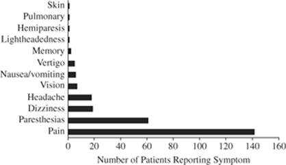
Manifestations of decompression sickness (DCS) can range from mild to severe (Fig. 39.2). The most common manifestations are joint pain and paresthesias. Although mild cases can progress to severe, severe manifestations almost always occur within 12 hours after surfacing.
|
|
|
Figure 39.2. Effect of hyperbaric oxygen (HBO) on intracranial pressure (ICP) and brain PO2 in pigs after air embolism. Top panel: HBO initially at 2.8 atmospheres absolute (ATA) (U.S. Navy Table 6) reduces ICP compared with no treatment, whether it is started 3 minutes or 60 minutes after embolization. Bottom panel: Brain tissue PO2 in the two groups of animals. For the 60-minute group, the closed circles represent PbrO2 in the first 10 minutes after embolization; the open circles represent PbrO2 in the first 10 minutes after the start of HBO. Values in lower panel are mean ± standard deviation. (Redrawn from van Hulst RA, Drenthen J, Haitsma JJ, et al. Effects of hyperbaric treatment in cerebral air embolism on intracranial pressure, brain oxygenation, and brain glucose metabolism in the pig. Crit Care Med. 2005;33:841–846.) |
|
|
|
Figure 39.3. Top: U.S. Navy (USN) Treatment Table 5. According to USN guidelines, this table may be used for symptoms involving skin (except for cutis marmorata), the lymphatic system, muscles and joints, with a normal neurologic exam, and when all symptoms have completely resolved within 10 minutes of reaching 2.8 atmospheres absolute (ATA). Bottom: USN Treatment Table 6. This table may be used for all types of decompression illness. Extensions (additional oxygen breathing cycles) can be administered at either treatment pressure (2.8 and 1.9 ATA). (Data from Navy Department. US Navy Diving Manual. Revision 4. Vol. 5: Diving Medicine and Recompression Chamber Operations. NAVSEA 0910-LP-103–8009. Washington, DC: Naval Sea Systems Command; 2005.) |
Treatment of Decompression Sickness and Arterial Gas Embolism
Prehospital Treatment
In addition to standard first aid principles, prehospital treatment of DCI consists of the administration of a high concentration of oxygen and fluid resuscitation. Oxygen administration reduces bubble size and can sometimes abolish symptoms and signs of decompression illness. A published study has provided epidemiologic evidence for its efficacy (93). Use of high concentrations of oxygen (preferably 100%) is recommended until definitive treatment is available. Periodic air breaks to reduce toxicity may be appropriate (e.g., 5 minutes every 30 minutes). The administration of oxygen for longer than 12 hours should be based upon the severity of the injury or the presence of hypoxemia breathing room air.
Both head-down and lateral decubitus positions have been recommended based on animal studies (94,95). However, the hemodynamic response to venous gas embolism is unaffected by body position (96,97), and prolonged head-down position may exacerbate cerebral edema (98). Supine position is therefore recommended, also because patient access and supportive therapies can be more easily administered in this position.
Hospital Treatment
Standard treatment of gas embolism includes airway and ventilatory management, maintaining a high PaO2 and normal PaCO2 (99) (Fig. 39.3), and support of arterial pressure. Like other forms of neurologic injury, it is recommended that when managing neurologic DCI, both hyperthermia and hyperglycemia (>140–185 mg/dL, 7.8–10.3 mM/L) should be avoided or treated (100).
Physical Removal of Gas
Physical removal of gas after massive arterial gas embolism has been described in cardiopulmonary bypass (101,102). Venous gas embolism has been successfully treated with chest compression (103) and aspiration through catheters in the right atrium (104,105) or pulmonary artery (106).
Recompression
Although symptomatic improvement can be obtained with oxygen at 1 ATA, the definitive treatment of both forms of decompression illness is hyperbaric oxygen. The safety and efficacy of HBO for the treatment of divers was initially shown 70 years ago (6). Since then, treatment protocols have been empirically developed that have been shown to have a high degree of success with a low probability of oxygen toxicity (107). The most widely used treatment protocols (“tables”) were developed by the U.S. Navy and promulgated via the Diving Manual (108) (Fig. 39.4). Both U.S. Navy Treatment Tables 5 and 6 use 100% oxygen breathing periods (“O2 cycles”) interspersed with air breathing periods (“air breaks”) at 2.8 and 1.9 ATA in a two-step pattern (see Fig. 39.4). Guidelines are available to administer additional O2 cycles (“extensions”) at both pressures (108). The vast majority, if not all cases, of DCI can be adequately treated using U.S. Navy treatment tables.
|
|
|
Figure 39.4. Symptoms of decompression illness in a series of recreational divers. (Redrawn from Divers Alert Network. Annual Diving Report. Durham, NC: Divers Alert Network; 2006.) |
The U.S. Navy tables were designed for use in multiplace chambers, where air breaks can easily be administered by discontinuing O2. Since monoplace chambers were designed to be compressed with 100% O2, shorter alternate treatment tables were designed for their use (109,110) (Fig. 39.5). Although direct comparisons with U.S. Navy tables have never been performed, case series suggest that these tables are efficacious for DCI (110). Monoplace chambers fitted with an air supply and delivery system can be used to administer treatment according to traditional Navy tables (111).
|
|
|
Figure 39.5. Hart-Kindwall monoplace treatment table. This table was designed for use in monoplace chambers without the capability of administering air breaks. Except for the lack of air breaks and limited ability for extension, it is similar to U.S. Navy Table 5, with a shorter time at 2.8 atmospheres absolute (ATA) and longer time at 1.9 ATA. (Data from Boerema I, Meyne NG, Brummelkamp WH, et al. Life without blood. J Cardiovasc Surg [Torino]. 1960;1:133–146.) |
Adjunctive Measures
In the 19th and early 20th century, recompression was the only treatment administered to patients with decompression illness. While hyperbaric oxygen remains the definitive treatment of bubble disease, there is increasing recognition that adjunctive therapies such as correction of hypovolemia may also be important (112).
Fluids
Severe decompression sickness is often associated with capillary leak, intravascular volume depletion, and hemoconcentration. The Undersea and Hyperbaric Medical Society (UHMS) recommends (level 1C) fluid administration to replenish intravascular volume, reverse hemoconcentration, and support blood pressure (113). Measures that augment cardiac preload such as supine position, head-down tilt, and water immersion (114) significantly increase the rate of inert gas washout. Thus, even in divers who are not dehydrated, there may be some benefit to extra fluid loading. Intravenous isotonic fluids without glucose (e.g., lactated Ringer solution, normal saline, or colloids) are recommended for severe DCI. Patients with mild symptoms may be treated with oral hydration fluids. For “chokes” (cardiorespiratory decompression sickness, in which high bubble loads cause pulmonary edema), animal studies suggest that aggressive fluid resuscitation can exacerbate pulmonary edema. Thus, for the patient with chokes, aggressive fluid resuscitation may not be warranted, particularly if advanced life support modalities such as endotracheal intubation and mechanical ventilation are not immediately available. For isolated AGE, in which the pathology is limited to cerebral infarction, aggressive fluid administration is also unwarranted.
Anticoagulants
Intravascular bubbles can induce platelet accumulation, adherence, and thrombus formation. Indeed, in a canine model of arterial gas embolism, therapeutic anticoagulation promoted a return in a short-term outcome: evoked potential amplitude, but only when heparin was combined with prostaglandin I2 (PGI2) and indomethacin (115). In this model, heparin alone was ineffective. In other experiments, heparin given either prophylactically or therapeutically to dogs with DCI was not beneficial (116). Furthermore, tissue hemorrhage can occur in decompression illness involving the spinal cord (117,118,119), brain (120,121), and inner ear (122,123). Thus, full therapeutic anticoagulation is not recommended.
Although anticoagulants are not indicated for the primary injury in DCI, patients with leg immobility due to DCI-induced spinal cord injury are at increased risk of deep vein thrombosis (DVT) and pulmonary thromboembolism (PE). Standard prophylactic anticoagulant measures, typically low-molecular-weight heparin (LMWH), are therefore recommended as soon as feasible after the onset of injury. Full anticoagulation is appropriate for established DVT/PE. If LMWH is contraindicated, elastic stockings or intermittent pneumatic calf compression is recommended, although their efficacy in preventing DVT or thromboembolism in DCI is unknown. Recommendations have been extrapolated from guidelines for traumatic spinal cord injury; neither their efficacy nor safety in neurologic DCI has been specifically confirmed. Thus, when facilities exist, a screening test for DVT a few days after injury is appropriate (113).
Lidocaine
The administration of lidocaine for arterial gas embolism is supported by several animal studies (124). No controlled human studies in accidental AGE have been performed. However, gas emboli are frequently observed in cardiopulmonary bypass. In this setting, two studies have demonstrated a beneficial effect of lidocaine administered in traditional antiarrhythmic doses on postoperative neurocognitive function (125,126). Another study has shown benefit for nondiabetics but not for diabetics (127). Human data directly pertinent to DCI are confined to three cases of decompression sickness or arterial gas embolism, published as case reports, which appeared to benefit from intravenous lidocaine (128,129). The UHMS does not recommend the routine use of lidocaine for DCI; however, recommendations have been made for its dosing (113). An appropriate end point is a serum concentration suitable for an antiarrhythmic effect (2–6 mg/L).
Nonsteroidal Anti-inflammatory Drugs
These drugs are commonly used empirically for treatment of bends pain that does not completely resolve with recompression. A randomized, controlled trial has been published in which tenoxicam, a nonselective cyclo-oxygenase inhibitor, was compared with placebo. Tenoxicam or placebo was administered during the first air break of the first hyperbaric treatment and continued daily for 7 days. Using as an end point the number of hyperbaric treatments required to achieve complete relief of symptoms or a clinical “plateau” of effect, the tenoxicam group required a median of two treatments versus three for the placebo group. The outcome at 6 weeks was not different (130). The UHMS guidelines have assigned nonsteroidal anti-inflammatory drugs a level 2B recommendation (113).
Corticosteroids
Unless given prophylactically, corticosteroids have not been shown to be of benefit in animal models of DCI (131,132,133). In a pig study, methylprednisolone treatment did not protect against severe DCS, and the treated animals had a greater mortality (134). In the absence of human trials of corticosteroids in DCI and the lack of benefit in animal studies, corticosteroids are not recommended.
Perfluorocarbons
Perfluorocarbons (PFCs) are a family of chemically inert, water-insoluble, synthetic compounds with a high solubility for both inert gases and oxygen, which may eventually become available for human use as blood substitutes. Intravenous injection of PFC emulsions could augment oxygen delivery to ischemic tissues with impaired circulation and facilitate inert gas washout from tissues (135). Indeed, beneficial effects have been observed in animal studies of both decompression sickness and gas embolism (136,137,138,139,140). There may also be a benefit from the surfactant properties in the treatment of intravascular gas bubbles (141).
Arterial Gas Embolism and Decompression Sickness Treatment Summary
Immediate treatment of AGE or DCS includes standard principles of first aid, including the administration of oxygen and fluids during transport to a hyperbaric chamber. If the patient is in an extremely remote location from which transport is not feasible and the manifestations are minor, if the patient's condition does not progress for 24 hours, and if the neurologic exam is normal, the risk of emergent transport may exceed the risk of conservative treatment (142).
Carbon Monoxide
Carbon monoxide (CO) is an important cause of unintentional poisoning fatalities in the United States each year (143). CO binds to hemoproteins, including hemoglobin and myoglobin, interfering with oxygen transport. It also binds to the mitochondrial cytochrome C oxidase in the electron transport chain (similar to cyanide), impairing oxidative phosphorylation, stopping the cell's energy production, and resulting in cellular hypoxia (144,145,146) and oxidative stress (147). In addition, CO exposure induces intravascular platelet–neutrophil activation (148). CO-related oxidative stress can cause chemical alterations in myelin basic protein (149), triggering immune-mediated neurologic deficits.
The symptoms and signs of CO poisoning include headache (or tightness across forehead), weakness, nausea and vomiting, syncope, tachycardia, tachypnea, and encephalopathy. Myocardial ischemia is also a common finding.
For survivors of this poisoning, the most debilitating results can be the late neurologic sequelae. These are often cognitive problems such as a decrement in short-term memory (150,151,152). Some patients improve clinically and then deteriorate several days after the event.
HBO therapy is known to accelerate the elimination of CO (153,154). Pace et al. found that the half-life of CO was longest when breathing air (214 minutes). Half-life decreased to 42 minutes breathing 100% O2 at 1 ATA and further to 18 minutes with 100% O2 at 2.5 ATA (153). The reduction in half-life may be important in preventing cell death by allowing mitochondrial adenosine triphosphate (ATP) production to resume before the cell would have otherwise died (144,155). In animal studies, HBO administration after acute CO exposure appears to minimize the lipid peroxidation in the brain, which occurs during or after removal of CO (147), and results in more rapid repletion of brain energy stores (155).
A double-blind randomized control trial carried out by Weaver et al. indicates that HBO therapy can prevent the occurrence of the late neurologic sequelae of CO poisoning if the patients are treated within 24 hours of the exposure (152).
All patients should be initially treated with 100% normobaric oxygen. HBO therapy is usually reserved for patients who have more severe poisoning, as determined by high HbCO level (e.g. ≥25%), loss of consciousness, or other neurologic manifestations, or myocardial ischemia, arrhythmias, or other cardiac abnormalities (152,154,156,157,158). A systematic analysis of 163 patients with CO poisoning who did not receive HBO revealed the following two risk factors for sequelae: older age and longer CO exposure (159). However, some patients without these risk factors also developed sequelae. The authors concluded that, in addition to other indications, regardless of HbCO level or loss of consciousness, anyone older than 36 years with symptoms should receive HBO.
Pregnant women should be treated according to maternal indications. Pregnant women may therefore have an HbCO level that is 10% to 15% less than that of the fetus. There is evidence that short periods of HBO therapy are not dangerous to the fetus or mother (160).
Cyanide
Cyanide leads to hypoxia on a cellular level by rapidly binding to mitochondrial cytochrome oxidase. Inhalation of high concentrations of cyanide (270 ppm) is rapidly fatal in humans (with blood levels reaching 3 µg/mL), whereas ingestion of cyanide is less rapidly fatal (161). When very low doses of cyanide are absorbed (whole blood levels of 0.5–2.53 µg/mL), tachycardia and decreased level of consciousness are possible (161,162).
There are very few studies and case reports of the use of HBO therapy in the treatment of cyanide poisoning (163,164,165,166,167). This is likely due partly to the effectiveness of chemical treatments (with sodium nitrite and thiosulfate) but also possibly related to the fact that the bonding of cyanide to the mitochondria's cytochrome C oxidase is not an oxygen-dependent mechanism. Chemical treatment of cyanide poisoning leads to the formation of methemoglobin. Utilizing HBO therapy to increase the amount of circulating dissolved oxygen has been shown to have both prophylactic and antagonistic effects on cyanide poisoning in rabbits (166). Human case reports also hint that HBO therapy may be useful when the response to chemical antidotes has been incomplete (165).
Hydrogen Sulfide
Like CO and cyanide, hydrogen sulfide (H2S) reacts with mitochondrial cytochrome C oxidase, impairing electron transport. This is not an oxygen-dependent mechanism. The rationale for using HBO therapy is the same as for cyanide poisoning, in that HBO therapy can increase the dissolved fraction of oxygen. Use of HBO therapy for H2S poisoning is based on two case reports suggesting a positive benefit (168,169).
Carbon Tetrachloride
Carbon tetrachloride (CCl4) is a CNS depressant, hepatotoxin, and nephrotoxin, with renal failure being the most common cause of death from very high-level exposures (170). In the setting of CCl4 poisoning of the rat, HBO has been shown to improve survival (171), decrease liver necrosis (172), decrease conversion of CCl4 to toxic free-radical metabolites (173,174), and decrease CCl4 metabolite-induced lipid peroxidation (175). One case report describes an obtunded patient treated with HBO for presumed CO poisoning. There was no historical evidence for CO exposure; the patient improved, regained consciousness, and admitted to ingestion of a normally lethal dose of 250 mL of CCl4 (176).
Necrotizing Infections
Clostridial Infections
This soil-based anaerobic organism causes a type of rapidly progressive disease known as gas gangrene, which, if left untreated, is almost uniformly fatal. In most cases, it is introduced to the human via accidental trauma. The most common species that cause the disease are Clostridium perfringens (80%–90%), Clostridium oedematiens, and Clostridium septicum. These organisms release α-toxin, which is a lecithinase related to the form found in snake, bee, and scorpion venoms, causing a liquefaction necrosis (177).
These organisms lack antioxidant defenses and therefore are susceptible to HBO therapy. The first to report this finding was Brummelkamp et al. in 1961 (178,179). Around the same time, it was discovered that at 3 ATA, α-toxin production quickly ceases (180); since then, animal studies (181) and meta-analyses of human case series support the use of HBO (182). If treatment is initiated within 24 hours of diagnosis, disease-specific mortality can be as low as 5% (177).
The typical HBO treatment schedule varies between 2.5 and 3 ATA for 90 minutes, with three treatments in the first 24 hours, followed by two treatments per day at 2 to 3 ATA until clinical stability. Aggressive surgical debridement and antibiotic therapy are also essential.
Nonclostridial Bacterial Infections
These are often necrotizing infections, usually polymicrobial, including at least one anaerobic species. These infections often follow local trauma and are enhanced by both local ischemia and reduced host defenses (many patients are diabetic with atherosclerosis) (183). The mainstays of therapy are surgical debridement and antibiotics. Individual case series and meta-analyses support the use of HBO as an adjunct (182,184,185). The HBO treatment schedule is similar to that of clostridial disease.
Mucormycosis
Rhinocerebral mucormycosis is a rare but devastating invasive disease of the head and neck with 30% to 50% or greater mortality when treated, often found in immunocompromised patients such as diabetics in ketoacidosis, or patients receiving antineoplastic agents and/or steroids (186). It is primarily treated with wide debridement and amphotericin B. Due to the rarity of this disease, randomized trials have not been performed. Several case reports have suggested that HBO therapy may be an effective adjunct (187,188,189). Recommended treatment protocol is 2 to 2.5 ATA for 2 hours, twice daily, for 40 to 80 treatments (190).
Severe Anemia
Hyperbaric oxygen increases dissolved oxygen in the plasma and thus enhances arterial oxygen content. Tissue oxygen delivery can therefore be supported acutely, even in the absence of hemoglobin. Therefore, HBO at 2 to 3 ATA can be used for temporary support of severely anemic patients if definitive therapy in the form of cross-matched blood is not immediately available (191). Evidence that intermittent repetitive HBO is effective therapy for patients who refuse blood has no basis in controlled outcome studies (192).
Head Injury
Evidence in animal studies suggests that HBO can prevent secondary injury after head trauma (193). HBO does reduce intracranial pressure after head injury (194), presumably due to cerebral vasoconstriction, but it is logistically very difficult to transport and monitor such patients for HBO. Although randomized studies have demonstrated a reduction in mortality with HBO treatment, the proportion of patients with good long-term results is not increased (194,195).
Thermal Injury
In a series of patients with carbon monoxide poisoning due to coal mine explosions and fire, those treated for CO poisoning with HBO who also had burns showed more rapid healing and less infection than others who did not receive HBO (196). Since then, some studies have supported its use (197,198), but others have failed to demonstrate a significant beneficial effect of HBO (199,200,201,202). In the randomized prospective study by Brannen et al. (202), twice-daily HBO at 2 ATA for 90 minutes had no effect on mortality or length of stay, although one of the authors reported in the discussion that HBO reduced the fluid loss, and the patients appeared to heal earlier. HBO appeared to reduce the volume of fluid required for initial resuscitation. A systematic review of the published evidence did not support the routine use of HBO in thermal burns (203). It should be noted that thermal burns are often accompanied by acute carbon monoxide poisoning for which HBO is indicated.
Myocardial Infarction
Increasing the blood O2 content using HBO causes bradycardia, as well as a reduction in cardiac output (204) and myocardial O2 consumption (23). HBO has been shown to improve wall motion abnormalities in patients with resting myocardial ischemia (205). In a rabbit model after 30 minutes of left coronary occlusion, HBO at 2.5 ATA reduced infarct size when administered either during or immediately after occlusion (206). A pilot randomized prospective study revealed lower peak creatine phosphokinase (CPK) levels and shorter time to pain relief with tissue plasminogen activator (tPA) with a single 2 ATA HBO treatment versus tPA and O2 at 1 ATA delivered via face mask (207). The complete study revealed small, statistically insignificant differences in favor of HBO, but was underpowered to detect differences in mortality (208).
Stroke
A series of 13 patients with stroke treated with HBO at 2 to 3 ATA within 5 hours of onset was published by Heyman et al. (209). At that time, no imaging was available to exclude hemorrhage. Nevertheless, of 13 patients treated within 5 hours of symptom onset, nine improved during HBO treatment, and two stuporous patients with hemiparesis or hemiplegia improved dramatically immediately upon exposure to HBO and maintained their improvement permanently. The use of HBO in stroke is supported by animal studies demonstrating smaller infarct volume, reduced edema, and attenuation of hemorrhagic transformation (38,39,44,210,211,212,213,214). Human studies have not been encouraging (215,216,217), possibly because few if any patients since Heyman's study have been treated within the same short time frame. Routine use of HBO in this context will have to await further human outcome studies.
Support of Arterial Oxygenation
HBO has been reported as a method of attempting to support arterial blood oxygenation in respiratory distress syndrome (RDS) of the newborn, with disastrous results because of pulmonary oxygen toxicity (218). HBO is occasionally used for short periods to support oxygenation during therapeutic lung lavage (219,220,221).
Sedation and General Anesthesia during Hyperbaric Treatment
Anesthetic agents may be required for surgery while in a saturation diving system (e.g., offshore), for therapeutic lung lavage, or for sedation during mechanical ventilation. Inhaled agents can be used with conventional anesthetic vaporizers, which deliver a constant partial pressure of agent, irrespective of chamber pressure. Nitrous oxide can be used as a sole agent at increased pressure, although it induces several disagreeable side effects, including tachypnea, tachycardia, hypertension, diaphoresis, muscle rigidity, catatonic jerking of the extremities, eye opening, and opisthotonus. It is also associated with severe nausea and vomiting after recovery (222). Nitrous oxide must be avoided entirely in helium atmospheres because its administration induces intravascular bubble formation due to isobaric counterdiffusion through the skin (223). Nitrous oxide should also be avoided even at 1 ATA in patients who have recently scuba dived or experienced decompression illness. In such patients, tissue bubbles may be present, which could enlarge due to nitrous oxide diffusion and cause symptoms (224).
Inside hyperbaric chambers, intravenous agents such as propofol, ketamine, midazolam, and narcotics are preferred because their use avoids atmospheric pollution. Pressure-induced reversal of anesthesia is not significant up to 10 ATA, and if it occurs at higher pressures, it can be offset by appropriate titration.
Hyperbaric Chamber Operation
Types of Hyperbaric Chambers
Monoplace
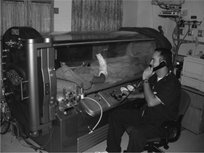
As implied by the name, these chambers have space for only one average-sized adult. Generally speaking, modern chambers of this type are cylindrical in shape and made of a large (approximately 0.6–1 m internal diameter and 2.1–2.3 m long) clear acrylic tube with a cap on one end and entry/exit hatch on the other. Patients slide into the chamber through the hatch to rest supine while they receive HBO therapy (Fig. 39.6).
Other than their small size, these chambers differ from their multiplace counterparts (described below) in that they are pressurized with 100% oxygen (in most cases) and are generally limited to no more than approximately 3 ATA operating pressure. This limitation makes them unsuitable for some high-pressure treatment tables occasionally used for some types of decompression illness. Monoplace chambers can be fitted such that air breaks can be administered using a tight-fitting mask.
|
|
|
Figure 39.6. Monoplace chamber. This type of chamber has room for one patient or a tender with a small child. Chamber atmosphere is 100% O2. The chamber is constructed of transparent Plexiglas to allow observation. Through-hull penetrators in the door on the left can be seen and allow monitoring, intravenous fluid administration, and control of a ventilator inside the chamber. (Photograph courtesy of Dr. Lindell Weaver.) |
A challenge with the use of these chambers is lack of direct access to the patient. However, almost all monitoring and ventilatory care (invasive blood pressure monitoring, mechanical ventilation, chest tube management, etc.) previously only available to patients in multiplace chambers can now be delivered in monoplace chambers (111,225).
Multiplace
These chambers can hold two or more patients/tenders. They exist in many shapes and sizes, usually large cylindrical or spherical shapes made of high-quality steel. Most of these chambers have a personnel lock as well, which allows patients or medical staff to exit or enter the chamber while it is at pressure. Transfer locks allow medicines, materials, and food to be moved into or out of the chamber. Patients are generally accompanied in the chamber by a tender or nurse, who can attend to the needs of the patient during the treatment. Administration of all critical care modalities is relatively easy inside a multiplace chamber (Fig. 39.7).
Due to their sturdier construction, multiplace chambers are generally able to withstand much higher pressures than their acrylic monoplace counterparts, and thus can be used for a wider range of treatment pressures.
Minimization of Fire Hazards and Atmosphere Control
Hyperbaric chambers are unique among medical equipment in that the nurse, tender, or physician is frequently also inside the treatment vessel (chamber) with the patient (in the case of multiplace chambers) and not easily accessible in the event of an emergency. The environment must be carefully managed to ensure atmosphere quality, with specified limits for oxygen and carbon dioxide, and to eliminate sources of ignition such as matches and cigarette lighters. Cotton suits are worn by patients and staff. Oil-based cosmetics and/or wigs (frequently made of synthetic materials) are prohibited (226).
|
|
|
Figure 39.7. Patient treatment in a multiplace chamber. |
Additionally, stretchers and equipment must have the petroleum-based lubricants removed from their wheels and other lubricated parts. Any other objects with petroleum-based lubricants must be cleaned of these lubricants prior to chamber treatment. At the time of this writing, there has not been a reported fire in a hyperbaric chamber that has resulted in a loss of life in the United States, although several such incidents have occurred overseas.
Ventilatory Care
Mechanical Ventilation
Certain precautions must be taken when diving a mechanically ventilated patient in a hyperbaric chamber. First of all, the ventilator must be approved for hyperbaric use. They should be fluidically or pneumatically controlled. Electrically driven ventilators are arguably less safe than ones using pneumatic or fluidic control. Although not commonly used at very high chamber pressures (6 ATA), ventilators powered by compressed oxygen have an inlet PO2 of up to 4,560 mm Hg (227), which can present a significant fire hazard.
As pressure rises, so does the gas density, which leads to a corresponding increase in airway resistance. Unless the ventilator is volume cycled, the tidal volumes may drop as pressure rises (227). Therefore, tidal volumes should be monitored closely (228).
Prior to chamber pressurization, inflating the endotracheal cuff with water or saline will prevent leakage due to cuff volume compression.
Suction
Since the chamber is at pressure, suction can be created simply by venting a hose to the outside world attaching a regulator to a through-hull penetrator. Normal hospital equipment can be modified for this use (229). In patients with copious secretions or ventilated patients, it is preferable to perform deep suctioning immediately prior to both compression and decompression of the chamber. This removes any mucous plugs that could contribute to air trapping.
Chest Tube Management
Conventional water seal or one-way valve pleural drainage systems operate satisfactorily inside hyperbaric chambers, with or without applied suction. During chamber decompression, expansion of gas volume within the tubing connecting the chest tube with the drainage system is automatically vented via the water seal or one-way valve. On the other hand, during chamber compression, the same gas volume is compressed and the connecting tubing and gas-containing space on the patient side of the water seal will tend to collapse, therefore producing high negative intrapleural pressures. Standard commercially available pleural evacuation systems have a manually activated pressure relief valve, which should be activated during the compression phase to relieve this excessive negative pressure.
Intravenous Infusion Devices
Several different IV infusion devices have been tested inside multiplace hyperbaric chambers and found to deliver fluid accurately. While it is the policy of some facilities not to use electrical equipment inside a chamber, others minimize a fire hazard by purging the device with 100% nitrogen. For monoplace use, the IV infusion device must be outside the chamber. Glass IV bottles should be avoided in order to prevent explosion during decompression due to expansion of any contained air bubble.
Arterial Blood Gas Measurement
Arterial blood gas analysis can be performed inside a multiplace hyperbaric chamber using an analyzer adapted for hyperbaric use. Alternatively, blood samples can be decompressed and analyzed at 1 ATA. The latter procedure is simpler, but subject to error. While pH and PCO2 are relatively stable during decompression, PO2 usually exceeds ambient pressure outside the chamber, and thus it tends to decline rapidly as oxygen is released from solution. Reasonably accurate values can be obtained if the sample is analyzed immediately after decompression (230).
Alternatively, it is possible to predict arterial PO2 during HBO therapy from a 1 ATA arterial blood gas measurement using the following equations. All that is needed is a 1 ATA blood gas measurement (at known FiO2), the HBO treatment pressure in ATA (PATA, usually between 2 and 3 ATA), barometric pressure (Pb, in mm Hg, usually near 760 mm Hg), the vapor pressure of water at body temperature (PH2O, at or near 47 mm Hg), the respiratory exchange ratio (usually 0.8), and PaCO2 and the following formulas:


where Pb is the barometric pressure outside the chamber; PaO2 (1 ATA) is the arterial PO2 at 1 ATA; PaO2 (1 ATA) is the alveolar PO2 at 1 ATA; FiO2 is the inspired O2 fraction; R is the respiratory exchange ratio (usually 0.8); PATA is the ambient pressure in the chamber in ATA; and PCO2 is the arterial PCO2 measured at 1 ATA, assumed to be unchanged during HBO.
Patient Monitoring
Most monitoring modalities used in hyperbaric chambers are identical to those used in normobaric situations, with few exceptions. Whenever inflatable pressure bags are used, such as for invasive blood pressure measurement, as the chamber pressurizes, the volume of air and the pressure in the pressure bag decreases, and thus one must periodically pump it up during compression; when decompressing the chamber, the air in the pressure bag expands, which must be released periodically to avoid rupture. For the same reason, pulmonary artery catheter balloons should be left open to the atmosphere during chamber compression and decompression.
Invasive pressure monitoring (or any monitoring where an electrical signal is transmitted via cable/wire) can be performed using through-hull penetrators to connect the transducer inside the chamber with the preamplifier outside.
Standard stethoscopes and sphygmomanometers can be used without difficulty in a multiplace chamber. Mercury pressure gauges should be avoided to prevent chamber contamination in the event of breakage.
Cardiac Arrest and Defibrillation
If a patient requires cardioversion or defibrillation while receiving HBO treatment, it is necessary to have through-hull penetrators for the high-voltage cables connecting an outside defibrillator with the paddles inside. Use of a low-impedance gel will prevent sparks or heat buildup at the site of paddle contact. Careful design and testing are necessary to confirm adequacy of energy delivery. The only alternative is to decompress the chamber and cardiovert or defibrillate at 1 ATA.
Tenders, Nurses, and Other Chamber Staff Considerations
Inside tenders in a multiplace chamber will take up nitrogen. While there is no requirement for a decompression stop for typical 2 ATA/2 hour or 2.5 ATA/90 minute treatments, many facilities require their staff to breathe 100% oxygen during decompression to reduce the very small risk of DCS. Additionally, repetitive exposures within a short time to even these low pressures may incur some risk of DCS. Minimum time intervals between hyperbaric exposures for staff are routine. Longer treatments or higher treatment pressures generally mandate specific decompression or oxygen breathing requirements for the inside staff. Emergency decompression from such exposures due to patient instability may therefore place the accompanying tender at risk. In the event of such an emergency, the tender should be immediately recompressed. The most widely accepted management schedule is described on p. 9–13 of the U.S. Navy Diving Manual (108).
Critical Care in a Hyperbaric Chamber in the Field
Field chambers are used in the offshore oil industry and at some remote inland dive sites. Divers injured due to decompression illness or trauma may require critical care in this setting. This is particularly the case for divers decompressing from saturation dives, in which an injured diver may require many days of decompression before he or she can be transferred to a hospital. Tracheal intubation, chest tube insertion, mechanical ventilation, hemodynamic and CNS monitoring, and treatment of convulsions may all be necessary (231). Portable radiographs can be obtained by passing an x-ray beam through a Plexiglas port, with the x-ray plate inside the chamber (232).
Hyperbaric Treatment Complications
Barotrauma
Otic
As many as 17% of all HBO therapy patients report ear pain with compression, making otic barotraumas the most common complication of HBO therapy (74). This is the result of difficulty with middle ear pressure equalization (i.e., eustachian tube opening). As the chamber is compressed, the increased pressure on the tympanic membrane can cause it to stretch medially just as when one dives in a swimming pool. Only rarely does this result in perforation of the tympanic membrane in awake patients, as they are able to notify the chamber operator of their progressive discomfort. Most often, there is unilateral otic discomfort associated with an erythematous tympanic membrane that heals over the following 5 to 7 days. In patients who are unable to adequately perform a Valsalva maneuver required to equalize pressure (i.e., sedated, intubated, with eustachian tube/sinus dysfunction, or with tracheostomy tube), bilateral myringotomies with or without tube placement are performed. Because myringotomies heal in 2 to 3 days, for patients unable to equalize, bilateral tympanostomy tubes are normally placed in patients who are expected to receive repetitive treatments for longer.
Sinus
Sinus barotrauma (“sinus squeeze”) is a relatively infrequent occurrence, which occurs in patients with active sinus infection, allergic rhinitis, or nasal polyps. During pressure change, the patient will feel discomfort in the region of the affected sinus, particularly if it is the frontal sinus (233). Sinus squeeze can usually be prevented using topical decongestants such as oxymetazoline and slow compression of the chamber (233).
Pulmonary
Although this is far more frequent with scuba diving, it can also occur rarely in dry hyperbaric chambers, causing AGE (234), pneumomediastinum, or pneumothorax. Patients with cystic or bullous disease are presumably at risk; however, many such patients have received HBO without complication. In patients with a pre-existing pneumothorax, tube decompression is recommended, especially if the patient is to be treated in a monoplace chamber.
Pulmonary Edema
Peripheral vasoconstriction induced by HBO and the resulting increase in afterload can precipitate pulmonary edema in patients with impaired ventricular function (235).
Evaluation of a Patient for Hyperbaric Oxygen Therapy
In assessing a patient for hyperbaric therapy, two aspects need to be evaluated: Potential efficacy of treatment and risk of adverse effects. Indications for hyperbaric oxygen therapy as determined by the Undersea and Hyperbaric Medical Society (7) are listed in Table 39.1. A second factor is the predicted arterial PO2, which must be within a therapeutic range during HBO therapy (>1,000 mm Hg). If a patient has pulmonary gas exchange impairment that precludes attainment of an arterial PO2 that is sufficiently high, then HBO is unlikely to be effective. A method for predicting arterial oxygenation during HBO makes use of the relative constancy of the ratio of arterial to alveolar PO2 (PaO2/PAO2 ratio) as described above (Eq. 2 and 3). Finally, the assessment must include evaluating for the risk of pulmonary barotrauma. During decompression, pulmonary cysts or bullae can rupture (234), although such complications are extremely rare. Patients with untreated pneumothorax usually require a tube thoracostomy, unless immediate chest decompression can be performed. Patients with a pneumothorax for whom monoplace treatment is planned require prophylactic chest tube insertion irrespective of the size of the pneumothorax. Patients in heart failure in whom left ventricular function may not be able to tolerate an increase in afterload are also at risk (235).
Susceptibility to otic barotrauma and occasionally to sinus barotrauma also plays a role in determining fitness for HBO therapy. Obtunded patients are especially at risk of otic barotrauma, and many practitioners advocate prophylactic myringotomy.
Summary
Although hyperbaric oxygen therapy has limited indications, it represents definitive therapy for some critically ill patients, especially those with gas bubble disease (decompression sickness or gas embolism) and carbon monoxide poisoning. The available evidence also strongly suggests that it is an effective adjunct in necrotizing soft tissue infections. HBO can be safely administered to the critically ill patient using an appropriately equipped hyperbaric chamber and implementing standard monitoring and supportive measures.
References
1. Simpson A. Compressed Air, as a Therapeutic Agent, in the Treatment of Consumption, Asthma, Chronic Bronchitis, and Other Diseases. Edinburgh: Sutherland & Knox; 1857.
2. Junod VT. Recherches physiologiques et thérapeutiques sur les effets de la compression et de la raréfaction de l'air, tant sur le corps que sur les membres isolés. Rev Med Fr Étrange. 1834;3:350–368.
3. Phillips JL. The Bends. New Haven, CT: Yale University; 1998.
4. Zuntz N. Zur Pathogenese und Therapie der durch rasche Luftdruckänderungen erzeugten Krankheiten. Fortschr Med. 1897;15:632–639.
5. Behnke AR, Shaw LA. The use of oxygen in the treatment of compressed air illness. Navy Med Bull. 1937;35:1–12.
6. Yarbrough OD, Behnke AR. The treatment of compressed air illness using oxygen. J Ind Hyg Toxicol. 1939;21:213–218.
7. Gesell LB, ed. Hyperbaric Oxygen Therapy: A Committee Report. Durham, NC: Undersea and Hyperbaric Medical Society; 2008.
8. Whalen RE, Saltzman HA, Holloway DH Jr, et al. Cardiovascular and blood gas responses to hyperbaric oxygenation. Am J Cardiol. 1965;15:638–646.
9. McMahon TJ, Moon RE, Luschinger BP, et al. Nitric oxide in the human respiratory cycle. Nat Med. 2002;8:711–717.
10. Boerema I, Meyne NG, Brummelkamp WH, et al. Life without blood. J Cardiovasc Surg (Torino). 1960;1:133–146.
11. Bergofsky EH, Wang MC, Yamaki T, et al. Tissue oxygen and carbon dioxide tensions during hyperbaric oxygenation. JAMA. 1964;189:841–844.
12. Bergofsky EH, Bertun P. Response of regional circulations to hyperoxia. J Appl Physiol. 1966;21:567–572.
13. Bird AD, Telfer AB. Effect of hyperbaric oxygen on limb circulation. Lancet. 1965;13:355–356.
14. Berry JM, Doursout MF, Butler BD. Effects of hyperbaric hyperoxia on cardiac and regional hemodynamics in conscious dogs. Aviat Space Environ Med. 1998;69:761–765.
15. Demchenko IT, Boso AE, Bennett PB, et al. Hyperbaric oxygen reduces cerebral blood flow by inactivating nitric oxide. Nitric Oxide. 2000;4:597–608.
16. Dooley JW, Mehm WJ. Noninvasive assessment of the vasoconstrictive effects of hyperoxygenation. J Hyperbar Med. 1990;4:177–187.
17. Sheikh AY, Rollins MD, Hopf HW, et al. Hyperoxia improves microvascular perfusion in a murine wound model. Wound Repair Regen. 2005;13:303–308.
18. Hahnloser PB, Domanig E, Lamphier E, et al. Hyperbaric oxygenation: alterations in cardiac output and regional blood flow. J Thorac Cardiovasc Surg. 1966;52:223–231.
19. Villanucci S, Di Marzio GE, Scholl M, et al. Cardiovascular changes induced by hyperbaric oxygen therapy. Undersea Biomed Res (Suppl). 1990;17:117.
20. Demchenko IT, Boso AE, Natoli MJ, et al. Nitric oxide is involved in the mechanism of HBO-induced cerebral vasoconstriction. Undersea Hyperb Med. 1998;25(Suppl):54.
21. Demchenko IT, Oury TD, Crapo JD, et al. Regulation of the brain's vascular responses to oxygen. Circ Res. 2002;91:1031–1037.
22. Hordnes C, Tyssebotn I. Effect of high ambient pressure and oxygen tension on organ blood flow in conscious trained rats. Undersea Biomed Res. 1985;12:115–128.
23. Savitt MA, Rankin JS, Elberry JR, et al. Influence of hyperbaric oxygen on left ventricular contractility, total coronary blood flow, and myocardial oxygen consumption in the conscious dog. Undersea Hyperb Med. 1994;21:169–183.
24. Zamboni WA, Roth AC, Russell RC, et al. Morphological analysis of the microcirculation during reperfusion of ischemic skeletal muscle and the effect of hyperbaric oxygen. Plast Reconstr Surg. 1993;91:1110–1123.
25. Helps SC, Meyer-Witting M, Rilley PL, et al. Increasing doses of intracarotid air and cerebral blood flow in rabbits. Stroke. 1990;21:1340–1345.
26. Helps SC, Gorman DF. Air embolism of the brain in rabbits pre-treated with mechlorethamine. Stroke. 1991;22:351–354.
27. Miljkovic-Lolic M, Silbergleit R, Fiskum G, et al. Neuroprotective effects of hyperbaric oxygen treatment in experimental focal cerebral ischemia are associated with reduced brain leukocyte myeloperoxidase activity. Brain Res. 2003;971:90–94.
28. Chen MF, Chen HM, Ueng SW, et al. Hyperbaric oxygen pretreatment attenuates hepatic reperfusion injury. Liver. 1998;18:110–116.
29. Tjarnstrom J, Wikstrom T, Bagge U, et al. Effects of hyperbaric oxygen treatment on neutrophil activation and pulmonary sequestration in intestinal ischemia-reperfusion in rats. Eur Surg Res. 1999;31:147–154.
30. Yamada T, Taguchi T, Hirata Y, et al. The protective effect of hyperbaric oxygenation on the small intestine in ischemia-reperfusion injury. J Pediatr Surg. 1995;30:786–790.
31. Zamboni WA, Wong HP, Stephenson LL. Effect of hyperbaric oxygen on neutrophil concentration and pulmonary sequestration in reperfusion injury. Arch Surg. 1996;131:756–760.
32. Haapaniemi T, Nylander G, Sirsjo A, et al. Hyperbaric oxygen reduces ischemia-induced skeletal muscle injury. Plast Reconstr Surg. 1996;97:602–607; discussion 8–9.
33. Zamboni WA, Roth AC, Russell RC, et al. The effect of acute hyperbaric oxygen therapy on axial pattern skin flap survival when administered during and after total ischemia. J Reconstr Microsurg. 1989;5:343–347.
34. Hong JP, Kwon H, Chung YK, et al. The effect of hyperbaric oxygen on ischemia-reperfusion injury: an experimental study in a rat musculocutaneous flap. Ann Plast Surg. 2003;51:478–487.
35. Bosco G, Yang ZJ, Nandi J, et al. Effects of hyperbaric oxygen on glucose, lactate, glycerol and anti-oxidant enzymes in the skeletal muscle of rats during ischaemia and reperfusion. Clin Exp Pharmacol Physiol. 2007;34:70–76.
36. Buras JA, Reenstra WR. Endothelial-neutrophil interactions during ischemia and reperfusion injury: basic mechanisms of hyperbaric oxygen. Neurol Res. 2007;29:127–131.
37. Riccio M, Pangrazi PP, Campodonico A, et al. Combined use of WEB2170 and HBO therapy can reduce ischemia and reperfusion injury to the skeletal muscle in a rabbit model. Microsurgery. 2007;27:43–47.
38. Henninger N, Kuppers-Tiedt L, Sicard KM, et al. Neuroprotective effect of hyperbaric oxygen therapy monitored by MR-imaging after embolic stroke in rats. Exp Neurol. 2006;201:316–323.
39. Schabitz WR, Schade H, Heiland S, et al. Neuroprotection by hyperbaric oxygenation after experimental focal cerebral ischemia monitored by MRI. Stroke. 2004;35:1175–1179.
40. Gurer A, Ozdogan M, Gomceli I, et al. Hyperbaric oxygenation attenuates renal ischemia-reperfusion injury in rats. Transplant Proc. 2006;38:3337–3340.
41. Kalns J, Lane J, Delgado A, et al. Hyperbaric oxygen exposure temporarily reduces Mac-1 mediated functions of human neutrophils. Immunol Lett. 2002;83:125–131.
42. Thom SR. Effects of hyperoxia on neutrophil adhesion. Undersea Hyperb Med. 2004;31:123–131.
43. Thom SR, Mendiguren I, Hardy K, et al. Inhibition of human neutrophil beta2-integrin-dependent adherence by hyperbaric O2. Am J Physiol. 1997;272:C770–C777.
44. Veltkamp R, Siebing DA, Sun L, et al. Hyperbaric oxygen reduces blood-brain barrier damage and edema after transient focal cerebral ischemia. Stroke. 2005;36:1679–1683.
45. Lou M, Chen Y, Ding M, et al. Involvement of the mitochondrial ATP-sensitive potassium channel in the neuroprotective effect of hyperbaric oxygenation after cerebral ischemia. Brain Res Bull. 2006;69:109–116.
46. Mader JT, Brown GL, Guckian JC, et al. A mechanism for the amelioration by hyperbaric oxygen of experimental staphylococcal osteomyelitis in rabbits. J Infect Dis. 1980;142:915–922.
47. Mader JT, Adams KR, Sutton TE. Infectious diseases: pathophysiology and mechanisms of hyperbaric oxygen. J Hyperbar Med. 1987;2:133–140.
48. Zamboni WA, Mazolewski PJ, Erdmann D, et al. Evaluation of penicillin and hyperbaric oxygen in the treatment of streptococcal myositis. Ann Plast Surg. 1997;39:131–136.
49. Carraway MS, Piantadosi CA. Oxygen toxicity. Respir Care Clin N Am. 1999;5:265–295.
50. Freeman BA, Crapo JD. Biology of disease: Free radicals and tissue injury. Lab Invest. 1982;47:412–426.
51. Gerschman R, Gilbert DL, Nye SW, et al. Oxygen poisoning and x-irradiation: a mechanism in common. Science. 1954;119:623–626.
52. Demchenko IT, Atochin DN, Boso AE, et al. Oxygen seizure latency and peroxynitrite formation in mice lacking neuronal or endothelial nitric oxide synthases. Neurosci Lett. 2003;344:53–56.
53. Demchenko IT, Boso AE, Whorton AR, et al. Nitric oxide production is enhanced in rat brain before oxygen-induced convulsions. Brain Res. 2001;917:253–261.
54. Demchenko IT, Piantadosi CA. Nitric oxide amplifies the excitatory to inhibitory neurotransmitter imbalance accelerating oxygen seizures. Undersea Hyperb Med. 2006;33:169–174.
55. Donald KW. Oxygen poisoning in man, I & II. BMJ. 1947;1:667–672, 712–717.
56. Doherty MJ, Hampson NB. Partial seizure provoked by hyperbaric oxygen therapy: possible mechanisms and implications. Epilepsia. 2005;46:974–976.
57. Bleiberg B, Kerem D. Central nervous system oxygen toxicity in the resting rat: postponement by intermittent oxygen exposure. Undersea Biomed Res. 1988;15:337–352.
58. Chavko M, McCarron RM. Extension of brain tolerance to hyperbaric O2 by intermittent air breaks is related to the time of CBF increase. Brain Res. 2006;1084:196–201.
59. Harabin AL, Survanshi SS, Weathersby PK, et al. The modulation of oxygen toxicity by intermittent exposure. Toxicol Appl Pharmacol. 1988;93:298–311.
60. Lambertsen CJ. Extension of oxygen tolerance in man: philosophy and significance. Exp Lung Res. 1988;14(Suppl):1035–1058.
61. Vote DA, Doar O, Moon RE, et al. Blood glucose meter performance under hyperbaric oxygen conditions. Clin Chim Acta. 2001;305:81–87.
62. Clark JM, Lambertsen CJ. Pulmonary oxygen toxicity: a review. Pharmacol Rev. 1971;23:37–133.
63. Bardin H, Lambertsen CJ. A Quantitative Method for Calculating Cumulative Pulmonary Oxygen Toxicity. Use of the Unit Pulmonary Toxicity Dose (UPTD). Philadelphia: Institute of Environmental Medicine, University of Pennsylvania; 1970.
64. Clark JM. Oxygen toxicity. In: Bennett PB, Elliott DH, eds. The Physiology and Medicine of Diving. Philadelphia: WB Saunders; 1993:121–169.
65. Harabin AL, Homer LD, Weathersby PK, et al. An analysis of decrements in vital capacity as an index of pulmonary oxygen toxicity. J Appl Physiol. 1987;63:1130–1135.
66. Clark JM, Lambertsen CJ. Rate of development of pulmonary O2 toxicity in man during O2 breathing at 2.0 Ata. J Appl Physiol. 1971;30:739–752.
67. Goldiner PL, Carlon GC, Cvitkovic E, et al. Factors influencing postoperative morbidity and mortality in patients treated with bleomycin. BMJ. 1978;1:1664–1667.
68. Nygaard K, Smith-Erichsen N, Hatlevoll R, et al. Pulmonary complications after bleomycin, irradiation and surgery for esophageal cancer. Cancer. 1978;41:17–22.
69. Thompson CC, Bailey MK, Conroy JM, et al. Postoperative pulmonary toxicity associated with mitomycin C therapy. South Med J. 1992;85:1257–1259.
70. Anderson B Jr, Shelton DL. Axial length in hyperoxic myopia. In: Bove AA, Bachrach AJ, Greenbaum LJ Jr, eds. Underwater and Hyperbaric Physiology IX Proceedings of the Ninth International Symposium on Underwater and Hyperbaric Physiology. Bethesda, MD: Undersea and Hyperbaric Medical Society; 1987:607–611.
71. Palmquist BM, Philipson B, Barr PO. Nuclear cataract and myopia during hyperbaric oxygen therapy. Br J Ophthalmol. 1984;68:113–117.
72. Behnke AR, Forbes HS, Motley EP. Circulatory and visual effects of oxygen at 3 atmospheres pressure. Am J Physiol. 1936;114:436–442.
73. Clark JM, Thom SR. Oxygen under pressure. In: Brubakk AO, Neuman TS, eds. Physiology and Medicine of Diving. London: Saunders; 2003:376.
74. Plafki C, Peters P, Almeling M, et al. Complications and side effects of hyperbaric oxygen therapy. Aviat Space Environ Med. 2000;71:119–124.
75. Junod T. Recherches sur les effets physiologiques et thérapeutiques de la compression et de raréfaction de 1'air, tant sur le corps que les membres isolés. Ann Gén Med sér 2, 1835;9:157–172.
76. Behnke AR, Thomas RM, Motley EP. The psychologic effects from breathing air at 4 atmospheres pressure. Am J Physiol. 1935;112:554–558.
77. Muth CM, Shank ES. Gas embolism. N Engl J Med. 2000;342:476–482.
78. Fukaya E, Hopf HW. HBO and gas embolism. Neurol Res. 2007;29:142–145.
79. Morris WP, Butler BD, Tonnesen AS, et al. Continuous venous air embolism in patients receiving positive end-expiratory pressure. Am Rev Respir Dis. 1993;147:1034–1037.
80. Kaufman BS, Kaminsky SJ, Rackow EC, et al. Adult respiratory distress syndrome following orogenital sex during pregnancy. Crit Care Med. 1987;15:703–704.
81. Nossum V, Koteng S, Brubakk AO. Endothelial damage by bubbles in the pulmonary artery of the pig. Undersea Hyperb Med. 1999;26:1–8.
82. Dutka AJ, Kochanek PM, Hallenbeck JM. Influence of granulocytopenia on canine cerebral ischemia induced by air embolism. Stroke. 1989;20:390–395.
83. Hallenbeck JM, Dutka AJ, Tanishima T, et al. Polymorphonuclear leukocyte accumulation in brain regions with low blood flow during the early postischemic period. Stroke. 1986;17:246–253.
84. Levin LL, Stewart GJ, Lynch PR, et al. Blood and blood vessel wall changes induced by decompression sickness in dogs. J Appl Physiol. 1981;50:944–949.
85. Chryssanthou C, Springer M, Lipschitz S. Blood-brain and blood-lung barrier alteration by dysbaric exposure. Undersea Biomed Res. 1977;4:117–129.
86. Zwirewich CV, Müller NL, Abboud RT, et al. Noncardiogenic pulmonary edema caused by decompression sickness: rapid resolution following hyperbaric therapy. Radiology. 1987;163:81–82.
87. Nossum V, Hjelde A, Brubakk AO. Small amounts of venous gas embolism cause delayed impairment of endothelial function and increase polymorphonuclear neutrophil infiltration. Eur J Appl Physiol. 2002;86:209–214.
88. van Hulst RA, Lameris TW, Haitsma JJ, et al. Brain glucose and lactate levels during ventilator-induced hypo- and hypercapnia. Clin Physiol Funct Imaging. 2004;24:243–248.
89. van Hulst RA, Lameris TW, Hasan D, et al. Effects of cerebral air embolism on brain metabolism in pigs. Acta Neurol Scand. 2003;108:118–124.
90. van Hulst RA, Haitsma JJ, Lameris TW, et al. Hyperventilation impairs brain function in acute cerebral air embolism in pigs. Intensive Care Med. 2004;30:944–950.
91. Mann C, Boccara G, Fabre JM, et al. The detection of carbon dioxide embolism during laparoscopy in pigs: a comparison of transesophageal Doppler and end-tidal carbon dioxide monitoring. Acta Anaesthesiol Scand. 1997;41:281–286.
92. Benson J, Adkinson C, Collier R. Hyperbaric oxygen therapy of iatrogenic cerebral arterial gas embolism. Undersea Hyperb Med. 2003;30:117–126.
93. Longphre JM, Denoble PJ, Moon RE, et al. First aid normobaric oxygen for the treatment of recreational diving injuries. Undersea Hyperb Med. 2007;34:43–49.
94. Atkinson JR. Experimental air embolism. Northwest Med. 1963;62:699–703.
95. Van Allen CM, Hrdina LS, Clark J. Air embolism from the pulmonary vein. Arch Surg. 1929;19:567–599.
96. Mehlhorn U, Burke EJ, Butler BD, et al. Body position does not affect the hemodynamic response to venous air embolism in dogs. Anesth Analg. 1994;79:734–739.
97. Geissler HJ, Allen SJ, Mehlhorn U, et al. Effect of body repositioning after venous air embolism. An echocardiographic study. Anesthesiology. 1997;86:710–717.
98. Dutka AJ. Therapy for dysbaric central nervous system ischemia: adjuncts to recompression. In: Bennett PB, Moon RE, eds. Diving Accident Management. Bethesda, MD: Undersea and Hyperbaric Medical Society; 1990:222–234.
99. van Hulst RA, Klein J, Lachmann B. Gas embolism: pathophysiology and treatment. Clin Physiol Funct Imaging. 2003;23:237–246.
100. Adams HP Jr, del Zoppo G, Alberts MJ, et al. Guidelines for the early management of adults with ischemic stroke: a guideline from the American Heart Association/American Stroke Association Stroke Council, Clinical Cardiology Council, Cardiovascular Radiology and Intervention Council, and the Atherosclerotic Peripheral Vascular Disease and Quality of Care Outcomes in Research Interdisciplinary Working Groups: the American Academy of Neurology affirms the value of this guideline as an educational tool for neurologists. Stroke. 2007;38:1655–1711.
101. Brown JW, Dierdorf SF, Moorthy SS, et al. Venoarterial cerebral perfusion for treatment of massive arterial air embolism. Anesth Analg. 1987;66:673–674.
102. Stark J, Hough J. Air in the aorta: treatment by reversed perfusion. Ann Thorac Surg. 1986;41:337–338.
103. Ericsson JA, Gottlieb JD, Sweet RB. Closed-chest cardiac massage in the treatment of venous air embolism. N Engl J Med. 1964;270:1353–1354.
104. Michenfelder JD, Martin JT, Altenburg BM, et al. Air embolism during neurosurgery. An evaluation of right-atrial catheters for diagnosis and treatment. JAMA. 1969;208:1353–1358.
105. Bowdle TA, Artru AA. Treatment of air embolism with a special pulmonary artery catheter introducer sheath in sitting dogs. Anesthesiology. 1988;68:107–110.
106. Marshall WK, Bedford RF. Use of a pulmonary-artery catheter for detection and treatment of venous air embolism: a prospective study in man. Anesthesiology. 1980;52:131–134.
107. Thalmann ED. Principles of US Navy recompression treatments for decompression sickness. In: Moon RE, Sheffield PJ, eds. Treatment of Decompression Illness. Kensington, MD: Undersea and Hyperbaric Medical Society; 1996:75–95.
108. Navy Department. US Navy Diving Manual. Revision 4. Vol. 5: Diving Medicine and Recompression Chamber Operations. NAVSEA 0910-LP-103-8009. Washington, DC: Naval Sea Systems Command; 2005.
109. Hart GB, Strauss MB, Lennon PA. The treatment of decompression sickness and air embolism in a monoplace chamber. J Hyperbar Med. 1986;1:1–7.
110. Cianci P, Slade JB Jr. Delayed treatment of decompression sickness with short, no-air-break tables: review of 140 cases. Aviat Space Environ Med. 2006;77:1003–1008.
111. Weaver LK. Monoplace hyperbaric chamber use of U.S. Navy Table 6: a 20-year experience. Undersea Hyperb Med. 2006;33:85–88.
112. Moon RE, ed. Adjunctive Therapy for Decompression Illness. Kensington, MD: Undersea and Hyperbaric Medical Society; 2003.
113. Undersea and Hyperbaric Medical Society. UHMS Guidelines for Adjunctive Therapy of DCI. In: Moon RE, ed. Adjunctive Therapy for Decompression Illness. Kensington, MD: Undersea and Hyperbaric Medical Society; 2003:184–189.
114. Balldin UI. Effects of ambient temperature and body position on tissue nitrogen elimination in man. Aerosp Med. 1973;44:365–3670.
115. Hallenbeck JM, Leitch DR, Dutka AJ, et al. Prostaglandin I2, indomethacin and heparin promote postischemic neuronal recovery in dogs. Ann Neurol. 1982;12:145–156.
116. Reeves E, Workman RD. Use of heparin for the therapeutic/prophylactic treatment of decompression sickness. Aerosp Med. 1971;42:20–23.
117. Broome JR. Association of CNS hemorrhage with failure to respond to recompression treatment—implications for management of refractory cases of decompression illness. In: Moon RE, Sheffield PJ, eds. Treatment of Decompression Illness. Kensington, MD: Undersea and Hyperbaric Medical Society; 1996:364–373.
118. Elliott DH, Hallenbeck JM, Bove AA. Venous infarction of the spinal cord in decompression sickness. J Roy Nav Med Serv. 1974;60:66–71.
119. Palmer AC, Blakemore WF, Payne JE, et al. Decompression sickness in the goat: nature of brain and spinal cord lesions at 48 hours. Undersea Biomed Res. 1978;5:275–286.
120. Gorman DF, Browning DN. Cerebral vaso-reactivity and arterial gas embolism. Undersea Biomed Res. 1986;13:317–335.
121. Waite CL, Mazzone WF, Greenwood ME, et al. Cerebral air embolism I. Basic studies. US Naval Submarine Medical Center Report No. 493. Panama City, FL: US Navy Submarine Research Laboratory; 1967. Report No.: 493.
122. Landolt JP, Money KE, Topliff ED, et al. Pathophysiology of inner ear dysfunction in the squirrel monkey in rapid decompression. J Appl Physiol. 1980;49:1070–1082.
123. McCormick JG, Holland WB, Brauer RW, et al. Sudden hearing loss due to diving and its prevention with heparin. Otolaryngol Clin North Am. 1975;8:417–430.
124. Mitchell SJ. Lidocaine in the treatment of decompression illness: a review of the literature. Undersea Hyperb Med. 2001;28:165–174.
125. Mitchell SJ, Pellett O, Gorman DF. Cerebral protection by lidocaine during cardiac operations. Ann Thorac Surg. 1999;67:1117–1124.
126. Wang D, Wu X, Li J, et al. The effect of lidocaine on early postoperative cognitive dysfunction after coronary artery bypass surgery [comment]. Anesth Analg. 2002;95:1134–1141, table of contents.
127. Mathew J, Grocott H, Phillips-Bute B, et al. Lidocaine does not prevent cognitive dysfunction after cardiac surgery. Anesth Analg. 2004;98(Suppl):SCA13.
128. Cogar WB. Intravenous lidocaine as adjunctive therapy in the treatment of decompression illness. Ann Emerg Med. 1997;29:284–286.
129. Mitchell SJ, Benson M, Vadlamudi L, et al. Cerebral arterial gas embolism by helium: an unusual case successfully treated with hyperbaric oxygen and lidocaine. Ann Emerg Med. 2000;35:300–303.
130. Bennett M, Mitchell S, Dominguez A. Adjunctive treatment of decompression illness with a non-steroidal anti-inflammatory drug (tenoxicam) reduces compression requirement. Undersea Hyperb Med. 2003;30:195–205.
131. Francis TJR, Dutka AJ, Clark JB. An evaluation of dexamethasone in the treatment of acute experimental spinal decompression sickness. In: Bove AA, Bachrach AJ, Greenbaum LJ, Jr, eds. Underwater and Hyperbaric Physiology IX Proceedings of the Ninth International Symposium on Underwater and Hyperbaric Physiology. Bethesda, MD: Undersea and Hyperbaric Medical Society; 1987:999–1013.
132. Dutka AJ, Mink RB, Pearson RR, et al. Effects of treatment with dexamethasone on recovery from experimental cerebral arterial gas embolism. Undersea Biomed Res. 1992;19:131–141.
133. Francis TJR, Dutka AJ. Methylprednisolone in the treatment of acute spinal cord decompression sickness. Undersea Biomed Res. 1989;16:165–174.
134. Dromsky DM, Toner CB, Fahlman A, et al. Prophylactic treatment of severe decompression sickness with methylprednisolone. Undersea Hyperb Med. 1999;26(Suppl):15.
135. Novotny JA, Bridgewater BJ, Himm JF, et al. Quantifying the effect of intravascular perfluorocarbon on xenon elimination from canine muscle. J Appl Physiol. 1993;74:1356–1360.
136. Mahon RT, Dainer HM, Nelson JW. Decompression sickness in a swine model: isobaric denitrogenation and perfluorocarbon at depth. Aviat Space Environ Med. 2006;77:8–12.
137. Dainer H, Nelson J, Brass K, et al. Short oxygen prebreathing and intravenous perfluorocarbon emulsion reduces morbidity and mortality in a swine saturation model of decompression sickness. J Appl Physiol. 2007;102:1099–1104.
138. Dromsky DM, Spiess BD, Fahlman A. Treatment of decompression sickness in swine with intravenous perfluorocarbon emulsion. Aviat Space Environ Med. 2004;75:301–305.
139. Yoshitani K, de Lange F, Ma Q, et al. Reduction in air bubble size using perfluorocarbons during cardiopulmonary bypass in the rat. Anesth Analg. 2006;103:1089–1093.
140. Zhu J, Hullett JB, Somera L, et al. Intravenous perfluorocarbon emulsion increases nitrogen washout after venous gas emboli in rabbits. Undersea Hyperb Med. 2007;34:7–20.
141. Eckmann DM, Armstead SC, Mardini F. Surfactants reduce platelet-bubble and platelet-platelet binding induced by in vitro air embolism. Anesthesiology. 2005;103:1204–1210.
142. Mitchell SJ, Doolette DJ, Wachholz CJ, et al., eds. Management of Mild or Marginal Decompression Illness in Remote Locations. Durham, NC: Divers Alert Network; 2005.
143. Centers for Disease Control and Prevention (CDC). Unintentional poisoning deaths—United States, 1999–2004. MMWR Morb Mortal Wkly Rep. 2007;56:93–96.
144. Brown SD, Piantadosi CA. Reversal of carbon monoxide-cytochrome c oxidase binding by hyperbaric oxygen in vivo. Adv Exp Med Biol. 1989;248:747–754.
145. Brown SD, Piantadosi CA. In vivo binding of carbon monoxide to cytochrome c oxidase in rat brain. J Appl Physiol. 1990;68:604–610.
146. Chance B, Erecinska M, Wagner M. Mitochondrial responses to carbon monoxide toxicity. Ann N Y Acad Sci. 1970;174:193–204.
147. Thom S. Antagonism of carbon monoxide-mediated brain lipid peroxidation by hyperbaric oxygen. Toxicol Appl Pharmacol. 1990;105:340–344.
148. Thom SR, Bhopale VM, Han ST, et al. Intravascular neutrophil activation due to carbon monoxide poisoning. Am J Respir Crit Care Med. 2006;174:1239–1248.
149. Thom SR, Bhopale VM, Fisher D. Hyperbaric oxygen reduces delayed immune-mediated neuropathology in experimental carbon monoxide toxicity. Toxicol Appl Pharmacol. 2006;213:152–159.
150. Werner B, Back W, Akerblom H, et al. Two cases of acute carbon monoxide poisoning with delayed neurological sequelae after a “free” interval. J Toxicol Clin Toxicol. 1985;23:249–265.
151. Thom S, Taber R, Mendiguren I, et al. Delayed neuropsychologic sequelae after carbon monoxide poisoning: prevention by treatment with hyperbaric oxygen. Ann Emerg Med. 1995;25:474–480.
152. Weaver LK, Hopkins RO, Chan KJ, et al. Hyperbaric oxygen for acute carbon monoxide poisoning. N Engl J Med. 2002;347:1057–1067.
153. Pace N, Strajman E, Walker EL. Acceleration of carbon monoxide elimination in man by high pressure oxygen. Science. 1950;111:652–654.
154. Piantadosi CA. Diagnosis and treatment of carbon monoxide poisoning. Respir Care Clin N Am. 1999;5:183–202.
155. Brown SD, Piantadosi CA. Recovery of energy metabolism in rat brain after carbon monoxide hypoxia. J Clin Invest. 1992;89:666–672.
156. Ernst A, Zibrak JD. Carbon monoxide poisoning. N Engl J Med. 1998;339:1603–1608.
157. Tibbles PM, Edelsberg JS. Hyperbaric-oxygen therapy. N Engl J Med. 1996;334:1642–1648.
158. Thom SR. Hyperbaric-oxygen therapy for acute carbon monoxide poisoning. N Engl J Med. 2002;347:1105–1106.
159. Weaver LK, Valentine KJ, Hopkins RO. Carbon monoxide poisoning: risk factors for cognitive sequelae and the role of hyperbaric oxygen. Am J Respir Crit Care Med. 2007;176:491–497.
160. Van Hoesen KB, Camporesi EM, Moon RE, et al. Should hyperbaric oxygen be used to treat the pregnant patient for acute carbon monoxide poisoning? A case report and literature review. JAMA. 1989;261:1039–1043.
161. Meyers RA, Thom SR. Carbon monoxide and cyanide poisoning. In: Kindwall EP, ed. Hyperbaric Medicine Practice. Flagstaff, AZ: Best Publishing; 1994:344–366.
162. Cope C, Abramowitz S. Respiratory responses to intravenous sodium cyanide, a function of the oxygen-cyanide relationship. Am Rev Respir Dis. 1960;81:321–328.
163. Carden E. Hyperbaric oxygen in cyanide poisoning. Anaesthesia. 1970;25:442–443.
164. Goodhart GL. Patient treated with antidote kit and hyperbaric oxygen survives cyanide poisoning. South Med J. 1994;87:814–816.
165. Litovitz TL, Larkin RF, Myers RA. Cyanide poisoning treated with hyperbaric oxygen. Am J Emerg Med. 1983;1:94–101.
166. Takano T, Miyazaki Y, Nashimoto I, et al. Effect of hyperbaric oxygen on cyanide intoxication: in situ changes in intracellular oxidation reduction. Undersea Biomed Res. 1980;7:191–197.
167. Way JL, End E, Sheehy MH, et al. Effect of oxygen on cyanide intoxication. IV. Hyperbaric oxygen. Toxicol Appl Pharmacol. 1972;22:415–421.
168. Smilkstein MJ, Bronstein AC, Pickett HM, et al. Hyperbaric oxygen therapy for severe hydrogen sulfide poisoning. J Emerg Med. 1985;3:27–30.
169. Whitcraft DD 3rd, Bailey TD, Hart GB. Hydrogen sulfide poisoning treated with hyperbaric oxygen. J Emerg Med. 1985;3:23–25.
170. ATSDR. Toxicological Profile for Carbon Tetrachloride. U.S. Dept. of Health and Human Services PHS, Agency for Toxic Substances and Disease Registry; 2005.
171. Burkhart KK, Hall AH, Gerace R, et al. Hyperbaric oxygen treatment for carbon tetrachloride poisoning. Drug Saf. 1991;6:332–338.
172. Marzella L, Muhvich K, Myers RA. Effect of hyperoxia on liver necrosis induced by hepatotoxins. Virchows Arch B Cell Pathol Incl Mol Pathol. 1986;51:497–507.
173. Burk RF, Lane JM, Patel K. Relationship of oxygen and glutathione in protection against carbon tetrachloride-induced hepatic microsomal lipid peroxidation and covalent binding in the rat. Rationale for the use of hyperbaric oxygen to treat carbon tetrachloride ingestion. J Clin Invest. 1984;74:1996–2001.
174. Burk RF, Reiter R, Lane JM. Hyperbaric oxygen protection against carbon tetrachloride hepatotoxicity in the rat. Association with altered metabolism. Gastroenterology. 1986;90:812–818.
175. Bernacchi A, Myers R, Trump BF, et al. Protection of hepatocytes with hyperoxia against carbon tetrachloride-induced injury. Toxicol Pathol. 1984;12:315–323.
176. Truss CD, Killenberg PG. Treatment of carbon tetrachloride poisoning with hyperbaric oxygen. Gastroenterology. 1982;82:767–769.
177. Heimbach RD. Gas gangrene. In: Kindwall EP, ed. Hyperbaric Medicine Practice. Flagstaff, AZ: Best Publishing; 1994:374–388.
178. Brummelkamp WH, Boerema I, Hoogendyk L. Treatment of clostridial infections with hyperbaric oxygen drenching. A report on 26 cases. Lancet. 1963;1:235–238.
179. Brummelkamp WH, Hogendijk J, Boerema I. Treatment of anaerobic infections (clostridial myositis) by drenching tissues with oxygen under high atmospheric pressure. Surgery. 1961;49:299–302.
180. Van Unnik AJM. Inhibition of toxin production in Clostridium perfringens in vitro by hyperbaric oxygen. Antoine von Leeuwenhoek. 1965;31:181–186.
181. Demello FJ, Haglin JJ, Hitchcock CR. Comparative study of experimental Clostridium perfringens infection in dogs treated with antibiotics, surgery and hyperbaric oxygen. Surgery. 1973;73:936–941.
182. Clarke LA, Moon RE. Hyperbaric oxygen in the treatment of life threatening soft tissue infections. Respir Care Clin N Am. 1999;5:203–219.
183. Bakker DJ. Selected aerobic and anaerobic soft tissue infections diagnosis and the use of hyperbaric oxygen as an adjunct. In: Kindwall EP, ed. Hyperbaric Medicine Practice. Flagstaff, AZ: Best Publishing; 1994:396–415.
184. Riseman JA, Zamboni WA, Curtis A, et al. Hyperbaric oxygen therapy for necrotizing fasciitis reduces mortality and the need for debridements. Surgery. 1990;108:847–850.
185. Dahm P, Roland FH, Vaslef SN, et al. Outcome analysis in patients with primary necrotizing fasciitis of the male genitalia. Urology. 2000;56:31–35; discussion 5–6.
186. Blitzer A, Lawson W, Meyers BR, et al. Patient survival factors in paranasal sinus mucormycosis. Laryngoscope. 1980;90:635–648.
187. Price JC, Stevens DL. Hyperbaric oxygen in the treatment of rhinocerebral mucormycosis. Laryngoscope. 1980;90:737–747.
188. Couch L, Theilen F, Mader JT. Rhinocerebral mucormycosis with cerebral extension successfully treated with adjunctive hyperbaric oxygen therapy. Arch Otolaryngol Head Neck Surg. 1988;114:791–794.
189. Ferguson BJ, Mitchell TG, Moon R, et al. Adjunctive hyperbaric oxygen for treatment of rhinocerebral mucormycosis. Rev Infect Dis. 1988;10:551–559.
190. Farmer JC, Kindwall EP. Use of adjunctive hyperbaric oxygen in the management of fungal disease. In: Kindwall EP, ed. Hyperbaric Medicine Practice. Flagstaff, AZ: Best Publishing; 1994:582–586.
191. Hart GB, Lennon PA, Strauss MD. Hyperbaric oxygen in exceptional acute blood-loss anemia. J Hyperbar Med. 1987;2:205–210.
192. Van Meter KW. A systematic review of the application of hyperbaric oxygen in the treatment of severe anemia: an evidence-based approach. Undersea Hyperb Med. 2005;32:61–83.
193. Palzur E, Vlodavsky E, Mulla H, et al. Hyperbaric oxygen therapy for reduction of secondary brain damage in head injury: an animal model of brain contusion. J Neurotrauma. 2004;21:41–48.
194. Rockswold SB, Rockswold GL, Vargo JM, et al. Effects of hyperbaric oxygenation therapy on cerebral metabolism and intracranial pressure in severely brain injured patients. J Neurosurg. 2001;94:403–411.
195. Rockswold GL, Ford SE, Anderson DC, et al. Results of a prospective randomized trial for treatment of severely brain-injured patients with hyperbaric oxygen. J Neurosurg. 1992;76:929–934.
196. Ikeda K, Ajiki H, Nagao H, et al. Experimental and clinical use of hyperbaric oxygen in burns. In: Wada J, Iwa T, eds. Proceedings of the 4th International Congress on Hyperbaric Medicine. Baltimore, MD: Williams & Wilkins; 1970:377–380.
197. Hart GB, O'Reilly RR, Broussard ND, et al. Treatment of burns with hyperbaric oxygen. Surg Gynecol Obstet. 1974;139:693–696.
198. Niezgoda JA, Cianci P, Folden BW, et al. The effect of hyperbaric oxygen therapy on a burn wound model in human volunteers. Plast Reconstr Surg. 1997;99:1620–1625.
199. Perrins DJD. A failed attempt to limit tissue destruction in scalds of pig skins with hyperbaric oxygen. In: Wada J, Iwa T, eds. Proceedings of the 4th International Congress on Hyperbaric Medicine. Baltimore, MD: Williams & Wilkins; 1970:381–387.
200. Cianci P, Lueders HW, Lee H, et al. Adjunctive hyperbaric oxygen therapy reduces length of hospitalization in thermal burns. J Burn Care Rehabil. 1989;10:432–435.
201. Cianci P, Williams C, Lueders H, et al. Adjunctive hyperbaric oxygen in the treatment of thermal burns. An economic analysis. J Burn Care Rehabil. 1990;11:140–143.
202. Brannen AL, Still J, Haynes M, et al. A randomized prospective trial of hyperbaric oxygen in a referral burn center population. Am Surg. 1997;63:205–208.
203. Villanueva E, Bennett MH, Wasiak J, et al. Hyperbaric oxygen therapy for thermal burns. Cochrane Database Syst Rev. 2004:CD004727.
204. Whalen R, Saltzman H, Holloway D, et al. Cardiovascular and blood gas responses to hyperbaric oxygenation. Am J Cardiol. 1965;15:638–646.
205. Swift PC, Turner JH, Oxer HF, et al. Myocardial hibernation identified by hyperbaric oxygen treatment and echocardiography in postinfarction patients: comparison with exercise thallium scintigraphy. Am Heart J. 1992;124:1151–1158.
206. Sterling DL. Hyperbaric oxygen limits infarct size in ischemic rabbit myocardium in vivo. Circulation. 1993;88:1931–1936.
207. Shandling AH, Ellestad MH, Hart GB, et al. Hyperbaric oxygen and thrombolysis in myocardial infarction: the “HOT MI” pilot study. Am Heart J. 1997;134:544–550.
208. Stavitsky Y, Shandling AH, Ellestad MH, et al. Hyperbaric oxygen and thrombolysis in myocardial infarction: the “HOT MI” randomized multicenter study. Cardiology. 1998;90:131–136.
209. Heyman A, Saltzman HA, Whalen RE. The use of hyperbaric oxygenation in the treatment of cerebral ischemia and infarction. Circulation. 1966;33 (Suppl II):20–27.
210. Veltkamp R, Warner DS, Domoki F, et al. Hyperbaric oxygen decreases infarct size and behavioral deficit after transient focal cerebral ischemia in rats. Brain Res. 2000;853:68–73.
211. Gunther A, Kuppers-Tiedt L, Schneider PM, et al. Reduced infarct volume and differential effects on glial cell activation after hyperbaric oxygen treatment in rat permanent focal cerebral ischaemia. Eur J Neurosci. 2005;21:3189–3194.
212. Veltkamp R, Siebing DA, Heiland S, et al. Hyperbaric oxygen induces rapid protection against focal cerebral ischemia. Brain Res. 2005;1037:134–138.
213. Qin Z, Karabiyikoglu M, Hua Y, et al. Hyperbaric oxygen-induced attenuation of hemorrhagic transformation after experimental focal transient cerebral ischemia. Stroke. 2007;38:1362–1367.
214. Veltkamp R, Bieber K, Wagner S, et al. Hyperbaric oxygen reduces basal lamina degradation after transient focal cerebral ischemia in rats. Brain Res. 2006;1076:231–237.
215. Anderson DC, Bottini AG, Jagiella WM, et al. A pilot study of hyperbaric oxygen in the treatment of human stroke. Stroke. 1991;22:1137–1142.
216. Nighoghossian N, Trouillas P, Adeleine P, et al. Hyperbaric oxygen in the treatment of acute ischemic stroke. A double-blind pilot study. Stroke. 1995;26:1369–1372.
217. Rusyniak DE, Kirk MA, May JD, et al. Hyperbaric oxygen therapy in acute ischemic stroke: results of the Hyperbaric Oxygen in Acute Ischemic Stroke Trial Pilot Study. Stroke. 2003;34:571–574.
218. Cochran WD, Levison H, Muirhead DM Jr, et al. A clinical trial of high oxygen pressure for the respiratory-distress syndrome. N Engl J Med. 1965;272:347.
219. Camporesi EM, Moon RE. Hyperbaric oxygen as an adjunct to therapeutic lung lavage in pulmonary alveolar proteinosis. In: Bove AA, Bachrach AJ, Greenbaum LJ Jr, eds. Underwater and Hyperbaric Physiology IX Proceedings of the Ninth International Symposium on Underwater and Hyperbaric Physiology. Bethesda, MD: Undersea and Hyperbaric Medical Society; 1987:955–960.
220. Biervliet JD, Peper JA, Roos CM, et al. Whole lung lavage under hyperbaric conditions: 1. The monitoring. Adv Exp Med Biol. 1992;317:115–120.
221. van der Kleij AJ, Peper JA, Biervliet JD, et al. Whole lung lavage under hyperbaric conditions: 2. Monitoring tissue oxygenation. Adv Exp Med Biol. 1992;317:121–124.
222. Russell JB, Snider MT, Richard RB, et al. Hyperbaric nitrous oxide as a sole anesthetic agent in humans. Anesth Analg. 1990;70:289–295.
223. Lambertsen CJ, Idicula J. A new gas lesion syndrome in man, induced by “isobaric gas counterdiffusion”. J Appl Physiol. 1975;39:434–443.
224. Acott CJ, Gorman DF. Decompression illness and nitrous oxide anaesthesia in a sports diver. Anaesth Intensive Care. 1992;20:249–250.
225. Weaver LK, Howe S. Noninvasive Doppler blood pressure monitoring in the monoplace hyperbaric chamber. J Clin Monit. 1991;7:304–308.
226. NFPA. National Fire Protection Association, Standards for Healthcare Facilities. Document 99, Chapter 20; 2005.
227. Moon RE, Bergquist LV, Conklin B, et al. Monaghan 225 ventilator use under hyperbaric conditions. Chest. 1986;89:846–851.
228. Weaver LK, Greenway L, Elliott CG. Performance of the Sechrist 500A hyperbaric ventilator in a monoplace hyperbaric chamber. J Hyperbar Med. 1988;3:215–225.
229. Weaver LK. Operational use and patient care in the monoplace hyperbaric chamber. Respir Care Clin N Am. 1999;5:51–92.
230. Weaver LK, Howe S. Normobaric measurement of arterial oxygen tension in subjects exposed to hyperbaric oxygen. Chest. 1992;102:1175–1181.
231. Van Meter K. Medical field management of the injured diver. Respir Care Clin N Am. 1999;5:137–177.
232. Booth L. Details of an X-ray system for use in diving medicine. In: Smith G, ed. Proceedings of the Sixth International Congress on Hyperbaric Medicine. Aberdeen: Aberdeen University Press; 1979:443–444.
233. Kindwall EP. Contraindications and side effects to hyperbaric oxygen treatment. In: Kindwall EP, ed. Hyperbaric Medicine Practice. Flagstaff, AZ: Best Publishing; 1994:46–54.
234. Wolf HK, Moon RE, Mitchell PR, et al. Barotrauma and air embolism in hyperbaric oxygen therapy. Am J Forensic Med Pathol. 1990;11:149–153.
235. Weaver LK, Churchill S. Pulmonary edema associated with hyperbaric oxygen therapy. Chest. 2001;120:1407–1409.
236. Leitch DR, Green RD. Pulmonary barotrauma in divers and the treatment of cerebral arterial gas embolism. Aviat Space Environ Med. 1986;57:931–938.
237. Moon RE, Gorman DF. Treatment of the decompression disorders. In: Neuman TS, Brubakk AO, eds. The Physiology and Medicine of Diving. New York, NY: Elsevier Science; 2003:600–650.
238. Moon RE, Sheffield PJ. Guidelines for treatment of decompression illness. Aviat Space Environ Med. 1997;68:234–243.
239. Piantadosi CA. The role of hyperbaric oxygen in carbon monoxide, cyanide and sulfide intoxication. Prob Resp Care. 1991;4:215–231.
240. Berk RF, Lane JM, Patel K. Relationship of oxygen and glutathione in protection against carbon tetrachloride-induced hepatic microsomal lipid peroxidation and covalent binding in the rat. Rationale for the use of hyperbaric oxygen to treat carbon tetrachloride ingestion. J Clin Invest. 1984;74:1996–2001.
241. Bakker DJ, van der Kleij AJ. Soft tissue infections including clostridial myonecrosis: diagnosis and treatment. In: Oriani G, Marroni A, Wattel F, eds. Handbook on Hyperbaric Medicine. New York: Springer-Verlag; 1996:343–361.
242. Bakker DJ, van der Kleij AJ. Clostridial myonecrosis. In: Oriani G, Marroni A, Wattel F, eds. Handbook on Hyperbaric Medicine. New York: Springer-Verlag; 1996:362–385.
243. Escobar SJ, Slade JB, Jr., Hunt TK, et al. Adjuvant hyperbaric oxygen therapy (HBO2) for treatment of necrotizing fasciitis reduces mortality and amputation rate. Undersea Hyperb Med 2005;32:437–443.
244. Zamboni WA, Kindwall EP. Author's reply to: still unproved in necrotising fasciitis. BMJ. 1993;307:936.
245. Yohai RA, Bullock JD, Aziz AA, et al. Survival factors in rhino-orbital-cerebral mucormycosis. Surv Ophthalmol. 1994;39:3–22.
246. Bouachour G, Cronier P, Gouello JP, et al. Hyperbaric oxygen therapy in the management of crush injuries: a randomized double-blind placebo-controlled clinical trial. J Trauma. 1996;41:333–339.
247. Nemiroff PM. Synergistic effects of pentoxifylline and hyperbaric oxygen on skin flaps. Arch Otolaryngol Head Neck Surg. 1988;114:977.
248. Zamboni WA, Roth AC, Russell RC, et al. The effect of hyperbaric oxygen on reperfusion of ischemic axial skin flaps: a laser Doppler analysis. Ann Plast Surg. 1992;28:339–341.
249. Jansen HM, Zuurmond WW, Roos CM, et al. Whole-lung lavage under hyperbaric oxygen conditions for alveolar proteinosis with respiratory failure. Chest. 1987;91:829–832.
250. Niu AKC, Yang C, Lee HC, et al. Burns treated with adjunctive hyperbaric oxygen therapy—a comparative study in humans. J Hyperbar Med. 1987;2:75–85.
251. Bevers RF, Bakker DJ, Kurth KH. Hyperbaric oxygen treatment for haemorrhagic radiation cystitis. Lancet. 1995;346:803–805.
252. Farmer JC Jr, Shelton DL, Angelillo JD, et al. Treatment of radiation-induced tissue injury by hyperbaric oxygen. Ann Otol Rhinol Laryngol. 1978;87:707–715.
253. Ferguson BJ, Hudson WR, Farmer JC, Jr. Hyperbaric oxygen therapy for laryngeal radionecrosis. Ann Otol Rhinol Laryngol. 1987;96:1–6.
254. Norkool DM, Hampson NB, Gibbons RP, et al. Hyperbaric oxygen therapy for radiation-induced hemorrhagic cystitis. J Urol. 1993;150:332–334.
255. Fife CE, Buyukcakir C, Otto GH, et al. The predictive value of transcutaneous oxygen tension measurement in diabetic lower extremity ulcers treated with hyperbaric oxygen therapy: a retrospective analysis of 1,144 patients. Wound Repair Regen. 2002;10:198–207.
256. Broughton G 2nd. Management of the brown recluse spider bite to the glans penis. Mil Med. 1996;161:627–629.
257. Maynor ML, Moon RE, Klitzman B, et al. Brown recluse spider envenomation: a prospective trial of hyperbaric oxygen therapy. Acad Emerg Med. 1997;4:184–192.
258. Divers Alert Network. Annual Diving Report. Durham, NC: Divers Alert Network; 2006.
259. van Hulst RA, Drenthen J, Haitsma JJ, et al. Effects of hyperbaric treatment in cerebral air embolism on intracranial pressure, brain oxygenation, and brain glucose metabolism in the pig. Crit Care Med. 2005;33:841–846.