Maureane Hoffman
Normal coagulation represents a balance between intact local hemostatic mechanisms in response to injury and the regulatory mechanisms that prevent systemic extension of coagulation. Plasma proteins and cellular components are both necessary for appropriate hemostasis. The coagulation reactions are normally localized on cell surfaces by specific receptors on the cells and protease inhibitors in the fluid phase. The process of hemostasis can be divided into primary hemostasis (formation of an initial platelet plug), secondary hemostasis (stabilization of the platelet plug in a fibrin polymer), and fibrinolysis (dissolution of the clot to allow healing or remove a thrombus) (Table 49.1). Disorders of coagulation result when local coagulation pathways are impaired, the protective mechanisms to prevent intravascular coagulation (thrombosis) are inadequate, or these protective measures overrespond and interfere with local hemostasis. Hemostasis can also be impaired by alterations in the local environment, such as acidosis or hypothermia. The components of the coagulation system can be assessed in clinical laboratory tests. However, no test is currently available that reflects the overall adequacy of the hemostatic process.
Points of Emphasis
· Primary hemostasis refers to the adhesion of circulating platelets at the site of injury. In many instances the initial platelet plug, assisted by local vasoconstriction, is sufficient to stop bleeding from small-caliber vessels.
· Secondary hemostasis involves the activation of plasma coagulation proteins leading to generation of thrombin on the activated platelet surface. This process culminates in stabilization of the initial platelet plug in a crosslinked fibrin meshwork.
· The hemostatic process is prevented from extending inappropriately through the vasculature by antithrombotic regulatory mechanisms.
· The role of fibrinolysis is to remove clots that are formed within the vasculature and to degrade hemostatic clots to allow normal progression of wound healing.
· The events in the coagulation process are regulated by the cell surfaces on which they take place.
· Laboratory tests can evaluate aspects of hemostatic function, but no laboratory tests provide a global assessment of the risk of bleeding or hemostatic adequacy.
· Local tissue conditions, such as hypothermia and acidosis, can strongly influence the effectiveness of the hemostatic process, even in the presence of adequate levels of coagulation factors and platelets.
Primary Hemostasis
Components
Primary hemostasis is composed of several important activities: reflex vasoconstriction after vascular injury, platelet adhesion to extracellular matrix components, platelet activation, and degranulation. These are modulated by biochemical and physiologic stimuli. Therefore, when any agents interfere with any of these steps, increased bleeding can result.
Vasculature
Constituents of the vessel wall are important components of primary hemostasis. In the baseline state vascular endothelial cells provide a nonthrombogenic interface with the circulating blood. An injury, however, exposes extracellular matrix proteins in the subendothelial and perivascular tissues (such as collagen, fibronectin, von Willebrand factor [vWF], thrombospondin, and laminin) that mediate platelet adhesion. The perivascular tissues also express significant levels of tissue factor (TF) activity, which initiates the process of thrombin generation on the adjacent platelet surfaces and leads to formation of a fibrin clot. Vascular injury typically also triggers reflex vasoconstriction, which assists the coagulation process in staunching bleeding.
Platelets
Platelets adhere at a site of injury and, in concert with local vasoconstriction, provide initial hemostasis. Once hemostasis is achieved by these mechanisms, the subsequent stabilization of the platelet plug in a fibrin meshwork can proceed more effectively than if bleeding continues. Initial hemostasis may be established even if a defect in the coagulation protein cascade is present. However, the platelet plug is insufficient to provide long-term hemostasis, and delayed rebleeding will occur if it is not reinforced by a stable fibrin clot during secondary hemostasis.
Platelets not only plug the vascular defects at a site of injury, but also provide the specialized cell surface on which activation of many of the coagulation proteins takes place. As they circulate in an unactivated state, platelets do not support activity of the coagulation proteases. However, when activated, they rapidly undergo a variety of alterations that allow them to support and regulate procoagulant activity.
Unactivated platelets express a very low level of phosphatidylserine, the primary procoagulant phospholipid, on their surfaces. Upon activation, phosphatidylserine is rapidly translocated from the inner to the outer leaflet of the platelet plasma membrane. It is then available to support binding and activity of the coagulation complexes (1).
|
Table 49.1 Overview of the components involved in normal hemostasis |
|
|
The platelet plasma membrane also provides a ready source of substrate (arachidonic acid) for the synthesis of prostaglandins and thromboxanes—compounds that modulate many of the functions of platelets (2). Platelet aggregation, granule release, and reflex vasoconstriction all are influenced by prostaglandins and thromboxanes.
Membrane receptors for collagen and other subendothelial and adhesive proteins are present on the platelet membrane (3,4) and can mediate binding of unactivated platelets at sites of injury. These binding events can transmit an activation signal to the platelet. However, full platelet activation probably also requires stimulation by thrombin that is produced as the coagulation reactions are initiated. Other platelet surface receptors, such as glycoprotein (GP) IIb/IIIa, the receptor for fibrinogen, rapidly change conformation from an inactive to an active form upon platelet activation (5). This allows the activated platelets to aggregate with fibrinogen serving as a bridge between platelets.
Platelet degranulation occurs somewhat more slowly after activation than do membrane surface changes. Dense and α-granules within the platelet cytoplasm contain numerous components that play a role in the coagulation process, such as factors V, VIII, and XIII; fibrinogen; von Willebrand factor; protease inhibitors; and platelet agonists (adenosine diphosphate [ADP], epinephrine, and serotonin). Secretion of these platelet agonists further enhances platelet activation and hemostasis. Platelet granules also contain chemotactic factors, growth factors, and other cytokines that play a role in the inflammatory response and wound healing.
Function in Normal Coagulation
When the integrity of a blood vessel is disrupted, the subendothelial tissues are exposed. Within seconds, platelets begin to adhere to the subendothelial binding sites both directly and indirectly via fibrinogen and von Willebrand factor. Exposure of extravascular tissue factor simultaneously initiates secondary hemostasis as discussed below.
Platelets are “activated” both by collagen and by small amounts of thrombin generated on nearby TF-bearing cells, resulting in release of the α-granule and dense granule contents and the production of thromboxane. These events occur within seconds after platelet adhesion and have a positive feedback effect on the procoagulant response. Vasoconstriction and formation of a platelet plug establish initial hemostasis within minutes if the injured vessel is not of very large caliber.
Laboratory Evaluation
A careful history should be taken before beginning a laboratory evaluation of any bleeding disorder. This should include an assessment of the duration, pattern, and severity of bleeding problems, including whether the bleeding is spontaneous or associated with trauma or surgery. A lifelong bleeding tendency may suggest a congenital disorder, but an onset in adulthood does not necessarily exclude a congenital problem. In obtaining a history of bleeding pattern, it is necessary to determine whether a true hemorrhagic disorder exists. In this regard, it is often helpful to assess if the bleeding is out of proportion to the degree of trauma, or whether blood transfusions were required for relatively minor surgical procedures. Since many drugs and foods can affect platelet function, a complete drug history is also important. Platelet-mediated bleeding disorders usually result in a mucocutaneous bleeding pattern, with ecchymosis, petechiae, purpura, epistaxis, and gingival bleeding commonly observed (6), in contrast to coagulation protein disorders, in which deep tissue bleeding and hemarthroses are more common.
Abnormalities of primary hemostasis can be due to either quantitative or qualitative platelet defects. The microscopic review of a peripheral blood smear allows one to estimate circulating numbers of platelets if a blood count is not available. Each platelet visualized per high-power oil-immersion field approximates 15,000 platelets per microliter of whole blood. A normal count is roughly 150,000 to 400,000/µL. However, 50,000 to 100,000/µL is usually sufficient for hemostasis if platelet function is normal. The cause of reduced circulating platelet numbers cannot be ascertained from a smear review, and bone marrow aspiration and biopsy are typically required to distinguish decreased platelet production from increased destruction.
Bleeding Time
Qualitative abnormalities of platelet function can be assessed by several different techniques, and algorithms for the evaluation of such disorders have been developed (7). The template bleeding time can provide an assessment of primary hemostasis in patients with a suspected platelet abnormality (8). However, the bleeding time has sometimes been used as a preoperative screening test of overall hemostasis. In the absence of a history of a bleeding disorder, the bleeding time is not a useful predictor of the risk of hemorrhage associated with surgical procedures, nor does a normal bleeding time exclude the possibility of excessive hemorrhage associated with invasive procedures (9).
In the several modern variations of the bleeding time test, a disposable template is used to make a standardized incision on the forearm after inflating a blood pressure cuff to 40 mm Hg on the upper arm. The time required for bleeding to stop is measured in a standardized fashion. Simple as it sounds, the bleeding time test requires a significant degree of skill and experience to perform reproducibly. Qualitative defects in platelet function, von Willebrand factor deficiency, afibrinogenemia, marked thrombocytopenia, and abnormalities in vascular collagen all can result in a prolonged bleeding time (normal is less than 10 minutes). The bleeding time result also depends on orientation and size of the incision, site of the incision, skin quality, skin temperature, operator technique, and patient cooperation. Disorders of coagulation (secondary hemostasis) do not generally affect the bleeding time.
Platelet Aggregation Tests
Platelet function can be assessed in the laboratory by aggregation studies. This approach is considered the gold standard in assessing qualitative platelet defects. Platelet-rich plasma is incubated with a platelet agonist, such as ADP, collagen, epinephrine, or thrombin, resulting in platelet activation and consequent aggregation. Aggregation is measured as an increase in light transmission through the platelet suspension. An analysis of the aggregation curves with different agonists often provides information on the nature of the platelet defect. Platelet aggregation studies also require an extremely skilled and experienced operator. In addition, preanalytic variables such as the blood collection technique and transport conditions can significantly impair platelet function. Aggregation studies cannot be done reliably if the platelet count is less than 100,000/µL. A “false normal” on platelet aggregation testing is very unlikely, but abnormal platelet aggregation results should be confirmed by repeat testing, if possible.
PFA-100
Semi-automated techniques have made platelet function testing much more readily available by removing the requirement for an extremely experienced operator. The oldest and most well-established of these is the PFA-100 (10). This instrument uses a disposable test cartridge to simulate primary hemostasis. Inside the cartridge is a membrane coated with collagen and epinephrine and/or ADP. An anticoagulated blood sample is aspirated through an aperture in the membrane and the time required for occlusion of the aperture is measured. While the PFA-100 test appears to be a useful screen for platelet dysfunction, there is no consensus that it is the replacement test for the bleeding time. Neither the bleeding time nor the PFA-100 is able to predict the likelihood that a patient will bleed excessively during an invasive procedure.
There are many other commercial tests that measure particular aspects of platelet aggregation or clot formation. Some are still only being used for research, while others are being used in selected clinical settings. The VerifyNow Aspirin Assay (formerly Ultegra RPFA-ASA), for instance, is a test that detects platelet dysfunction due to aspirin effects (11). A VerifyNow P2Y12 test is now available to monitor the effects of the antiplatelet agent clopidogrel (12), and VerifyNow IIb/IIIa Assay to monitor the effects of abciximab and other anti-IIb/IIIa agents (11). Plateletworks is a testing method used to monitor changes in platelet function by measuring aggregation ability (13). This is not a comprehensive list, and new tests are appearing regularly. However, none of them has emerged as being useful for the global assessment of platelet function at this time.
|
Table 49.2 Procoagulants of secondary hemostasis |
|
|
Secondary Hemostasis
Components
From a biochemical standpoint, the individual components of secondary hemostasis can be classified by their structural features into three groups: the contact factors, the vitamin K–dependent factors, and the thrombin-sensitive factors. Table 49.2 lists each of these groups and their respective factors that are involved in hemostasis. Proteins C and S are vitamin K–dependent; however, they function as anticoagulant rather than procoagulant proteins. Protein Z is a procoagulant; its precise role in hemostasis remains to be clarified.
Contact Factors
Factor XI (FXI), FXII, high-molecular-weight kininogen (HK), and prekallikrein (PK) are referred to as the “contact factors.” Patients who are deficient in FXII, HK, or PK do not have a clinical bleeding tendency, although their activated partial thromboplastin time (aPTT; see below) is prolonged.
When factor XII contacts a negatively charged surface, such as the “activator” in the aPTT or subendothelial collagen, its activation from a zymogen form begins. It undergoes a conformational change that provides partial activation and, more importantly, renders it susceptible to further activation by PK. HK, a precursor of bradykinin, is also involved in the factor XII activation process. In a reciprocal action, activated FXII (FXIIa) in turn cleaves and activates PK to kallikrein, which activates still more FXII. Kallikrein and FXIIa are now thought to primarily have a role in the activation of plasminogen (discussed in the section on fibrinolysis) and in inflammatory responses rather than in normal hemostasis. However, there is evidence that the contact factors can play a role in promoting thrombosis under some circumstances.
By contrast, FXI is clearly involved in normal hemostasis, since its deficiency is associated with a clinical bleeding tendency. It is a zymogen precursor of a serine protease that circulates in complex with the nonenzymatic cofactor HK. While FXI is activated by FXIIa in the aPTT assay, it is activated by thrombin during hemostasis in vivo. Both thrombin and FXI bind to sites on the platelet surface glycoprotein Ib, and this interaction facilitates FXI activation.
Vitamin K–dependent Factors
The vitamin K–dependent coagulation factors include prothrombin (factor II) and factors VII, IX, and X. In addition, three other vitamin K–dependent plasma proteins that are involved in normal coagulation have been identified: Proteins C, S, and Z. With the exception of protein S, the vitamin K–dependent factors are zymogens, which are inert precursors of serine proteases that must be proteolytically activated to express their enzymatic activity. Their activity is dramatically enhanced by binding to a specific cofactor that does not have enzymatic activity of its own. Thus, TF is the cofactor for FVIIa, FVIIIa for FIXa, FVa for FXa, and protein S for activated protein C. Thrombin is fully active to clot fibrinogen and activate platelets in the absence of any cofactor. However, binding to a cofactor on the surface of endothelial cells, thrombomodulin, changes its activity so that it is no longer procoagulant, but rather activates protein C, which has anticoagulant/antithrombotic effects. The complex of activated protein C/protein S cleaves and inactivates the cofactors FVa and FVIIIa. This means that any thrombin that escapes from the vicinity of an injury and reaches healthy endothelium initiates antithrombotic responses, so that clotting is not propagated throughout the vascular tree.
The vitamin K–dependent factors all have a homologous protein structure (14). Each of them has an amino-terminal γ-carboxy glutamic acid (Gla) domain with 9 to 12 Gla residues. The negatively charged Gla residues bind calcium ions and maintain the Gla domain in an appropriate conformation to mediate binding of the protein to lipid membranes. Thus, calcium chelators such as ethylenediaminetetraacetic acid (EDTA) and citrate exert their anticoagulant effects by preventing the binding of Gla-containing factors to membranes.
Each vitamin K–dependent factor is posttranslationally modified by a γ-glutamyl carboxylase that catalyzes carboxylation of glutamic acid to form the Gla residues. This carboxylase requires oxygen, carbon dioxide, and the reduced form of vitamin K for its action. For each glutamyl residue that is carboxylated, one molecule of reduced vitamin K is converted to the epoxide form. A separate enzyme complex, the vitamin K epoxide reductase (15), converts the epoxide form of vitamin K back to the reduced form. Warfarin (Coumadin) blocks the activity of the reductase and prevents recycling of vitamin K back to the reduced form. Warfarin thus blocks γ-glutamyl carboxylation, with the result that a heterogeneous population of undercarboxylated forms of the vitamin K–dependent factors appears in circulation. These undercarboxylated forms have reduced coagulant activity. Since warfarin blocks recycling of vitamin K rather than blocking the carboxylase itself, the effects of warfarin can be reversed by administration of vitamin K.
Protein Z functions as a cofactor for a protein Z–dependent serine protease inhibitor (ZPI) (16). Protein Z actually binds to FXa and facilitates its inhibition by ZPI, and thus protein Z/ZPI appear to have an anticoagulant function. This is supported by the finding that deficiency of protein Z is associated with a thrombotic tendency in a mouse model (17). The full physiologic and pathophysiologic roles of protein Z/ZPI are not yet understood, but their levels appear to contribute to the risk of thrombosis in humans.
Thrombin-sensitive Factors
The thrombin-sensitive factors are so named because of their susceptibility to proteolysis by thrombin, which in most cases leads to their enzymatically active forms. These include fibrinogen and factors V, VIII, and XIII. The central importance of thrombin formation to normal coagulation and its regulation is again emphasized by these interactions, along with others discussed previously. Factors V and VIII have homologous structures, but factor XIII and fibrinogen are structurally very different.
Conversion of fibrinogen to fibrin, and its subsequent polymerization and incorporation into the matrix of a platelet plug, represents the end point for secondary hemostasis. Fibrinogen is a comparatively large, complex coagulation protein. Conversion to fibrin occurs when thrombin symmetrically cleaves two pairs of short polypeptides, fibrinopeptides A and B, from its amino terminal ends. This exposes cryptic binding sites, allowing spontaneous polymerization of fibrin. As fibrin is polymerized and becomes insoluble, it is also stabilized by the action of activated factor XIII, which forms covalent crosslinks between fibrin monomers. FXIII is activated by the action of thrombin as well. In the absence of FXIII, the fibrin polymers that are formed are unstable and cannot provide sufficient matrix rigidity to the primary platelet plug. Deficiency of this factor results in delayed bleeding as a consequence of early clot breakdown.
FV is also proteolytically activated by thrombin. Recall, too, that FV is also present in platelet α-granules and is secreted and expressed on the platelet membrane surface, where many of the coagulation reactions occur. In fact, the platelet as a source of FVa for initial in vivo coagulation is probably more important than the FVa provided by plasma.
FVIII circulates in complex with very-high-molecular-weight multimers of the adhesive protein vWF. Von Willebrand factor stabilizes the inherently unstable FVIII as well as mediating platelet adhesion to extracellular matrix components under high shear conditions. By binding vWF to the platelet surface, GPIb also localizes FVIII to the platelet surface, where it is activated and released from vWF by thrombin. It then complexes with its partner protease, FIXa, to form the complex that will activate FX on the platelet surface.
Function in Normal Coagulation
The Coagulation Cascade
In the 1960s two groups proposed a similar model of the interactions of the coagulation factors. Each clotting factor was thought to exist as a proenzyme that was converted to an active enzyme by proteolysis. In these “cascade” or “waterfall” models, sequential activation of the plasma clotting factors served to progressively amplify a procoagulant signal and ultimately lead to a burst of thrombin generation (18,19). The original model was subsequently modified to include the observation that some of the procoagulants were cofactors that did not have enzymatic activity.
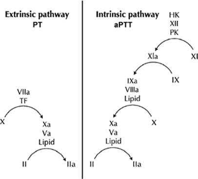
Initially, the scheme of coagulation only included what we now call the “intrinsic” pathway—so named because all of its components are present in the blood. While it had long been known that tissue extracts (thromboplastin) could trigger blood clotting, the intrinsic pathway was thought to be of primary physiologic importance because deficiency of FVIII or FIX was known to result in a severe bleeding condition (hemophilia). Later the tissue factor or “extrinsic” pathway was added to the model to produce the familiar coagulation cascade as shown in Figure 49.1.
|
|
|
Figure 49.1. The cascade model of coagulation. PT, prothrombin time; TF, tissue factor; aPTT, activated partial thromboplastin time; HK, high-molecular-weight kininogen; PK, prekallikrein. |
In the modern cascade model the intrinsic pathway is initiated by activation of the contact factors. FXIa can then activate FIX, which then acts with its cofactor, FVIII, on a phospholipid surface to activate FX. The extrinsic pathway consists of FVIIa and TF. Both pathways can activate FX, which, in complex with its cofactor FVa on a phospholipid surface, converted prothrombin to thrombin. Our current screening coagulation tests follow the outline of this model: The prothrombin time (PT) test is initiated by adding TF, calcium, and phospholipid to plasma and measuring the time required for clotting. The aPTT is initiated by adding a charged surface and phospholipid to plasma to allow activation of the contact factors (the “activation” phase). The plasma is then recalcified and the time to clotting is measured. Thus, the cascade model of coagulation is useful as a guide to interpreting the PT and aPTT.
However, the cascade model has severe limitations as a model of physiology. The key observation that the FVIIa/TF complex activated not only FX but also FIX (20) suggested that the intrinsic and extrinsic pathways were in fact linked. Several groups demonstrated points of interaction between the pathways and suggested that they could not operate as independent and redundant systems, as the cascade model implied. Furthermore, this model fails to provide insight into why patients with coagulation abnormalities bleed. For example, patients with deficiencies of the contact factors have a prolonged aPTT only and no clinical bleeding tendency. Patients with FXI deficiency have a similarly prolonged aPTT and variable bleeding. Patients with classic hemophilia A (FVIII deficiency) and hemophilia B (FIX deficiency) have a less prolonged aPTT but a significant bleeding tendency.
A Cell-based Model of Coagulation
It was recognized from the earliest studies of coagulation that cells were important participants in the coagulation process. Of course, it is clear that normal hemostasis is not possible in the absence of platelets. In addition, TF is an integral membrane protein and thus its activity is normally associated with cells. Since different cells express different levels of pro- and anticoagulant proteins as well as having different complements of receptors for components of hemostasis, simply representing the cells involved in coagulation as phospholipid vesicles overlooks the important contributions of cells in directing hemostasis in vivo. Thus, a model that gives insight into the physiology and pathophysiology of hemostasis requires due consideration to the role of living cells in the process. A cell-based model of coagulation has been proposed (21), which views the coagulation process as occurring in a series of steps that take place on two different cell surfaces: the TF-bearing cells and platelets. This model is illustrated in Figure 49.2.
Initiation
The initiation step takes place on the TF-bearing cell surface, which is exposed to blood when an injury occurs (Fig. 49.2A). Functioning as a receptor for FVII, the transmembrane protein TF promotes conversion of the FVII to its proteolytically active form, FVIIa. The FVIIa/TF complex catalyzes conversion of FIX and FX to their active forms, FIXa and FXa, respectively. On the TF-bearing cell, the FXa formed during this step combines with its cofactor FVa and forms a prothrombinase complex sufficient to generate a small amount of thrombin in the milieu of the TF-bearing cell. This small quantity of thrombin is not sufficient to clot fibrinogen. It does, however, promote platelet activation and the conversion of FV, FVIII, and FXI to their active forms in the next step.
Amplification
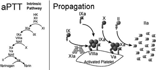
The amplification step (Fig. 49.2B) takes place on the platelet surface, where the small amount of thrombin formed during initiation can bind to specific receptors. Protease-activated receptors (PAR-1 and PAR-4) mediate platelet activation. Thrombin also binds to GPIb, which facilitates the activation of FVIII and its release from vWF, as well as activation of FXI. Platelet activation leads to secretion of partially activated FV from platelet α-granules, with thrombin acting to complete its conversion to FVa. Cofactors Va and VIIIa are quickly bound to specific sites on the platelet surface once they are activated. Factor IXa activated by the FVIIa/TF complex is capable of diffusing from the TF-bearing cell to adjacent activated platelet surfaces, where it binds to its cofactor to form FVIIIa/FIXa complexes. At the end of the amplification step, activated platelets have bound activated factors in preparation to produce large amounts of thrombin.
Propagation
During the propagation step (Fig. 49.2C), plasma FX is converted to FXa on the activated platelet surface, where it can move into direct association with its cofactor FVa while remaining surface bound. The platelet surface FXa/FVa acts on plasma prothrombin to produce a thrombin burst of sufficient magnitude to clot fibrinogen.
The role of FXI in hemostasis has been a point of some controversy, since even severe FXI deficiency does not result in a hemorrhagic tendency as severe as that seen in severe FVIII or IX deficiency. This can be explained if FXI is viewed as an “enhancer” or “booster” of thrombin generation during the propagation phase. FXIa activates additional FIXa on the platelet surface to supplement FIXa/FVIIIa complex formation and enhance platelet surface FXa and thrombin generation. Thus, FXI is not essential for platelet-surface thrombin generation, as are FIX and FVIII, and its deficiency does not compromise hemostasis to the degree seen in FIX and FVIII deficiency.
|
|
|
Figure 49.2. A cell-based model of coagulation. TFPI, tissue factor pathway inhibitor; vWF, von Willebrand factor. |
Fibrin Polymerization
Thrombin now cleaves two pairs of short polypeptides from fibrinogen, and the resulting fibrin monomer rapidly and spontaneously polymerizes. Thrombin also activates FXIII, and FXIIIa in turn catalyzes covalent linkages between fibrin monomers, vastly increasing the tensile strength of the fibrin mesh that now stabilizes the initial platelet plug.
Laboratory Evaluation
The cell-based model of coagulation shows us why the “extrinsic” and “intrinsic” pathways are not redundant. Let us consider the “extrinsic” pathway to consist of the FVIIa/TF complex working with the FXa/Va complex, and the “intrinsic” pathway to consist of FXIa working with the complexes of factors VIIIa/IXa and factors Xa/Va. As illustrated in the right hand panel of Figure 49.3, the “extrinsic” pathway operates on the TF-bearing cell to initiate the coagulation process. By contrast, as shown in Figure 49.4, the “intrinsic” pathway operates on the activated platelet surface to produce the burst of thrombin that causes formation of the fibrin clot. Thus, these two pathways are specialized to carry out functions on different cell surfaces. The PT can be considered to be a test of the adequacy of the components of the initiating pathway, while the aPTT is a test of the components needed for platelet surface thrombin generation. Each can provide useful information about the cause of abnormal bleeding, but either can be expected to give us an overall view of hemostatic function or accurately predict the risk of bleeding.
|
|
|
Figure 49.3. The prothrombin time (PT) tests the extrinsic/initiation pathway. TF, tissue factor. |
Prothrombin Time
Although the first step in identifying and classifying disorders of secondary hemostasis is always a careful history and physical examination, the focus here is on the laboratory evaluation of clotting disorders. The most commonly ordered screening test of clotting is PT. In this test, plasma is incubated with a thromboplastin reagent that contains TF and phospholipid vesicles to bind FVII from the plasma and activate the extrinsic pathway (Fig. 49.3). Acquired or congenital deficiencies of factors VII, X, V, prothrombin or fibrinogen can prolong the PT. The PT is also commonly used to monitor oral anticoagulant therapy, that is, warfarin. Any acquired or congenital deficiencies of factors VII, X, or V; thrombin; or fibrinogen can also prolong the PT.
Activated Partial Thromboplastin Time
The aPTT is another commonly measured parameter of coagulation. Plasma is incubated with a reagent that provides a negatively charged surface for activation of the contact factors of coagulation. The reagent also contains phospholipid vesicles and calcium, which are required for activity of the distal portion of the intrinsic pathway. As shown in the left-hand panel of Figure 49.4, the aPTT is sensitive to a deficiency of any of the factors in the intrinsic and common pathways.
|
|
|
Figure 49.4. The activated partial thromboplastin time (aPTT) tests the intrinsic/platelet surface pathway. HK, high-molecular-weight kininogen; PK, prekallikrein. |
Thrombin Time
The thrombin time assesses both the quantitative and qualitative aspects of the conversion of fibrinogen to fibrin. Thrombin is added to plasma, and the time until appearance of a fibrin clot is measured. The myriad disorders associated with either availability or function of fibrinogen are assessed with this test. It is exquisitely sensitive to the presence of heparin and inhibitors of fibrin monomer polymerization (i.e., fibrin degradation products, dysfibrinogens). The reptilase time also provides an assessment of fibrinogen and its function. Like thrombin, the reptilase enzyme (derived from snake venom) cleaves polypeptides from fibrinogen, leading to activation and fibrin monomer formation. Unlike the thrombin time, however, the reptilase time is not affected by heparin.
Specific Factor Assays
Factor assays are also available for most of the important coagulation proteins involved in the clotting of blood. The general concept behind these assays involves providing the specific substrate for the factor that is suspected to be deficient. Subsequent time to clotting is then measured and clotting factor activity is reported as a percentage of normal activity.
“Global” Tests of Coagulation
As the critical role of cells in coagulation has become more clear, a number of coagulation tests have become available that can be run on whole blood or platelet-rich plasma.
Because the cells do not have to be separated from the plasma for these assays and some are suitable for point-of-care testing, some of these assays also have a much quicker turnaround time than tests run in the clinical laboratory. Such assays include several variations on the venerable thromboelastogram (TEG) (22,23); the Thrombogram, which measures the pattern of thrombin generation in platelet-rich plasma (24); the Hemodyne, which measures fibrin clot strength and platelet contraction (25); the Clot Signature Analyzer, which uses non–anticoagulated blood (26); and others. One or more of these assays seem likely to play an important role in assessing hemostatic parameters that are not well reflected in the PT and aPTT. However, none of the currently available whole blood or platelet-rich plasma tests has emerged as clearly superior to its competitors.
An additional test that deserves mention is the activated clotting time (ACT). It is not considered a test of “global” hemostasis, but does use whole blood and is performed as point-of-care testing (27). It is similar to a whole blood aPTT because clotting is initiated with charged materials that activate the contact pathway, such as celite, kaolin, or a combination of celite, kaolin, and glass. The ACT is commonly used for monitoring blood anticoagulant response to heparin during cardiopulmonary bypass and cardiac catheterization. However, some other anticoagulants and other agents, such as aprotinin, can affect the ACT. Furthermore, the test does not reflect the anticoagulant effect of some agents, such as low-molecular-weight heparins. Since the test has not been standardized, different reagent/instrument systems may give different results under the same clinical conditions. In spite of these caveats, the ACT has proven to be useful in the cardiac cath lab and during cardiac surgery when a single system is used consistently and local “cutoff” levels have been determined for assessing adequate anticoagulation and adequate reversal by protamine.
Physiology and Biochemistry of Fibrinolysis
Components
The major enzyme component of the fibrinolytic system is plasminogen, the zymogen form of plasmin. It is primarily synthesized in the liver and circulates in the plasma at 10- to 20-mg/dL concentrations under basal conditions. Plasminogen concentrations, however, can greatly increase in response to inflammatory states, paralleling changes seen in fibrinogen levels. Plasmin is the final effector of the fibrinolytic system, which degrades the fibrin clot. Plasmin cleaves fibrin (and also fibrinogen) at multiple sites, resulting in several by-products, known clinically as fibrin(ogen) degradation products (FDPs). After polymerization of fibrin, factor XIIIa crosslinks between the D-domains of adjacent fibrin monomers. When plasmin digests crosslinked fibrin, a product is produced that is unique to fibrin cleavage, called the d-dimer. This can be measured and quantitated (discussed later).
Plasmin is produced by the action of two plasminogen activators (PAs): Urokinase-type plasminogen activator (uPA) and tissue-type plasminogen activator (tPA). Although tPA is present in many tissues, its release by vascular endothelial cells is key in the activation of fibrinolysis. Several drugs, including vasopressin, 1-desamino-8-D-arginine vasopressin (DDAVP), and epinephrine can enhance vessel wall production of tPA. Pathophysiologic stimuli such as tissue anoxia or endothelial trauma also result in enhanced synthesis and secretion of tPA. Plasminogen is found at a much higher plasma concentration than the PAs. Therefore, the availability of the two PAs generally determines the extent of plasmin formation. The activity of plasmin is regulated by a plasma serine protease inhibitor: Antiplasmin. The PAs are regulated by circulating plasminogen activator inhibitors (PAIs), primarily PAI-1.
Function in Normal Coagulation
Even as the fibrin clot is being formed in the body, the fibrinolytic system is being initiated to disrupt it. If a clot is formed in the vicinity of an intact endothelial layer, fibrinolysis is activated as a protective mechanism. tPA release from endothelial cells is provoked by thrombin as well as venous occlusion (28). Both the secreted tPA and plasma plasminogen bind to the evolving fibrin polymer, which dramatically enhances the activation of plasminogen by tPA. Plasmin is formed in the vicinity of the thrombus and acts to degrade it.
When a hemostatic clot is formed at a site of tissue injury, fibrinolysis must still take place to remove the clot during wound healing. PAs released from leukocytes, probably primarily uPA, begin the fibrinolytic process in this case.
Thrombin activatable fibrinolysis inhibitor (TAFI) is a zymogen that can be activated (TAFIa) by thrombin or plasmin (29). As fibrin is degraded by plasmin, C-terminal lysines are exposed that enhance activation of additional plasminogen to plasmin. TAFIa removes the C-terminal lysines from fibrin and thereby inhibits the cofactor activity of fibrin for plasminogen activation.
Fibrinolysis is essential for removal of clots during the process of wound healing as well as for removing intravascular clots that might otherwise be manifest as thrombosis. Intravascular deposition of fibrin is also associated with the development of atherosclerosis. Therefore, an effective fibrinolytic system tends to protect against the chronic process of atherosclerotic vascular disease as well as the acute process of thrombosis. Conversely, defects of fibrinolysis increase the risk of atherothrombotic disease. For example, elevated levels of PAI-1, an inhibitor of fibrinolysis, are associated with an increased risk of atherosclerosis and thrombosis (30), as are decreased levels of plasminogen (31).
Streptokinase and urokinase have been used clinically as exogenous activators of the fibrinolytic system to lyse occlusive thrombi that cause myocardial infarction and stroke. Streptokinase is a biologic product of certain Lancefield strains of β-hemolytic streptococci and is antigenic in nature. Its mechanism of plasminogen activation seems related to a nonenzymatic, nonproteolytic reaction through an alteration of the plasminogen active site that occurs on binding of the two molecules (streptokinase and plasminogen). The resultant complex directly cleaves a second molecule of plasminogen to its active form, plasmin. Urokinase was initially isolated from urine and fetal kidney cells but is now produced using recombinant DNA technology. In contrast to streptokinase, urokinase activates plasminogen through direct proteolytic action and is not antigenic. Recombinant human tPA (rh-tPA) also can be infused intravenously to activate fibrinolysis. Exogenously administered rh-tPA has identical actions as endogenously produced tPA.
After activation by any of the above described methods, excess fibrinolysis has the potential to interfere with normal hemostatic mechanisms. Since tPA normally activates plasminogen in the vicinity of fibrin, fibrinolytic activity is confined to the site of clot formation. However, plasmin can digest both fibrinogen and fibrin. When excess plasmin activity is produced or it is not inhibited appropriately, plasmin can degrade and deplete circulating fibrinogen. Thus, therapeutic activation of fibrinolysis produces a systemic lytic state with dose-dependent depletion of plasma fibrinogen.
Laboratory Evaluation
Tests that directly measure fibrinolytic function or activity are not as readily available as those that measure secondary hemostasis. In general, the PT, aPTT, and thrombin time are relatively insensitive to changes in fibrinolytic activity (including those induced by streptokinase or urokinase). Therefore, these tests are not useful in the diagnosis of increased fibrinolysis. An assay of plasma fibrinogen and fibrin degradation product levels provides some useful information concerning fibrin(ogen)olysis, if basal levels are known. These assays are not the tests of choice for either diagnosing or monitoring hyperfibrinolysis.
The test that has historically been most used to evaluate the possibility of hyperfibrinolysis is the euglobulin clot lysis time. However, it is not quantitative and is at best only a gross indicator of increased fibrinolytic activity. The test involves the precipitation of fibrinogen, plasminogen, and plasminogen activators by mixing a plasma sample with an acidic solution of low ionic strength. These precipitated proteins are then redissolved and allowed to clot, and the time to clot lysis is measured. A shortened time to clot lysis (less than 3 hours) indicates a state of increased fibrinolysis. Unfortunately, the degree of shortening of the euglobulin clot lysis time does not correlate with the extent of increased fibrinolysis. Further, difficulties in test interpretation can occur if the level of fibrinogen is low or if amounts of fibrin degradation products sufficient to impede clot formation are present. Because this test is very labor intensive and its interpretation can be problematic, it is now rarely performed.
The one assay that is commonly performed to monitor an aspect of fibrinolysis is the D-dimer assay. D-dimer is a specific proteolysis product of the action of plasmin on crosslinked fibrin. The presence of increased amounts of D-dimer in plasma or serum indicates that thrombin has been generated, fibrin has formed, and some degree of fibrinolysis has occurred. The D-dimer level is increased any time the coagulation response has been activated—whether appropriate or inappropriate. Thus, D-dimer levels are elevated following surgery or trauma, as well as in the setting of intravascular coagulation (thrombosis) and disseminated intravascular coagulation (DIC). This test is theoretically negative in instances of primary fibrinolysis, where fibrinogen—not crosslinked fibrin—is being degraded. The D-dimer assay can be extremely useful as part of an algorithm for the evaluation of pulmonary embolism—primarily when a normal D-dimer level can rule out thrombosis/embolism.
Coagulation Function in the Tissue Environment
The compartmentalization of the various aspects of coagulation, as presented here, is intended to ease the burden of conceptualization for the reader. In vivo coagulation, however, occurs in a tissue environment. While we have discussed, to some extent, the important role of the vessel wall and vascular endothelial cells in controlling the coagulation process, there are additional local attributes that can have a very significant impact on the effectiveness of hemostasis. First and foremost, hemostasis is unlikely to be effective when a very large vessel has been transected and primary hemostasis is unable to stop or slow the hemorrhage. This type of problem requires a surgical rather than a biochemical approach. In addition, two of the most important variables that can impair the effectiveness of the coagulation process are low temperature and pH.
Hypothermia
Hypothermia is defined as a body temperature of less than 35°C. Hypothermia occurs in a number of clinical settings, including both deliberate and accidental circumstances. In some instances, such as during cardiopulmonary bypass surgery, hypothermia is desired for its neuroprotective effects. However, hypothermia that accompanies severe trauma is associated with a significantly worse prognosis than either trauma or hypothermia alone. The primary risk of hypothermia in both regulated and nonregulated environments is that of abnormal bleeding. Mild hypothermia increases transfusion requirements, even during elective surgery (32). Several mechanisms have been proposed to contribute to impaired hemostasis associated with hypothermia, including reduced activity of clotting factors and platelets, activation of fibrinolysis, and endothelial injury (33). It appears that the defect in hemostasis observed in mild hypothermia (37°C–33°C) results primarily from impaired platelet adhesion/aggregation, but that reduced coagulant enzyme activity becomes a compounding factor of profound hypothermia (34). Of course, local hypothermia will not be reflected in the results of laboratory coagulation testing. Therefore, it is important for the clinician to consider the possibility that hypothermia may be contributing to bleeding in certain patients.
Metabolic Derangements
Like hypothermia, acidosis is also associated with worse survival in trauma and surgery patients. It can result from metabolic derangements that develop in the sickest patients. Excess lactic acid production associated with tissue hypoxia is the best recognized cause. Lactic acid is the end product of anaerobic metabolism and its level is related to oxygen availability. Acidosis can impair coagulation and worsen the risk of serious hemorrhage (35). Massive transfusion can exacerbate acidosis, since stored blood has a reduced pH.
Small changes in pH have a much greater impact on the effectiveness of the coagulation system than do small changes in temperature. Most of the coagulation proteases have a structure analogous to trypsin, a digestive enzyme. Trypsin has a pH optimum in the alkaline range (8.0–8.5), which suits it well to functioning in the alkaline environment of the small intestine. The coagulation proteases similarly have a pH optimum in the alkaline range, which does not equip them to work well in acidotic tissues. A drop in pH from 7.4 to 7.0 reduces the activity of the enzyme complex that activates thrombin by more than 70% (36). Acidosis is also not reflected in the results of laboratory coagulation testing and the clinician must be aware of the profound effect that modest changes in the pH can have on hemostasis.
Summary
The process of hemostasis is intricately intertwined with other aspects of the host response to injury. Laboratory testing is critical to the evaluation of a patient with a disorder of coagulation. However, laboratory testing must be coupled with a careful clinical evaluation and interpretation, including recognition of other conditions such as hypothermia and acidosis that can impact hemostasis.
References
1. Rosing J, van Rijn JL, Bevers EM, et al. The role of activated human platelets in prothrombin and factor X activation. Blood. 1985;65:319.
2. FitzGerald GA. Mechanisms of platelet activation: thromboxane A2 as an amplifying signal for other agonists. Am J Cardiol. 1991;68:11B.
3. Canobbio I, Balduini C, Torti M. Signaling through the platelet glycoprotein Ib-V-IX complex. Cell Signal. 2004;16:1329.
4. Moroi M, Jung SM. Platelet glycoprotein VI: its structure and function. Thromb Res. 2004;114:221.
5. Bennett JS. Structure and function of the platelet integrin alphaIIbbeta3. J Clin Invest. 2005;115:3363.
6. Marcus A. Platelets and their disorders. In: Ratnoff O, Forbes C, eds. Disorders of Hemostasis. Philadelphia: WB Saunders; 1996:79.
7. Kottke-Marchant K, Corcoran G. The laboratory diagnosis of platelet disorders. Arch Pathol Lab Med. 2002;126:133.
8. Sramek R, Sramek A, Koster T, et al. A randomized and blinded comparison of three bleeding time techniques: the Ivy method, and the Simplate II method in two directions. Thromb Haemost. 1992;67:514.
9. Peterson P, Hayes TE, Arkin CF, et al. The preoperative bleeding time test lacks clinical benefit: College of American Pathologists' and American Society of Clinical Pathologists' position article. Arch Surg. 1998;133:134.
10. Mammen EF, Comp PC, Gosselin R, et al. PFA-100 system: a new method for assessment of platelet dysfunction. Semin Thromb Hemost. 1998;24:195.
11. Wheeler GL, Braden GA, Steinhubl SR, et al. The Ultegra rapid platelet-function assay: comparison to standard platelet function assays in patients undergoing percutaneous coronary intervention with abciximab therapy. Am Heart J. 2002;143:602.
12. Malinin A, Pokov A, Spergling M, et al. Monitoring platelet inhibition after clopidogrel with the VerifyNow-P2Y12(R) rapid analyzer: the VERIfy Thrombosis risk ASsessment (VERITAS) study. Thromb Res. 2006;119:277.
13. White MM, Krishnan R, Kueter TJ, et al. The use of the point of care Helena ICHOR/Plateletworks and the Accumetrics Ultegra RPFA for assessment of platelet function with GPIIB-IIIa antagonists. J Thromb Thrombolysis. 2004;18:163.
14. Roberts H, Monroe D, Hoffman M. Molecular biology and biochemistry of the coagulation factors, and pathways of blood coagulation. In: Beutler E, Lichtman M, Coller B, et al., eds. William's Hematology. New York: McGraw-Hill Publishing; 2000.
15. Li T, Chang CY, Jin DY, et al. Identification of the gene for vitamin K epoxide reductase. Nature. 2004;427:541.
16. Han X, Fiehler R, Broze GJ Jr. Isolation of a protein Z-dependent plasma protease inhibitor. Proc Natl Acad Sci U S A. 1998;95:9250.
17. Yin ZF, Huang ZF, Cui J, et al. Prothrombotic phenotype of protein Z deficiency. Proc Natl Acad Sci U S A. 2000;97:6734.
18. Macfarlane RG. An enzyme cascade in the blood clotting mechanism, and its function as a biological amplifier. Nature. 1964;202:498.
19. Davie EW, Ratnoff OD. Waterfall sequence for intrinsic blood clotting. Science. 1964;145:1310.
20. Østerud B, Rapaport SI. Activation of factor IX by the reaction product of tissue factor and factor VII: additional pathway for initiating blood coagulation. Proc Natl Acad Sci U S A. 1977;74:5260.
21. Hoffman M, Monroe DM 3rd. A cell-based model of hemostasis. Thromb Haemost. 2001;85:958.
22. De Nicola P, Mazzetti GM. How to interpret a thromboelastogram. Minerva Med. 1956;47:2043.
23. Robert Valeri C, Ragno G. In vitro testing of platelets using the thromboelastogram, platelet function analyzer, and the clot signature analyzer to predict the bleeding time. Transfus Apher Sci. 2006;35:33.
24. Al Dieri R, Peyvandi F, Santagostino E, et al. The thrombogram in rare inherited coagulation disorders: its relation to clinical bleeding. Thromb Haemost. 2002;88:576.
25. Reid TJ, Snider R, Hartman K, et al. A method for the quantitative assessment of platelet-induced clot retraction and clot strength in fresh and stored platelets. Vox Sang. 1998;75:270.
26. Fricke W, Kouides P, Kessler C, et al. A multicenter clinical evaluation of the Clot Signature Analyzer. J Thromb Haemost. 2004;2:763.
27. Doherty TM, Shavelle RM, French WJ. Reproducibility and variability of activated clotting time measurements in the cardiac catheterization laboratory. Catheter Cardiovasc Interv. 2005;65:330.
28. Szymanski LM, Pate RR, Durstine JL. Effects of maximal exercise and venous occlusion on fibrinolytic activity in physically active and inactive men. J Appl Physiol. 1994;77:2305.
29. Bajzar L, Manuel R, Nesheim ME. Purification and characterization of TAFI, a thrombin-activable fibrinolysis inhibitor. J Biol Chem. 1995;270:14477.
30. Huber K, Christ G, Wojta J, et al. Plasminogen activator inhibitor type-1 in cardiovascular disease. Status report 2001. Thromb Res. 2001;103(Suppl 1):S7.
31. Xiao Q, Danton MJ, Witte DP, et al. Plasminogen deficiency accelerates vessel wall disease in mice predisposed to atherosclerosis. Proc Natl Acad Sci U S A. 1997;94:10335.
32. Schmied H, Kurz A, Sessler DI, et al. Mild hypothermia increases blood loss and transfusion requirements during total hip arthroplasty. Lancet. 1996;347:289.
33. Kirkpatrick AW, Chun R, Brown R, et al. Hypothermia and the trauma patient. Can J Surg. 1999;42:333.
34. Wolberg AS, Meng ZH, Monroe DM 3rd, et al. A systematic evaluation of the effect of temperature on coagulation enzyme activity and platelet function. J Trauma. 2004;56:1221.
35. Cosgriff N, Moore EE, Sauaia A, et al. Predicting life-threatening coagulopathy in the massively transfused trauma patient: hypothermia and acidoses revisited. J Trauma. 1997;42:857.
36. Meng ZH, Wolberg AS, Monroe DM 3rd, et al. The effect of temperature and pH on the activity of factor VIIa: implications for the efficacy of high-dose factor VIIa in hypothermic and acidotic patients. J Trauma. 2003;55:886.