Jamie Taylor
Clifford S. Deutschman
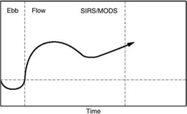
In 1794 John Hunter wrote, “There is a circumstance attending accidental injury which does not belong to disease—namely, that the injury has in all cases a tendency to produce both the disposition and the means of a cure.” This first described the stress response, a biphasic physiologic response that, when uninterrupted by complications, has predictable characteristics and lasts 7 to 10 days (Fig. 53.1). When not altered by intervention or complications, the stress response is initiated by a global depression of energy expenditure and metabolism. This 24-hour phase that occurs immediately following injury is followed by a period of hypermetabolism that characteristically persists for 5 to 7 days. The driving force for this second phase appears to lie with the need to mount an immune or inflammatory response to combat infection and facilitate repair of damaged tissues. Most markers for ongoing inflammation and metabolism peak on postinjury day 2 and return to baseline around day 7. Although the intensity of the response may change or the time course may be altered by intervention, these events must take place or the organism will not survive.
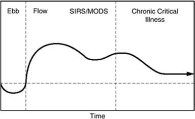
It is possible for the normal stress response to be altered by coexisting disease or interrupted by adverse events such as recurrent bleeding, systemic inflammatory response syndrome (SIRS), or progression of SIRS into sepsis. The organism then may enter a state of persistent hypermetabolism. This continued inflammation has been postulated to lead to organ dysfunction and immune incompetence (Fig. 53.2). The onset of immune incompetence marks a transition from a hyperfunctional to a hypofunctional state in which patients are at higher risks for nosocomial infections and demonstrate a pervasive endocrinopathy (Fig. 53.3). It is from this state that most intensive care unit (ICU) deaths occur (1).
|
|
|
Figure 53.1. The stress response. (Reprinted with permission from Kohl BA, Deutschman CS. The inflammatory response to surgery and trauma. Curr Opin Crit Care. 2006;12(4):325–332.) |
The Normal Stress Response
David P. Cuthbertson was perhaps the first to study the host response to injury. In 1929, while working at Glasgow University, Cuthbertson was charged with the duty of investigating why fractures of the distal femur were slow to heal. He discovered that if prolonged immobilization occurred postinjury, the urinary excretion of sulphur, nitrogen, phosphorus, and calcium was elevated. As an aside to these studies, Cuthbertson noted that body temperature followed a characteristic pattern (Fig. 53.1). In the first 24 hours following the fracture, temperature decreased. Following this period, temperature rose, peaking on postinjury day 3 and returning to baseline by postinjury day 7. He correlated this change in temperature with alterations in oxygen consumption and carbon dioxide production. Further, he noted that the sulphur: nitrogen ratio closely matched that of muscle. This led to Cuthbertson's proposal of a paradigm by which the body responded to injury. If the damage to the patient is not immediately fatal, there is a compensatory reaction in which vasoconstriction shunts blood away from the periphery and to the central organs, most notably the heart and brain. This promotes short-term survival. Hypothermia and oliguria are associated with a global decrease in oxygen consumption and energy expenditure. In an effort to expand plasma volume and avoid failed oxygen delivery, the body conserves salt and water by increasing aldosterone secretion (2). These effects are seen throughout the body in the first 24 hours after injury. Cuthbertson termed this sequence of events the “ebb” phase or traumatic shock. When it becomes clear that death is not imminent, a second aspect of the response emerges. The key to these reactions is an attempt to repair tissue damage, a process that is accomplished via the activity of white blood cells (WBCs). Slowed circulation in the ebb phase allows WBCs to move toward the periphery and adhere to the endothelium. Neutrophils react first, with macrophages following. With restoration of the circulation, the process becomes active. It is characterized by phagocytosis and lysis of bacterial, viral, or fungal invaders and removal of cellular debris. In addition, macrophages, lymphocytes, and antigen-presenting cells (APCs) secrete proteins called cytokines. To a great extent, these are growth factors that facilitate repair of damaged tissue. This process requires enormous amounts of energy, with a 2- to 20-fold increase in oxygen consumption and resting energy expenditure (REE). Body temperature rises and oxygen consumption and carbon dioxide production increase (2,3). Monk et al. showed an increase in REE of up to 55% above predicted in trauma patients (4). Some studies suggest that survival is dependent on this ability to maintain hypermetabolism and adequate oxygen utilization (3,5,6,7,8). Because WBCs are more or less obligate glucose users, there is an associated increase in glucose requirements (9). After the first 24 hours, hepatic glycogen stores are depleted and a source of de novo glucose is required. This is generated by hepatic gluconeogenesis. Cuthbertson proposed that the body was able to provide its own source of nutrients by breaking down protein, a theory that has since been validated. This provides substrate for gluconeogenesis and constituent amino acids for synthesis of hepatic proteins and repair in the area of injury. However, adequate mobilization is not enough to ensure substrate delivery. Therefore, the response includes capillary dilatation to increase flow and improve delivery. Unfortunately, due to thrombosis in damaged tissue, most injured areas are avascular. To allow substrate delivery to these regions, capillary tight junctions separate, allowing fluid and substrate to “leak” from the vasculature. Increased vascular permeability results in redistribution of extracellular fluid and plasma proteins to form edema and exudate (10). Glucose and other nutrients move down their concentration gradients across the extracellular matrix to areas of damage. Removal of waste requires an increase in renal blood flow and glomerular filtration to enable excretion of amino acid degradation products. The liver detoxifies nitrogenous wastes by the production of urea; metabolizes alanine, lactate, and glycerol through gluconeogenesis; and produces acute-phase proteins that bind metabolic by-products and limit the activity of proteolytic enzymes secreted by activated WBCs (1). Because this process increases delivery to nearly every part of the body, Cuthbertson termed this part of the response the “flow” phase.
|
|
|
Figure 53.2. Continued inflammation, which has been postulated to lead to organ dysfunction and immune incompetence. SIRS, systemic inflammatory response syndrome; MODS, multiple organ dysfunction syndrome. |
|
|
|
Figure 53.3. The onset of immune incompetence marks a transition from a hyperfunctional to a hypofunctional state in which patients are at higher risks for nosocomial infections and demonstrate a pervasive endocrinopathy. SIRS, systemic inflammatory response syndrome; MODS, multiple organ dysfunction syndrome. |
Work by Cuthbertson and Francis C. Moore demonstrated that initiation of the flow phase is in part hormonally modulated. An initial dramatic release of endogenous catecholamines (11) is supplemented by alterations in the somatotropic system (growth hormone and insulinlike growth factor) such that anabolism is postponed and energy substrates are redirected to vital organs. Both the thyroid and the gonadal axes are suppressed. Adrenocorticotropin hormone (ACTH) secretion is heightened by increased corticotropin-releasing hormone (CRH), arginine vasopressin (AVP), catecholamines, angiotensin II, serotonin, and some inflammatory cytokines (interleukin [IL]-1, IL-2, IL-6, and tumor necrosis factor [TNF]). ACTH stimulates the adrenal glands to produce glucocorticoids and mineralocorticoids. Glucocorticoids and glucagon promote glucogenesis and glycogenolysis and induce peripheral insulin resistance, leading to increased glucose production (12). This in turn increases insulin secretion, producing an “insulin-resistant” state.
Tissue repair is initiated by these activities. Traumatized tissue and hemorrhage initiate platelet accumulation and activation. The coagulation cascade is triggered by both the intrinsic and extrinsic pathways. This serves to sustain and enhance immune cell migration and activation. In addition, fibroblasts at the edge of the wound divide, migrate toward the center, and produce collagen. Surviving capillaries bud, and these new capillaries also migrate toward the center. Eventually the wound edges will fuse and consist of vascularized granulation tissue (13). This process is thought to be mediated by an increase in fibroblast growth factors, epidermal growth factor, platelet-derived growth factor, and vascular endothelial growth factor (14).
This massive mobilization of defense mechanisms may also affect normal tissue. Therefore, one of the most important characteristics of the normal stress response is that it is a balance between the inflammatory and the anti-inflammatory systems. This involves the proinflammatory cytokine TNF, released by the activated macrophage. When TNF “spills over” from the interstitium into the bloodstream, it stimulates the medullary reticular formation and the hypothalamus in the brain. This activation of the hypothalamic-pituitary-adrenal axis ultimately causes increased anti-inflammatory activity. For example, glucocorticoids released as a result of this process limit the negative biologic consequences caused by inflammation (15). TNF also stimulates the dorsal vagal complex and alters the efferent vagal output. This is in part responsible for “sickness” behavior (i.e., anorexia and fever) (16,17,18). More importantly, however, is neuromodulation of the immune response. The “inflammatory reflex” or the cholinergic anti-inflammatory pathway occurs when proinflammatory cytokines such as IL-1 and TNF stimulate the parasympathetic nervous system through receptors on the vagus nerve. The afferent input travels to the nucleus solitarius and is relayed to the dorsal motor nucleus, resulting in an increase in acetylcholine release at cholinergic nerve terminals in the areas of inflammation. Activated macrophages have acetylcholine receptors that, when stimulated, decrease the release of proinflammatory cytokines (19,20). This balance of the systems is crucial in limiting the stress response.
Moore et al. also observed changes in the size of body fluid compartments. In a normal stress response, catabolism is accompanied by an increase in the vascular space (recruitment); expansion of the extravascular, extracellular space; and a decrease in the intracellular compartment. This process ends 4 to 5 days after injury with a shift to anabolism (11). The vasculature contracts, fluid is removed from the extracellular space and either moves back into cells or is excreted by the kidneys, and the intracellular shift is accompanied by an influx of protein and electrolytes. While the physiologic “signal” that initiates this transition is still unknown, it is telling that the transition occurs at the completion of the first wave of angiogenesis. The generation of a new vascular highway obviates the need for nutrient concentration gradients, increased vascular permeability, and water and electrolyte conservation. Conservation of salt and water is no longer a priority and a brisk diuresis results. In addition, as the intracellular space expands, increases in intracellular anions and cations are required. These must come from the vascular space. Therefore, resolution of catabolism is accompanied by decreases in serum levels of potassium, magnesium, and phosphate ions and, therefore, diuresis and decreases in serum electrolytes are the hallmarks of resolution of the stress response.
The cellular immune response to normal inflammatory stimuli involves neutrophils, monocytes (macrophages), lymphocytes, and APCs. Neutrophils are recruited to areas of injury early in the process. This is stimulated in part by protein antigens and chemoattractant molecules (chemokines) released by endothelial cells and fixed tissue macrophages. Their function is removal of cellular debris by phagocytosis and secretion of lytic molecules such as digestive enzymes and free radicals. While the influx of neutrophils is self-limited, lasting about 48 hours, the rest of the response, which starts within hours of neutrophil influx, may be more persistent. It consists of infiltration by macrophages, APCs, and lymphocytes. Macrophages are of key importance in innate immunity, a process that is nonspecific and involves natural barriers such as skin, natural killer cells, and chemicals in the blood that act immediately upon antigen introduction. Macrophages respond to stimulation with phagocytosis of foreign or damaged material and secretion of cytokines that stimulate inflammation and also function as growth factors. In addition, they contribute to adaptive immunity by presenting antigens, a function that also is served by APCs. Antigen-presenting cells such as dendritic cells capture antigens, transport them to lymph nodes, and present them to T cells, initiating cell-mediated immunity. Follicular dendritic cells have a similar function except that they present antigens to B cells and therefore initiate humoral immunity. Lymphocytes are the prime components of the adaptive immune response. They have specific receptors for antigens. B cells produce antibodies and are mediators of humoral immunity. T cells recognize peptide fragments of protein antigens bound to APCs and are involved in cell-mediated immunity. T cells can be further divided into CD4 cells, which enhance or inhibit the immune response; CD8 cells, which lyse other cells with intracellular pathogens; and natural killer (NK) cells, which do not express antigen receptors and contribute to innate immunity.
Adaptive immunity is antigen specific and can be divided into five phases. The first phase is presentation of the antigen to a B or T cell by an APC. In the second phase, B and T cells are activated, undergoing clonal expansion, differentiation, and antibody production. Antigens are eliminated in the third or effector phase. Decline is the fourth phase: The stimulus has been removed and there is apoptosis of immune cells and phagocytosis of cellular debris. The last phase involves the surviving immune cells acquiring memory (21). As the process proceeds, there is a change in the phenotype of CD4 T cells that is profoundly important. The catabolic phase of inflammation is characterized by an abundance of CD4 cells of the type 1 helper T cell (Th1) phenotype. This results in secretion of proinflammatory cytokines such as IL-2, TNF-α, and interferon-γ. The switch to anabolism is accompanied by a predominance of type 2 helper T cells (Th2), which secrete anti-inflammatory cytokines such as IL-4 and IL-10. In a normal stress response, immune function declines and the transition from Th1 to Th2 occurs by the fourth or fifth day. The switch from Th1 to Th2 may be hormonally mediated. It is known that cortisol and androgens, which are secreted in great quantities in catabolism, stimulate Th2 cell production.
One clinical manifestation of this is in the blood. That is, early in the response there is a mixed leukocytosis with neutrophil predominance. This gives way to a macrophage/lymphocyte-rich pattern and is followed by an overall decline in the white blood cell count.
Deviation from the Normal Stress Response
The stress response is considered to be adaptive and vital in order to survive an injury. However, many aspects of the process may become excessive or unbalanced. This converts an adaptive response into a pathologic one. Risk factors that predispose to the development of an abnormal response include inadequate or delayed resuscitation, persistent inflammatory or infectious sources, baseline organ dysfunction, age older than 65 years, immunosuppression, alcohol abuse, malnutrition, and invasive instrumentation (22). There are two such common occurrences in the surgical or trauma population. The first is hemorrhage, uncorrected fluid loss, or underresuscitation. These result in a recurrence of shock, with vasoconstriction, decreased perfusion and cardiac output, and impaired tissue substrate delivery. The treatment is identification and treatment of the underlying cause accompanied by correction of the fluid imbalance.
The second common abnormality is prolonged hypermetabolism. Persistence of a hyperdynamic circulation and secretion of immunologic markers; elevations of serum potassium, magnesium, and phosphate; and/or marginal urine output indicate a prolonged or renewed stress response and warn of a new or recurrent abnormality. This state commonly is referred to as systemic inflammatory response syndrome. If SIRS is suspected to be from an infectious cause, then the condition is referred to as sepsis or the sepsis syndrome. Initially, the normal stress response, SIRS, and sepsis may mimic each other. Differentiation lies in the time course and in the etiology of the metabolic perturbations. The definitions of SIRS, sepsis, and severe sepsis, as formulated in 2001, are detailed in Table 53.1.
Sepsis is the most common cause of death in noncardiac intensive care units. The increasing use of broad-spectrum antibiotics, immunosuppression therapy, and invasive technology may be responsible (23). Recent studies estimate the incidence of sepsis in the United States as 240 to 300 cases of severe sepsis per 100,000 people, an increase from 74 cases per 100,000 people in 1979. The mortality rate ranges from 17.9% for sepsis, 28.6% for severe sepsis, and up to 50% in those with septic shock and comorbid conditions. The etiology of sepsis has also changed over time. In the 1970s and 1980s, Gram-negative bacteria were the predominant cause of sepsis. Gram-positive organisms are now the leading pathogens, with fungal organisms on the rise (24).
|
Table 53.1 Diagnostic criteria for sepsis in adults |
|||||||
|
Sepsis likely predisposes to organ dysfunction. When this dysfunction is overt, the process is referred to as multiple organ dysfunction syndrome (MODS). However, abnormalities in MODS seem to be confined to cellular and organ dysfunction as histology and infrastructure are preserved. The most proximal defect that has been identified to date in SIRS/sepsis/MODS is an abnormality of oxygen utilization at the subcellular level. Two theories to explain this have been advanced. The first is that there is an impairment of microcirculatory autoregulation. Vasodilation of some vascular beds coexists with vasoconstriction of others, causing a maldistribution of flow and, therefore, oxygen (25). The second theory assumes adequate perfusion but an alteration in cellular metabolism with an inability to extract and use oxygen (26). This is supported by recent studies demonstrating a defect in mitochondrial function (27,28,29). In either case, the result is a block in cellular metabolism. As a result, the ability of cells and organs to respond to external stimuli may be lost. For example, there is a progressive loss of hormonal responsiveness. The liver becomes unresponsive to insulin and glucagons and the cardiovascular system to catecholamines (30,31,32). As such, the hyperfunctional state cannot be maintained. It has been proposed that cells enter a “preservation mode” in which viability is maintained but the capacity to communicate with each other is lost. Cellular interaction is lost and thus organ function is compromised (33,34,35). Acute lung injury (ALI) progresses to acute respiratory distress syndrome (ARDS) and hypotension from vasoplegia is compounded by cardiac dysfunction that requires vasopressors and inotropic and chronotropic support. Renal function decreases to a point that renal replacement therapy must be considered and hepatic dysfunction results in severe ascites and coagulopathy, both having the potential to lead to a profound encephalopathy. The immune system is one of the most important systems to be affected. The development of immune incompetence coupled with a pervasive endocrinopathy places the patient at a higher risk for nosocomial infections, and it is in this state that most deaths from sepsis occur.
The issue of immune incompetence requires further discussion. Historically, sepsis has been viewed as a condition ruled by uncontrolled inflammation. However, an increasing number of studies indicate that sepsis is in fact a state of inflammatory failure (36,37,38,39). More specifically, sepsis is associated with an alteration in the adaptive immune response (40). The early phase of sepsis resembles normal stress in that there is a hormonal milieu that stimulates Th2 responses and these responses are observed. Indeed, studies have shown that patients with sepsis have increased Th2 cells and IL-10 and that these levels predict mortality (41,42). However, as sepsis progresses, there is a profound endocrinopathy and progressive anergy (12). That is, chronic critical illness is associated with a loss of T-cell responsiveness on any level (43). This may reflect enhanced lymphocyte apoptosis (44,45,46). Hotchkiss et al. also demonstrated that there were decreased levels of follicular dendritic cells, B cells, and CD4 T cells at the time of death of septic patients, resulting in impaired antigen presentation, antibody production, and B-cell and macrophage stimulation (44,45,46). The ultimate result of immunosuppression is the development of sequential infections, often invoking the decision to withdraw therapy.
One major contributor to the development of complications from inflammation is the presence of comorbidities. Chronic comorbid conditions are present in over 50% of patients with sepsis and are associated with an increase in mortality (47,48,49). Diseases reported to increase the risk of the normal stress response developing into sepsis are diabetes mellitus (DM), human immunodeficiency virus (HIV), chronic liver disease, and cancer (47). Esper et al. conducted a historical cohort study that reviewed patients with the diagnosis of sepsis in U.S. acute care hospitals from 1979 to 2003, characterizing the type and source of infections and comorbid diseases. They found that men were more likely than women and African Americans were more likely than Caucasians to develop sepsis. Non-Caucasian patients who were septic were more likely to have concomitant DM, HIV, chronic renal failure, and alcohol abuse. Caucasians had higher incidences of cancer and chronic obstructive pulmonary disease (COPD). The presence of one comorbidity increased the risk of developing at least one organ system failure by 30%. Those with two comorbidities had a 39% chance and those with three or more had a 45% chance of developing acute organ failure (50).
It is not difficult to imagine how baseline insufficiencies affect the stress response. For example, the ability to maintain a circulatory system capable of providing oxygen and nutrients to areas of injury is paramount to survival. In the setting of underlying coronary artery disease (CAD), this ability may be impaired. Kern et al. (51) found that patients with CAD have a significantly decreased cardiac index and oxygen delivery and, not surprisingly, an increased oxygen extraction ratio during sepsis. They also showed that these patients had increased endothelial adhesion molecule expression, which may correlate with the severity of sepsis, shock, and organ failure and predict poor outcome (52,53). Chronic pulmonary disease, regardless of the etiology, increases the chance of intubation and the requirement for prolonged ventilatory support. Intubation places the patient at risk for ventilator-associated pneumonia, aspiration, and respiratory muscle atrophy. A patient with chronic renal or liver failure is at risk for anemia, coagulopathies, and immunosuppression prior to being injured. With an impaired functional reserve in vital organs and responses, the stress response to injury has a high likelihood of progressing to a state of prolonged critical illness.
Treatment or Prevention
It is logical that treatment of pre-existing disorders and comorbidities will alter the stress response. Indeed, a number of studies have examined the role of perioperative β-blockade and concluded that, in appropriate patients, outcome is improved. More problematic are attempts to alter the course of the prolonged state that constitutes SIRS, sepsis, MODS, and chronic critical illness. Despite promising animal data, most approaches have failed in patients. The successes are notable. Herndon et al. showed that in the pediatric burn population, resting energy expenditure decreased and net muscle protein balance increased with administration of propranolol. However, there have been several experiments conducted with mice in septic or hemorrhagic shock showing an increase in mortality from immunosuppression after β-blockade (54,55).
Adequate analgesia via epidural and intravenous use of agents such as opiates, α-blockers, nonsteroidal anti-inflammatory drugs, and local anesthetics have been shown to both decrease inflammation and improve immune function (56,57,58,59). Early goal-directed resuscitation has been shown to improve outcome in a single-center trial (60). A multicenter trial in the United States is beginning. Similarly, a protocol using insulin infusions to maintain serum glucose levels between 80 and 110 mg/dL has been shown to reduce mortality and complications in a surgical ICU in a single institution (61). However, similar results were not observed in the medical ICU in the same institution (62) or in an as yet unpublished multicenter European trial (GLUCONTROL, confirmed by personal communication). The benefit of insulin appeared to be confined to patients in the ICU for more than 3 to 5 days. This would suggest that insulin is one of many hormones that become ineffective in chronic critical illness.
Summary
Injury is present in the form of elective surgery, trauma, infection, and medical illnesses such as pancreatitis. It is crucial for the clinician to understand the underlying course of events that comprise the stress response. This enables the detection of deviations from normal physiology. Although intervention may be useful in reducing the extremes of the stress response and limiting the untoward impact of comorbidities, balance is the hallmark of a normal response. Pre-existing disease and persistent hypermetabolism offset this balance and pathologic conditions prevail. Both anti-inflammatory and inflammatory strategies may offer therapeutic benefit to attenuating the abnormalities, but most therapy has proved disappointing.
References
1. Kohl BA, Deutschman CS. The inflammatory response to surgery and trauma. Curr Opin Crit Care. 2006;12(4):325–332.
2. Clowes G, et al. Energy metabolism and proteolysis in traumatized and septic man. Surg Clin North Am. 1976;56:1169–1184.
3. Cerra FB, et al. Correlations between metabolic and cardiopulmonary measurements in patients after trauma, general surgery, and sepsis. J Trauma. 1979;19(8):621–629.
4. Monk DN, et al. Sequential changes in the metabolic response in critically injured patients during the first 25 days after blunt trauma. Ann Surg. 1996;223(4):395–405.
5. Russell JA, et al. Oxygen delivery and consumption and ventricular preload are greater in survivors than in nonsurvivors of the adult respiratory distress syndrome. Am Rev Respir Dis. 1990;141(3):659–665.
6. Shoemaker WC, et al. Physiologic patterns in surviving and nonsurviving shock patients. Use of sequential cardiorespiratory variables in defining criteria for therapeutic goals and early warning of death. Arch Surg. 1973;106(5):630–636.
7. Hayes MA, et al. Oxygen transport patterns in patients with sepsis syndrome or septic shock: influence of treatment and relationship to outcome. Crit Care Med. 1997;25(6):926–936.
8. Hayes MA, et al. Response of critically ill patients to treatment aimed at achieving supranormal oxygen delivery and consumption. Relationship to outcome. Chest. 1993;103(3):886–895.
9. Woolf N. Pathology: Basic and Systemic. Philadelphia: WB Saunders; 1998:41–62.
10. Cuthbertson DP. Post-traumatic metabolism: a multidisciplinary challenge. Surg Clin North Am. 1978;58(5):1045–1054.
11. Moore F, Olsen K, McMurrey J. The Body Cell Mass and Its Supporting Environment. Philadelphia: WB Saunders; 1978.
12. Van den Berghe G, de Zegher F, Bouillon R. Clinical review 95: acute and prolonged critical illness as different neuroendocrine paradigms. J Clin Endocrinol Metab. 1998;83(6):1827–1834.
13. Hunt TK, et al. Studies on inflammation and wound healing: angiogenesis and collagen synthesis stimulated in vivo by resident and activated wound macrophages. Surgery. 1984;96(1):48–54.
14. Frank S, et al. Regulation of vascular endothelial growth factor expression in cultured keratinocytes. Implications for normal and impaired wound healing. J Biol Chem. 1995;270(21):12607–12613.
15. Landry DW, Oliver JA. The pathogenesis of vasodilatory shock. N Engl J Med. 2001;345(8):588–595.
16. Goehler LE, et al. Vagal immune-to-brain communication: a visceral chemosensory pathway. Auton Neurosci. 2000;85(1-3):49–59.
17. Hermann GE, et al. c-Fos generation in the dorsal vagal complex after systemic endotoxin is not dependent on the vagus nerve. Am J Physiol Regul Integr Comp Physiol. 2001;280(1):R289–299.
18. Emch GS, Hermann GE, Rogers RC. TNF-alpha activates solitary nucleus neurons responsive to gastric distension. Am J Physiol Gastrointest Liver Physiol. 2000;279(3):G582–586.
19. Borovikova LV, et al. Vagus nerve stimulation attenuates the systemic inflammatory response to endotoxin. Nature. 2000;405(6785):458–462.
20. Bernik TR, et al. Pharmacological stimulation of the cholinergic antiinflammatory pathway. J Exp Med. 2002;195(6):781–788.
21. Abbas AK, Lichtman AH. Immunology: Function and Disorders of the Immune System. 2nd ed. Philadelphia: Saunders; 2004:6–11.
22. Orbach S, Weiss Y, Deutschman CS. Care of the patient with sepsis or the systemic inflammatory response syndrome. In: Murray M, et al., eds. Critical Care Medicine: Perioperative Management. Philadelphia: Lippincott-Raven Publishers; 2002.
23. Levy MM, et al. 2001 SCCM/ESICM/ACCP/ATS/SIS international sepsis definitions conference. Crit Care Med. 2003;31(4):1250–1256.
24. Angus DC, Pereira CA, Silva E. Epidemiology of severe sepsis around the world. Endocr Metab Immune Disord Drug Targets. 2006;6(2):207–212.
25. Ince C, Sinaasappel M. Microcirculatory oxygenation and shunting in sepsis and shock. Crit Care Med. 1999;27(7):1369–1377.
26. Cerra FB. The systemic septic response: concepts of pathogenesis. J Trauma. 1990;30(12 Suppl):S169–174.
27. Levy RJ, et al. Competitive and noncompetitive inhibition of myocardial cytochrome C oxidase in sepsis. Shock. 2004;21(2):110–114.
28. Deutschman CS, Levy RJ, Weiss YG. Glutamine and heat shock proteins: one more approach to lung injury. Crit Care Med. 2005;33(6):1422–1424.
29. Singer M, Brealey D. Mitochondrial dysfunction in sepsis. Biochem Soc Symp. 1999;66:149–166.
30. Clemens MG, et al. Insulin resistance and depressed gluconeogenic capability during early hyperglycemic sepsis. J Trauma. 1984;24(8):701–708.
31. Deutschman CS, De Maio A, Clemens MG. Sepsis-induced attenuation of glucagon and 8-BrcAMP modulation of the phosphoenolpyruvate carboxykinase gene. Am J Physiol. 1995;269(3 Pt 2):R584–591.
32. Breslow MJ, et al. Effect of vasopressors on organ blood flow during endotoxin shock in pigs. Am J Physiol. 1987;252(2 Pt 2):H291–300.
33. Godin PJ, Buchman TG. Uncoupling of biological oscillators: a complementary hypothesis concerning the pathogenesis of multiple organ dysfunction syndrome. Crit Care Med. 1996;24(7):1107–1116.
34. Buchman TG. The community of the self. Nature. 2002;420(6912):246–251.
35. Buchman TG, et al. Complex systems analysis: a tool for shock research. Shock. 2001;16(4):248–251.
36. Lederer JA, Rodrick ML, Mannick JA. The effects of injury on the adaptive immune response. Shock. 1999;11(3):153–159.
37. Oberholzer A, Oberholzer C, Moldawer LL. Sepsis syndromes: understanding the role of innate and acquired immunity. Shock. 2001;16(2):83–96.
38. Ertel W, et al. Downregulation of proinflammatory cytokine release in whole blood from septic patients. Blood. 1995;85(5):1341–1347.
39. Venet F, et al. Both percentage of gammadelta T lymphocytes and CD3 expression are reduced during septic shock. Crit Care Med. 2005;33(12):2836–2840.
40. Shelley O, et al. Interaction between the innate and adaptive immune systems is required to survive sepsis and control inflammation after injury. Shock. 2003;20(2):123–129.
41. Opal SM, DePalo VA. Anti-inflammatory cytokines. Chest. 2000;117(4):1162–1172.
42. Gogos CA, et al. Pro- versus anti-inflammatory cytokine profile in patients with severe sepsis: a marker for prognosis and future therapeutic options. J Infect Dis. 2000;181(1):176–180.
43. Meakins JL, et al. Delayed hypersensitivity: indicator of acquired failure of host defenses in sepsis and trauma. Ann Surg. 1977;186(3):241–250.
44. Hotchkiss RS, et al. Apoptotic cell death in patients with sepsis, shock, and multiple organ dysfunction. Crit Care Med. 1999;27(7):1230–1251.
45. Hotchkiss RS, et al. Depletion of dendritic cells, but not macrophages, in patients with sepsis. J Immunol. 2002;168(5):2493–2500.
46. Hotchkiss RS, et al. Sepsis-induced apoptosis causes progressive profound depletion of B and CD4+ T lymphocytes in humans. J Immunol. 2001;166(11):6952–6963.
47. Angus DC, et al. Epidemiology of severe sepsis in the United States: analysis of incidence, outcome, and associated costs of care. Crit Care Med. 2001;29(7):1303–1310.
48. Alberti C, et al. Epidemiology of sepsis and infection in ICU patients from an international multicentre cohort study. Intensive Care Med. 2002;28(2):108–121.
49. Martin GS, et al. The epidemiology of sepsis in the United States from 1979 through 2000. N Engl J Med. 2003;348(16):1546–1554.
50. Esper AM, et al. The role of infection and comorbidity: factors that influence disparities in sepsis. Crit Care Med. 2006;34(10):2576–2582.
51. Kern H, et al. Increased endothelial injury in septic patients with coronary artery disease. Chest. 2001;119(3):874–883.
52. Sessler CN, et al. Circulating ICAM-1 is increased in septic shock. Am J Respir Crit Care Med. 1995;151(5):1420–1427.
53. Boldt J, et al. Circulating adhesion molecules in the critically ill: a comparison between trauma and sepsis patients. Intensive Care Med. 1996;22(2):122–128.
54. Schmitz D, et al. beta-Adrenergic blockade during systemic inflammation: impact on cellular immune functions and survival in a murine model of sepsis. Resuscitation. 2007;72(2):286–294.
55. Oberbeck R, et al. Influence of beta-adrenoceptor antagonists on hemorrhage-induced cellular immune suppression. Shock. 2002;18(4):331–335.
56. Akural EI, et al. The effects of pre-emptive epidural sufentanil on human immune function. Acta Anaesthesiol Scand. 2004;48(6):750–755.
57. Wu CT, et al. The effect of epidural clonidine on perioperative cytokine response, postoperative pain, and bowel function in patients undergoing colorectal surgery. Anesth Analg. 2004;99(2):502–509.
58. Volk T, et al. Postoperative epidural anesthesia preserves lymphocyte, but not monocyte, immune function after major spine surgery. Anesth Analg. 2004;98(4):1086–1092.
59. Molina PE. Opioids and opiates: analgesia with cardiovascular, haemodynamic and immune implications in critical illness. J Intern Med. 2006;259(2):138–154.
60. Rivers E, et al. Early goal-directed therapy in the treatment of severe sepsis and septic shock. N Engl J Med. 2001;345(19):1368–1377.
61. van den Berghe G, et al. Intensive insulin therapy in the critically ill patients. N Engl J Med. 2001;345(19):1359–1367.
62. Van den Berghe G, et al. Intensive insulin therapy in the medical ICU. N Engl J Med. 2006;354(5):449–461.
63. Curi R, et al. Molecular mechanisms of glutamine action. J Cell Physiol. 2005;204(2):392–401.