Marc A. Simon
Michael R. Pinsky
This work is supported in part by NIH grants HL007820, HL067181, and HL073198, and the NIH Roadmap Multidisciplinary Clinical Research Career Development Award Grant (K12 RR023267) from the National Institutes of Health.
Immediate Concerns
Definition
Cardiogenic shock is a major and frequently fatal complication of a variety of acute and chronic disorders that results in a primary impairment of the ability of the heart to maintain adequate tissue perfusion despite sufficient intravascular volume.
Essential Diagnostic Tests and Procedures
1. Bedside clinical criteria that provide evidence of reduced organ perfusion include oliguria, confusion, peripheral cyanosis, and evidence of peripheral vasoconstriction.
2. An accurate definition of cardiogenic shock also requires persistence of the shock state after correction of extracardiac conditions, such as hypovolemia or a variety of metabolic abnormalities including significant disturbances in acid-base metabolism, electrolyte abnormalities, or arrhythmias.
3. The pulmonary artery occlusion pressure (PAOP) is frequently in excess of 18 mm Hg, and the cardiac index (CI) is usually less than 2.2 L/minute/m2.
Initial Therapy
1. Cardiogenic shock in the setting of acute myocardial infarction warrants pharmacologic intervention to limit infarct size and includes using heparin, aspirin, nitrates, β-blockers, calcium channel blockers, or a combination thereof. Primary coronary artery stenting is now recommended for patients with ST elevation or left bundle branch block who develop shock within 36 hours of acute myocardial infarction and are suitable for revascularization that can be performed within 18 hours of shock onset (1). Thrombolytic therapy may be used if early revascularization is not available (2).
2. Hemodynamic management includes optimization of preload and afterload and augmentation of contractility, when appropriate, with agents such as dobutamine, dopamine, norepinephrine, digitalis preparations, or phosphodiesterase inhibitors.
3. Surgical intervention in myocardial infarction has been used to limit infarct size by direct revascularization or correction of mechanical defects of an acute ischemic event such as ventricular septal defects (VSDs), acute mitral insufficiency, free wall rupture, or left ventricular aneurysm.
4. Mechanical assist devices such as the intra-aortic balloon pump (IABP) are used as temporizing measures to optimize blood pressure, cardiac output, and tissue perfusion in patients with cardiogenic shock while further diagnostic procedures and disease staging are performed. Newer percutaneous ventricular assist devices (VADs) providing 2 to 5 L/minute blood flow are now available and hold promise for rapid restoration of peripheral perfusion.
Classification
A variety of classification schemes have been proposed for the division of circulatory shock according to etiology and underlying hemodynamic mechanisms. Circulatory shock can be subdivided into four distinct classes on the basis of underlying mechanism plus hemodynamics; these classes should be considered and excluded before establishing a definite diagnosis of cardiogenic shock.
Hypovolemic Shock
Hypovolemic shock results from volume loss caused by conditions such as gastrointestinal bleeding or extravasation of plasma.
Obstructive Shock
Obstructive shock results from impedance of the circulatory channels by an intrinsic or extrinsic obstruction. Pulmonary embolism, dissecting aneurysm, and pericardial tamponade result in obstructive shock.
Distributive Shock
Distributive shock is caused by conditions such as direct arteriovenous shunting and is characterized by decreased resistance or increased venous capacity from the vasomotor dysfunction.
Cardiogenic Shock
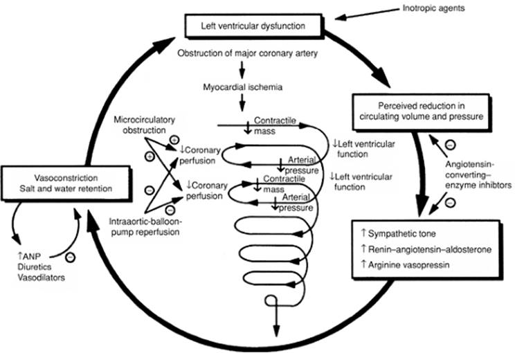
Cardiogenic shock is characterized by primary myocardial dysfunction resulting in the inability of the heart to maintain an adequate cardiac output with subsequent compromising of metabolic requirements (Fig. 56.1). The most common etiologies are myocardial infarction and cardiomyopathy with a superimposed hemodynamic stress.
|
|
|
Figure 56.1. Neurohumoral and mechanical events that lead to death in patients with cardiogenic shock. ANP, atrial natriuretic peptide. (Used with permission from Pasternak RC, Braunwald E. Acute myocardial infarction. In: Wilson JD, Braunwald E, Isselbacher KJ, eds. Harrison's Principles of Internal Medicine. 12th ed. Vol. 1. New York: McGraw-Hill; 1991:953–964; and Francis GS. Neuroendocrine manifestations of congestive heart failure. Am J Cardiol. 1988;62[Suppl]:9A–13A.) |
Etiology
In order to understand the therapeutic approaches used to support left ventricular (LV) ejection and aid acutely decompensated hearts, it is important to understand the mechanisms underpinning LV systole. Systolic ventricular function is determined by preload, afterload, and contractility. Preload is the wall stress on the left ventricle prior to ejection. Operationally, we use LV end-diastolic volume to reflect this wall stress. Since measures of volumes can be difficult at the bedside, LV end-diastolic pressure, left atrial pressure, or pulmonary artery occlusion pressure are often used as surrogates for LV end-diastolic volume. Afterload is the maximal LV wall stress during ejection. By LaPlace's law, wall stress is proportional to the product of LV radius of curvature and transmural pressure. Under normal conditions, maximal LV afterload occurs at the instant of aortic value opening. Contractility is a more difficult term to define and quantify. A reasonable definition is the amount of force capable of being produced by the contracting myocardium (3). On a cellular level, contractility is related to the integrity of the actin–myocin coupling, intracellular calcium (Ca2+) flux rate, and quantity. Functionally, one measures contractility by varying preload and afterload. Numerous measures have been attempted to quantify contractility with varying degrees of success depending upon the degree of true independence they have from preload or afterload. Measures of contractility include the maximal rate of isovolumic pressure development (dP/dtmax), the Frank-Starling law relating peak systolic activity (defined as either maximal developed pressure, volume ejected, or the product of the two) directly to end-diastolic volume (4), and left ventricular end-systolic pressure–volume relation (ESPVR) derived from pressure–volume loops. Systolic performance is the ability of the left ventricle to empty. This is a function of end-systolic volume; a commonly used calculation is the LV ejection fraction (effective ejection fraction in the case of valvular regurgitation).
The most common etiology of cardiogenic shock is acute myocardial infarction (MI) with a resultant loss of approximately 40% of functioning myocardium. Following myocardial infarction, the final infarct size has been shown to correlate with the degree of LV dysfunction (5). Loss of myocardial function may occur in one massive MI or may result in a cumulative loss of pump function caused by serial smaller infarcts. Cardiogenic shock more commonly results from infarction of the left ventricle, although recent clarification of the potential role of the right ventricle in the precipitation of the shock state has been recognized. Additionally, acute mechanical complications of myocardial infarction such as mitral insufficiency, free wall rupture, and acute VSD may result in cardiogenic shock during the peri-infarct period, as does the late development of left ventricular aneurysm (Table 56.1). Other causes of cardiogenic shock include end-stage or fulminant cardiomyopathy, myocarditis, acute chordal rupture causing valvular regurgitation, obstruction to left ventricular ejection (severe aortic stenosis or hypertrophic cardiomyopathy) or left ventricular filling (mitral stenosis or left atrial myxoma), or severe septic shock with myocardial depression.
|
Table 56.1 Contributing factors to the development of cardiogenic shock in myocardial infarction |
|
|
Left Ventricular Acute Myocardial Infarction
Reduction in left ventricular performance is one of the major complications of ischemic heart disease. Several classifications that attempt to standardize the clinical and hemodynamic presentation of myocardial infarction have been proposed to aid in determining prognosis and the therapeutic approaches in patients with established cardiogenic shock or those who have the potential to progress to the shock state.
The Killip classification uses pure clinical bedside evaluation of the patient to establish prognostic indicators to predict the mortality associated with an acute myocardial infarction using the physical findings of congestive heart failure (6).
· Class I patients developed no overt signs of congestive heart failure, and these individuals had a low in-hospital mortality rate. This subgroup represented approximately 40% to 50% of all patients who presented with an acute MI. The in-hospital fatality rate was approximately 6%.
· Class II patients demonstrated evidence of impaired ventricular function as manifest by persistent bibasilar rales and an audible third heart sound. This subset of patients accounted for approximately 30% to 40% of patients with acute MI. The in-hospital mortality rate of 17% was triple relative to class I patients.
· Class III patients were characterized by the development of acute pulmonary edema, which was seen in approximately 10% to 15% of patients admitted to the hospital. A significant mortality rate of 38% was seen in this group treated conservatively before the thrombolytic era.
|
Table 56.2 Hemodynamic subsets and mortality in myocardial infarction |
||||||||||||
|
||||||||||||
· Class IV patients had established cardiogenic shock with hypotension and signs of organ hypoperfusion. Cardiogenic shock occurred in 5% to 10% of infarct patients in this series but was associated with a high in-hospital mortality rate of 80%, which was a function of both severity of the underlying illness plus the limited availability of definitive treatment at the time this classification was proposed.
The group at Cedars Sinai Medical Center, Los Angeles, also developed a clinical classification of heart failure associated with acute MI, which was subsequently refined by the availability of invasive hemodynamic monitoring using pulmonary artery catheters (PACs) (7) (Table 56.2). The Cedars Sinai classification also subdivided patients with acute myocardial into four subsets based on the measurement of the PAOP, CI, and clinical assessment.
Class I patients had no clinical evidence of pulmonary congestion or tissue hypoperfusion. Hemodynamic parameters measured in these subjects revealed the PAOP to be less than 18 mm Hg and the CI to be in excess of 2.2 L/minute/m2. The advent and widespread use of pulmonary artery catheters clarified the concept of the ideal wedge that established the impact of diastolic dysfunction secondary to acute ischemia, with resultant impaired relaxation and elevated filling pressures being required to maintain adequate cardiac output.
Class I patients accounted for 25% of subjects admitted to the coronary care unit, and there was a low in-hospital mortality rate of 1%. Patients who on clinical grounds demonstrated no evidence of hypoperfusion or pulmonary congestion would not be expected to benefit from invasive cardiac monitoring. Frequent clinical reassessments and close attention paid to blood pressure and evidence of organ perfusion would represent adequate care.
Class II patients demonstrated pulmonary congestion as manifest by only an elevated PAOP greater than 18 mm Hg with an associated normal cardiac index. Class II patients accounted for approximately 25% of patients admitted to the coronary care unit, but an 11% mortality rate was associated with this group. Mild pulmonary congestion is transiently seen in a significant percentage of patients admitted to the coronary care unit and has a multifactorial etiology. Diastolic dysfunction induced by ischemia with retrograde transmission of elevated filling pressures into the pulmonary venous circuit results in extravasation of fluid into the pulmonary bed when hydrostatic pressure exceeds oncotic pressure. Ischemic papillary muscle dysfunction with mild degrees of mitral insufficiency is also a potential cause of pulmonary congestion in this subgroup. Physical examination of these patients reveals mild to moderate rales and potentially an audible third heart sound associated with radiographic evidence of pulmonary venous hypertension. Dyspnea and orthopnea are the main symptoms superimposed on the clinical presentation of myocardial ischemia. Treatment in this group is centered on reduction of filling pressures to a level that relieves pulmonary venous congestion but does not result in an overzealous reduction of filling pressures below the ideal wedge as the reduced cardiac contractility will require some increased filling volume and pressure to maintain adequate stroke volume and perfusion pressure (Starling mechanism). Excessive diuresis should be assiduously avoided, especially in patients who were euvolemic before the onset of their infarct. Despite signs of pulmonary congestion, patients presenting with acute pulmonary congestion frequently are not intravascularly volume overloaded, and diuretic therapy may reduce filling pressures to a level that would impair cardiac output. It is often difficult to ascertain at the bedside which patients are actually euvolemic and which are hypervolemic. Afterload reduction therapy will benefit both groups of patients and may allow time to assess total effective circulating blood volume by indirect measures, such as the existence of hyponatremia, peripheral edema, and S4 gallop. Inotropic agents should be considered in such a situation so that pulmonary congestion can be relieved by diuresis if afterload reduction is not immediately effective since the increased inotropic state mitigates against a reduction in cardiac output induced by any reduction in cardiac filling pressures. Oxygenation should be maintained with adequate arterial saturation that may be monitored by oximetry. Vasodilator therapies in the form of nitroglycerin or inotropic agents with vasodilating capacity such as dobutamine are effective to return the hemodynamic parameters to normal. The usefulness and risk–benefit ratio of invasive hemodynamic monitoring in this subgroup of patients are controversial, although these patients frequently may be managed on clinical grounds.
Class III patients are characterized predominantly by clinical evidence of hypoperfusion. Hemodynamic monitoring reveals a PAOP less than 18 mm Hg and a cardiac index of less than 2.2 L/minute/m2. The class III subgroup accounted for approximately 15% of patients with acute MI and was associated with a 23% mortality rate. Patients in this subgroup may be extremely difficult to manage on clinical grounds, and treatment can be facilitated by invasive hemodynamic monitoring to establish the volume status. Relative hypovolemia is determined by measuring the pulmonary artery occlusion pressure, which falls below that of the ideal wedge as predicted in ischemic states. Excessive diuresis is extremely problematic in this group of patients and may excessively decrease cardiac output because of the pre-existent relative hypovolemia. Class III patients require restoration of intravascular volume to increase filling pressures to a degree that ensures adequate cardiac output and organ perfusion.
Class IV patients demonstrated elevated PAOP in excess of 18 mm Hg and a depressed cardiac index of less than 2.2 L/minute/m2 and frequently manifested signs of cardiogenic shock with clinical evidence of organ hypoperfusion and dysfunction. This subgroup accounted for approximately 35% of patients with MI and was associated with an in-hospital mortality rate of approximately 50%. Class IV patients may have a mechanical defect such as acute mitral insufficiency, free wall rupture, or VSD underlying the acute myocardial infarction; these are discussed separately. Oxygenation with the potential assisted ventilation in addition to inotropic and judicious use of vasodilator support is the recommended therapy in these subgroups.
Right Ventricular Infarction
Although isolated right ventricular (RV) infarction is rare, evidence of RV infarction and RV dysfunction is found in up to half of all infarcts and is clinically significant in nearly half of all inferior infarcts (8,9). The clinical diagnosis of RV infarction should be considered when elevated jugular venous pressure is accompanied by hypotension while the lung fields are clear. But the diagnosis may be difficult to establish clinically unless hemodynamic measurements, special electrocardiographic leads, echocardiography, or nuclear imaging are performed (10). Right-sided precordial leads obtained by electrocardiography that demonstrates at least 1-mm ST elevation is approximately 70% sensitive in the diagnosis of RV infarction and confers a particularly poor prognosis (11). Echocardiography is an easily obtainable noninvasive study that demonstrates RV dilation and impairment of wall motion of the right ventricle. Radionuclide angiography currently is considered to be the most sensitive means to diagnose RV infarction, although more recent data suggest that magnetic resonance imaging is comparable (12,13). A decrease in RV ejection fraction that is associated with wall motion abnormalities is more than 90% sensitive in the diagnosis of an RV infarction. Hemodynamic studies that are supportive of significant ischemic involvement of the right ventricle are manifest by increases in right atrial pressures plus demonstration of resistance to diastolic filling, as shown by blunting of the y-descent that follows tricuspid valve opening. A “square root” sign or “dip and plateau” pattern in the diastolic pressure curve is commonly demonstrated in RV infarctions but is not specific and may be associated with pericardial tamponade or restrictive cardiomyopathy (14).
The Should We Emergently Revascularize Occluded Coronaries for Cardiogenic Shock (SHOCK) trial registry reported on the clinical characteristics of patients presenting with isolated RV shock (15). Patients with RV shock compared to LV shock were younger and had a lower prevalence of previous MI (25.5% vs. 40.1%), a lower prevalence of anterior MI (11% vs. 59%), and less multivessel disease (34.8% vs. 77.8%). As expected, the infarct-related vessel involved the right coronary artery more in RV shock (96% of cases) versus LV shock (27% of cases). These patients had a shorter median time between myocardial infarction and the diagnosis of shock (2.9 vs. 6.2 hours) compared to patients with left ventricular shock. Right atrial pressure was a highly significant distinguishing factor of right from left ventricular shock (mean pressure 23.0 ± 9.9 vs. 14.2 ± 7.4, p = 0.0001), while all other hemodynamic measures were similar. Interestingly, in-hospital mortality was not significantly different between RV and LV shock (53.1% vs. 60.8%). Improvement in survival due to revascularization was similar between groups and multivariate analysis revealed that RV shock was not an independent predictor of lower in-hospital mortality (odds ratio 1.07, 95% confidence interval 0.54–2.13). This similarity in survival was despite patients with RV shock being younger; thus, RV shock may carry a worse prognosis.
Cardiogenic shock in patients with RV infarction frequently represents a substantial loss of functioning myocardium and carries a poor prognosis. RV infarction accompanied by cardiogenic shock is frequently associated with a variety of conduction abnormalities, including a high-grade atrioventricular block or significant rhythm disturbances. The treatment of RV infarction complicated by cardiogenic shock centers around maintaining RV filling pressures and assurance of adequate volume. Hemodynamic measurements may facilitate the estimate of volume loading required. Nitrates, diuretics, and other predominantly vasodilating compounds should be avoided. Atrial fibrillation is frequently poorly tolerated by these patients and may require immediate electrical cardioversion. The use of digitalis in acute RV infarction, even in the presence of atrial fibrillation, is controversial. Adequate inotropic support with vasodilating inotropic agents such as dobutamine is used if cardiac output fails to optimize after adequate volume loading. Percutaneous revascularization should be considered as it has been shown to improve outcomes (16).
Mechanical Defects
A variety of mechanical defects may be associated with cardiogenic shock in the peri-infarction stage (Table 56.3). Myocardial infarction resulting in cardiogenic shock from the appearance of mechanical defects such as acute mitral insufficiency, VSD, or free wall rupture represents a major complication and requires aggressive diagnostic and therapeutic interventions if the patient is expected to survive. Despite improvements in imaging techniques plus mechanical assist devices and emergency surgery, the mortality from these complications remains extremely high.
Acute Mitral Insufficiency
The mitral valve is a complicated apparatus and consists of the valvular annulus, leaflets, chorda tendineae, and papillary muscles plus potential functional alterations from involvement of the adjacent myocardium. Abnormalities affecting any of the components of the mitral valve may result in acute or chronic mitral insufficiency. The mitral valve annulus may be dilated and contribute to mitral insufficiency, although this complication is primarily associated with cardiomyopathies or connective tissue diseases such as Marfan syndrome rather than an acute myocardial infarction. Calcification of the mitral valve annulus is common in the elderly and may alter coaptation of the mitral valve leaflets and result in mitral incompetence.
|
Table 56.3 Complications of myocardial infarction |
||||||||||||||||||||||||||||||||||||
|
||||||||||||||||||||||||||||||||||||
Acute mitral insufficiency caused by involvement of the valvular leaflets is associated with infective endocarditis from necrotizing organisms such as Staphylococcus aureus or Enterococcus, resulting in destruction of the valvular apparatus. Traumatic penetrating injuries that involve the valve itself are rare. Rupture of the chorda tendineae may also be seen in endocarditis or a variety of connective tissue diseases, including myxomatous degeneration or Marfan syndrome.
Chordal rupture that results in severe impairment of left ventricular function depends on the number of involved structures and the rapidity with which the rupture occurs. Mitral insufficiency in the peri-infarction state may result from involvement of the surrounding myocardium or papillary muscles. Papillary muscles located adjacent to the infarction zone may simply become dysfunctional because of alteration of synchrony of contraction related to ischemia or frank rupture from ischemic necrosis.
The degree of mitral insufficiency is a function of the degree of involvement and anatomic competence. The two papillary muscles (posteromedial and anterolateral papillary muscles) have different ischemic vulnerabilities because of the blood supply from the coronary arteries. The anatomic vascular supply represents end arteries that are solely supplied by terminal portions of the coronaries, thus rendering the papillary muscles vulnerable to ischemic involvement during an acute myocardial infarction. Papillary muscle dysfunction may result from intermittent ischemia during unstable angina or myocardial infarction with involvement of the adjacent myocardium (17). Papillary muscle dysfunction is characterized by mild flow murmurs, which may be grade I or grade II by auscultation. The anterolateral papillary muscle has a dual blood supply, which provides partial protection during ischemia. The diagonal branches of the left anterior descending and marginal branches from the circumflex supply blood to the anterolateral papillary muscle. The posteromedial papillary muscle is generally supplied solely from the posterior descending branch of the right coronary artery, increasing its vulnerability to ischemic-related dysfunction.
Significant ischemia involving the papillary muscle that results in complete rupture with fulminant mitral insufficiency is generally fatal because of the marked volume load ejected retrograde into the left atria and pulmonary venous bed (18). However, if the major ischemic-related necrosis is distal and only involves rupture of the head of the papillary muscle, the resultant mitral insufficiency may be tolerated hemodynamically long enough to allow recognition, proper diagnosis, and surgical intervention. Mild ischemic involvement of the papillary muscle may be increased in hemodynamic significance in the presence of pre-existing left ventricular dilation, which alters the ability of the mitral leaflets to coapt. Severe ischemic-related mitral insufficiency is more frequently a result of posteromedial papillary muscle necrosis resulting from inferior or posterior myocardial infarctions, although one third of cases may result from anterior infarction (19,20). Less than half of cases present with electrocardiographic evidence of ST elevation or Q waves (20). Right ventricular papillary muscle rupture may occur but is uncommon. Involvement of papillary muscles in the right ventricle results in tricuspid insufficiency, which if severe may result in right ventricular failure.
Papillary muscle rupture is a relatively uncommon complication and occurs in approximately 1% of patients having an acute ischemic event. The incidence has decreased in the thrombolytic era (21). After acute MI with cardiogenic shock, the incidence of acute severe mitral regurgitation is 6.9% (22). The peak incidence of papillary muscle rupture is within the first week, with the majority occurring between days 3 and 5 after an acute MI. The diagnosis of papillary muscle rupture may be suspected on physical examination and has been facilitated with the advent of hemodynamic monitoring and echocardiography.
The physical examination in acute mitral insufficiency secondary to papillary muscle rupture differs from the findings associated with chronic valvular regurgitation. In the acute setting a palpable thrill is uncommon and the radiation of the murmur differs from chronic conditions. The systolic murmur is soft, is decrescendo, generally ends before the second heart sound, and is best audible at the base of the heart as opposed to the apex with radiation to the neck or the top of the head.
Echocardiography and Doppler ultrasound has been a major advance in the diagnosis of acute mitral insufficiency and its clinical separation from other mechanical lesions associated with a new murmur (23). The left atrium and left ventricle are generally of normal size, and the ejection fraction is increased and frequently hyperdynamic. The mitral leaflet flails and may prolapse into the left atrium. Doppler ultrasound with color flow study determines the presence and severity of mitral insufficiency and presence of an intracardiac shunt and quantifies the degree of mitral regurgitation. Data from the Should We Emergently Revascularize Occluded Coronaries for Cardiogenic Shock (SHOCK) trial, which randomized patients with cardiogenic shock within 36 hours of an acute myocardial infarction, has shown that the severity of mitral regurgitation quantified by Doppler echocardiography is an independent predictor of survival (24).
Pulmonary artery catheter placement with measurement of PAOP and cardiac output is useful in mitral insufficiency. The presence of a regurgitant wave in the PAOP tracing may be visible in acute mitral regurgitant lesions, especially when there is no evidence of a step-up in oxygen concentration in the right atria or right ventricle. Pulmonary artery catheterization is not necessary for diagnosis, but the use of invasive monitoring allows optimization of cardiac output, filling pressures, and adjustment of inotropic, vasodilator, and diuretic therapy on the basis of induced changes in pressures.
Ventricular Septal Defect
Rupture of the interventricular septum may present in a similar clinical manner as mitral insufficiency with the abrupt onset of congestive heart failure plus a new murmur, making the two conditions difficult to separate on clinical grounds. Rupture of the interventricular septum also occurs in the first week after the acute ischemic event with a peak incidence occurring between days 3 and 5. The prevalence rate of acute VSDs after an infarction is difficult to accurately determine but occurs within the range of 0.5% to 2.0% and is the cause of death in approximately 5% of all fatal MIs. Incidence of VSD as a cause of cardiogenic shock after acute myocardial infarction in the SHOCK trial registry was 3.9% (22). Blood supply to the septum is supplied by septal perforating branches of the left anterior descending vessel and acute VSD is more common in anterior myocardial infarctions. These patients frequently have multivessel disease and are older patients experiencing an initial myocardial infarction (25).
The diagnosis of acute VSD may be inferred on clinical grounds but frequently requires more sophisticated evaluation to accurately diagnose and quantify the defect, which is located in the muscular septum and may be multiple. The physical examination in acute VSD depends on the magnitude of the shunt, which is, in turn, a function of the size of the ventricular defect, right ventricular compliance, pulmonary artery pressures, and inotropic state. A significant VSD is associated with the characteristic findings of shock in addition to a new holosystolic murmur associated with a precordial thrill. A precordial thrill may be palpated in approximately 50% of patients with an acute VSD and is a function of the magnitude of pressure gradient between the two chambers.
The diagnosis of VSD and its separation from acute mitral insufficiency has been facilitated by the advent of noninvasive and invasive diagnostic procedures. Two-dimensional echocardiography combined with Doppler flow study generally identifies a significant defect (26). Contrast echocardiography using microbubble techniques also may aid in the diagnosis of acute VSD and establish the presence of an intracardiac shunt. Pulmonary artery catheterization demonstrates the absence of a V wave in the pulmonary wedge tracing and an increase in oxygen saturation by ~10% in the right ventricle compared with the right atrium. The mortality rate for septal defects is significant, with approximately 25% of patients dying within the first 24 hours and a 50% mortality rate at 1 week. Less than 10% survive 1 year when treated solely with medical therapy (27). When occurring in the setting of cardiogenic shock, in-hospital mortality has been reported as high as 87% (22).
Free Wall Rupture and Tamponade
Free wall rupture is a major complication of myocardial infarction and is difficult to diagnose premortem. The prevalence of this complication is unknown but may occur in up to 8% of all myocardial infarctions with approximately one third occurring in the first 24 hours after the onset of the ischemic event and the peak incidence between days 5 and 7 (28). The SHOCK trial registry reported a 1.4% incidence of free wall rupture as a cause of cardiogenic shock after acute MI (22).
Rupture of the free wall is a major cause of mortality in acute ischemic events and is associated with large transmural infarcts with inadequate collateral circulation. This serious complication occurs more commonly in elderly hypertensive patients. Involvement of the left ventricle is the rule, although free wall rupture involving the right ventricle has been reported. Rupture of the free wall is frequently associated with the ventricular remodeling process in which a segmental infarction results in elevated left ventricular and diastolic pressure with expansion of the infarcted area. Expansion involves thinning of the affected area with regional hypertrophy in the adjacent region surrounding the infarct. A disproportionate dilatation occurs in the infarcted area and the risk of free wall rupture is enhanced with high shearing forces and elevated pressures. Free wall rupture generally occurs in the border zone between the infarcted area and the normal surrounding myocardium. The advent of thrombolytic therapy has been postulated to potentially increase the risk of free wall rupture, although this has not been definitely confirmed. Thrombolytic therapy may actually minimize the extent of myocardial necrosis and decrease free wall rupture. The use of agents such as corticosteroids, previously used to blunt inflammatory response and infarct size, has been associated with increased risk of free wall rupture.
Cardiac rupture is a catastrophic event resulting in sudden cardiac death unless a pseudoaneurysm forms. Hemopericardium with cardiac tamponade is difficult to diagnose early enough to institute definitive therapy. Cardiac tamponade after acute myocardial infarction also may be secondary to hemorrhagic pericarditis, but massive hemopericardium is usually due to cardiac rupture with rapid development of electromechanical dissociation and death. The diagnosis of free wall rupture is difficult but should be suspected with sudden hypotension, elevated jugular venous pressures, muffled heart sounds, and a pulsus paradoxus. Echocardiography can document the presence of pericardial fluid and occasionally demonstrates the perforated free wall (29,30). The classic signs of tamponade are present on echocardiography and are caused by the rising intrapericardial pressure compressing the right atrium and right ventricle, resulting in equalization of pressures and right ventricular diastolic collapse. Definitive therapy involves pericardiocentesis plus volume and pressure support with early surgical intervention being necessary for salvage. Untreated free wall rupture is universally fatal, although isolated instances of successful aggressive intervention with surgical therapy have been reported (31).
Left Ventricular Aneurysm
Left ventricular aneurysm is a relatively common complication of MI and may occur in up to 15% of survivors (32). A true aneurysm has a wide base with the ventricular walls composed entirely of myocardium, compared with a pseudoaneurysm, which generally has a narrow base with the walls consisting of pericardium and thrombotic debris. True aneurysms have a relatively low risk of free wall rupture but are associated with increased mortality due to sudden death from ventricular arrhythmias, emboli from mural thrombus, and progressive loss of left ventricular function (33). Aneurysms may develop early in the postinfarction period and can be asymptomatic or present with significant deterioration of left ventricular function. The presence of left ventricular aneurysm may be inferred by persistent ST elevation in the absence of chest pain or enzyme leakage (34).
Echocardiography demonstrating dyskinesis is a valuable tool in diagnosing aneurysms, as is left ventricular angiography. Left ventricular angiography demonstrates paradoxic systolic distention during ventricular contraction. Successful treatment of the aneurysm may be achieved with resection of the involved myocardium, frequently in combination with saphenous vein or mammary artery bypass grafting because of the high associated prevalence of multivessel coronary artery disease. Surgical resection has been advocated in the presence of arrhythmias to eliminate the substrate for ventricular tachycardia, but electrophysiologic mapping techniques are necessary to demonstrate that the origin of the arrhythmia arises from the left ventricular aneurysm.
Clinical Manifestations
The clinical manifestations of cardiogenic shock are a function of the underlying cause, and mechanical defects must be aggressively sought because of the need for definitive therapy. Clinical recognition of the shock syndrome frequently requires prompt and aggressive stabilization procedures to be instituted before the definitive diagnosis of the underlying etiology (Fig. 56.2). A history and physical examination should be obtained with special attention to mental status, jugular venous pulsations, quality and intensity of heart sounds, presence and localization of a murmur, and presence of oliguria. Diagnostic tests such as electrocardiogram, portable chest radiograph, arterial blood gases, and echocardiography frequently provide adequate clinical information to make a diagnosis and initiate stabilization therapy. A quarter of patients presenting with cardiogenic shock secondary to predominant left ventricular dysfunction do not have evidence of pulmonary congestion (35).
Therapy
Percutaneous Revascularization
Prior to 1999, interventions for the management of cardiogenic shock complicating acute MI were not systematically studied. The landmark SHOCK trial demonstrated that a strategy of early revascularization by angioplasty or surgery reduced mortality from 63% to 50% at 6 months (36). This finding has resulted in a major paradigm shift in the management of cardiogenic shock. The first branch-point in the decision algorithm is whether or not shock is present in the setting of an acute MI. If shock is present, patients should undergo immediate coronary angiography with percutaneous intervention if feasible.
The SHOCK trial studied patients with onset of shock within 36 hours of an MI and randomized the patients to immediate revascularization versus initial medical stabilization. Almost all patients required inotropes or vasopressors. Treatment in the revascularization group (64% of patients) was angioplasty or stenting (stents were not available at the beginning of the trial in 1993, but were actively used by the end of the trial in 1998) and coronary artery bypass graft surgery in 36%. In a subgroup analysis, survival was similar between percutaneous and surgically revascularized patients (55.6% vs. 57.4% at 30 days and 51.9% vs. 46.8% at 1 year, respectively) despite a higher incidence of diabetes and multivessel disease in those patients surgically revascularized (37).
Thrombolytic therapy was used in 49% of patients in the revascularization group and in 63% of the medical therapy group. There was no difference in survival at 30 days (53.3% in the revascularization group vs. 44.0% in the medical therapy group), likely a result of improved medical therapy. Age older than 75 years was associated with significantly higher 30-day mortality. Follow-up reports have shown persistent benefit to early revascularization with survival rates of 47% versus 34% at 1 year and 33% versus 20% at 6 years (38,39). Of the patients surviving to hospital discharge (143/302), 6-year survival was 62% versus 44% (39).
|
|
|
Figure 56.2. Management algorithm for cardiogenic shock. ACE-I, angiotensin-converting enzyme inhibitor; ARB, angiotensin II receptor blocker; PA, pulmonary artery. |
An exciting aspect of the SHOCK trial was the registry that was created from patients screened for enrollment but not randomized, whose data was reported separately in a dedicated supplement to the Journal of the American College of Cardiology in September 2000. This registry described 1,190 patients and is the largest prospectively collected database for cardiogenic shock (22). Etiology of shock from the registry was left ventricular failure (78.5%), acute severe mitral regurgitation (6.9%), ventricular septal rupture (3.9%), isolated right ventricular shock (2.8%), and tamponade from free wall rupture (1.4%). Electrocardiographic site of infarction was anterior (55%), inferior (46%), posterior (19%), lateral (32%), and apical (11%), with multiple sites present in half of the cases. There was ST elevation, Q waves, or new left bundle branch block in 79% of cases. Systolic blood pressure averaged 88 mm Hg with a mean heart rate of 96 beats per minute. Of the subset of patients with invasive hemodynamics measured, PAOP was 23 mm Hg, cardiac index was 2.08 L/minute/m2, and left ventricular ejection fraction was 33%. In-hospital mortality averaged 60% and ranged from 55% for acute severe mitral regurgitation, isolated right ventricular shock, and tamponade to 87% for ventricular septal rupture. Of the 717 patients who underwent coronary angiography, 15.5% had significant left main stenosis and 53.4% had three-vessel disease. Coronary artery disease severity also correlated with in-hospital mortality: No or single-vessel disease was associated with a 35% mortality rate as compared with three-vessel disease with a mortality rate of 50.8% (40).
Since the SHOCK trial, stenting has replaced angioplasty alone as the primary treatment for ischemic coronary artery disease due to its reduced incidence of restenosis. One recent case series has shown that stenting for cardiogenic shock decreased mortality compared to angioplasty alone (from 68% to 43%) (41). Primary coronary artery stenting is now recommended for patients with ST elevation or left bundle branch block who develop shock within 36 hours of acute myocardial infarction and are suitable for revascularization that can be performed within 18 hours of shock onset (1). Thrombolytic therapy may be used if early revascularization is not available (2).
While drug-eluting stents, which slowly elute a pharmacologic agent (currently either sirolimus or paclitaxel), are now widely used instead of bare metal stents due to their proven efficacy in reducing the incidence of restenosis, to date they have not been studied in the setting of cardiogenic shock (42,43).
Pharmacologic Limitation of Infarct Size
Several pharmacologic interventions have been used during acute myocardial infarction to minimize the extent of irreversible ischemic damage and decrease the likelihood of subsequent development of cardiogenic shock. Quantitative measurements of the extent of myocardial damage by electrocardiographic mapping and creatine kinase (CK) release are imprecise and frequently limit quantitative assessment of the potential therapeutic impact of pharmacologic interventions. Calcium channel blockers, β-blockers, and nitrates have been the main agents that have undergone clinical analysis to minimize myocardial damage, whereas a variety of experimental or uncommonly used therapies have been evaluated in small-scale clinical trials. Nitrates are complex pharmacologic agents with arterial and venodilating activity in addition to other potential beneficial effects, such as alteration of prostacyclin metabolism. Nitrates, when administered as topical, oral, or sublingual agents, are predominately venodilators with subsequent venous pooling, decreased venous return, and lowering of PAOP. Reduction in venous return and optimization of PAOP decrease left ventricular volume and improve subendocardial perfusion, thus reducing wall stress with the potential for minimizing infarct extent. Nitrates also have effects on systemic vascular resistance and epicardial coronary arteries, with resultant reduction of impedance to left ventricular ejection and increase in coronary blood flow.
Intravenously administered nitroglycerin has a more balanced arterial and venodilating effect. Clinical trials demonstrate that intravenous nitroglycerin administered at a level to decrease mean aortic pressure by 10% (44) results in a decrease in extension of MI and improves left ventricular ejection fraction and survival (45). Intravenous nitrates minimize the magnitude of infarct size as monitored by CK, and alter infarct expansion with reduction in the subsequent remodeling process and progression to congestive heart failure. Intravenous nitrates are potent vasodilators and require careful blood pressure monitoring to prevent significant hypotension and paradoxical bradycardia. Nitrates may result in a beneficial redistribution of coronary flow to the subendocardium without the coronary steal syndrome, a major detriment of other potent intravenous vasodilators such as nitroprusside.
Calcium channel blockers are important agents in managing patients with classic and vasospastic angina. The calcium channel blocking agents decrease systemic vascular resistance, decrease oxygen demand, and increase coronary flow, improving the balance between supply and demand. At pharmacologic doses, these agents also may have other potentially beneficial effects including antiplatelet activity.
Despite the documented beneficial effect of these agents in hypertension and angina, calcium channel blockers have not been proven to be beneficial in the treatment of MI and do not definitely limit infarct size. Studies using nifedipine have been unable to demonstrate benefit in patients with acute MI. Diltiazem has been advantageous in non–Q-wave infarction in the Diltiazem Reinfarction study (46). However, the Multicenter Diltiazem Postinfarction trial was not able to document a benefit to the administration of diltiazem in the postinfarction state when compared with placebo (47). Subgroup analysis demonstrated a mortality benefit with diltiazem therapy when no pulmonary congestion was present. However, mortality was increased when diltiazem was administered to subjects whose infarction was complicated by pulmonary congestion, implying that this agent should not be used in patients with cardiogenic shock. Studies performed in Denmark using intravenous verapamil followed by oral administration did not demonstrate a benefit. Later studies using only oral verapamil demonstrated a mortality reduction, although these trials have not been reconfirmed (48). Currently, the evidence for using calcium channel blockers for the treatment of acute MI to limit infarct size and progression to cardiogenic shock is limited.
β-Adrenergic blocking agents have been used in treating hypertension, atrial fibrillation, and a variety of ischemic conditions. β-Blockers act predominantly by decreasing myocardial oxygen demand caused by the negative chronotropic and inotropic activities of these agents. β-Blockers may have several other potentially beneficial effects including antiplatelet activity, regression of left ventricular hypertrophy, and reduction in sudden cardiac death. Clinical trials using β-blockade in acute MI have yielded conflicting results. The Goteborg trial administered metoprolol or placebo to subjects having an acute myocardial infarction and demonstrated a significant reduction in mortality at 90 days in the group randomly assigned to β-blocker therapy (49). Early administration of metoprolol was associated with a reduction in estimated infarct size, which presumably has an effect on early and long-term survival. Despite the fact that β-blockers are not commonly used as antiarrhythmic agents, there was a documented decrease in sudden cardiac death in the β-blocker group, which has been shown to be secondary to an increase in the ventricular fibrillatory threshold.
The Metoprolol in Acute Myocardial Infarction trial was able to demonstrate that the early administration of intravenous metoprolol followed by oral maintenance dose in acute MI was associated with a decrease in mortality in a high-risk subgroup of infarct patients (50). A subgroup study of the Goteborg Metoprolol trial found that early treatment with metoprolol in patients with suspected acute MI and signs of heart failure resulted in significantly reduced mortality at 3 months (10% vs. 19%), which persisted to 1 year (14% vs. 27%), compared to those who did not receive metoprolol (51). Propranolol, a noncardioselective β-blocker, has not uniformly been demonstrated to decrease mortality or limit infarct size when administered early in acute MI patients. However, the Beta-Blocker Heart Attack trial demonstrated reduced mortality when propranolol was administered after the acute phase of the infarction had subsided (52). Intravenous atenolol was studied in the First International Study of Infarct Survival Trial and demonstrated a 15% reduction in the early mortality of infarct patients who were given oral atenolol after the intravenous loading doses (53). β-Blockers also have been combined with thrombolytic therapy to limit infarct size. The Thrombolysis in Myocardial Infarction trial (TIMI II-B) studied the impact of three 5-mg boluses of metoprolol administered at 5-minute intervals followed by oral metoprolol compared with thrombolysis plus oral metoprolol. The TIMI II-B trial demonstrated a decrease in nonfatal reinfarctions and recurrent ischemic episodes in the group who received immediate intravenous metoprolol followed by oral therapy compared with the delayed subgroup. More recently, the CAPRICORN (Carvedilol Post Infarct Survival Control in Left Ventricular Dysfunction) trial studied carvedilol (6.25–25 twice per day in addition to standard therapy of aspirin, angiotensin converting enzyme [ACE] inhibition, and thrombolysis) versus placebo in a high-risk group of acute MI patients (n = 1,959) with an LV ejection fraction of ≤40%. Patients were treated for a mean of 1.3 years. All-cause mortality was lower in the carvedilol group than in the placebo group (54). Patients who had echocardiography demonstrated a significantly higher LV ejection fraction and decreased LV end-systolic volume in the carvedilol group at 6 months (55). Another post hoc analysis of the CAPRICORN study found that carvedilol suppressed atrial arrhythmias (2.3% vs. 5.4%) as well as ventricular arrhythmias (0.9% vs. 3.9%) compared to the control group (56). The beneficial effect on ventricular remodeling, in addition to the antiarrhythmic effect, may be one of the mechanisms by which carvedilol decreased mortality after acute MI in patients treated with ACE inhibitors. Use of β-blockers in acute myocardial infarction, while now standard of care, must be undertaken with caution because of the potential of precipitating atrioventricular block, reactive airways disease, and hypotension (57).
ACE inhibitors have been administered orally and intravenously in clinical trials to halt progression to congestive heart failure in the SAVE (Survival and Ventricular Enlargement) and Consensus-II (Cooperative New Scandinavian Enalapril Survival Study) trials. The SAVE study used captopril in over 2,000 patients who had an acute anterior MI when enrolled during the period from 3 to 16 days after the acute myocardial event (58). All patients with ejection fractions <40% were randomized to receive oral captopril or a placebo. Patients receiving captopril demonstrated less congestive heart failure, fewer recurrent MIs, fewer hospitalizations, and improved mortality over a 42-month period. The Consensus-II trial used intravenous enalapril in the early phase of infarction followed by oral enalapril, but there was no mortality benefit when compared with placebo (59). A review of the major post-MI heart failure trials such as SAVE, AIRE (Acute Infarction Ramipril Efficacy), and TRACE (Trandolapril Cardiac Evaluation), between 1992 and 1995, calculated that ACE inhibitors produced a relative risk reduction of 16% while β-blockade in addition to ACE inhibition in the CAPRICORN trial demonstrated an additional relative risk reduction of 23% (60). Oral ACE inhibitors are attractive agents because of their effects on hemodynamics, microcirculation, and angiotensin-mediated vasoconstriction and should be administered especially in anterior infarcts with significant reductions in ejection fraction unless there are contraindications (hyperkalemia, known drug sensitivity).
Selective aldosterone blockade with eplerenone for patients with LV ejection fraction of ≤40% after acute MI has been studied in one large, placebo-controlled trial, EPHESUS (Eplerenone Post-Acute Myocardial Infarction Heart Failure Efficacy and Survival Study). Eplerenone (in addition to treatment with β-blockers and ACE inhibitors) reduced all-cause mortality by 15%, cardiovascular mortality by 17%, heart failure hospitalizations by 23%, and sudden cardiac death by 21% (61). These outcomes were even more marked in the subgroup of patients with LV ejection fraction of ≤30%. In this group, all-cause mortality was reduced by 21%, cardiovascular mortality by 23%, sudden cardiac death by 33%, and heart failure mortality or hospitalization by 25% (62).
Angiotensin receptor blockade for LV ejection fraction of ≤40% after acute MI has been studied in the VALIANT (Valsartan in Acute Myocardial Infarction Trial) study (63). This was a multicenter, double-blind, randomized, active-controlled, parallel-group study comparing the efficacy and safety of long-term treatment with valsartan, captopril, and their combination in high-risk patients after MI. This compared three treatment groups consisting of patients receiving standard therapy plus valsartan (n = 4,909), valsartan plus captopril (n = 4,885), or captopril alone (n = 4,909). Valsartan treatment alone resulted in similar outcomes as captopril treatment alone and thus these agents can be used interchangeably. The combination of valsartan plus captopril increased the rate of adverse events (hypotension and renal dysfunction more commonly with valsartan; cough, rash, and taste disturbance more commonly with captopril) with no change in survival.
Adjunctive antiplatelet therapy with the glycoprotein IIb/IIIa inhibitor abciximab during emergent coronary artery stenting for cardiogenic shock has been shown to reduce mortality from 43% to 33% in one case series (41). The glycoprotein IIb/IIIa inhibitor eptifibatide used for non–ST-elevation myocardial infarction or unstable angina in the PURSUIT (Platelet Glycoprotein IIb/IIIa in Unstable Angina: Receptor Suppression in Using Integrelin Therapy) study has been shown to reduce 30-day mortality in the subset of patients developing cardiogenic shock (64). Clopidogrel in addition to aspirin is now standard of care after percutaneous coronary intervention and has been shown to decrease 1-year mortality in the setting of ST-elevation myocardial infarction (65). However, clopidogrel significantly increases the risk of postoperative bleeding in patients requiring surgical intervention.
Several agents have been used in small studies as adjunctive therapy in acute MI but have not reached widespread clinical use. Myocardial damage may be potentiated by the presence of reactive oxygen radicals, and free radical scavengers such as superoxide dismutase or catalase may provide potential benefit. Free radical scavengers have been shown to be effective when administered before the onset of experimental infarcts and definitive clinical studies are currently ongoing.
Glucose insulin potassium infusions (polarizing solution) have been used for several years to reduce infarct size by altering free fatty acid metabolism (66). Polarizing solution consists of 300 g of glucose, 50 units of regular insulin, and 80 mmol of potassium in 1 L of water delivered at 1.5 mL/kg/hour. Ejection fraction and wall motion abnormalities have been noted to improve after administering this solution, resulting in decreased mortality. Polarizing solution has not been studied extensively in double-blind, placebo-controlled trials and routine administration of this solution has not reached clinical acceptance.
Hyaluronidase may have anti-inflammatory activity and modulate the immune response postulated to play some role in the extent of infarct size. Hyaluronidase has been administered in small clinical studies and was associated with improved mortality and decreased development of Q waves implying myocardial salvage. There are no large-scale clinical trials available (67).
Thrombolysis
Thrombolysis induced by pharmacologic agents or direct angioplasty is an attractive treatment for re-establishing coronary perfusion to minimize the extent of myocardial infarction and progression to cardiogenic shock. The open artery hypothesis postulates that clinical outcome is dependent on maintaining adequate coronary perfusion to minimize ischemic damage mediated by vascular occlusion secondary to an intravascular thrombus. Recent trials of coronary thrombolysis, GISSI (Gruppo Italiano per lo Studio della Sopravvivenza nell'Infarto Miocardio), ISIS (International Study of Infarct Size), and GUSTO (Global Utilization of Strategies to Open Occluded Coronary Arteries), demonstrate the prevalence of cardiogenic shock in approximately 2% to 3% of acute MIs on arrival to the hospital with an additional 3% to 4% subsequently developing cardiogenic shock for a combined total of 7% (68,69). Early progression to cardiogenic shock is characterized demographically by elderly patients and the presence of anterior infarctions, low ejection fractions, diabetes, and previous MIs. Despite the theoretical attractiveness of administering recombinant tissue plasminogen activators or streptokinase in patients with established or impending cardiogenic shock, the mortality associated with cardiogenic shock remains high despite thrombolytic therapy, with the survival rate being only 35% as reported in the GISSI-I and GISSI-II trials (70). Prompt administration of thrombolytic agents within the first hour of acute MI may result in improved survival rates if reperfusion of the infarct-related artery can be sustained. Low coronary perfusion pressures in cardiogenic shock may play a potential role in the poor clinical outcome of these patients after thrombolytic therapy.
In vitro experimental infarct studies with reduced perfusion pressure have shown decreased diffusion of thrombolytic agents into clots with resultant impaired fibrinolysis (71). Enhanced pressure increases the rate of dissolution of an intravascular thrombus, implying that in cardiogenic shock with systemic hypotension, a reduced transcoronary pressure gradient may decrease efficacy of thrombolytic agents. The metabolic abnormalities associated with cardiogenic shock including lactic acidosis also may alter the conversion of plasminogen to plasmin and limit the efficacy of these drugs in clot lysis. Failure from lytic agents to sustain vascular patency in patients with cardiogenic shock is an indication for early cardiac catheterization and direct angioplasty if no contraindications exist. Persistent hypotension, nonevolving ST elevation, continuing clinical evidence of myocardial ischemia, CK elevations, and clinical instability are potential indications for rescue coronary angioplasty, which may result in increased survival (72). Rescue angioplasty has not been systematically studied in randomized controlled trials comparing it to thrombolytic therapy. If thrombolytic therapy does not result in establishment of coronary perfusion, angioplasty should be considered as a therapeutic option. The SHOCK trial reported that 49% of patients in the revascularization group received thrombolytic therapy and the early intervention group had a survival benefit (see section above) (36). Additionally, the SHOCK trial reported a survival benefit due to thrombolytic therapy (in-hospital mortality of 54% vs. 64%) (73). Cardiogenic shock secondary to mechanical defects such as papillary muscle dysfunction also has been treated successfully with percutaneous transluminal coronary angioplasty (PTCA), resulting in improved mitral regurgitation with resolution of cardiogenic shock (74).
Thrombolytic agents should be administered to all patients with acute MI who demonstrate evidence of the shock state if there are no contraindications and availability of a cardiac catheterization laboratory is >90 minutes. Failure of evidence of reperfusion is an indicator for rescue angioplasty.
Pharmacologic Agents
Inotropic Agents
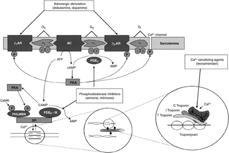
The effectiveness of various inotropic agents in cardiogenic shock depends on the cause and underlying pathophysiologic mechanism of the shock state. With systemic hypotension, adequate perfusion of the coronary arteries must be maintained (Fig. 56.3).
Dopamine
Dopamine is an endogenous catecholamine with positive inotropic properties secondary to stimulation of α- and β-receptors plus dopamine receptors, which have been divided into two subtypes: DA1 and DA2 (75,76). DA1 receptors are postsynaptic and induce dilation of the coronary, renal, and mesenteric vasculature. DA2 receptors are located in autonomic ganglia and in the postganglionic sympathetic nervous system. Stimulation of DA2 receptors blocks the release of endogenous catecholamines from intraneuronal storage sites. The effect of dopamine on α and β activity is dose related. Low infusion dosages of dopamine (2–5 µg/kg/minute) result in positive inotropic activity secondary to stimulation of the β1 receptors. α-Receptor stimulation occurs at dosages above 10 µg/kg/minute and results in a secondary increase in systemic vascular resistance caused by peripheral vasoconstriction. In addition to the inotropic effect, dopamine results in increased atrioventricular conduction from adrenergic stimulation. The effects of dopamine are thus dose dependent, and pharmacologic activity is a function of the amount of dopamine infused corrected for body weight. The individual response may be variable and unless the clinical situation warrants large pressor doses to maintain blood pressure, dopamine infusion should begin at a low rate (1 µg/kg/minute) and gradually be increased to clinical responsiveness. Cardiogenic shock with low tissue perfusion accompanied by hypotension may be treated in a more aggressive manner with progressively increasing doses of dopamine at 5-minute intervals.
Low-dose dopamine infusion results in stimulation of DA2 receptors and minimal or no changes in heart rate, cardiac output, or blood pressure. Stimulation of DA2 receptors results in renal vasodilation and increases glomerular filtration rate, renal blood flow, and sodium excretion. Reduction in cardiac output in shock frequently results in shunting of blood away from the renal vasculature and induction of a prerenal state with elevated blood urea nitrogen–to–creatinine ratios and sodium retention. Dopamine reverses the redistribution of cardiac output and increases the amount of sodium presented to the loop of Henle, which allows increased efficacy of diuretics such as furosemide or bumetanide.
Medium dosing ranges of dopamine (5–10 µg/kg/minute) result in an increase in cardiac output, which may also improve volume status by increasing renal blood flow. The cardiac effects of dopamine in this dosing range are secondary to stimulation of the β1-adrenergic receptors caused by a secondary release of norepinephrine. The effect of dopamine is indirect and depends on a pre-existent adequate storage level of endogenous catecholamines. Long-standing congestive heart failure is frequently associated with reduction in sympathetic receptors in the myocardium and the efficacy of dopamine may be limited if prolonged congestive heart failure was present before the shock syndrome. Dopamine infusion at this dose generally does not result in alterations of venous return secondary to venodilation, and right atrial and PAOP may not decrease. Dopamine may be combined with either direct vasodilating compounds or other inotropic agents such as dobutamine, which combine inotropism with vasodilation. Medium dosing range infusions of dopamine are generally safe and effective in maintaining blood pressure. Acid-base status and electrolyte levels should be optimized to avoid potential induction of arrhythmias with resultant malignant ventricular arrhythmias or marked sinus or supraventricular tachycardias, which would increase myocardial oxygen demand.
|
|
|
Figure 56.3. Mechanisms of action of inotropic drugs. (Used with permission from Garcia Gonzalez MJ, Dominguez Rodriguez A. Pharmacologic treatment of heart failure due to ventricular dysfunction by myocardial stunning: potential role of levosimendan. Am J Cardiovasc Drugs. 2006;6[2]:69–75.) β1AR, β1-adrenergic receptor; β2AR, β2-adrenergic receptor; AC, adenyl cyclase; AMP, adenosine monophosphate; ATP, adenosine triphosphate; Ca2+, calcium; CaMK, calmodulin-activated kinase; cAMP, cyclic AMP; Gi, inhibitory G protein with α, β, and γ subunits; Gs, stimulatory G protein with α, β, and γ subunits; P, phosphorus; PDEc, cytosolic phosphodiesterases; PDEp-III, particulate, SR-associated PDE III; PHLMBN, phospholamban; PKA, cAMP-dependent protein kinase A; SR, sarcoplasmic reticulum. |
High-range dopamine infusions (>10 µg/kg/minute) result in activation of α-adrenergic receptors and a secondary norepinephrine release with vasoconstriction and increased systemic vascular resistance. Patients in cardiogenic shock may need much higher doses of dopamine and ranges up to 50 µg/kg/minute have been used. Strict attention to volume status and repeated examinations for signs of excessive vasoconstriction are necessary. A central venous line is used for higher dopamine doses due to tissue necrosis should the solution extravasate. Dopamine may interact with certain coadministered drugs. Tricyclic antidepressants may increase the pressor response of direct-acting sympathomimetics and decrease the sensitivity to indirect-acting sympathomimetics. Because dopamine has direct and indirect effects on the vasculature, this agent should be used with caution, especially with overdoses of the tricyclic drugs (77). Although not commonly used, the rauwolfia alkaloids may potentiate the pressor response of direct-acting sympathomimetics resulting in hypertension. Monoamine oxidase inhibitors may increase pressor response of dopamine (78). Dopamine is an endogenous catecholamine that is degraded by catechol-o-methyltransferase and is not effective when administered in oral doses.
Dobutamine
As opposed to dopamine, which is an endogenous catechol and immediate precursor of norepinephrine and epinephrine, dobutamine is a synthetic agent that stimulates predominantly β1-adrenoreceptors (79) (Table 56.4). Dobutamine is a direct-acting agent, unlike dopamine, and does not require the presence or release of intramyocardial norepinephrine to modulate its effects. Mild activation of β2 and α receptors may be seen with this agent, but significantly less when compared with β1 receptors. Administration of dobutamine results in a direct inotropic stimulation plus a secondary reflex vasodilation with reduction of systemic vascular resistance and an increase in cardiac output.
The pharmacologic mechanism of dobutamine is complicated because of its asymmetric structure and racemic mixture. The positive and negative isomers have been evaluated as to their relative activities in in vitro studies, and it seems that the positive isomer is predominantly responsible for the activation of the β receptors. The administration of dobutamine alters stimulation of β receptors in a differential manner with an increased binding affinity for the predominantly cardiac β1-adrenergic receptors with a direct inotropic effect. The inotropic effects of this agent are not coupled with an increased rate of arrhythmias when compared with epinephrine and norepinephrine and there seems to be fewer adverse electrophysiologic effects when compared with dopamine. Although a mild vasodilator, there are no major effects on arterial blood pressure due to an increase in cardiac output and stroke volume. The increase in cardiac output results in improved renal blood flow and enhanced ability to excrete sodium and water. Dobutamine is effective in cardiogenic shock, assuming that the underlying etiology is not caused by valvular or subvalvular stenosis and the pharmacologic infusion does not result in significant hypotension, and this agent may be combined with dopamine to maintain blood pressure.
|
Table 56.4 Dobutamine |
|||||||||||||||||||||||||||
|
Norepinephrine
Norepinephrine is a powerful α-adrenergic agonist that results in significant peripheral vasoconstriction when administered within the usual dosage range of 2 to 8 µg/minute. Norepinephrine is generally instituted in the treatment of cardiogenic shock after failure of volume correction and dopamine to maintain adequate cardiac output and blood pressure (80). Norepinephrine is a naturally occurring catecholamine that has both α- and β1-adrenergic activity. Although generally associated with an increase in cardiac output, increases in systemic vascular resistance and mean aortic blood pressure may affect cardiac output adversely. The pressure work of the left ventricle and oxygen consumption are increased and blood may be shunted away from various organ beds because of volume redistribution secondary to catecholamine sensitivity. Oliguria and azotemia from impaired renal blood flow may be worsened secondary to the norepinephrine-mediated vasoconstriction. Norepinephrine has been associated with increased irritability of the ventricle with an increased electrical instability and potential adverse rhythm disorders. Clinical response will vary depending on the advantageous effects of increased perfusion pressure and cardiac output weighed against the detrimental effects of increased myocardial oxygen consumption and shunting from visceral organs.
Digitalis Preparations
The use of digitalis in general and cardiogenic shock specifically has been controversial because of theoretical objections involving the use of this agent and the lack of controlled clinical trials documenting a beneficial impact on mortality (81). Digitalis glycosides have complex mechanisms of action whose inotropic activity is modulated by increasing the availability of intracellular calcium secondary to inhibition of sodium–potassium ATPase. Inhibition of this ubiquitous enzyme, which is found not only in cardiac tissue but also in the central nervous system, gastrointestinal tract, and kidney, results in calcium influx by the activation of the sodium–calcium exchange mechanism. The level of free cytosolic calcium regulates the activity of tropomyosin with increased interactions between actin and myosin filaments and increased contractility. Alterations in contraction are caused by variations in levels of cytosolic calcium, which can be moved in and out of the sarcoplasmic reticulum.
The increase in cardiac output after administration of digitalis is modest when compared with the more powerful intravenous inotropes such as dobutamine, dopamine, and norepinephrine. Digitalis increases the refractory period at the atrioventricular node and decreases conduction velocity, resulting in a negative chronotropic effect in patients with atrial fibrillation. An advantage is that digitalis lacks the negative inotropic activity of other agents that have been used to slow the rate in atrial fibrillation, including β-blockers and calcium channel blockers such as diltiazem and verapamil (82). Digitalis increases vagal tone, decreases levels of norepinephrine in chronic heart failure possibly from decreased activity of the peripheral sympathetic nervous system, resets baroreceptor sensitivity, and may enhance natriuresis from increased cardiac output.
Digitalis withdrawal has been associated with worsening heart failure in a randomized, double-blind, placebo-controlled study of digitalis withdrawal in patients also treated with ACE inhibitors. However, the role of digitalis in cardiogenic shock is limited due to a modest increase in cardiac output, although the autonomic effects of this agent with decreases in the heart rate in atrial fibrillation are clinically beneficial.
Isoproterenol
Isoproterenol has both β1- and β2-adrenergic properties with increased myocardial contractility, heart rate, and cardiac output without vasoconstriction. The powerful chronotropic and inotropic activities of this agent increase myocardial work and oxygen. Isoproterenol is infrequently used in heart failure or cardiogenic shock unless the shock state is associated with bradyarrhythmias that do not respond to other therapies or with acute valvular insufficiency if blood pressure and volume status are maintained. Isoproterenol thus has a limited role in the acute management of cardiogenic shock.
Phosphodiesterase Inhibitors
Amrinone and milrinone are bipyridine derivatives that inhibit cellular levels of phosphodiesterase (83). Inhibition of this key enzyme results in increased levels of cyclic adenosine monophosphate (AMP) in cardiac muscle with resultant enhancement of protein phosphorylation by protein kinase with increased inotropic and chronotropic activities. The methylxanthines were known to nonspecifically inhibit phosphodiesterase activity and result in mild enhancement of the inotropic state. Both amrinone and milrinone have been shown in experimental and clinical studies to increase cardiac output in patients with severe congestive heart failure or cardiogenic shock (84).
Administering these agents results in reduction of central filling pressures and increases in stroke volume and cardiac output. The chronotropic effects of amrinone and milrinone are modest, but a mild increase in heart rate may be observed. Large doses may result in severe peripheral vasodilation, hypotension, and tachycardia. The phosphodiesterase inhibitors have been studied in patients with pump failure after myocardial infarctions, and at a dose of 200 µg/kg/hour, have been shown to improve cardiac function. Comparison in clinical trials of amrinone to other vasodilating inotropes such as dobutamine documented a greater decrease in systemic and pulmonary venous pressures in the group that received amrinone (85). The vasodilating activity of the phosphodiesterase inhibitors, while increasing cardiac output, may result in significant hypotension, requiring concomitant administration of sympathomimetic amines with at least partial α activity such as norepinephrine. The side effect profile of the phosphodiesterase inhibitors relates mainly to hematologic and gastrointestinal effects. Nausea, vomiting, and diarrhea occur in many patients. Thrombocytopenia is common with amrinone (approximately 15%), although the marked decreases in platelet counts to levels under 50,000 seems to be relatively rare and may require dose reduction. Milrinone is more potent on a milligram basis when compared with amrinone and also has effects on the inotropic state and ventricular relaxation. Incidence of thrombocytopenia seems less (<5%) than with amrinone. Enoximone is an imidazole derivative that also results in phosphodiesterase inhibition and increases levels of cyclic AMP and contractile force in isolated muscle preparations (86). Intravenous enoximone results in an increase in cardiac index with a decrease in right-sided filling pressures with minimal impacts on systemic vascular resistance and heart rate. Enoximone is currently undergoing a variety of controlled trials and seems to have a relatively mild side effect profile, and thrombocytopenia is uncommon with the use of this agent.
Glucagon
Glucagon is uncommonly used in cardiogenic shock but has a potential advantage in that it has a different mechanism of action from other sympathomimetic amines and does not require β-receptor stimulation to exert its inotropic effects (87,88). Glucagon is administered in a dosing range of 4 to 6 mg intravenously, which may be followed by a constant infusion of 4 to 12 mg/hour. Glucagon administration increases cardiac output by approximately 20%, which is associated with a decrease in peripheral vascular resistance with less myocardial oxygen demand when compared with norepinephrine. The indications for glucagon have not been delineated, although it seems justifiable to administer this agent to patients with cardiogenic shock who do not respond to conventional therapy or cannot tolerate other agents because of the development of significant arrhythmias or hematologic toxicity.
Levosimendan
Levosimendan is the first of a new class of inotropic agents called calcium sensitizers. Its mechanism of action involves increasing calcium sensitivity by binding to troponin C and stabilizing it in the calcium-induced conformation. This augments the effect of calcium binding to troponin C. Additionally, at high concentrations levosimendan inhibits phosphodiesterase 3, which also results in increased intracellular calcium concentration. These effects result in increased myocardial contraction associated with increased intracellular calcium transients (89). It improves myocardial contractility without increasing oxygen requirements and induces peripheral and coronary vasodilation with a potential antistunning, anti-ischemic effect (90). Given its vasodilatory properties, it is not primarily for cardiogenic shock but more for low-output heart failure. In addition to calcium sensitization, levosimendan also stimulates adenosine triphosphate (ATP)-sensitive potassium ion channels that are suppressed by intracellular ATP and acts synergistically with nucleotide diphosphates. This mechanism may contribute to the vasodilator action and may protect cardiomyocytes against ischemic damage (91). A loading dose of 6 to 24 mg/kg over 10 minutes followed by an infusion of 0.1 mg/kg/minute for 50 minutes, increased to 0.2 mg/kg/minute for an additional 23 hours, has been well tolerated (90). Initial clinical experience suggests that levosimendan causes dose-dependent increases in stroke volume and cardiac index, with minimum increase in heart rate (92). There are dose-dependent decreases in PAOP and right atrial, pulmonary arterial, and mean arterial pressures. The hemodynamic effects of levosimendan appear to be more pronounced than those seen with dobutamine (93) and sustained up to 24 hours after discontinuation of infusion due to an active metabolite (94). An initial clinical trial found no significant adverse events (92). Data from two published clinical trials indicate that levosimendan is associated with improved 6-month survival compared with dobutamine or placebo, although the studies were not powered to look at this outcome (93,95,96). There are several other trials not yet published but presented at national meetings that report a survival benefit of levosimendan compared with dobutamine or placebo. However, a 24-hour infusion of levosimendan had no effect on 6-month survival compared with dobutamine for patients with acutely decompensated heart failure in the Survival of Patients with Acute Heart Failure in need of Intravenous Inotropic Support (SURVIVE) trial reported at the American Heart Association Scientific Sessions in 2005 but not yet published (93). The European Society of Cardiology's 2005 guidelines on the diagnosis and treatment of acute heart failure include the use of levosimendan in patients with symptomatic low cardiac output secondary to systolic dysfunction without severe hypotension (97). This drug is not yet approved by the Food and Drug Administration (FDA), although it is available in some European countries.
Surgical Intervention
Surgical intervention in myocardial infarction has been used to limit infarct size by direct revascularization or to correct the mechanical defects of an acute ischemic event such as VSD, acute mitral insufficiency, free wall rupture, or left ventricular aneurysm. Surgical intervention for revascularization in acute myocardial infarction had been contraindicated on theoretic grounds because of the presumed high morbidity and mortality rates from cardiac catheterization and operative interventions during the unstable period of acute myocardial infarction. A variety of clinical studies determined that coronary bypass surgery could be performed in an expeditious manner with low mortality. Bypass surgery has been used as primary therapy in acute myocardial infarction with an overall operative rate of approximately 5% for transmural infarctions and a highly acceptable long-term mortality rate (98,99). Evidence is accumulating that early revascularization (<6 hours) by direct PTCA, intravenous or intracoronary thrombolytic agents, or bypass surgery in selected patients represents the treatment of choice. Congestive heart failure that occurs in the postmyocardial state may be amenable to revascularization by surgical interventions, although large-scale, controlled, randomized studies are lacking. However, several surgical series have reported on early and long-term survival of patients with an acute MI complicated by cardiogenic shock receiving coronary artery bypass surgery (98,99,100,101,102). Surgical intervention in cardiogenic shock is fraught with considerable clinical problems and requires the presence of surgically accessible and potentially viable myocardium. Surgical intervention has the advantage of re-establishing flow not only in the infarct-related artery, but also in vessels not involved in the acute ischemic process but significantly obstructed. Viability of the myocardium in the peri-infarction state may be difficult to determine secondary to problems with the acute delineation of stunned, hibernating, or irreversibly damaged myocardium. Nitroglycerin or dobutamine enhancement of ejection fraction is an indirect method of determining viability but is time consuming in a period where early revascularization is of prime importance.
Indications for surgical intervention in cardiogenic shock have not been completely delineated but should be considered in patients who fail to respond to volume correction and inotropic therapy. Failure of conventional medical interventions for cardiogenic shock should result in consideration of intra-aortic balloon counterpulsation, a temporizing measure before revascularization. Historically, emergent coronary artery bypass surgery preceded by placement of intra-aortic balloon pump has demonstrated improved survival rates in cardiogenic shock to approximately 75%. The SHOCK trial registry reported a 28% in-hospital mortality for the 290 patients undergoing coronary artery bypass surgery, which is comparable to other reported series (22,100). In a subgroup analysis of the SHOCK trial, survival was similar between percutaneous and surgically revascularized patients (55.6% vs. 57.4% at 30 days and 51.9% vs. 46.8%, respectively, at 1 year) despite a higher incidence of diabetes and multivessel disease in those patients surgically revascularized (37). Thus, surgical revascularization has an important role in patients with more extensive coronary artery disease.
Surgery for acute mitral insufficiency associated with cardiogenic shock in the postinfarction state is the only available definitive therapy. The impact of acute mitral insufficiency on left ventricular performance may be underestimated by studying the ejection fraction since the left ventricle ejects retrograde into the low-compliance left atrial and pulmonary venous system. Medical therapy with inotropic support and systemic peripheral vasodilation improves regurgitant flow as calculated by the regurgitant fraction. Severe mitral insufficiency is associated with a variety of adverse pathophysiologic changes that result in a poor survival after surgical intervention, but the results are significantly better than medical treatment that results in essentially 100% mortality if marked mitral insufficiency is associated with cardiogenic shock.
Surgical intervention is generally required for acute VSDs, which occur in the muscular portion of the interventricular septum and may be multiple. Two anatomic types of acute VSDs have been described. A VSD resulting from occlusion of a posterior descending coronary artery that arises from the right coronary is associated with a defect located in the inferobasilar region of the septum. Anteroseptal myocardial infarctions, which are associated with thrombotic occlusion of the left anterior descending, are associated with midapical to anterior defects in the septum. The physiologic impact of a left-to-right shunt is a function of the quantitative amount of involved myocardium plus associated left ventricular dysfunction, pulmonary artery pressures, and right ventricular compliance. A significant left-to-right shunt markedly decreases forward flow with poor peripheral perfusion and the clinical characteristics of cardiogenic shock. If the left ventricular end-diastolic pressure is markedly elevated, left-to-right shunting will also occur during diastole and is associated with an extremely high 24-hour mortality rate of approximately 25% (101,102). Medical treatment alone is associated with a 20% survival beyond 60 days and 1-year survival of less than 10%.
Surgical intervention in acute VSDs requires early and aggressive diagnostic and therapeutic interventions. Despite IABP and optimization with medical management, refinements in surgical technique have improved 1-year survival to 32% without coronary artery bypass. Evaluation of clinical trials that attempt to postpone therapy to improve the healing process have been questioned because this eliminates the most severely ill patients from definitive therapy and introduces a selection bias into the implications of therapy. Early surgical intervention with direct patch grafts plus coronary artery bypass may result in survival rates of up to 75%.
Free wall rupture of the left ventricle is a surgical disease even with a clotted hemopericardium tamponading further extravasation of blood into the pericardial space. The diagnosis of free wall rupture may be extraordinarily difficult on clinical grounds, and signs of pericardial tamponade should be actively sought. Pericardiocentesis with decompression of the pericardial space may be lifesaving in the short term but represents only a temporizing procedure. Cardiac rupture is essentially fatal, but surgical intervention may be successful with direct oversewing of the defect if recognized and managed in a timely fashion (103,104).
Left ventricular aneurysm as a cause of cardiogenic shock may require surgical intervention as a definitive therapy. The remodeling process, which begins after an acute ischemic event with regional thinning and expansion of the infarct zone, may result in progressive decrease in left ventricular performance and cardiogenic shock. If the aneurysmal dilation of the left ventricle involves more than 20% of the left ventricular mass, severe impairment of pumping ability ensues and potentially requires surgical intervention if there is a poor response to medical management including intra-aortic balloon pumping. Surgical intervention for aneurysms should be optimized in timing with adequate healing and fibrosis.
Mechanical Circulatory Support
The intra-aortic balloon pump has been in clinical use for over 20 years to increase diastolic coronary arterial perfusion and to decrease left ventricular afterload (105). The intra-aortic balloon pump is a temporizing measure that does not increase myocardial oxygen demand and results in reduction of ventricular diastolic volume and reduces pulmonary congestion with an increase in cardiac output. The intra-aortic balloon pump is the most widely used circulatory assist device in patients with cardiogenic shock because of the ease of insertion either percutaneously or surgically. Effective counterpulsation results in stabilization and potential reversal of the shock state with improvement in peripheral perfusion but does require an adequate systemic pressure and left ventricular performance to maximize its use. Profoundly hypotensive patients respond poorly to intra-aortic counterpulsation and the IABP has limited efficacy.
Balloon pumping in selected patients allows optimization of blood pressure, cardiac output, and tissue perfusion in patients with cardiogenic shock while further diagnostic procedures are performed. Hemodynamic effects of the IABP include the following (in percent change): Peak aortic systolic pressure (10%–15%), diastolic intra-aortic pressure (70%), arterial end-diastolic pressure (10%), peak ventricular pressure (10%), LV end-diastolic pressure (10%), dp/dt (10%), systemic vascular resistance (no change), mean arterial pressure (no change), cardiac index (10%–15%), and pulmonary capillary resistance (10%–15%). The intra-aortic balloon pump may be used prophylactically in patients with mechanical defects such as acute mitral insufficiency or VSD to increase coronary perfusion, allow time for healing, and restore cardiac output toward normal. The impact of intra-aortic balloon pumping on long-term survival is controversial and depends on the indications for insertion, hemodynamic status, and etiology of the cardiogenic shock. Patient selection is a key issue and early insertion of the intra-aortic balloon may result in increased clinical benefit rather than procrastination until overt low flow state has developed. The addition of the IABP to thrombolytic therapy for acute MI complicated by cardiogenic shock has been studied in a randomized clinical trial. There was no overall mortality benefit, but the subgroup of patients with Killip class III or IV benefitted with a 6-month mortality rate of 39% for combined therapy versus 80% for fibrinolysis alone (106). The SHOCK trial registry also reported a survival benefit with intra-aortic balloon pumping in addition to thrombolytic therapy (47% vs. 63% in-hospital mortality), but these results were heavily affected by higher revascularization rates in the group receiving the intra-aortic balloon pump (68% vs. 20%) (73).
Patients who are not expected to significantly benefit from an intra-aortic balloon pump are elderly patients, those with severe peripheral vascular disease, and those with large MIs exceeding 40% of the left ventricular myocardium. The overall survival rate of patients with cardiogenic shock treated with the intra-aortic balloon pump is approximately 40%. For subjects who required balloon insertion for large myocardial infarctions without a significant mechanical obstruction, the survival rate was only 27%. Complications may be documented in up to 30% of patients who undergo intra-aortic balloon pumping and relate mainly to local vascular problems, including surgical trauma, emboli, infection, and hemolysis.
Left ventricular assist devices (LVADs) function as prosthetic ventricles but require a sternotomy for insertion. Assist devices may be used to support left ventricular performance, right ventricular performance, or a combination, depending on the underlying condition. The indications for insertion of an LVAD are controversial, and traditionally have been reserved for medical and intra-aortic balloon pump failure and in the presence of the potentially salvageable myocardium and particularly as a bridge to cardiac transplantation. The Thoratec extracorporeal left ventricular assist device (Thoratec, Pleasanton, CA) has been used as a bridge to cardiac transplantation. Insertion of the Thoratec device in patients with severe left ventricular dysfunction allowed survival to transplant in approximately 75% of 29 patients (107). Similarly, in the Heartmate LVAD (Thoratec, Pleasanton, CA), survival to cardiac transplantation has been reported as 76% out of 97 patients (108). The REMATCH (Randomized Evaluation of Mechanical Assistance in the Treatment of Congestive Heart Failure) trial demonstrated improved outcomes in chronically ill patients too sick for cardiac transplantation as an alternative (destination therapy) to routine medical care (109). Hemodynamic unloading and myocardial rest after ventricular assist device (VAD) placement may lead to recovery of native cardiac function, allowing for removal of the device without cardiac transplantation (110,111,112,113,114,115,116). Ventricular assist device support is also associated with decreases in neurohormonal activation, alterations in myocyte calcium handling, and improvement in the proinflammatory cytokine milieu (117,118,119,120,121,122,123,124,125). Histologic analysis of the explanted heart at the time of transplantation demonstrated decreased fibrosis and myocyte size after VAD placement (121,126,127). Despite these salutatory changes as a result of VAD support, the frequency of bridge to recovery (BTR) in chronically supported subjects remains low, in the range of 3% to 10% in various series (111,114,115,128).
The Heartmate LVAD is approved for life-long support in patients deemed too ill for cardiac transplantation with several other devices undergoing clinical trials. There is retrospective data that early mechanical support as a bridge to transplantation after acute MI complicated by cardiogenic shock improves survival compared with a strategy of early revascularization (129). The technology has now advanced to include several other continuous flow pumps that offer the potential advantage of greater mechanical longevity, thus making them truly a lifelong option. Complications include hemolysis, thromboembolism, and infection, which have been decreased with increasing experience.
The Nimbus hemopump (Nimbus Medical, Inc., Rancho Cordova, CA) circumvented the problem associated with median sternotomy and allows a percutaneous placement of a cannula across the aortic valve, which is coupled to an extracorporeal power source. The Nimbus hemopump uses an Archimedes spiral screw valve that rotates at approximately 25,000 revolutions per minute without significant hemolysis (130). Although this pump is no longer available, there are several other percutaneous continuous rotary flow VADs, including the TandemHeart (Cardiac Assist, Pittsburgh, PA), Cancion (Orqis Medical, Lake Forest, CA), and Impella (Abiomed, Danvers, MA). The TandemHeart is currently FDA approved, while the Impella is approved in Europe and in clinical trial in the United States. The Cancion system is also in clinical trial in the United States.
Future and Adjunctive Therapies
Autologous Stem Cells
Stem cells offer the hope of biologically rebuilding damaged myocardium due to their ability to differentiate into cardiomyocytes. There has been a substantial amount of research into the biology of various stem cells and now several clinical trials have been reported, with mixed results. Most trials have looked at stem cells' delivery percutaneously by intracoronary catheter after acute myocardial infarction in numbers ranging from 30 to 100 patients. The BOOST (Bone Marrow Transfer to Enhance ST-elevation Infarct Regeneration) trial found 6% improvement in ejection fraction compared to control but no significant difference at 18 months (131,132). The ASTAMI (Autologous Stem Cell Transplantation in Acute MI) trial found no difference in ejection fraction at 4 and 6 months, although Janssens et al. reported improved regional wall motion and decreased infarct size (133,134). The TOPCARE-CHD (Transcoronary Transplant of Progenitor Cells after MI with Chronic Ischemic Heart Disease) trial found 2.9% improvement in ejection fraction at 3 months, while the REPAIR-AMI (Intracoronary Administration of Bone Marrow-derived Progenitor Cells in Acute Myocardial Infarction) trial reported a 2.5% improvement in ejection fraction at 4 months (135,136). Multiple studies are currently under way including evaluating the safety and efficacy of stem cells implanted during surgery for VAD installation as well as coronary artery bypass surgery with depressed ventricular function and percutaneously for chronic angina. While this is a very promising therapy, considerable issues remain, including the risk of generating an arrhythmic focus, the best cell type, the amount of local myocardial blood flow necessary, the best method to deliver the cells to the myocardium, and the number of cells necessary.
Clenbuterol
Clenbuterol is a β2-adrenergic receptor agonist that induces skeletal muscle hypertrophy and improves contraction. It also has been found to cause cardiomyocyte hypertrophy without apoptosis (137). In a recently reported single-center study, 15 patients requiring LVAD support were treated with clenbuterol in addition to lisinopril, carvedilol, spironolactone, and losartan (138). There was sufficient myocardial recovery to explant the LVAD in 11 of 15 patients, in whom 4-year survival was 89%, quality-of-life scores were almost normal, and mean left ventricular ejection fraction was 64%. These patients all had heart failure due to nonischemic cardiomyopathy without histologic evidence of active myocarditis. A multicenter trial in the United States is being planned.
Tissue-engineered Patches
Patches made from decellularized extracellular matrix may be another useful solution to biologic regeneration of myocardium. The patch retains biologically active substances such as growth factors providing paracrine as well as mechanical support for regrowth of cardiomyocytes. These devices are still in preclinical testing but have shown improvements in regional function in a myocardial infarction model (139).
Conclusions
Despite rapid advancement in pharmacologic thrombolytic therapy, mechanical revascularization techniques, and development of mechanical ventricular assist devices, cardiogenic shock remains a major clinical challenge with an associated high mortality. Improved survival in cardiogenic shock may be seen with an aggressive approach to diagnosis and management of the problem, with emphasis on early recognition and treatment of mechanical defects such as VSD, acute mitral insufficiency, and free wall rupture. Limitation of infarct size by minimizing the extent of infarcted tissue is the key component in all therapeutic strategies with the goal to maximize perfusion, limit irreversible cell death, and decrease potential for a secondary mechanical event.
Pearls
· Clinical criteria used to establish the diagnosis of cardiogenic shock include absolute or relative hypotension, which is defined as a systolic blood pressure less than 90 mm Hg or a blood pressure that has fallen to at least 30 mm Hg less than the individual's baseline blood pressure.
· Cardiogenic shock thus may be a complication in patients with chronic hypertension who have an acute cardiac event that results in a decrease in blood pressure, but not to the 90 mm Hg systolic level.
· If signs of organ dysfunction and tissue hypoperfusion accompany this condition, the individual qualifies for the diagnosis of cardiogenic shock.
· The exact incidence of cardiogenic shock is difficult to ascertain because of variability in diagnostic criteria and survival rates in the early phase of acute MI. The Multicenter Investigation of Limitation of Infarct Size trial (140) documented an incidence rate of cardiogenic shock in 7.5% of subjects who were admitted to the hospital after having an acute myocardial infarction, a constant value from 1975 to 1997 (141). The Global Utilization of Streptokinase and Tissue Plasminogen Activator for Occluded Arteries (GUSTO-I) trial, the GUSTO-III trial, and other thrombolytic trials have reported incidences of 5% to 10% (68,142).
· The mortality rate for cardiogenic shock in the setting of acute MI is exceedingly high despite significant improvements due to a strategy of early revascularization as reported in the SHOCK (Should We Emergently Revascularize Occluded Coronaries for Cardiogenic Shock) trial. Historically, mortality rates were 81% as originally reported by Killip in 1967 (6). Early revascularization by angioplasty or surgery has been shown to reduce mortality from 63% to 50% at 6 months in the SHOCK trial (36).
References
1. Smith SC Jr, Feldman TE, Hirshfeld JW Jr, et al. ACC/AHA/SCAI 2005 Guideline Update for Percutaneous Coronary Intervention–summary article: a report of the American College of Cardiology/American Heart Association Task Force on Practice Guidelines (ACC/AHA/SCAI Writing Committee to update the 2001 Guidelines for Percutaneous Coronary Intervention). Circulation. 2006;113(1):156.
2. Sanborn TA, Sleeper LA, Bates ER, et al. Impact of thrombolysis, intra-aortic balloon pump counterpulsation, and their combination in cardiogenic shock complicating acute myocardial infarction: a report from the SHOCK trial registry. Should we emergently revascularize occluded coronaries for cardiogenic shock? J Am Coll Cardiol. 2000;36(3 Suppl A):1123.
3. Noble MI. Problems concerning the application of concepts of muscle mechanics to the determination of the contractile state of the heart. Circulation. 1972;45:252.
4. Sagawa K, Maughan WL, Sunagawa K, et al. Cardiac Contraction and the Pressure-Volume Relationship. New York: Oxford University Press, Inc.; 1988.
5. Mathey D, Biefield W, Hanrath P, et al. Attempt to quantitate relation between cardiac function and infarct size in acute myocardial infarction. Br Heart J. 1974;36(3):271.
6. Killip T, Kimball JT. Treatment of myocardial infarction in a coronary care unit. Am J Cardiol. 1967;20:457.
7. Forrester JS, Diamond GA, Chatterjee K. Medical therapy of acute myocardial infarction by application of hemodynamic subsets. N Engl J Med. 1976;295:1356.
8. Andersen HR, Falk E, Nielsen D. Right ventricular infarction: frequency, size and topography in coronary heart disease: a prospective study comprising 107 consecutive autopsies from a coronary care unit. J Am Coll Cardiol. 1987;10(6):1223.
9. Kinch JW, Ryan TJ. Right ventricular infarction. N Engl J Med. 1994;330(17):1211.
10. Andersen HR, Nielsen D, Falk E. Right ventricular infarction. Am Heart J. 1989;117:82.
11. Zehender M, Kasper W, Kauder E, et al. Right ventricular infarction as an independent predictor of prognosis after acute inferior myocardial infarction. N Engl J Med. 1993;328:981.
12. Dell'Italia LJ, Starling MR, Crawford MH. Right ventricular infarction. J Am Coll Cardiol. 1984;4:931.
13. Sato H, Murakami Y, Shimada T, et al. Detection of right ventricular infarction by gadolinium DTPA-enhanced magnetic resonance imaging. Eur Heart J. 1995;16(9):1195.
14. Goldstein JA, Barzilai, Rosamond TL. Determination of hemodynamic compromise with severe right ventricular infarction. Circulation. 1990;82:359.
15. Jacobs AK, Leopold JA, Bates E, et al. Cardiogenic shock caused by right ventricular infarction: a report from the SHOCK registry. J Am Coll Cardiol. 2003;41:1273.
16. Bowers TR, O'Neill WW, Grines C, et al. Effect of reperfusion on biventricular function and survival after right ventricular infarction. N Engl J Med. 1998;338(14):933.
17. Tcheng JE, Jackman JD, Nelson CL. Outcome of patients sustaining acute ischemic mitral regurgitation during myocardial infarction. Ann Intern Med. 1992;117:18.
18. Sharma SK, Seckler J, Israel DH. Clinical angiographic and anatomic findings in acute severe ischemic mitral regurgitation. Am J Cardiol. 1992;70:277.
19. Sharma SK, Seckler J, Israel DH, et al. Clinical, angiographic and anatomic findings in acute severe ischemic mitral regurgitation. Am J Cardiol. 1992;70(3):277.
20. Thompson CR, Buller CE, Sleeper LA, et al. Cardiogenic shock due to acute severe mitral regurgitation complicating acute myocardial infarction: a report from the SHOCK Trial Registry. Should we use emergently revascularize occluded coronaries in cardiogenic shock? J Am Coll Cardiol. 2000;36(3 Suppl A):1104.
21. Lear J, Feinberg MS, Vered Z. Effect of thrombolytic therapy on the evaluation of significant mitral regurgitation in patients with a first inferior myocardial infarction. J Am Coll Cardiol. 1993;21:1661.
22. Hochman JS, Buller CE, Sleeper LA, et al. Cardiogenic shock complicating acute myocardial infarction–etiologies, management and outcome: a report from the SHOCK trial registry. Should we emergently revascularize occluded coronaries for cardiogenic shock? J Am Coll Cardiol. 2000;36:1063.
23. Kisdnuke A, Otsuji Y, Kuroiwa R. Two dimensional echocardiographic assessment of papillary muscle contractility in patients with prior myocardial infarction. J Am Coll Cardiol. 1993;21:932.
24. Picard MH, Davidoff R, Sleeper LA, et al. Echocardiographic predictors of survival and response to early revascularization in cardiogenic shock. Circulation. 2003;107:279.
25. Menon V, Webb JG, Hillis LD, et al. Outcome and profile of ventricular septal rupture with cardiogenic shock after myocardial infarction: a report from the SHOCK trial registry. Should we emergently revascularize occluded coronaries in cardiogenic shock? J Am Coll Cardiol. 2000;36(3 Suppl A):1110.
26. Harrison MR, MacPhail B, Gurley JC. Usefulness of color flow Doppler imaging to distinguish ventricular septal defect from acute mitral regurgitation complicating acute myocardial infarction. Am J Cardiol. 1989;64:697.
27. Gray JM, Sethna D, Matloff JM. The role of cardiac surgery in acute myocardial infarction with mechanical complications. Am Heart J. 106:723.
28. Bates RJ, Beutler S, Resnekov L. Cardiac rupture: challenge in diagnosis and management. Am J Cardiol. 1977;40:1231.
29. Assmann PE, Roelandt JR. Two dimensional and Doppler echocardiography in acute myocardial infarction and its complications. Ultrasound Med Biol. 1987;13:507.
30. Buda AJ. The role of echocardiography in the evaluation of mechanical complications of acute myocardial infarction. Circulation. 1991;84(Suppl 3):109
31. Pappas PJ, Cerndianu AC, Baldino WA, et al. Ventricular free rupture after myocardial infarction: treatment and outcome. Chest. 1991;99:892.
32. Visser CA, Kan G, David CK, et al. Echocardiographic cineangiographic correlation in detecting left ventricular aneurysm. Am J Cardiol. 1982;50:337.
33. Heras M, Sany G, Etriu A, et al. Does left ventricular aneurysm influence survival after acute myocardial infarction. Eur Heart J. 1990;11:441.
34. Aryan S, Varat MA. Persistent ST elevation and ventricular wall abnormalities: a two-dimensional echocardiographic study. Am J Cardiol. 1984;53:1142.
35. Menon V, White H, LeJemtel T, et al. The clinical profile of patients with suspected cardiogenic shock due to predominant left ventricular failure: a report from the SHOCK trial registry. Should we emergently revascularize occluded coronaries in cardiogenic shock? J Am Coll Cardiol. 2000;36(3 Suppl A):1071.
36. Hochman JS, Sleeper LA, Webb JG, et al. Early revascularization in acute myocardial infarction complicated by cardiogenic shock. SHOCK investigators. Should we emergently revascularize occluded coronaries for cardiogenic shock. N Engl J Med. 1999;341(9):625.
37. White HD, Assmann SF, Sanborn TA, et al. Comparison of percutaneous coronary intervention and coronary artery bypass grafting after acute myocardial infarction complicated by cardiogenic shock: results from the should we emergently revascularize occluded coronaries for cardiogenic shock (SHOCK) trial. Circulation. 2005;112:1992.
38. Hochman JS, Sleeper LA, White HD, et al. One-year survival following early revascularization for cardiogenic shock. JAMA. 2001;285(2):190.
39. Hochman JS, Sleeper LA, Webb JG, et al. Early revascularization and long-term survival in cardiogenic shock complicating acute myocardial infarction. JAMA. 2006;295(21):2511.
40. Wong SC, Sanborn T, Sleeper LA, et al. Angiographic findings and clinical correlates in patients with cardiogenic shock complicating acute myocardial infarction: a report from the SHOCK trial registry. Should we emergently revascularize occluded coronaries for cardiogenic shock? J Am Coll Cardiol. 2000;36(3 Suppl A):1077.
41. Chan AW, Chew DP, Bhatt DL, et al. Long-term mortality benefit with the combination of stents and abciximab for cardiogenic shock complicating acute myocardial infarction. Am J Cardiol. 2002;89(2):132.
42. Moses JW, Leon MB, Popma JJ, et al. Sirolimus-eluting stents versus standard stents in patients with stenosis in a native coronary artery. N Engl J Med. 2003;349:1315.
43. Stone GW, Ellis SG, Cox DA, et al. A polymer-based, paclitaxel-eluting stent in patients with coronary artery disease. N Engl J Med. 2004;350(3):221.
44. Flaherty JT, Becker LC, Bulkley BH. Randomized prospective trial of intravenous nitroglycerin in patients with AMI. Circulation. 1983;68:576.
45. Jugoutt BI, Warnica JW. Intravenous nitroglycerin therapy to limit myocardial infarct size, expansion and complications. Circulation. 1988;78:906.
46. Gibson RS, Boden WE, Theroux P, et al. Diltiazem and reinfarction in patients with non–Q wave myocardial infarction. N Engl J Med. 1986;315:423.
47. The Multicenter Diltiazem Postinfarction Trial Group. The effect of diltiazem on mortality and reinfarction after myocardial infarction. N Engl J Med. 1988;319:385.
48. The Danish Study Group on Verapamil in Myocardial Infarction. Treatment with verapamil during and after an acute myocardial infarction: a review based on Danish Verapamil Infarction Trials I and II. J Cardiovasc Pharmacol. 1991;18(Suppl 6):520.
49. Herlity J, Hjalmarson A, Swedberg K, et al. The influence of early intervention in acute myocardial infarction on long term mortality and morbidity assessed in the Goteborg Metoprolol trial. Int J Cardiol. 1986;10:291.
50. Hjalmarson A, Elmfeldt D, Herlitz J, et al. Effect on mortality of metoprolol in acute myocardial infarction. A double-blind randomised trial. Lancet. 1981;2:823.
51. Herlitz J, Waagstein F, Lindqvist J, et al. Effect of metoprolol on the prognosis for patients with suspected acute myocardial infarction and indirect signs of congestive heart failure (a subgroup analysis of the Goteborg Metoprolol trial). Am J Cardiol. 1997;80:40J.
52. Viscoli CM, Horowitz RI, Singer BH. Beta blockers after myocardial infarction. Ann Intern Med. 1993;118:99.
53. ISIS-I Collaborative Group. Mechanisms for the early mortality reduction produced by beta blockade started early in myocardial infarction: ISIS-I. Lancet. 1988;i(8591):921.
54. Dargie HJ. Effect of carvedilol on outcome after myocardial infarction in patients with left-ventricular dysfunction: the CAPRICORN randomised trial. Lancet. 2001;357(9266):1385.
55. Doughty RN, Whalley GA, Walsh HA, et al. Effects of carvedilol on left ventricular remodeling after acute myocardial infarction: the CAPRICORN Echo Substudy. Circulation. 2004;109(2):201.
56. McMurray J, Kober L, Robertson M, et al. Antiarrhythmic effect of carvedilol after acute myocardial infarction: results of the Carvedilol Post-Infarct Survival Control in Left Ventricular Dysfunction (CAPRICORN) trial. J Am Coll Cardiol. 2005;45:525.
57. Roberts R, Rogers WJ, Mueller HS, et al. Immediate versus deferred beta blockade following thrombolytic therapy in patients with acute myocardial infarction (TIMI-IIB). Circulation. 1991;83:422.
58. The SAVE Investigators. Effect of captopril on mortality and morbidity in patients with left ventricular dysfunction after myocardial infarction. N Engl J Med. 1992;327:669.
59. Swedburg K, Held P, Kjekshus J. Effects of early administration of enalapril on mortality in patients with acute myocardial infarction: results of the Co-operative New Scandinavian Enalapril Survival Study (Consensus-II). N Engl J Med. 1992;327:678.
60. McMurray J, Pfeffer MA. New therapeutic options in congestive heart failure: part I. Circulation. 2002;105(17):2099.
61. Pitt B, Remme W, Zannad F, et al. Eplerenone, a selective aldosterone blocker, in patients with left ventricular dysfunction after myocardial infarction. N Engl J Med. 2003;348(14):1309.
62. Pitt B, Gheorghiade M, Zannad F, et al. Evaluation of eplerenone in the subgroup of EPHESUS patients with baseline left ventricular ejection fraction <or=30%. Eur J Heart Fail. 2006;8(3):295.
63. Pfeffer MA, McMurray JJ, Velazquez EJ, et al. Valsartan, captopril, or both in myocardial infarction complicated by heart failure, left ventricular dysfunction, or both. N Engl J Med. 2003;349(20):1893.
64. Hasdai D, Harrington RA, Hochman JS, et al. Platelet glycoprotein IIb/IIIa blockade and outcome of cardiogenic shock complicating acute coronary syndromes without persistent ST-segment elevation. J Am Coll Cardiol. 2000;36(3):685.
65. Zeymer U, Gitt AK, Junger C, et al. Effect of clopidogrel on 1-year mortality in hospital survivors of acute ST-segment elevation myocardial infarction in clinical practice. Eur Heart J. 2006;27(22):2661.
66. Rackley CE, Russel RO, Rogers WJ. Glucose-insulin-potassium infusion: review of clinical experience. Postgrad Med. 1979;65:93.
67. Henderson A, Campbell RWF, Julian DG. Effect of a highly purified hyaluronidase preparation on electrocardiographic changes in acute myocardial infarction. Lancet. 1982;i:874.
68. Holmes DR Jr, Bates ER, Kleiman NS, et al. Contemporary reperfusion therapy for cardiogenic shock: the GUSTO-I trial experience. The GUSTO-I investigators. Global Utilization of Streptokinase and Tissue Plasminogen Activator for Occluded Coronary Arteries. J Am Coll Cardiol. 1995;26:668.
69. Grella RD, Becker RC. Cardiogenic shock complicating coronary artery disease: diagnosis, treatment and management. Curr Probl Cardiol. 1994;12:693.
70. Gruppo Italiano Per lo Studio Della Streptochinase Mell Infarcto Miocardio (GISSI). Effectiveness of intravenous streptokinase treatment in acute myocardial infarction. Lancet. 1986;i:397.
71. Cox RH. Mechanical aspects of large coronary arteries. In: Santamore WP, Bove AA, eds. Coronary Artery Disease. Baltimore: Urban and Schwartzenberg; 1982:19.
72. Strack RS, Califf RM, Hinohara R, et al. Survival and cardiac event rates in the first year after emergent coronary angioplasty for acute myocardial infarction. J Am Coll Cardiol. 1988;11:1141.
73. Sanborn TA, Sleeper LA, Bates ER, et al. Impact of thrombolysis, intra-aortic balloon pump counterpulsation, and their combination in cardiogenic shock complicating acute myocardial infarction: a report from the SHOCK trial registry. Should we emergently revascularize occluded coronaries for cardiogenic shock? J Am Coll Cardiol. 2000;36:1123.
74. Shawl FA, Forman MB, Punja S, et al. Emergent coronary angioplasty in the treatment of acute ischemic mitral regurgitation. J Am Coll Cardiol. 1989;14:986.
75. Mueller HS, Evans R, Ayres SM. Effects of dopamine on hemodynamics and myocardial metabolism in shock following acute myocardial infarction in man. Circulation. 1978;57:361.
76. Goldberg LO. Cardiovascular and renal actions of dopamine: potential clinical applications. Pharmacol Rev. 1972;21:1.
77. Teba L, Schiebel F, Dedhia HV, et al. Beneficial effect of norepinephrine in the treatment of circulatory shock caused by tricyclic antidepressant overdose. Am J Emerg Med. 1988;6:566.
78. Horowitz D, Goldberg LI, Sjoerdsm A. Increased blood pressure response to dopamine and norepinephrine produced by MAO inhibitors. J Lab Clin Med. 1960;56:747.
79. Sonnenblick EH, Frishman WH, Lyeintel TH. Dobutamine: a new synthetic cardioactive sympathetic amine. N Engl J Med. 1979;300:17.
80. Mueller H, Ayres S, Giarinelli S, et al. Effect of isoproterenol, L-norepinephrine and intra-aortic balloon counterpulsation on hemodynamics and myocardial metabolism in shock following myocardial infarction. Circulation. 1972;55:325.
81. Kelly RA, Smith TW. Digoxin in heart failure: implications of recent trials. J Am Coll Cardiol. 1993;22(Suppl 4):107A.
82. Sarter BH, Marchinski FE. Redefining the role of digoxin in atrial fibrillation. Am J Cardiol. 1992;69:71.
83. Honerjager P. Pharmacology of bipyridine phosphodiesterase III inhibitors. Am Heart J. 1991;121:1939.
84. Benotti JR, Grossman W, Braunwald E, et al. Hemodynamic assessment of amrinone. N Engl J Med. 1978;299:1373.
85. Klein M, Siskind S, Frishman W. Hemodynamic comparison of intravenous amrinone and dobutamine in patients with chronic congestive heart failure. Am J Cardiol. 1981;48:160.
86. Dage RC, Kariya T, Hsiek CP, et al. Pharmacology of enoximone. Am J Cardiol. 1987;60:100.
87. Goldstein RE, Skelton CL, Levey GS. Effects of chronic heart failure on the capacity of glucagon to enhance contractility and adenyl cyclase. Circulation. 1971;44:638.
88. Scholtz H. Inotropic drugs and their mechanisms of action. J Am Coll Cardiol. 1984;4:389.
89. Hasenfuss G, Pieske B, Castell M, et al. Influence of the novel inotropic agent levosimendan on isometric tension and calcium cycling in failing human myocardium. Circulation. 1998;98(20):2141.
90. De Luca L, Colucci WS, Nieminen MS, et al. Evidence-based use of levosimendan in different clinical settings. Eur Heart J. 2006;27(16):1908.
91. Yokoshiki H, Katsube Y, Sunagawa M, et al. The novel calcium sensitizer levosimendan activates the ATP-sensitive K+ channel in rat ventricular cells. J Pharmacol Exp Ther. 1997;283(1):375.
92. Slawsky MT, Colucci WS, Gottlieb SS, et al. Acute hemodynamic and clinical effects of levosimendan in patients with severe heart failure. Study investigators. Circulation. 2000;102(18):2222.
93. Mebazaa A, Barraud D, Welschbillig S. Randomized clinical trials with levosimendan. Am J Cardiol. 2005;96(6A):74G.
94. Kivikko M, Lehtonen L, Colucci WS. Sustained hemodynamic effects of intravenous levosimendan. Circulation. 2003;107(1):81.
95. Moiseyev VS, Poder P, Andrejevs N, et al. Safety and efficacy of a novel calcium sensitizer, levosimendan, in patients with left ventricular failure due to an acute myocardial infarction. A randomized, placebo-controlled, double-blind study (RUSSLAN). Eur Heart J. 2002;23(18):1422.
96. Follath F, Cleland JG, Just H, et al. Efficacy and safety of intravenous levosimendan compared with dobutamine in severe low-output heart failure (the LIDO study): a randomised double-blind trial. Lancet. 2002;360(9328):196.
97. Nieminen MS, Bohm M, Cowie MR, et al. Executive summary of the guidelines on the diagnosis and treatment of acute heart failure. The Task Force on Acute Heart Failure of the European Society of Cardiology. Eur Heart J. 2005;26:384.
98. De Wood MA, Heit J, Spores J, et al. Anterior transmural myocardial infarction: effects of surgical coronary reperfusion on global and regional left ventricular function. J Am Coll Cardiol. 1983;1:1223.
99. De Wood MA, Notske RN, Berg R, et al. Medical and surgical management of early Q wave myocardial infarction. J Am Coll Cardiol. 1989;14:65.
100. Bolooki H. Emergency cardiac procedures in patients in cardiogenic shock due to complications of coronary artery disease. Circulation. 1989;79:I137.
101. Gray RJ, Sethna D, Matloff JM. The role of cardiac surgery in acute myocardial infarction with mechanical complications. Am Heart J. 1983;10:723.
102. Fox AC, Glassman E, Isom OW. Surgically remediable complications of myocardial infarction. Prog Cardiovasc Dis. 1979;21:461.
103. Bates RJ, Beutler S, Resnekov L, et al. Cardiac rupture: challenge in diagnosis and management. Am J Cardiol. 1977;40:1231.
104. Shapiro I, Isakov A, Burke M, et al. Cardiac rupture in patients with acute myocardial infarction. Chest. 1987;92:219.
105. Cohn LH. The role of mechanical devices. J Cardiac Surg. 1990;5:278.
106. Ohman EM, Nanas J, Stomel RJ, et al. Thrombolysis and counterpulsation to improve survival in myocardial infarction complicated by hypotension and suspected cardiogenic shock or heart failure: results of the TACTICS Trial. J Thromb Thrombolysis. 2005;19:33.
107. Farrar DJ, Hill JD, Gray LA, et al. Heterotopic prosthetic ventricles as a bridge to cardiac transplantation. N Engl J Med. 1988;318:333.
108. McCarthy PM, Smedira NO, Vargo RL, et al. One hundred patients with the HeartMate left ventricular assist device: evolving concepts and technology. J Thorac Cardiovasc Surg. 1998;115(4):904.
109. Rose EA, Gelijns AC, Moskowitz AJ, et al. Long-term use of a left ventricular assist device for end-stage heart failure. N Engl J Med. 2001;345:1435.
110. Gorcsan J 3rd, Severyn D, Murali S, et al. Non-invasive assessment of myocardial recovery on chronic left ventricular assist device: results associated with successful device removal. J Heart Lung Transplant. 2003;22:1304.
111. Farrar DJ, Holman WR, McBride LR, et al. Long-term follow-up of Thoratec ventricular assist device bridge-to-recovery patients successfully removed from support after recovery of ventricular function. J Heart Lung Transplant. 2002;21:516.
112. Maybaum S, Stockwell P, Naka Y, et al. Assessment of myocardial recovery in a patient with acute myocarditis supported with a left ventricular assist device: a case report. J Heart Lung Transplant. 2003;22:202.
113. Khan T, Delgado RM, Radovancevic B, et al. Dobutamine stress echocardiography predicts myocardial improvement in patients supported by left ventricular assist devices (LVADs): hemodynamic and histologic evidence of improvement before LVAD explanation. J Heart Lung Transplant. 2003;22:137.
114. Mancini DM, Beniaminovitz A, Levin H, et al. Low incidence of myocardial recovery after left ventricular assist device implantation in patients with chronic heart failure. Circulation. 1998;98:2383.
115. Hetzer R, Muller JH, Weng Y, et al. Bridging-to-recovery. Ann Thorac Surg. 2001;71:S109.
116. Frazier OH, Delgado RM 3rd, Scroggins N, et al. Mechanical bridging to improvement in severe acute “nonischemic, nonmyocarditis” heart failure. Congest Heart Fail. 2004;10:109.
117. James KB, McCarthy PM, Thomas JD, et al. Effect of the implantable left ventricular assist device on neuroendocrine activation in heart failure. Circulation. 1995;92:II191.
118. Milting H, El Banayosy A, Kassner A, et al. The time course of natriuretic hormones as plasma markers of myocardial recovery in heart transplant candidates during ventricular assist device support reveals differences among device types. J Heart Lung Transplant. 2001;20:949.
119. Kucuker SA, Stetson SJ, Becker KA, et al. Evidence of improved right ventricular structure after LVAD support in patients with end-stage cardiomyopathy. J Heart Lung Transplant. 2004;23:28.
120. Terracciano CM, Hardy J, Birks EJ, et al. Clinical recovery from end-stage heart failure using left-ventricular assist device and pharmacological therapy correlates with increased sarcoplasmic reticulum calcium content but not with regression of cellular hypertrophy. Circulation. 2004;109:2263.
121. Barbone A, Holmes JW, Heerdt PM, et al. Comparison of right and left ventricular responses to left ventricular assist device support in patients with severe heart failure: a primary role of mechanical unloading underlying reverse remodeling. Circulation. 2001;104:670.
122. Bartling B, Milting H, Schumann H, et al. Myocardial gene expression of regulators of myocyte apoptosis and myocyte calcium homeostasis during hemodynamic unloading by ventricular assist devices in patients with end-stage heart failure. Circulation. 1999;100:II216.
123. Chaudhary KW, Rossman EI, Piacentino V 3rd, et al. Altered myocardial Ca2+ cycling after left ventricular assist device support in the failing human heart. J Am Coll Cardiol. 2004;44:837.
124. Torre-Amione G, Stetson SJ, Youker KA, et al. Decreased expression of tumor necrosis factor-alpha in failing human myocardium after mechanical circulatory support: a potential mechanism for cardiac recovery. Circulation. 1999;100:1189.
125. Goldstein DJ, Moazami N, Seldomridge JA, et al. Circulatory resuscitation with left ventricular assist device support reduces interleukins 6 and 8 levels. Ann Thorac Surg. 1997;63:971.
126. Bruckner BA, Stetson SJ, Perez-Verdia A, et al. Regression of fibrosis and hypertrophy in failing myocardium following mechanical circulatory support. J Heart Lung Transplant. 2001;20:457.
127. Zafeiridis A, Jeevanandam V, Houser SR, et al. Regression of cellular hypertrophy after left ventricular assist device support. Circulation. 1998;98:656.
128. Simon MA, Kormos RL, Murali S, et al. Myocardial recovery using ventricular assist devices: prevalence, clinical characteristics and outcomes. Circulation. 2005;112(Suppl I):I-32.
129. Tayara W, Starling RC, Yamani MH, et al. Improved survival after acute myocardial infarction complicated by cardiogenic shock with circulatory support and transplantation: comparing aggressive intervention with conservative treatment. J Heart Lung Transplant. 2006;25:504.
130. Merhige ME, Smalling RW, Cassidy D, et al. Effect of the hemopump left ventricular assist device on regional myocardial perfusion and function. Circulation. 1989;80(Suppl 3):158.
131. Wollert KC, Meyer GP, Lotz J, et al. Intracoronary autologous bone-marrow cell transfer after myocardial infarction: the BOOST randomised controlled clinical trial. Lancet. 2004;364:141.
132. Meyer GP, Wollert KC, Lotz J, et al. Intracoronary bone marrow cell transfer after myocardial infarction: eighteen months' follow-up data from the randomized, controlled BOOST (BOne marrOw transfer to enhance ST-elevation infarct regeneration) trial. Circulation. 2006;113:1287.
133. Janssens S, Dubois C, Bogaert J, et al. Autologous bone marrow-derived stem-cell transfer in patients with ST-segment elevation myocardial infarction: double-blind, randomised controlled trial. Lancet. 2006;367:113.
134. Lunde K, Solheim S, Aakhus S, et al. Intracoronary injection of mononuclear bone marrow cells in acute myocardial infarction. N Engl J Med. 2006;355:1199.
135. Assmus B, Honold J, Schächinger V, et al. Transcoronary transplantation of progenitor cells after myocardial infarction. N Engl J Med. 2006;355:1222.
136. Schächinger V, Erbs S, Elsässer A, et al. Intracoronary bone marrow-derived progenitor cells in acute myocardial infarction. N Engl J Med. 2006;355:1210.
137. Yacoub MH. A novel strategy to maximize the efficacy of left ventricular assist devices as a bridge to recovery. Eur Heart J. 2001;22:534.
138. Birks EJ, Tansley PD, Hardy J, et al. Left ventricular assist device and drug therapy for the reversal of heart failure. N Engl J Med. 2006;355:1873.
139. Kochupura PV, Azeloglu EU, Kelly DJ, et al. Tissue-engineered myocardial patch derived from extracellular matrix provides regional mechanical function. Circulation. 2005;112:I-144.
140. Hands ME, Rutherford JD, Muller JE, et al. The in-hospital development of cardiogenic shock after myocardial infarction: incidence, predictors of occurrence, outcome and prognostic factors. J Am Coll Cardiol. 1989;14:40.
141. Goldberg RJ, Samad NA, Yarzebski J, et al. Temporal trends in cardiogenic shock complicating acute myocardial infarction. N Engl J Med. 1999;340(15):1162.
142. Hasdai D, Holmes DR Jr, Topol EJ, et al. Frequency and clinical outcome of cardiogenic shock during acute myocardial infarction among patients receiving reteplase or alteplase. Results from GUSTO-III. Global Use of Strategies to Open Occluded Coronary Arteries. Eur Heart J. 1999;20(2):128.