Marianne E. Cinat
David B. Hoyt
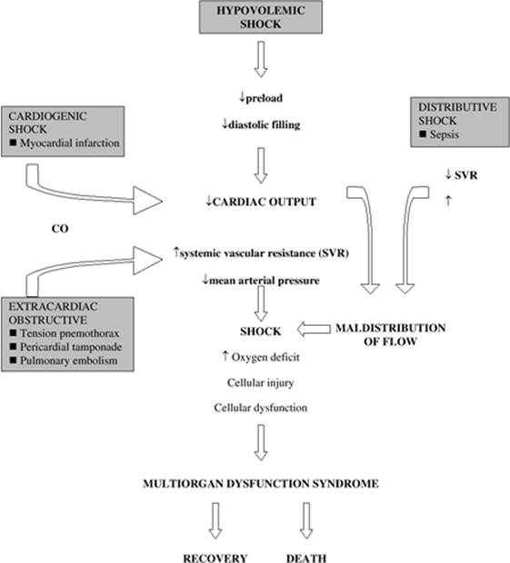
The definition of shock describes the final common pathway of many disease states: ineffective tissue perfusion, resulting in severe dysfunction of organs vital to survival. The most commonly used classification system for shock includes four categories based on hemodynamic characteristics (1):
1. Hypovolemic shock resulting from a decreased circulating blood volume in relation to the total vascular capacity and characterized by a reduction of diastolic filling pressures and volumes
2. Cardiogenic shock related to cardiac pump failure caused by loss of myocardial contractility/functional myocardium or structural/mechanical failure of the cardiac anatomy characterized by elevations of diastolic filling pressures and volumes
3. Extracardiac obstructive shock involving obstruction to flow in the cardiovascular circuit and characterized by either impairment of diastolic filling or excessive afterload
4. Distributive shock caused by loss of vasomotor control, resulting in arteriolar and venular dilation and characterized by increased cardiac output and decreased systemic vascular resistance after fluid resuscitation.
Although the hemodynamic characteristics of the various forms of shock may vary, the final common pathway—inadequate cellular perfusion—must be addressed early to prevent long-term sequelae and death (Fig. 58.1).
Hemorrhagic shock is a form of hypovolemic shock. It is a common, yet complicated, clinical condition that physicians are frequently called upon to evaluate and treat. Etiologies include trauma, postoperative bleeding, medical conditions, and iatrogenic causes. Diagnosis must be accurate and expedient. Therapy must be direct, efficient, and multifactorial in order to avoid the potential multisystem sequelae.
The purpose of this chapter is to address the immediate concerns for patients with hemorrhagic shock, as well as the etiology and epidemiology of this clinical condition, and to describe the pathophysiology, clinical features, and diagnostic and therapeutic approach to hemorrhagic shock. New and experimental therapies will also be introduced.
Immediate Concerns
The key steps in the approach to patients with hemorrhagic shock are listed in Table 58.1.
1. Early recognition. Early recognition requires astute clinical acumen to identify early systemic signs of hemorrhage and hypovolemic shock. Signs and symptoms include restlessness, anxiety, altered level of consciousness, shortness of breath, tachypnea, pallor, tachycardia, and oliguria. A decreased pulse pressure may also be observed along with decreased capillary refill due to peripheral vasoconstriction. Hypotension indicates significant volume depletion and may be a late clinical manifestation.
2. Important aspects in the patient history. An accurate history should be obtained expediently. For patients with traumatic injury, a thorough understanding of the mechanism of injury should be obtained, including the magnitude of blunt force trauma and/or the trajectory of the missile or object in penetrating trauma. In postoperative or postprocedural patients, the exact nature of the surgical procedure should be defined and potential sites of hemorrhage identified. In patients without recent surgery, risk factors for nonpostoperative, nontraumatic etiologies should also be sought (gastritis, peptic ulcer disease, atherosclerosis with aneurysmal disease). Significant comorbidities should also be delineated including coagulation disorders (von Willebrand, hemophilia), medical conditions associated with altered coagulation (cirrhosis, renal failure, iatrogenic vitamin K deficiency from parenteral nutrition or antimicrobials), or use of medications such as antiplatelet therapy and anticoagulants (Coumadin, heparin, low-molecular-weight heparin, or antimicrobials).
3. Initial action and intervention. The initial action taken in each case of hemorrhagic shock, regardless of etiology, should be directed at restoring circulating volume to ensure adequate tissue perfusion. Once the airway is secured and adequate ventilation is ensured, two peripheral large-bore intravenous catheters should be placed and fluid resuscitation begun. A blood sample should also be sent immediately for type and cross-match per institutional protocol. Initial resuscitation can include crystalloid, but should quickly be changed to blood products if signs of hypovolemia and ongoing hemorrhage persist. If a patient is in extremis and cross-matched blood products are not immediately available, type O blood (universal donor) should be immediately requested and transfused. For massive hemorrhage, clotting factors such as fresh frozen plasma, platelets, and cryoprecipitate should be prepared. The value of massive transfusion protocols to include predetermined ratios of clotting factors will be discussed later in this chapter.
4. Directed physical examination. Physical examination should be directed at obvious sources of external bleeding such as lacerations, extremity fractures, or surgical incisions. If identified, these should be immediately controlled. Physical signs of underlying liver disease should also be identified such as petechia, jaundice, ascites, angiomas, or testicular atrophy. Previous cardiac or carotid surgical incisions may hint toward concurrent antiplatelet or anticoagulant therapy. Evidence of retroperitoneal bleeding in patients with pancreatitis is marked by flank or periumbilical contusions.
5. Identify occult source of hemorrhage. If no obvious source of external bleeding is identified, a rapid evaluation should be performed to identify likely occult sources of bleeding. In the trauma patient, significant internal hemorrhage can occur in four defined regions: the thoracic cavity, the peritoneal cavity, the retroperitoneum, and extremity fractures. These areas can be rapidly assessed by chest radiograph, pelvic radiograph, a focused abdominal sonographic examination for trauma (FAST), and physical examination of extremities along with appropriate radiographs. In nontrauma patients without clear evidence of bleeding, the gastrointestinal tract should be rapidly evaluated via nasogastric tube, rectal examination, and endoscopy where appropriate. Additional diagnostic tests can be obtained based on clinical history, patient background, and condition. Abdominal aortic aneurysms can be identified on physical examination, by ultrasound, or by calcifications on abdominal radiograph. In rare selected instances, angiography may be used to identify and treat sources of hemorrhage not otherwise apparent (pelvic fractures, pancreatitis, lower gastrointestinal bleeding). This should only be instituted when a specific source of hemorrhage is highly likely and therapeutic intervention is sought. Computed tomography should never be sought in hemodynamically unstable patients with hemorrhage.
|
|
|
Figure 58.1. Final common pathway of shock. Hemorrhagic shock results in acute changes in circulating blood volume that culminates in a final common pathway shared by all classifications of shock. |
6. Expedite treatment. Once a source of bleeding is identified, a swift and directed treatment plan should be formulated and implemented without delay. Prolonged untreated hemorrhagic shock can lead to rapid decompensation and death if not appropriately identified and treated. Rapid intervention with surgical, angiographic, or endoscopic control of the hemorrhage is indicated, along with rapid correction of the underlying coagulopathy.
Epidemiology and Etiology
Hypovolemic shock can be due to hemorrhagic and nonhemorrhagic sources. Hemorrhage is the most frequent cause of hypovolemic shock and is most commonly due to blood loss after trauma or major surgery (Table 58.2). Following trauma, obvious external signs of injury and hemorrhage should be rapidly identified and controlled. As described above, the thoracic cavity, peritoneal cavity, and retroperitoneum should all be evaluated for occult hemorrhage.
|
Table 58.1 Key steps in the approach to a patient with hemorrhagic shock |
|
|
|
|
Table 58.2 Major etiologies of hemorrhagic shock |
|
|
Causes of hemorrhagic shock not due to trauma include a ruptured abdominal aortic aneurysm and gastrointestinal bleeding. Gastrointestinal bleeding can be caused by peptic ulcer disease, reflux esophagitis, variceal bleeding, erosive gastritis (stress ulcers), or an aortoduodenal fistula after vascular surgery. Prior manipulation by endoscopy or sphincterotomy can also lead to upper gastrointestinal bleeding. Lower gastrointestinal bleeding can result from diverticular disease, carcinoma, polyps, arteriovenous malformations, ischemia, or colitis. Pulmonary sources of hemorrhage can occur from tumor, tuberculosis, fungal infection, bronchiectasis, or tracheoinnominate fistula following tracheostomy. Hematuria from a tumor, trauma, or polycystic kidney disease is rare but can lead to hemorrhagic shock.
Nonhemorrhagic sources of hypovolemic shock can also occur. Although not the focus of this chapter, these are due to external fluid losses such as dehydration, vomiting, diarrhea, polyuria, uncontrolled diabetes mellitus leading to osmotic diuresis, and acute adrenocortical insufficiency. Disorders that lead to interstitial fluid redistribution such as thermal injury, trauma, and anaphylaxis can also lead to hypovolemic shock. Finally, disorders that cause increased vascular capacitance (venodilation) can lead to a relative hypovolemia and include sepsis, anaphylaxis, and the release of toxins/drugs leading to vasodilation.
Classification of Hemorrhagic Shock
Early diagnosis of hemorrhagic shock is imperative to avoid delay in treatment. However, clinical signs are relatively insensitive for small amounts of blood loss (2). There is a progressive hemodynamic deterioration with ongoing blood loss. This classic progression is delineated in Table 58.3. Total blood volume is estimated at approximately 70 mL/kg in the average adult, or nearly 5 L for a 70-kg person.
Class I
Class I hemorrhage is marked by a less than 750 mL estimated blood loss, or less than 15% of total circulating blood volume. There are minimal physical signs associated with this volume of blood loss. The patient may not have tachycardia, with a heart rate remaining less than 100 beats per minute; the systolic blood pressure and pulse pressure remain normal; the respiratory rate remains at 14 to 20 breaths per minute; and urine output remains adequate (>30 mL/hour). Only subtle physical signs such as delayed capillary refill and slight anxiety may exist.
Class II
Class II hemorrhage is marked by an estimated blood loss of 750 to 1,500 mL (or 15% to 30% of the total circulating blood volume). Physical signs begin to manifest during this stage of hemorrhage. Although the systolic blood pressure may be maintained, the patient usually becomes tachycardic (heart rate greater than 100 beats per minute), the pulse pressure begins to decrease, and capillary refill is delayed. The respiratory rate begins to increase (20–30 breaths per minute), urine output becomes diminished (20–30 mL/hour), and the patient becomes very anxious.
|
Table 58.3 Clinical classes of hemorrhagic shock |
||||||||||||||||||||||||||||||||||||||||||||||||||
|
Class III
Class III hemorrhage is marked by an estimated blood loss of >1,500 to 2,000 mL (or >30%–40% of total circulating blood volume). During this phase, significant hemodynamic compromise becomes apparent. Heart rate increases to >120 beats per minute, systolic blood pressure decreases, pulse pressure decreases, capillary refill decreases, tachypnea worsens with a respiratory rate of 30 to 40 breaths per minute, urine output drops to 5 to 15 mL/hour, and the patient becomes confused, showing further evidence of decreased perfusion of the central nervous system.
Class IV
Class IV hemorrhage is marked by an estimated blood loss of >2,000 mL (or >40% of total circulating blood volume). During this phase, most compensatory cardiovascular mechanisms have been maximized and total hemodynamic collapse is imminent. Signs of class IV hemorrhage include severe tachycardia with a heart rate >140 beats per minute, a decreased systolic blood pressure, a decreased pulse pressure, delayed capillary refill, significant tachypnea with a respiratory rate of >35 breaths per minute, minimal to no urine output, and severely altered mental status as marked by confusion and/or lethargy.
Potential Pitfalls
Despite these guidelines, several potential pitfalls exist that can make the diagnosis more difficult. Concurrent medication, such as β-blockers, may attenuate the physiologic response to hemorrhage. In the presence of β-blockade, tachycardia may be blunted or may not occur at all. Prior hydration status and use of diuretics can also alter the rate at which these signs present. Pregnant patients have a significantly increased total blood volume, and thus can lose up to 1,000 mL of blood before presenting with any clinical signs of hemorrhage. Blood is diverted from the placenta via vasoconstriction; the mother's total blood circulation is maintained at the expense of the fetus. Elderly patients may have atrial arrhythmias leading to a high ventricular response, making tachycardia less sensitive in this patient population. Concurrent use of antiplatelet or anticoagulant medication can cause relatively small injuries to bleed excessively, and identification and intervention may be delayed. Although unloading of the baroreceptors and activation of the sympathetic nervous system usually lead to tachycardia, some patients may respond to traumatic hemorrhage with bradycardia as a result of a vagal nerve–mediated transient sympathoinhibition due to acute and sudden blood loss (3,4,5,6,7,8,9). Finally, a significant reduction in skin blood flow (i.e., cool, clammy skin) is an early ominous sign of shock in view of selective cutaneous vasoconstriction (10). Intervention and resuscitation must be imminent upon presentation of these signs and symptoms.
Pathophysiology
Circulatory Changes
Hemorrhage results in a predictable pattern of events that begins with acute changes in circulating blood volume and culminates in a final common pathway shared by all classifications of shock (Fig. 58.1). Hemodynamically, hypovolemic shock is characterized by a fall in ventricular preload, resulting in decreased ventricular diastolic filling pressures and volumes. This in turn leads to a decrease in cardiac output and stroke volume (3,4,5,11,12,13,14,15). Following unloading of the cardiac baroreceptors and activation of the sympathetic nervous system, tachycardia ensues in an attempt to compensate for the decrease in cardiac output and stroke volume (12). The sympathetic output also results in vasoconstriction, leading to a decrease in pulse pressure. Greater variations in blood pressure will occur with the respiratory cycle due to an increased sensitivity of the underfilled heart to changes in venous return with varying intrathoracic pressure (16,17,18). The increased sympathetic tone may prevent a severe drop in arterial blood pressure initially. However, continued blood loss will ultimately result in hypotension and shock (3). Due to compensatory vasoconstriction, systemic vascular resistance rises early after the development of hypovolemic shock, but may fall in later stages, potentially heralding irreversibility and death (3,19,20).
The response to blood loss is a dynamic process that involves competing adaptive (compensatory) and maladaptive responses at each stage of development. Although intravascular volume replacement is always a necessary component of resuscitation in hypovolemic shock, the complex biologic response to the insult may progress to a point at which such resuscitation is insufficient to reverse the progression of the shock syndrome. For instance, patients who have sustained greater than a 40% loss of blood volume for 2 hours or more may not be able to be effectively resuscitated. Severe hemorrhage leads to a series of inflammatory mediator, cardiovascular, and organ responses that supersede the injury itself and ultimately drive recovery or death (3,19,20,21,22,23,24,25).
Oxygen Balance
Shock is characterized by an oxygen deficit in tissues and cells. The significance of the deficit and the extent of cellular injury can be quantified as a function of both the severity and the duration of the deficit—the greater the severity, the longer the duration, the worse the outcome of shock.
Oxygen delivery to tissues is determined by cardiac output and the oxygen content in arterial blood. Oxygen content refers to the number of milliliters of oxygen contained in 100 mL of blood (mL/dL) and is a function of the hemoglobin concentration, the oxygen saturation of hemoglobin, and the amount of oxygen dissolved in plasma (the calculation is [Hgb × 1.34 × O2 saturation] + [PaO2 × 0.0003]). During hemorrhage, as the cardiac output falls, oxygen delivery to the tissues also falls. Initially, the body will maintain sufficient uptake of oxygen by extracting more from the arterial blood. This will result in a fall in the mixed venous oxygen saturation (SvO2) with an increase in the arteriovenous oxygen content gradient (CaO2 – CvO2). Eventually, this compensatory mechanism also fails, and tissue hypoxia with lactic acidosis ensues. Cerebral and cardiac functions are maintained by diversion of blood flow from other organs (skin, muscle, and kidneys) (26). However, when these compensatory mechanisms are maximized, cardiac function and tissue oxygen delivery deteriorates further, and irreversible shock may develop (27).
Critical oxygen delivery is a function of cellular needs for oxygen and the ability of cells to extract oxygen from the arterial blood. Many factors contribute to this equation. During hemorrhage, tissue oxygen needs may increase due to increased respiratory muscle activity and increased catecholamine circulation (28). However, some evidence suggests that catecholamines down-regulate the metabolic needs of cells during hypovolemic shock (4,5,28,29,30). Regional blood flow is modified during hypovolemic shock in an attempt to maintain oxygen delivery to critical tissues (26,31). In addition, the individual needs of various tissues may vary during hemorrhagic shock. For instance, the oxygen needs of the kidney may decline during hemorrhage because a fall in renal perfusion leads to a fall in glomerular filtration and a decrease in energy-consuming tubular absorption (26). In contrast, the gut may experience an increased oxygen debt early due to the high oxygen need of the mucosa, along with redistribution of blood away from the gut to more critical tissues. This is the physiologic basis for gastric tonometry as a means of measuring the adequacy of resuscitation early following hemorrhage (32,33).
Oxygen extraction in tissues is influenced by the position of the oxyhemoglobin dissociation curve (34,35,36,37). Factors that improve the ability of tissues to extract oxygen from hemoglobin (i.e., shift the curve to the right) include acidosis, hypercarbia, hyperthermia, and decreased blood viscosity. However, in any extreme, each of these factors can be overcome by inadequate oxygen delivery and cardiovascular collapse. Interestingly, the oxyhemoglobin curve has been shown to shift to the left in critically ill patients (38). The presence of 2,3-diphosphoglycerate (DPG) in transfused blood has also been associated with a left shift of the oxyhemoglobin dissociation curve (39). Thus, although transfusions may increase the hemoglobin level, theoretically improving oxygen delivery, they may negatively affect the ability of tissues to extract oxygen from the hemoglobin.
The severity of oxygen debt during hypovolemic shock has been shown to be a major determinant of survival in animals and in patients following trauma, hemorrhage, and major surgery (20,27,40,41). A large oxygen debt has been associated with the development of acute respiratory distress syndrome (ARDS) and multiple organ dysfunction syndrome (MODS) (33,40,41,42,43,44). Conversely, a high oxygen delivery and uptake during resuscitation has been associated with improved survival (27,41,42,43,44,45,46). Whether increasing oxygen delivery to supranormal levels ultimately improves survival during resuscitation in critical illness remains controversial, and the medical literature has produced mixed results (27,33,43,44,47,48,49,50).
Cellular Response
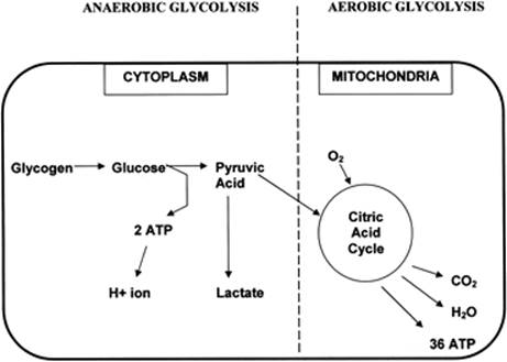
During hypovolemic shock, the oxygen deficit in the tissues causes a fall in the mitochondrial production and concentration of high-energy phosphates because of greater breakdown than production (51,52,53,54,55,56,57). This led many researchers to evaluate the utility of adenosine triphosphate (ATP) in the resuscitation of hemorrhagic shock (58,59). In the presence of sufficient oxygen, aerobic combustion of 1 mol of glucose yields 38 mol of energy-rich ATP. However, in the absence of sufficient oxygen, glucose taken up by the cells cannot be combusted because of insufficient uptake of pyruvate into the mitochondrial tricarboxylic acid cycle. Pyruvate is then converted to lactate within the cytoplasm. Anaerobic glycolysis yields only 2 mol of ATP, which is then hydrolyzed into hydrogen ion, ultimately leading to intracellular and extracellular metabolic acidosis (51,52,60,61,62) (Fig. 58.2). This process is ultimately a function of the severity and duration of regional hypoperfusion relative to oxygen demand and is more pronounced in some tissues (diaphragm, liver, kidney, gut) than in others (heart, skeletal muscle). Ultimately, a significant fall in the high-energy phosphates for a prolonged duration will lead to irreversible cellular injury and death.
The sequelae of low ATP production are profound. About 60% of the energy produced by respiring cellular mitochondria is needed to fuel the sodium-potassium (Na+-K+) pump of the cell. This pump controls the gradient in electrolyte concentrations and electric potential over the cell membrane. In the absence of sufficient ATP, the Na+-K+ pump is inhibited, resulting in an influx of sodium into the cell and efflux of potassium out of the cell. This in turn leads to cellular fluid uptake (51,53,63,64,65,66). Hyperkalemia may result due to potassium exchange between cells, the interstitial fluid, and vascular space.
Independent of the Na+-K+ pump, there may be a selective increase in cell membrane permeability for ions during hemorrhagic shock. Hypovolemic shock has been shown to lead to a rapid decrease in the transmembrane potential (with a less negative inner membrane potential), resulting in rapid electrolyte and fluid shifts across the membrane. Circulating heat shock proteins may also contribute to these changes independent of energy deficit (66,67,68,69,70,71).
|
|
|
Figure 58.2. Cellular mechanisms during anaerobic and aerobic glycolysis. In anaerobic conditions, pyruvic acid cannot enter the citric acid cycle within the mitochondria and is instead shunted to the production of lactate. This process produces only two molecules of adenosine triphosphate (ATP), as opposed to the 36 molecules of ATP produced from glucose in the mitochondria during aerobic glycolysis. Hydrolysis of ATP molecules in anaerobic conditions results in the production of hydrogen ions that cannot be cleared, leading to intracellular acidosis. (Adapted from Mizock BA, Falk JL. Lactic acidosis in critical illness. Crit Care Med. 1992;20[1]:80.) |
Finally, calcium (Ca2+) influx into cells and their mitochondria inhibits cellular respiration and ultimately contributes to cellular damage and swelling. Plasma levels of free Ca2+ may also fall. This may have profound consequences on the function of several organs during shock including the liver, kidney, heart, and vascular smooth muscle (64,65,72,73,74,75,76,77,78,79,80,81,82). Intracellular lysosomes lose their integrity, and proteolytic enzymes are released and contribute to cellular dysfunction and cell death.
The sum of the intracellular changes and alterations in signaling transduction pathways described above ultimately leads to the development of cellular dysfunction and multiple organ dysfunction syndrome, which may be irreversible (82). Laboratory investigations are aimed at novel resuscitation techniques involving substances that attenuate abnormalities of cellular signaling following hemorrhagic shock (58,59,77,78,79,80,81,82).
Neurohumoral Response
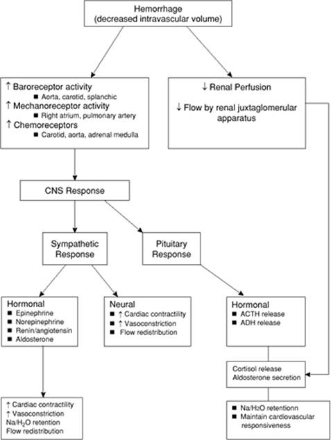
In response to hemorrhage and hypovolemia, a complex neurohumoral response is initiated in an attempt to maintain blood pressure and retain fluid. Decreased intravascular volume stimulates baroreceptors in the carotid body and aortic arch, along with mechanoreceptors in the right atrium. This stimulation leads to several neurohumoral responses (Fig. 58.3). Circulating catecholamines are liberated by activation of the sympathetic nervous system and the adrenal medulla. Direct sympathetic stimulation of the vessel wall leads to vasoconstriction. Angiotensin II is liberated via the renin–angiotensin–aldosterone system. Vasopressin (antidiuretic hormone [ADH]) is released by the pituitary in hypovolemic shock and leads to vasoconstriction. Finally, decreased cardiac filling pressures reduce cardiac secretion of α-atrial natriuretic peptide (ANP), thereby reducing the vasodilatory and diuretic effects of ANP.
Macrocirculation
During loss of circulating blood volume, mechanisms are initiated to counteract the fall in cardiac output and oxygen delivery by facilitating a redistribution of peripheral blood flow (26). Regional autoregulation takes place via a delicate balance of endogenous vasodilators and vasoconstrictors. Endothelial cells produce potent vasodilators such as endothelium-derived relaxing factor (nitric oxide [NO]), heme oxygenation–derived carbon monoxide (CO), and metabolic byproducts in tissues, including carbon dioxide (CO2), potassium, and adenosine (25,83,84,85,86,87,88,89). Some authors describe that inhibition of NO early following hemorrhage ameliorates early hypotension and improves mortality (90,91,92,93,94). Conversely, other authors describe endothelial dysfunction in organs with diminished NO production (95,96). Endothelin is a potent endothelial cell–derived vasoconstrictor that is released upon catecholamine stimulation or hypoxia (97). The overall increase in systemic peripheral vascular resistance is distributed differently among various organs in the body (31). Vasoconstriction also occurs in the venous vasculature, increasing return of available blood to the heart (14,98). The complex interplay of these mechanisms for vasodilation and vasoconstriction ultimately determines the regional redistribution of blood flow to organs following hemorrhagic shock. The redistribution of blood flow results in a greater share of oxygen delivery to organs with high obligatory metabolic demands (heart and brain), and a lesser share to those with fewer demands including the skin, skeletal muscle, kidney, intestine, and pancreas (5,31,86,99,100,101).
Microcirculation
One of the most important determinants of tissue perfusion during shock is the response and function of the microvasculature, which is defined as vessels less than 100 to 150 µm in diameter. Although arteries and medium-sized arterioles constrict in response to the extrinsic control mechanisms described above, terminal arterioles, venules, and capillaries remain unaffected and are more controlled by local metabolic factors.
|
|
|
Figure 58.3. Neurohormonal response to hemorrhage. Hemorrhage results in a decrease in the circulating intravascular volume, which initiates a complex cascade of compensatory events. CNS, central nervous system; ACTH, adrenocorticotropic hormone; ADH, antidiuretic hormone. |
Alterations in microvascular function and flow are effected through precapillary and postcapillary sphincters, which are sensitive to both extrinsic and intrinsic control mechanisms. Exchange of metabolites and compartmental regulation of fluids occurs at the capillary level. Therefore, alteration of tone of the pre- and postcapillary sphincters can have significant effects on microcirculatory function (102,103,104). Failure to dilate sphincters supplying metabolically active tissues may result in ischemia and anaerobic metabolism with lactate production. Increased precapillary tone, as seen with sympathetic stimulation, results in increased blood pressure systemically and decreased hydrostatic pressure locally. In fact, the microvascular arterioles may even dilate in response to the above vasoconstriction due to release of metabolic byproducts of underperfusion (carbon dioxide, hydrogen ion, etc.). The decrease in hydrostatic pressure locally then leads to redistribution of fluid from the interstitium to the circulation. Conversely, increased postcapillary tone (relative to precapillary tone) results in vascular pooling of blood and loss of fluid to the interstitium (as a result of increased hydrostatic pressure). This increased hydrostatic pressure may become accentuated in response to crystalloid resuscitation, leading to interstitial edema (104). Finally, hemorrhage and shock have also been shown to induce increased permeability of capillaries, leading to interstitial fluid leak during resuscitation (105,106).
Hypovolemic shock and hemorrhage also induce the expression of endothelial adhesion molecules on neutrophils and endothelium (63,107). This results in neutrophil adherence and “rolling” of cells within the capillary bed (108,109,110,111,112,113). Capillary flow then diminishes and may also impair red blood cell flow. While this decrease in transit time may augment the ability of tissue to extract oxygen, it may also lead to microvascular thrombosis and further tissue ischemia (114,115).
Metabolic and Hormonal Response
The early hyperglycemic response to trauma/hemorrhage is the combined result of enhanced glycogenolysis, caused by the hormonal response to stress including elevated epinephrine, cortisol, and glucagon levels; increased gluconeogenesis in the liver, partly mediated by glucagon; and peripheral resistance to the action of insulin (51,116). Increased gluconeogenesis in the liver, and to a lesser extent in the kidneys, follows increased efflux of amino acids, such as alanine and glutamine from the muscle to the liver, due to a breakdown of muscle protein. The latter is evidenced by increased urinary losses of nitrogen and a negative nitrogen balance. Lactate produced in muscle can also be converted to glucose in the liver (117). Increased epinephrine also results in skeletal muscle insulin resistance, sparing glucose for use by glucose-dependent organs such as the heart and brain. Later in shock, hypoglycemia may ensue, possibly because of glycogen depletion or hepatic ischemia (51,117,118). Fatty acids are increased early in shock, but later levels fall (116). Without energy for glycolysis, the cell depends on lipolysis and the autodigestion of intracellular protein for energy. Initially, ketone bodies and the branched-chain amino acids are used as alternative fuel sources. Without oxygen, these sources become inefficient, leading to hypertriglyceridemia, increased β-hydroxybutyric acid and acetoacetate levels, and changes in the amino acid concentration pattern. As these metabolic changes occur, set in motion by cellular hypoxia and promoted by systemic hormonal changes, structural changes occur within individual cells (119).
Organ Perfusion and Function during Hemorrhage
Heart
The heart is a critical organ in the pathophysiology of shock. At baseline, myocardial oxygen extraction is almost maximal; therefore, increased cardiac work must be met by increased coronary blood flow. When coronary perfusion is compromised, as it is during systemic hypotension, cardiac function suffers. In the presence of sympathetic stimulation, blood flow from the endocardium is redistributed toward the epicardium, impairing cardiac performance (120). Underlying coronary artery disease, arrhythmias, hypoxemia, and acidosis can add to cardiac dysfunction. In the absence of coronary stenosis, myocardial necrosis/infarction is unusual in hypovolemic shock. Rather, the heart plays a participatory role in which it is unable to compensate fully for arterial hypotension caused by hypovolemia, vasodilation, and other factors.
Under basal aerobic conditions, 60% of energy comes from fat (free fatty acids and triacylglycerides), 35% from carbohydrates, and 5% from amino acids and ketone bodies. However, during anaerobic conditions imposed by hypoxemia or ischemia, the myocardium shifts to glycolysis. Anaerobic glycolysis, however, is insufficient to meet cardiac work demands for any length of time because the myocardial glycogen stores, as an alternative fuel source, are minimal and rapidly depleted.
Brain
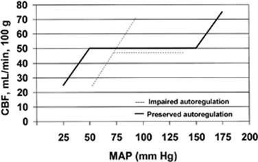
Like the heart, the brain almost exclusively depends on perfusion, rather than changes in extraction, to meet its oxidative metabolic needs. Protective mechanisms, collectively referred to as autoregulation, have evolved to guard perfusion. Pressure autoregulation refers to the ability of the brain to maintain total and regional cerebral blood flow (CBF) nearly constant despite large changes in systemic arterial blood pressure (Fig. 58.4) (121). Cerebral function seems to be maintained until the mean arterial pressure drops below 50 to 60 mm Hg (122). The factors that control cerebral autoregulation are not completely understood, but seem to include local carbon dioxide and oxygen tension, and the so-called Bayliss effect (i.e., contraction or dilation of arteriolar smooth muscle in the presence of increased or decreased intravascular pressure).
|
|
|
Figure 58.4. Autoregulation. Cerebral pressure autoregulation refers to the ability of the brain to maintain total cerebral blood flow (CBF) nearly constant between a broad range of mean arterial pressures (MAPs). Cerebral function seems to be maintained until the mean arterial pressure drops below 50 mm Hg. However, in the presence of traumatic brain injury, autoregulation may become impaired, and the brain may be more susceptible to changes in mean arterial pressure. |
In the presence of neurotrauma, autoregulation is impaired and the brain is exquisitely sensitive to secondary insults, such as hypoxia and hypotension. Hemorrhagic shock and resuscitation may also impair autoregulation because of endothelial cell dysfunction and diminished NO-dependent vasodilator reactivity, so that the brain may experience an oxygen deficit along with metabolic and functional deterioration (52,87). However, the vulnerability of the brain to anoxic injury is uncertain and appears variable. The adequacy and the method of resuscitation can critically influence postischemic recovery. These observations have motivated investigation of specific brain resuscitation regimens (123,124,125,126). However, there are no conclusive data that one modality provides improved outcomes.
Lungs
Hypovolemic shock is associated with a rise in minute ventilation marked by tachypnea, hyperventilation, and a fall in arterial PCO2 (28,30,127,128,129,130). These changes are usually due to a decrease in pulmonary perfusion, leading to an increase in dead space ventilation. Thus, a higher minute ventilation is necessary for a given CO2 production (28,30,127). In addition, minute ventilation may need to increase further in order to compensate for a metabolic acidosis following accumulation of lactic acid in the blood. The imbalance between the increased demands of the diaphragm and reduced blood flow in shock may finally lead to respiratory muscle fatigue and respiratory failure, requiring intubation (28). Therefore, early airway control is imperative in patients with severe hemorrhagic shock.
Hemorrhagic shock requiring massive transfusion also increases the risk of acute respiratory distress syndrome (129,130,131,132). Contributing factors include release of proinflammatory mediators; activation of neutrophils in the lungs and other organs after reperfusion; contusion and/or ischemia-reperfusion of the lung; pulmonary microemboli of neutrophils, platelets, and fat particles from long bone fractures; and induction of transfusion-related acute lung injury (TRALI), which is discussed later in this chapter.
Kidney
Oliguria, as defined by a urinary output of less than 0.5 mL/kg/hour, is a cardinal manifestation of shock. However, the pathogenesis of shock-related oliguria is more complex than mere renal hypoperfusion (133,134,135). Blood flow to the kidney is rarely reduced below 40% to 50% of normal levels, even in the face of more severe reductions in overall cardiac output. Thus, the decreased glomerular filtration rate results from additional mechanisms. Sympathetic stimulation, circulating catecholamines, angiotensin, and locally produced prostaglandins contribute to afferent arteriolar vasoconstriction. These compounds promote the redistribution of blood flow away from cortical glomeruli toward the renal medulla (65). Vasodilation of the efferent arteriole may amplify these changes. The net effect is a decreased glomerular filtration rate and a decrease in the energy needs of the kidney. Additional fluid (and salt) conservation is promoted by the effects of aldosterone and antidiuretic hormone.
If renal hypoperfusion persists, the cortical kidney will become ischemic. Three pathologic changes are observed: (a) tubular necrosis with back-diffusion of glomerular infiltrate, (b) tubular obstruction by casts or other cellular debris, and (c) tubular epithelial damage with consequent interstitial edema and tubular collapse. Following hypovolemia and renal ischemia, these pathologic changes may be secondary events (i.e., reperfusion injury) that can amplify but rarely initiate acute renal failure. The presence of these pathologic changes partially explains why restoration of normal hemodynamic function does not often lead to an immediate improvement in renal function. Although irreversible renal failure from shock alone is rare, fluid and electrolyte balance are often supported by dialysis although normal perfusion has been restored.
Intestine
During hypovolemic shock, blood flow from intestine is redistributed to other organs. The decrease in blood flow to the gut is relatively greater than the decrease in cardiac output due to the local vasoconstriction caused by catecholamines, vasopressin, and angiotensin II (4,5,26,136,137,138,139). Ischemic injury to the gut is manifested primarily by interstitial fluid sequestration and hemorrhage or necrosis of the mucosal lining, and is most prominent in the stomach (139). Ulcer formation (140) with exsanguinating hemorrhage can occur several days after normal hemodynamic function has been restored (141). Breakdown of the gut epithelium creates a port of entry for translocation of bacteria or deleterious bacterial products (endotoxin) (142,143). These factors may be important in the pathogenesis of irreversible shock (144) by releasing mediators to the systemic circulation. The determination of mucosal pH via tonometry has been described as a potential indicator of the therapeutic response and a marker of MODS (145,146).
Liver
Hepatic perfusion declines during hypovolemic shock because of diminished portal and hepatic arterial blood flow, roughly in proportion to the fall in cardiac output (57,61,109,147,148,149,150). Clinical manifestations of ischemic liver injury are not usually apparent in the early stages of hemorrhagic shock, as the organ participates in the release of acute-phase reactants. As hepatic cells die, they release characteristic enzymes (i.e., aspartate aminotransferase, alanine aminotransferase) (151). Occasionally, an obstructive picture with elevated bilirubin and alkaline phosphatase predominates. Later, the synthesis of coagulation factors, albumin, and prealbumin may deteriorate (152,153). Less clinically obvious is the impairment in the reticuloendothelial system function. Impaired hepatic clearance functions and reticuloendothelial system failure contribute to continued circulation of vasoactive substances that can perpetuate shock. Hepatic ischemia may result in a diminished capacity for metabolism of drugs and for gluconeogenesis from lactate and amino acids, contributing to hypoglycemia in the late stages of hypovolemic shock. The capacity to clear gut-derived endotoxin and lactate may also decrease, and the ischemic liver produces lactate (154). The appearance of “shock liver” with massive hepatocellular necrosis is unusual and presents mainly in patients with pre-existing liver conditions (155).
Spleen
The spleen contracts during hypovolemic shock, probably due to an increased sympathetic tone, which results in the release of red blood cells into the circulation (5,26). Changes in hematocrit during the early phase of bleeding probably underestimate the severity of plasma losses. The spleen also releases stored platelets.
Pancreas
The importance of the pancreas in the clinical picture in hemorrhagic shock has not been fully established. Older studies have demonstrated that the pancreas becomes severely ischemic during hypovolemic shock (156). Recently, much work has been done to better elucidate the role of the pancreas following hemorrhage and reperfusion. Preliminary data suggest that following hemorrhage, the mucosal barrier of the intestine becomes ischemic and therefore has increased permeability to pancreatic enzymes. These digestive enzymes then gain access to the wall of the intestine, initiating self-digestion of submucosal extracellular matrix proteins and interstitial cells. This initiates the generation and release of a host of strong inflammatory mediators, which may contribute to the multiorgan dysfunction syndrome. Recent investigations are focusing on protease inhibition in the intestinal lumen as a means of attenuating the inflammatory response following hemorrhage (157,158,159,160,161,162,163).
Inflammatory Response and Tissue Injury
A detailed discussion of the inflammatory and immune response to trauma and hemorrhage is beyond the scope of this chapter. However, several general concepts can be introduced. Following hemorrhage and resuscitation, macrophages, including lung macrophages and Kupffer cells in the liver, may release proinflammatory cytokines including tumor necrosis factor (TNF)-α and interleukin (IL)-1, -6, and -8. During reperfusion, cytokines may induce and amplify the inflammatory response to ischemia and may further induce local and remote organ damage (148,164,165,166,167,168,169,170,171,172,173,174). The reperfused gut, for example, may, together with the liver, be a source of systemically circulating cytokines, and possibly endotoxin. Release of mediators into the mesenteric lymph, portal, or systemic circulations during reperfusion may have deleterious effects on remote organs, such as the lungs, due to neutrophil activation and adherence, leading to pulmonary vascular injury with increased permeability (165,168,169). Circulating levels of proinflammatory cytokines may thus be of predictive value for remote organ damage, including ARDS, after trauma and hemorrhage in patients (167,170).
Arachidonic acid makes up 20% of cell membranes and is released from these membranes in response to a multitude of stimuli that activate phospholipase A2 and C, and is then metabolized via one of two major enzyme systems. The cyclo-oxygenase pathway results in the production of thromboxanes and prostaglandins, while the lipoxygenase pathway produces leukotrienes. Thromboxane has potent vasoconstricting properties on both the pulmonary and splanchnic circulation, promotes aggregation of thrombocytes and neutrophils, causes bronchoconstriction, and can lead to increased vascular permeability. The prostaglandins have varied effects. Prostacyclin (prostaglandin I2 [PGI2]) has potent vasodilating properties and inhibits thrombocyte and neutrophil aggregation (175). PGE2 and PGD2 also have vasodilating properties, while other prostaglandins (PGF2α) are potent vasoconstrictors. Leukotrienes, which are produced by the lipoxygenase pathway, cause vasoconstriction and increased capillary permeability and attract neutrophils (175). Thromboxane, prostaglandins, and leukotrienes interact with other mediators in a complex fashion (175,176,177,178,179). Vasoconstricting prostaglandins may be involved in the tissue damage during ischemia/reperfusion. Vasodilating prostaglandins may be involved in the vasodilated state of terminal hypovolemic shock (3,175,176,177,178,179).
Platelet-activating factor (PAF) is a nonprotein phospholipid, which is secreted by many cells including platelets, endothelial cells, and inflammatory cells. It is a major mediator of the pulmonary and hemodynamic effects of endotoxin. The major systemic effects of PAF are vasodilatation, cardiac depression, and enhancement of capillary leak. Its complex interactions with other mediators are still poorly understood (180).
Antigen–antibody complexes activate the complement cascade, and complement fragments thus generated can interact with other cytokines to promulgate the inflammatory response. Complement activation can yield potent vasodilating and leukoattractant substances (175,177,181,182).
Oxygen radicals, such as hydrogen peroxide and superoxide anion, are released by activated neutrophils in response to a variety of stimuli. They are also released when xanthine oxidase is activated after reperfusion in ischemia-reperfusion models. These highly reactive products lead to cell membrane dysfunction, increased vascular permeability, and release of eicosanoids (183,184,185,186).
This inflammatory process results in the local accumulation of activated inflammatory cells, which release various local toxins such as oxygen radicals, proteases, eicosanoids, platelet-activating factor, and other substances. When unregulated, such accumulations can cause tissue injury. The initial attachment of neutrophils to the vascular endothelium at an inflammatory site is facilitated by the interaction of adherence molecules on the neutrophil and endothelial cell surfaces (108,187,188,189,190,191,192).
Immune Function following Hemorrhage and Resuscitation
Despite the initiation of the inflammatory cascade, hypovolemic shock and resuscitation depress the immune system by suppressing the function of lymphocytes, macrophages, and neutrophils, depressing both humoral and cellular immune responses, decreasing antigen presentation and delayed hypersensitivity to skin-test antigens, and increasing susceptibility to sepsis (63,166,193,194,195,196,197,198). The immune consequences of hemorrhage and resuscitation differ among cell populations, however, with some cells expressing enhanced (199,200,201) and others diminished inflammatory responses (202,203). Hormone may also influence immune response (204,205). The immunosuppression after hypovolemic shock may also be potentiated by the release of anti-inflammatory cytokines (IL-10) (206,207,208) and soluble cytokine receptors (receptor antagonists) for the proinflammatory cytokines (203,209,210).
Management of Hemorrhagic Shock
Trauma is by far the most common etiology for hemorrhagic shock. While other causes do exist, management priorities are similar regardless of the source of bleeding. Diagnosis, evaluation, and management must often occur simultaneously. A methodical approach is necessary to optimize outcome. Unique to hemorrhagic shock, as opposed to other forms of shock, is that definitive management frequently requires surgical or procedural intervention to cease bleeding. The diagnostic pathway and interventions pursued become part of the resuscitation pathway. What follows is a summary of the interventions, diagnostic studies, monitoring strategies, and resuscitation techniques for hemorrhagic shock.
Immediate Management
Airway and Breathing
When approaching any patient in shock, the sequence of events should be to address the issues of airway, breathing, and circulation—also known as the “ABCs” (211). Most patients with fully developed shock require tracheal intubation and mechanical ventilation, even if acute respiratory failure has not yet developed. Studies have shown that during shock, the respiratory muscles require a disproportionate percent of the cardiac output (28). Failure to mount a hyperventilatory response to a metabolic acidosis is a significant predictor of the need for subsequent intubation in trauma patients (212). Mechanical ventilation allows flow to be redistributed, lessens the work of breathing, may help reverse lactic acidosis, and supports the patient's airway until other therapeutic measures can be effective. Tracheal intubation is also required if there is evidence of mental status changes, such that airway protection is questionable. Evidence of hypoxemia and/or hypoventilation is also an absolute requirement for early intubation.
Perhaps most complex is the patient with evidence of compensated hemorrhagic shock whose mental status is still intact. In this type of patient, clinical acumen is imperative. If the initial response to resuscitation is sustained (i.e., “a responder”—see below), then close observation of the airway may be appropriate while additional workup and treatment are pursued. However, in a patient who is not responsive or has a transient response (see below) to fluid resuscitation, control of the airway early is necessary prior to respiratory collapse (212). In addition, if diagnostic and therapeutic interventions, such as angiography and embolization, are required during resuscitation to control hemorrhage, early airway control should be obtained.
Once the airway is secured, it is important to closely monitor techniques of ventilation. Studies have shown that there is a tendency of rescue and medical personnel to hyperventilate patients during resuscitation (213,214). Hyperventilated patients have been shown to have an increased mortality when compared to nonhyperventilated patients in the setting of severe traumatic brain injury (214). Animal studies have supported this information, showing that cardiac output increases with hypoventilation and decreases with hyperventilation and positive end-expiratory pressure (PEEP) (215,216). Thus, adequate appropriate ventilator strategies are imperative early in hemorrhagic shock to optimize tissue perfusion and outcome.
Circulation
The management steps to restore adequate circulation are threefold:
1. Secure access to the bloodstream in order to initiate infusion of fluids and blood products
2. Control obvious sources of hemorrhage and prevent ongoing hemorrhage
3. Assess extent of shock and hemorrhage
Intravenous Access
Access to the bloodstream should be obtained expediently. Two peripheral large-bore intravenous catheters (18 gauge or larger) are necessary. If cannulation of a peripheral vein is difficult due to collapse, then central venous access should be secured. In the presence of trauma to the torso, venous access above and below the diaphragm is preferable. When obtaining intravenous access, it is important to note that the maximal rate of infusion via a catheter is directly proportional to the diameter of the catheter and indirectly proportional to the length. Therefore, a 9 French percutaneous introducer sheath will infuse fluids more rapidly than a 7 French triple-lumen catheter. A large-bore peripheral intravenous catheter will also infuse fluids more rapidly than a 7 French triple lumen catheter due to a shorter length and less resistance. In pediatric patients, an intraosseous access may be necessary. This is only recommended for children under the age of 6 and should only be used until an alternative source of venous access is obtained.
Control Obvious Hemorrhage Immediately
Resuscitation of the bleeding patient requires early identification of potential bleeding sources followed by prompt action to minimize blood loss, restore tissue perfusion, and achieve hemodynamic stability. This is particularly important in the trauma patient where multiple sources may be involved. Wound compression is the initial maneuver to control an exsanguinating wound. For massive soft tissue injuries, placing a tourniquet proximally may decrease hemorrhage and allow resuscitation prior to definitive control. Fractures should be splinted or placed in traction. Evidence of pelvic instability or hemorrhage may be temporized by a sheet, a pelvic binder, an external fixator, or a pelvic C-clamp (217,218,219,220). In the presence of massive trauma, patients may present with coagulopathy in the emergency department and this should be preemptively addressed. The same principles should be applied to nontraumatic hemorrhagic shock, such as gastrointestinal bleeding and ruptured aortic aneurysms: rapidly identify and attenuate the obvious sources of hemorrhage.
Initiate Resuscitation and Assess Extent of Bleeding: Responders and Nonresponders
The traditional classification of hemorrhagic shock was discussed earlier (Table 58.3). While this is a useful guideline for determining the extent of blood loss for a given patient, perhaps more important in determining an appropriate treatment algorithm is the patient's response to resuscitation. Following hemorrhage, resuscitation should be initiated with 2 L of lactated Ringer solution or isotonic crystalloid solution. The response to this initial fluid bolus will provide critical insight as to the presence of ongoing hemorrhage and need for surgical intervention (Table 58.4) (221).
Rapid responders become hemodynamically normal and remain this way following the initial fluid bolus. This group of patients has likely lost <20% of their total circulating blood volume, and ongoing aggressive resuscitation is not necessary. Intravenous fluids can be lowered to maintenance rates while additional workup proceeds. Blood should still be sent for type and cross-match and should be made available. Retrospective studies have shown that patients with field hypotension who become normotensive on arrival to the emergency department have increased morbidity, mortality, need for operation, and admission rate to the intensive care unit (ICU) (222,223,224). Approximately 15% of these patients will need transfusion, with 37% requiring therapeutic surgery (224). Hence, even a brief episode of hypotension can be a marker for significant underlying injury.
Transient responders represent a group of patients who initially respond to a 2-L bolus of crystalloid, but then begin to show signs of deterioration when intravenous fluid infusion is lowered to maintenance levels. These patients have likely lost 20% to 40% of their circulating blood volume, and either have ongoing blood loss or inadequate resuscitation. Continued fluid resuscitation and initiation of blood transfusion are indicated. A transient response to blood infusion indicates ongoing hemorrhage. Rapid surgical intervention or angioembolization (225,226,227,228) to control hemorrhage is immediately indicated.
Nonresponders represent patients who fail to respond to crystalloid and blood administration in the emergency department. These patients have likely lost >40% of their circulation blood volume and have ongoing hemorrhage. Immediate control of hemorrhage is necessary via surgical intervention or angioembolization in the face of significant pelvic fractures (225,226,227,228). On rare occasions, failure to respond to fluid administration may be due to pump failure as a result of blunt cardiac injury, cardiac tamponade, tension pneumothorax, or myocardial infarction. Central venous pressure monitoring and cardiac ultrasonography may help differentiate between various etiologies in this setting.
|
Table 58.4 Response to initial fluid resuscitation and patient management |
||||||||||||||||||||||||||||||||||||
|
||||||||||||||||||||||||||||||||||||
Emergency Department Resuscitative Thoracotomy
Resuscitative thoracotomy is occasionally indicated for exsanguinating hemorrhage. In trauma, indications for resuscitative thoracotomy include (a) patients with penetrating thoracic injuries who arrive pulseless, but with myocardial electrical activity, and (b) blunt trauma patients who have vital signs on arrival but then sustain a witnessed arrest or onset of pulseless electrical activity. Specific recommendations are listed in Table 58.5. Therapeutic maneuvers that can be attained with a resuscitative thoracotomy include (a) evacuation of pericardial blood causing tamponade, (b) direct control of exsanguinating thoracic or cardiac hemorrhage, (c) open cardiac massage, and (d) cross-clamping of the descending aorta to slow blood loss below the diaphragm and improve perfusion to the heart and brain.
Depending on the cause of injury, the overall mortality rate in these situations is extremely high (229,230,231). The highest survival rates are found in patients with isolated cardiac injury without loss of vital signs (approximately 35%). Some reports of thoracic aortic cross-clamping for exsanguinating intra-abdominal hemorrhage have reported survival rates of nearly one third (231,232), but this is mainly in the setting of penetrating abdominal trauma. Survival rates following resuscitative thoracotomy in blunt trauma are dismal, ranging from 0% to 5%. Aortic cross-clamping should be viewed as an adjunct to other initial hemorrhage control measures. It has not been established whether thoracic aortic clamping should be performed before or after the abdominal incision, or whether thoracic or intra-abdominal aortic cross-clamping is more effective (233). However, when aortic cross-clamping is deemed necessary for continuous bleeding or low blood pressure, the prognosis is generally poor (234). No clinical data exist for the use of emergency room thoracotomy in nontrauma infradiaphragmatic bleeding.
Adjunctive Measures
Historical teachings have been that tilting a patient into head-down position (i.e., Trendelenburg) diverts blood volume into the central circulation and improves venous return, thereby improving stroke volume and cardiac output in hypovolemic shock. However, recent studies do not show any significant redistribution of blood volume centrally (235). In fact, the head-down position can worsen gas exchange and cardiac function. Therefore, the Trendelenburg position is no longer recommended as a resuscitative technique. If this type of measure is deemed desirable, raising the legs above the level of the heart should be adequate (13).
|
Table 58.5 Indications and contraindications for emergency department resuscitative thoracotomy |
||||||||||||
|
||||||||||||
The use of pneumatic antishock garments (PASGs, previously military antishock trousers [MAST]) currently has a limited role in the management of hypotensive trauma patients. Although their use was almost universal for hemorrhage control in the late 1970s and 1980s, recent studies have demonstrated that they have no effect on patients with thoracic injury. In fact, some evidence suggests that mortality is higher when PASGs are applied (236,237). No survival advantage has been demonstrated in the pediatric population, although there may be a small survival benefit in children with a systolic blood pressure of less than 50 mm Hg (238). The main utility of PASGs currently is as a temporizing agent to stabilize pelvic fractures.
Fluid Resuscitation
Careful attention to fluid resuscitation is necessary during management of hemorrhagic shock to optimize outcome. It is still unclear which type of fluid should be employed in the initial treatment of the bleeding patient.
Colloids versus Crystalloids
Several meta-analyses have shown an increased risk of death in patients resuscitated with colloids as compared with crystalloids (239,240,241,242,243) during hemorrhagic shock. While three of these studies suggested that the effect was particularly significant in the trauma population (239,242,243), the results of a recent meta-analysis showed no significant difference (244). A recent trial evaluating 4% albumin versus 0.9% normal saline in nearly 7,000 ICU patients showed that albumin administration was not associated with worse outcome. There was a trend, however, toward higher mortality in the trauma subgroup that received albumin (p = 0.06) (245). The difficulty with interpreting these meta-analyses and the individual studies is that they are very heterogenous. Each evaluates different patient populations and resuscitation strategies, and mortality may not always be a primary end point. However, given these results, crystalloid resuscitation is currently the accepted standard as initial therapy for hemorrhagic shock.
Many synthetic colloid solutions such as hetastarch and dextran have also been associated with coagulopathy. Recent research suggests that hetastarch solutions with a high mean molecular weight and a high C2/C6 ratio suppress coagulation more than solutions with rapidly degradable low-molecular-weight colloids (246,247,248). This coagulopathy may be produced by one of several potential mechanisms including a reduction in von Willebrand factor, platelet dysfunction, reduced factor VII levels, and an interaction with fibrinogen (249,250).
Crystalloid solutions are not without side effects. Resuscitation with fluids that contain supraphysiologic concentrations of chloride can lead to hyperchloremic acidosis. This can be significant in patients where lactic acidosis may already be present. Lactated Ringer solution contains a more physiologic concentration of chloride (109 mEq/L) than normal saline (NS 154 mEq/L), and therefore may be the preferred choice. Animal studies have also shown that resuscitation with normal saline can lead to more coagulopathy and increased blood loss than resuscitation with lactated Ringer solution (251).
Massive resuscitation with crystalloid fluids alone can lead to several significant complications including cardiac and pulmonary complications, gastrointestinal dysmotility, coagulation abnormalities, and immunologic dysfunction (252). Reports of lactated Ringer solution and normal saline increasing reperfusion injury and leukocyte adhesion suggest that crystalloid resuscitation may worsen acidosis and coagulopathy in severely injured patients and possibly increases the risk of ARDS, systemic inflammatory response syndrome (SIRS), and multiorgan failure (MOF) (252,253,254,255,256). Abdominal compartment syndrome has been clearly associated with excessive use of crystalloid resuscitation (50,257,258,259,260,261). Recently, there has been increased focus on early use of blood products in order to minimize crystalloid use in the resuscitation of hemorrhagic shock (262). Finally, resuscitation strategies that focus on early aggressive fluid resuscitation to normalize blood pressure before bleeding is controlled may result in increased hemorrhage and increased mortality. This has led some authors to suggest that “hypotensive resuscitation” should be the goal until the source of hemorrhage is controlled (263,264,265). However, the exact goals for mean arterial pressure and trigger points for bleeding have not been established. The potential adverse sequelae when used in patients with associated injuries or comorbidities (i.e., severe closed head injury) have not been clearly established (266).
In light of these potential sequelae of resuscitation, future research should focus on improvement in fluid composition and adjuncts to the administration of large volumes of fluid (266). Current strategies should focus on the type of fluid delivered, the rate of delivery, the timing of delivery, and the prevention of sequelae from large-volume resuscitation.
Preventing Hypothermia
All fluids during resuscitation from hemorrhagic shock should be warmed to prevent hypothermia. Equipment is now available that allows the rapid infusion of blood and/or crystalloids at warmed temperatures (i.e., up to 750 mL fluid per minute warmed to over 37°C). This newer equipment is more effective and efficient and results in fewer complications associated with earlier models (such as air embolism and bacterial contamination) (267). Other techniques during resuscitation that can be used to prevent hypothermia in the acutely hemorrhaging patient include warming the circuit on the ventilator in ventilated patients, ensuring the patient is covered with warm blankets at all times following exposure and thorough examination, warming the resuscitation and operating rooms, using external warming blankets such as the Baer hugger during resuscitation and in the operating room, and using warm water blankets on the operating room table during exploratory operations. Hypothermia is clearly associated with increased mortality following resuscitation from hemorrhagic shock (268), and every attempt to prevent or minimize its occurrence and severity should be employed.
Diagnostic Approach
If no obvious source of external bleeding is identified, a rapid evaluation should be performed to identify likely occult sources of bleeding. In the trauma patient, significant internal hemorrhage can occur in four defined regions: the thoracic cavity, the peritoneal cavity, the retroperitoneum, and extremity fractures. These areas can be rapidly assessed via chest radiograph, a pelvic radiograph, FAST, and physical examination of extremities along with appropriate radiographs. In-depth coverage of the diagnosis of abdominal trauma is provided in a later chapter of this book. In nontrauma patients without clear evidence of bleeding, the gastrointestinal tract should be rapidly evaluated via nasogastric tube, rectal examination, and endoscopy where appropriate. Additional diagnostic tests can be obtained based on clinical history, patient background, and condition. Abdominal aortic aneurysms can be identified on physical examination, by ultrasound, or by calcifications on abdominal radiograph. In selected instances, angiography may be used to identify and treat sources of hemorrhage not otherwise apparent (pelvic fractures, pancreatitis, lower gastrointestinal bleeding) (225,226,227,228,269,270,271,272,273,274,275,276,277). This should only be instituted when a specific source of hemorrhage is highly likely and therapeutic intervention is sought. Computed tomography should never be sought in hemodynamically unstable patients with hemorrhage.
Laboratory Testing and Monitoring of Resuscitation
Measurement of Bleeding
Hematocrit/Hemoglobin
Hemoglobin and hematocrit measurements have long been part of the basic diagnostic workup of patients with hemorrhage and/or trauma. However, in patients with rapid bleeding, a single hematocrit measurement on presentation to the emergency department may not reflect the degree of hemorrhage. In a short transport or presentation time, prior to initiation of resuscitation, the body's compensatory mechanisms for fluid retention and resorption into the vascular space have not taken place, and initial hematocrit levels may remain stable despite significant blood loss. A retrospective study of 524 trauma patients (278) determined that the initial hematocrit had a sensitivity of only 0.50 for detecting patients with an extent of traumatic hemorrhage requiring surgery. The diagnostic value is further confounded by the administration of intravenous fluids and red cell concentrates during resuscitation (278,279,280,281).
Two prospective observational studies determined the sensitivity of serial hematocrit measurements for detecting patients with severe injury (282,283). In the first study (282), the authors compared values of hematocrit at admission and 15 minutes and 30 minutes following arrival to the emergency department. A normal hematocrit on admission did not preclude significant injury. The mean change in hematocrit levels between arrival and 15 minutes, and 15 minutes and 30 minutes was not significantly different in patients with or without serious injuries. However, a decrease of hematocrit by >6.5% at 15 and 30 minutes had a high specificity for injury (0.93–1.0), but a low sensitivity (0.13–0.16).
Another prospective observational study examined the utility of serial hematocrit measurements during the initial 4 hours following admission (283). A significant limitation to this study is that they removed patients who required a blood transfusion in order to eliminate confounding variables. In the remaining 494 patients, a decrease in hematocrit of more than 10% between admission and 4 hours was highly specific for severe injury (0.92–0.96), but again, it was not sensitive (0.09–0.27).
Overall, decreasing hematocrit levels over time may reflect continued bleeding. However, patients with significant bleeding may maintain their hematocrit level, especially in the absence of resuscitation. Conversely, hematocrit levels may also be confounded by aggressive fluid resuscitation early during resuscitation (278,279,280,281). An initial hematocrit level will help to identify patients who present with pre-existing anemia who may have a lower threshold for hemorrhage. The hematocrit level should be used in conjunction with other measures of perfusion in order to determine the presence of occult hemorrhage.
Measurements of Perfusion
Lactate
Lactate was initially suggested as a diagnostic parameter and prognostic indicator of hemorrhagic shock in the 1960s (284). Substantial data exist that lactate levels as a marker of tissue oxygen debt can predict outcome in various forms of shock (60,285,286,287,288). In 1983, Vincent et al. performed a prospective study on 27 patients with circulatory shock and concluded that changes in lactate concentrations provided an early and objective evaluation of a patient's response to therapy (289). However, its overall utility has been questioned by some because it is felt to be a late marker of tissue hypoperfusion, can be affected by hepatic function, and can be influenced by glycolysis and alkalosis (290,291,292,293).
Despite these concerns, data do exist showing that the amount of lactate produced by anaerobic glycolysis is an accurate indirect marker of oxygen debt, tissue hypoperfusion, and the severity of hemorrhagic shock (294,295,296,297,298,299,300,301,302,303,304,305). In many forms of shock, arterial lactate levels above 2 mEq/L have been associated with increased mortality (286,287,300,305). However, during hemorrhage, not only is the initial lactate level important, but also the rate of clearance (298,299). Two prospective studies confirm this. In one prospective observational study (298), 76 patients with multiple trauma were analyzed with respect to clearance of lactate between survivors and nonsurvivors over 48 hours. If lactate normalized within 24 hours, survival was 100%. Survival decreased to 77.8% if normalization occurred within 48 hours, and to 13.6% in those in whom lactate levels remained elevated above 2 mEq/L for more than 48 hours. This was confirmed in another prospective study of 129 trauma patients (299) in which initial lactate levels were higher in nonsurvivors. A prolonged time to normalization (>24 hours) was associated with the development of posttraumatic organ failure. Finally, venous lactate has been shown to be an excellent approximation for arterial lactate in acute trauma patients and is a useful marker for significant injury (306).
Taken together, these studies suggest that both the initial lactate level and the rate of clearance are reliable indicators of morbidity and mortality following trauma. However, whether lactate should be used as an end point of resuscitation or is merely a marker of tissue ischemia has not been clearly established.
Base Deficit
Base deficit values derived from arterial blood gas analysis have also been shown to provide an indirect estimation of tissue acidosis due to impaired perfusion (294,296,297,300,301,302,303). However, base deficit can be affected by resuscitation fluids (hyperchloremic metabolic acidosis) and exogenous administration of sodium bicarbonate. Despite these potential drawbacks, initial base deficit has been shown in several retrospective studies to correlate with transfusion requirements, organ dysfunction, morbidity, and mortality following trauma (307,308,309,310,311,312,313). The magnitude and severity of the base deficit also correlates to outcome, and is useful in both pediatric and elderly patients (307,310,311,312). Base deficit has been shown to be a better predictor of outcome than pH alone following traumatic injury (309). Recently, serum bicarbonate levels have been shown to be an appropriate surrogate for arterial base deficit in the ICU (314,315).
Lactate versus Base Deficit
Although many studies have shown that both base deficit and serum lactate levels correlate with outcome following trauma and hemorrhage, these two parameters do not always correlate with each other (304,316). In fact, lactate has been found to be a superior predictor of mortality as compared to base deficit in a recent study of patients in the intensive care unit following trauma (304). Both base deficit and lactate have been shown to correlate to outcome in nontraumatic etiologies of hemorrhagic shock (268,317). Given that there are confounding variables following trauma that can affect measured levels of both lactate and base deficit, independent assessment of both parameters along with the patient's clinical condition is recommended for the evaluation of shock in trauma patients.
Measurement of Coagulopathy
Standard Coagulation Studies
Coagulopathy associated with hemorrhagic shock may be due to one of several etiologies: (a) iatrogenic, in which a dilutional coagulopathy develops due to inadequate resuscitation with clotting factors and blood products; (b) premorbid, in which some patients may have a pre-existing coagulopathy due to underlying disease (such as cirrhosis, hemophilia, von Willebrand, renal failure, etc.); and (c) acute traumatic coagulopathy, in which coagulopathy after trauma is common. Traditional teachings have been that this coagulopathy is not inherent, but rather iatrogenic, due to dilution from intravenous fluid therapy, massive blood transfusions, progressive hypothermia, and acidosis. Recent literature has now determined that an inherent acute traumatic coagulopathy is present in up to 30% of patients who present to the emergency department immediately following trauma and is an independent predictor of morbidity and mortality (318,319,320,321). Therefore, acute measurement of coagulation parameters during resuscitation from hemorrhagic shock is indicated.
|
Table 58.6 Transfusion guidelines for patients with coagulopathy and hemorrhage or traumatic injury |
||||||||||||||
|
Traditional studies of coagulation include prothrombin time (PT), activated partial thromboplastin time (aPTT), fibrinogen level, and platelet count. Although no tightly controlled trials have been performed, current recommendations for therapeutic end points in hemorrhagic shock include maintaining PT and aPTT at less than 1.5 times the normal value, maintaining a platelet count of >100 in patients with active bleeding or traumatic brain injury, and maintaining a fibrinogen level of >1 g/L (229,322,323) (Table 58.6).
While these laboratory studies are standard, they do present several drawbacks. To begin, in vivo coagulation depends on the interaction between platelets and coagulation factor enzymes. Laboratory values of PT and aPTT are performed on platelet-poor plasma and fail to evaluate the cellular interactions of clotting. PT and aPTT measurements also do not take into account hypothermia-induced coagulopathy because samples are warmed prior to measurement. Platelet and fibrinogen assays give numerical values, but fail to assess function. Finally, each of these tests takes time, up to 30 to 45 minutes. This lag time makes these studies clinically inefficient because when the results become available, they may not truly reflect the patient's clinical condition. During resuscitation, actively bleeding patients are in a constant state of flux. Alternative point-of-care testing such as the iSTAT handheld analyzer can provide rapid bedside results, but is currently limited to activated clotting time (ACT) and PT/international normalized ratio (INR) (250). The clinical implications of acute trauma coagulopathy and clinical testing are discussed further later in this chapter.
Thromboelastograph Analyzer
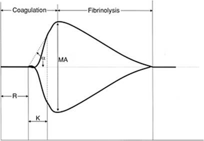
The thromboelastograph (TEG) analyzer is a bedside machine that provides a functional evaluation of overall coagulation on whole blood at the same temperature as the patient. The TEG has been shown to be a more sensitive measure of coagulation disorders than standard coagulation measures (324). The thromboelastograph assay provides a tracing that measures clotting (R value), clot formation (α angle), clot strength (maximum amplitude [MA]), and clot lysis (LY 30) (Fig. 58.5). Elongation of the R value represents a deficiency in coagulation factors. The α angle represents the rate of fibrin accumulation and cross-linking, which can be affected by fibrinogen function and, to a lesser degree, platelet function. The MA is a measure of clot strength and is affected primarily by platelets and, to a lesser degree, fibrinogen. A study investigating the utility of the TEG in trauma patients found that 65% of patients were hypercoagulable and 10% were hypocoagulable. Of the seven hypocoagulable patients, only one had an elevated PT and PTT, but six of seven required blood transfusion (325). Only the Injury Severity Score (ISS) and TEG were predictive of early transfusion.
|
|
|
Figure 58.5. Thromboelastogram. The thromboelastograph (TEG) analyzer is a bedside machine that provides a functional evaluation of overall coagulation on whole blood at the same temperature as the patient. The thromboelastograph assay provides a tracing that measures time to clot formation (R value), speed to a certain clot strength (K value), rate of clot formation (α angle), overall clot strength (maximum amplitude [MA]) and clot lysis (LY 30). |
A large volume of literature exists describing the use of the TEG in various settings including trauma, transplant, and cardiac surgery (250,325,326,327,328,329,330). However, despite its many advantages, the TEG has not become the standard of care for measurement of coagulopathy. Using the TEG, whole blood samples must be run within 3 to 4 minutes of collection, necessitating the presence of multiple machines in critical areas of the hospital. Quality control of each of these machines is work intensive. Differences due to age, gender, blood collection sites, and sample stability have been raised (331,332,333,334,335,336). Finally, accurate readings require appropriate processing, and intensive ongoing education of hospital staff would be necessary to ensure accurate results. Although the real-time functional results of routine TEG analysis would be clinically useful, the current processing and maintenance requirements make it impractical for routine use.
Rapid Definitive Control of Bleeding
Multiple studies have confirmed that patients in need of emergency surgery for ongoing hemorrhage have a better survival if the elapsed time to definitive care is minimized (337,338,339,340,341,342,343,344,345,346,347). Those patients with unnecessary delays in diagnosis and definitive treatment will have increased morbidity and mortality (344). Although there are no prospective randomized trials confirming this, multiple retrospective studies provide ample data to confirm the validity of this strategy. In trauma, early surgical control of hemorrhage has been associated with improved survival in penetrating vascular injuries (337), duodenal injuries (338), and polytrauma patients in extremis (339). A multicenter retrospective review of over 500 deaths in the operating room concluded that delayed transfer to the operating room was a cause of death that could be avoided by shortening the time to diagnosis and resuscitation (348). Similar results have been documented in the treatment of patients with ruptured abdominal aortic aneurysms who are hemodynamically unstable (345,346,347). The benefit for rapid transport time to the operating room is not as dramatic for patients who are hemodynamically stable following ruptured abdominal aortic aneurysm (AAA), implying that ongoing hemorrhage has been arrested in this group of patients.
The development of trauma systems has significantly contributed to improved trauma outcomes by triaging more severely injured patients to hospitals that have systems in place to rapidly diagnose, resuscitate, and definitively treat patients with hemorrhagic shock (340,341,342,343). The implementation of trauma systems has resulted in improved outcomes in severely injured patients, decreased time to operating room in hypotensive patients, decreased complications, decreased hospital length of stay, and decreased mortality, especially in patients with severe injury as measured by an ISS of >15 (343). Definitive prompt care is critical to optimize outcomes in patients with trauma and hemorrhage.
Lethal Triad of Resuscitation: Hypothermia, Acidosis, and Coagulopathy
Patients with severe hemorrhagic shock requiring massive resuscitation are at risk for exhaustion of their physiologic reserves, leading to irreversible shock and the inability to recover despite ongoing resuscitation. The common denominator in these patients is the development of the “lethal triad,” “bloody vicious cycle,” or “spiral of death”—terms used to describe the combination of profound acidosis, hypothermia, and coagulopathy (Fig. 58.6). Each of these factors has been independently associated with increased risk of death (268,303,319,349,350,351,352,353,354). There also seems to be a cumulative synergistic effect for each of these risk factors in patients with hemorrhagic shock. In one retrospective study of 39 patients with abdominal packing for surgically uncontrollable bleeding (351), five risk factors for death were identified: pH <7.18, temperature ≤33°C, PT ≥16, PTT ≥50, and transfusion greater than 10 units of blood. Patients with zero to one risk factor had an 18% mortality, two to three risk factors 83% mortality, and four to five risk factors 100% mortality. Similar findings were reported by Cosgriff et al., who identified risk factors for the development of life-threatening coagulopathy (352). Patients with an ISS of >25, pH <7.1, temperature less than 34°C, and systolic blood pressure ≤70 mm Hg had a 98% chance of developing life-threatening coagulopathy, whereas patients with none of these risk factors had a 1% chance of developing coagulopathy.
|
|
|
Figure 58.6. Lethal triad of hemorrhagic shock. The development of acidosis, hypothermia, and coagulopathy during resuscitation from hemorrhagic shock is described as the “lethal triad,” “bloody vicious cycle,” or “spiral of death.” Each of these factors has been independently associated with mortality. There is a cumulative synergistic effect for each of these variables, such that irreversible shock may develop if all factors are present. |
The development of profound acidosis, hypothermia, and coagulopathy is a lethal combination in patients during the resuscitation from hemorrhagic shock. Resuscitation strategies should be designed at limiting the development of these complications.
Damage Control Laparotomy
Damage control laparotomy (355) is a concept that was initially introduced by Pringle in 1908 when he described the use of hepatic sutures over packs to control bleeding. In 1913, Halsted detailed the procedure and modified its techniques. During World War II, damage control laparotomy fell out of favor and was not reintroduced until 1955 when Madding et al. reported the use of packs to temporize intraoperative bleeding, but felt that they needed to be removed prior to abdominal closure. In the 1970s, Ledgerwood had successful case reports using abdominal packing to control bleeding following trauma. However, the modern era of damage control laparotomy is attributed to Stone et al. who, in 1983, described the techniques of abbreviated laparotomy, packing to control hemorrhage, and deferred definitive surgical repair of injuries until coagulation had been established (356). Since then, a number of authors have described the beneficial effects of damage control laparotomy (357,358,359,360,361,362,363,364,365,366). Although retrospective, studies have documented a nearly 50% decrease in operative times for the most severely injured patients treated by this approach and salvage rates of 20% to 60% in patients who would have formerly died in the operating room (351,353,358,367).
The principle of damage control surgery is to obtain rapid control of hemorrhage and contamination, with early completion of the operation, with a goal to restore normal physiology as opposed to normal anatomy (Table 58.7). The ultimate goal is to prevent patients from exhausting their physiologic reserves by developing the “lethal triad of death,” or profound hypothermia, acidosis, and coagulopathy. Damage control surgery has three basic components. First, an abbreviated laparotomy for control of bleeding, control of contamination, and restitution of blood flow are necessary. The goal is to achieve these end points as quickly as possible without spending unnecessary time on traditional organ repairs that can be performed at a later time. The abdomen is packed and a temporary abdominal closure is performed (Fig. 58.7). The second component involves treatment in the intensive care unit that is focused on core rewarming, correction of acidosis, and reversal of coagulopathy, as well as optimizing ventilation and hemodynamic status. This phase will typically last for 24 to 48 hours. The third component is definitive surgical repair that is performed only when target parameters have been achieved. This sequence may require several operative interventions to attain definitive repair, each with a goal of preserving the physiologic reserve of the patient.
|
Table 58.7 Damage control laparotomy and damage control resuscitation |
||||
|
||||
|
|
||||
|
Figure 58.7. Open abdomen with temporary abdominal closure. A key component to damage control laparotomy is to perform an abbreviated laparotomy for control of bleeding, control of contamination, and restitution of blood flow. The goal is to achieve these end points as quickly as possible to avoid the development of irreversible shock and the lethal triad of acidosis, hypothermia, and coagulopathy. Frequently, the abdomen is temporarily closed and the fascia left open to prevent the development of abdominal compartment syndrome during resuscitation. Re-exploration usually takes place 24 to 48 hours later, and every few days thereafter until the fascia is closed. Occasionally, the fascia cannot be reapproximated due to loss of domain. In this circumstance, a ventral hernia remains, which can be repaired at a later date (usually 6–12 months postinjury). |
Damage Control Resuscitation
Damage control resuscitation (DCR) is a term that has recently been coined to describe a specific strategy during the resuscitation phase of trauma care (250,368,369). It should be initiated within minutes of presentation and is meant to preemptively address issues associated with resuscitating critically injured patients: prevention of hypothermia, acidosis, and coagulopathy. Damage control resuscitation involves two components: hypotensive resuscitation and hemostatic resuscitation (Table 58.7).
Hypotensive Resuscitation
Hypotensive resuscitation refers to the concept that fluid should be administered at a rate that returns the systolic blood pressure to a safe but lower than normal pressure until operative control of bleeding can be established. The traditional treatment of hemorrhaging patients has used early and aggressive fluid administration to restore blood volume. However, this approach may increase hydrostatic pressure on the wound or injured vessel, leading to dislodgement of blood clots, a dilution of coagulation factors, and undesirable cooling of the patient. Low-volume fluid resuscitation, or “permissive hypotension,” may avoid the adverse effects of early aggressive resuscitation while maintaining a level of tissue perfusion adequate for short periods. This strategy has been suggested historically for the management of ruptured abdominal aortic aneurysm patients (370), and has recently regained attention in the trauma population (263,264,265,266,371,372,373,374,375). It has shown promise in animal and human trials (263,265), but has yet to be confirmed in large-scale prospective randomized human clinical trials. Overall, data have been mixed (264). A recent animal trial suggests that increases in blood pressure are well tolerated without exacerbating hemorrhage when they are achieved gradually and with a significant delay following the injury (376). Finally, a recent Cochrane Database review found that there was not conclusive evidence from randomized controlled trials for or against early or larger volumes of intravenous fluid resuscitation in uncontrolled hemorrhage (377).
Although the concept of permissive hypotension seems promising in some circumstances, further work needs to be done. In addition, it requires extraordinarily tight control by an experienced physician who is guiding fluid resuscitation moment to moment. Hypotensive resuscitation should not be considered in patients with traumatic brain injury and spinal cord injury where adequate cerebral perfusion pressure is crucial to ensure tissue oxygenation (229,375). It should also be carefully considered in elderly patients and may be contraindicated in patients with a history of chronic hypertension (229). At the present time, it is considered experimental, and should be employed only in specific circumstances with experienced physicians.
Hemostatic Resuscitation
Conventional resuscitation practice for damage control has focused on rapid reversal of acidosis and prevention of hypothermia. Surgical techniques are aimed at controlling hemorrhage and contamination rapidly, with definitive repair occurring following hemodynamic stabilization. However, direct treatment of coagulopathy early has been relatively neglected, and has been viewed as a byproduct of resuscitation, hemodilution, and hypothermia. Delay in availability of blood products due to current blood banking techniques has also hindered the ability to employ immediate resuscitation with clotting factors.
It has now been demonstrated that acute traumatic coagulopathy is present in 25% to 30% of critically injured patients on arrival to the emergency department (318,319,320,321). The presence of coagulopathy may be even higher in patients with severe closed head injury, with an incidence of 21% to 79% when stratified by ISS (378). It has also been shown that the presence of early coagulopathy is an independent predictor of mortality following trauma (319). Early acute traumatic coagulopathy appears to be due to alterations in the thrombomodulin–protein C pathway rather than consumption of coagulation factors (321); however, additional work needs to be done to clarify these mechanisms.
Hemostatic resuscitation employs blood components early in the resuscitation process to restore both perfusion and normal coagulation function while minimizing crystalloid use. Lactated Ringer solution and normal saline resuscitation have been shown to increase reperfusion injury and leukocyte adhesion (252,253,254,255,256,379). As such, standard crystalloid resuscitation may worsen, presenting acidosis and coagulopathy in severely injured patients. Several retrospective studies in trauma have shown that survival is associated with an increased use of clotting factors (352,367). Many other studies have recommended more aggressive use of clotting factors to treat and correct underlying coagulopathy (380,381,382,383,384,385,386,387,388).
Although additional prospective studies need to be completed, there seems to be increased literature supporting early aggressive resuscitation with clotting factors while minimizing crystalloid use during massive resuscitation. Current military experience has moved to using thawed plasma as the primary resuscitation fluid in at least a 1:1 or 1:2 ratio with packed red blood cells (368,387,388,389). Continued resuscitation occurs with a massive transfusion protocol at a ratio of 6 units of plasma, 6 units of packed red blood cells, 6 units of platelets, and 10 units of cryoprecipitate. Recombinant factor VIIa is occasionally used along with early red cell transfusion to promote early hemostasis (368,389).
Massive Transfusion
Definition
In the 1970s, massive transfusion was defined as greater than 10 units of blood transfused in a 24-hour period of time, and survival rates were dismal (6.6%) (390). Over the last two decades, however, survival rates have improved, and the criteria to define massive transfusion have evolved (367,383,384,391,392,393,394,395,396). Recent reports use variable end points, increasing the number of transfusions to greater than 20 units in 24 hours (383) or defining transfusions during the entire hospital stay (392). Most recently, investigators have reported outcomes on patients receiving over 50 units of blood in 48 hours (367).
|
Table 58.8 Survival following massive transfusion |
||||||||||||||||||||||||||||||||||||||||||||||||
|
Survival
As technology and blood banking procedures have improved, patient outcomes following massive transfusion have also improved (397). Although early survival rates were dismal, recent reviews report survival rates as high as 60% in patients requiring over 50 units of blood in the early resuscitation period (Table 58.8). Moreover, many of these patients can ultimately return to work (75% of survivors), and survival in elderly patients has also been reported in several studies (367,392,394,395). This dramatic improvement in survival over the past several decades can be attributed to many factors including an improved understanding of the consequences of massive resuscitation (268,349,352), improved technology for massive resuscitation (i.e., rapid transfusion with warmed fluids) (267), increased use of damage control techniques (353,367), improved trauma systems (343), improved transfusion practices during resuscitation (367), and improved blood banking techniques (367). Based on these results, massive transfusion in trauma patients receiving over 50 units of blood in the acute period following injury is justified, with acceptable survival and functional capacity following discharge (367,383,384,391,392,393,394,395,396,397).
Transfusion Protocols
Historically, massive transfusion protocols have been developed to assist clinicians in the resuscitation of hemorrhaging patients. However, a recent review of massive transfusion protocols globally revealed wide variation in practice (389). Most protocols recommend empiric strategies of 1 unit of fresh frozen plasma (FFP) for every 4 to 10 units of packed red blood cells (PRBCs) and 1 plateletpheresis for every 10 to 20 units of PRBCs (380). Organizational guidelines recommended transfusing to laboratory end points (322). However, recent literature suggests that a more aggressive approach is warranted (368,380,387,388). During ongoing hemorrhage, the clinical situation of the patient changes too rapidly to depend on laboratory values sent 20 to 45 minutes prior. Thus, empiric strategies to correct or avoid coagulopathy need to be employed. These strategies need to be modified for two clinical conditions: (a) ongoing hemorrhage and (b) coagulopathy following hemorrhage control.
Resuscitation during Ongoing Hemorrhage
Recent data have established that a significant number of patients have an acute traumatic coagulopathy on presentation to the emergency department immediately following injury. Moreover, it appears that traditional strategies for resuscitation have been inadequate and unable to reverse this coagulopathy early following trauma (387). Several retrospective clinical studies suggest that aggressive early transfusion of clotting factors (fresh frozen plasma, platelets, and cryoprecipitate) is associated with increased survival (352,367,380,385,388).
Randomized prospective controlled trials evaluating therapy for massive hemorrhage are challenging. However, recently two studies utilizing computer modeling have suggested that more aggressive resuscitation is necessary to correct coagulopathy. In the first study, the optimal replacement ratios were 2:3 of FFP to PRBCs and 8:10 of platelets to PRBCs (398). Interestingly, a second computer model found similar results, suggesting a transfusion rate of 1:1 for FFP per unit of blood transfused if there is no pre-existing coagulopathy, and a ratio of 1.5 units of FFP per unit of PRBCs if coagulopathy exists on presentation (399). Moreover, both of these mathematical models underestimate the potential need for clotting factor replacement because only dilutional coagulopathy is taken into account; there is no assessment for the relative contributions of consumption, acidosis, and hypothermia, which are frequently seen in the acute trauma patient.
Based on these data, old transfusion strategies appear to be fundamentally flawed in the acutely hemorrhaging patient. Current recommendations suggest that, in patients with ongoing hemorrhage, empiric transfusion should occur in the ratio of 1:1:1 (1 unit of fresh frozen plasma to 1 unit of packed red blood cells to 1 unit of platelets) (Table 58.9) (368,380,387,388). Since platelets are usually supplied pooled as 6 to 10 units or as a plateletpheresis, which is equivalent to 6 to 10 units, patients should receive a platelet transfusion for every 6 to 10 units of PRBCs. When blood products are available, crystalloids should be avoided in the acutely hemorrhaging patients as they can worsen coagulopathy.
It is important to note that while this is the recommended transfusion protocol for patients with traumatic injury and hemorrhage, it is unclear if the same blood products and clotting factors are necessary in nontrauma patients with acute hemorrhage (i.e., intra-operative, gastrointestinal, ruptured aneurysm) (322,400,401,402,403). In blood loss during elective surgery, the situation is more controlled and resuscitation can be initiated immediately. A premorbid coagulopathy may or may not exist. Goals in resuscitation in this circumstance are similar: maintain adequate tissue perfusion to avoid acidosis, correct coagulopathy, and prevent hypothermia.
|
Table 58.9 Recommended empiric massive transfusion protocol for acute ongoing hemorrhage |
||
|
Resuscitation Once Hemorrhage Is Controlled
Once hemorrhage is controlled, goal-directed transfusion can be pursued based on laboratory data and clinical variables. Many clinicians will work to achieve normal coagulation parameters for 24 hours post injury and control of hemorrhage (PT INR <1.5, platelet count >100 × 109 per liter) (387). In a patient who no longer shows evidence of medical or surgical bleeding, traditional guidelines for transfusion therapy can be employed (322). These include using red blood cell transfusion for symptomatic anemia or a hemoglobin concentration of less than 7 g/dL; FFP or prothrombin complex concentrate for a prothrombin time >1.5 times normal; fresh frozen plasma for an activated partial thromboplastin time >1.5 times normal; cryoprecipitate for a fibrinogen level <80 to 100 mg/dL; and platelet transfusion for a platelet count <50 × 109 per liter (Table 58.6).
Complications of Massive Transfusion
Despite acceptable survival rates, there are several known complications to massive transfusion (Tables 58.10 and 58.11) (397,404,405,406,407,408,409). Physicians caring for patients who require massive transfusion must anticipate, identify, and rapidly treat these potential complications in order to optimize outcome.
Disordered hemostasis following massive transfusion is a known complication of massive blood transfusion (397,400,401). Stored blood is lacking in factors V and VIII. These factors degrade over time in stored blood, and thus become deficient in the massively transfused patient. This can contribute to the coagulopathy seen following massive transfusion. Dilutional thrombocytopenia also occurs during massive transfusion, and is more common after 1.5 times the normal blood volume is transfused. However, thrombocytopenia can occur earlier, especially if there is disseminated intravascular coagulation, pre-existing thrombocytopenia, or a consumptive coagulopathy. As discussed previously, resuscitation of hemorrhagic shock must include clotting factors and platelets to avoid ongoing coagulopathy.
|
Table 58.10 Noninfectious transfusion-associated complications |
|
|
Oxygen delivery to tissues is also affected by blood transfusion. Transfused blood tends to have a higher affinity for oxygen, thus leading to decreased oxygen delivery to tissues. Longer storage periods for blood lead to a reduction in red cell deformability, altered red cell adhesiveness, and other red cell storage lesions. These changes reduce red blood cell viability after transfusion, reduce tissue oxygen availability, and promote the inflammatory response, specifically neutrophil priming and pulmonary endothelial cell activation.
Systemic inflammation and potential tissue injury may also be induced by the transfusion of aged blood. Transfusion of aged blood (>14 days of storage) in the first 6 hours of resuscitation has been shown to be an independent risk factor for postinjury multiorgan failure (410) and is associated with delayed apoptosis of neutrophils (411), increased infection rates (412), and a longer ICU stay (413). This may be particularly significant in large trauma centers and transplantation centers where older blood is preferentially distributed because of their high-volume use.
|
Table 58.11 Infectious transfusion-associated complications |
||||||||||||||||||
|
||||||||||||||||||
Alloimmunization can occur when an immunocompetent host develops an immune response to donor antigens. The antigens most often involved include the human leukocyte antigen (HLA) class I and II on platelets and leukocytes, granulocyte-specific antigens, platelet-specific antigens, and red blood cell–specific antigens. Consequences of alloimmunization include a refractory response to platelet transfusion, posttransfusion purpura, neonatal alloimmune thrombocytopenia, acute intravascular hemolytic transfusion reaction, hemolytic disease in newborns, and febrile nonhemolytic reactions against granulocytes. Clinical manifestations can be minor, such as fever, leading to active bleeding and hemolysis, which can be fatal. Workup and treatment vary, depending on the severity of the reaction (414,415).
Metabolic and electrolyte disturbances can also occur following massive transfusion (397). Citrate toxicity can occur in patients with abnormal liver function or in whom the administration of blood is very rapid. The healthy adult liver will metabolize 3 g of citrate every 5 minutes. Each unit of blood contains approximately 3 g of citrate. Therefore, transfusion rates higher than 1 unit every 5 minutes can exceed the liver's capacity to handle this overload. Citrate then binds to calcium and can lead to clinical hypocalcemia. Patients may exhibit temporary tetany and hypotension. Calcium replacement should occur concurrently during massive transfusion.
Electrolyte disturbances such as hyperkalemia or hypokalemia can occur with massive transfusion. The longer the shelf life, the higher the potassium concentration; sometimes concentrations may even exceed 30 mmol/L. Unless very large amounts of blood are transfused, hyperkalemia is generally not a problem. On the other hand, as red cells begin active metabolism, intracellular uptake of potassium begins, and hypokalemia may result.
Acid-base disturbances can also occur with massive blood transfusions. Stored blood contains lactate at levels up to 30 to 40 mmol/L. In addition, citric acid is present and may be metabolized to bicarbonate, resulting in severe metabolic alkalosis. Conversely, the patient's overall condition and tissue hypoperfusion may actually lead to metabolic acidosis.
Although rare, blood transfusion can also result in the induction of acquired inhibitors of coagulation. The most common antibodies are directed against coagulation factor VIII. This can result in massive bleeding, which is difficult and costly to treat. The main goals of treatment are to stop hemorrhage and remove the inhibitor. Factor VIII concentrate is used only for life-threatening circumstances. Successful elimination of the anti-VIII antibody has been accomplished with the use of oral immunosuppressants such as cyclophosphamide and prednisone (416).
Transfusion-related acute lung injury
TRALI is a devastating complication of transfusion that consists of a syndrome that includes dyspnea, hypotension, bilateral pulmonary edema, and fever. Its incidence is reported to be between 0.04% and 0.06% (or approximately 1 in 2,000). Clinically, it resembles acute respiratory distress syndrome. Criteria for the diagnosis of TRALI include:
· Acute lung injury (ALI) as defined by acute onset, hypoxemia (PaO2: FiO2 ratio ≤300), bilateral infiltrates on frontal chest radiograph, and no evidence of left atrial hypertension or circulatory overload
· No pre-existing ALI before transfusion
· Occurs during or within 6 hours of transfusion
· No temporal relationship to an alternative risk factor for ALI (i.e., burns, aspiration, multiple trauma, cardiopulmonary bypass, sepsis, etc.)
If an additional risk factor exists, then possible TRALI is diagnosed (Table 58.12) (417,418,419,420).
The pathogenesis can be either immune (antibody) mediated or nonimmune mediated (Table 58.13) (418,419,420,421,422). Immune-mediated TRALI is most common and is due to the presence of leukocyte antibodies in the donor transfusion (421,422). These antibodies form immune complexes that are deposited in the pulmonary vascular bed, leading to release of vasoactive substances, leakage of fluid into alveolar spaces, activation of complement, leukostasis, and activation of polymorphonuclear neutrophils. Immune-mediated TRALI occurs more commonly with fresh frozen plasma than with platelet concentrates, is associated with multiparous female donors (423), can occur in healthy recipients, and is usually severe, requiring mechanical ventilation in 70% of individuals. Non–immune-mediated TRALI is thought to be due to the presence of biologically active lipids in the donor transfusion (418,419,420). It occurs with stored platelet concentrates more commonly than stored red cells, occurs predominantly in critically ill patients with a primed immune system, and is usually mild and transient, requiring only supplemental oxygen support. Treatment of TRALI is generally supportive and includes ventilatory and hemodynamic assistance. There are no data to support the use of corticosteroids, and additional blood component therapy should be given only if transfusion needs exist. The diagnosis of TRALI in the patients requiring massive transfusion is difficult because of the many other etiologies also present that can lead to acute lung injury (424,425).
|
Table 58.12 Criteria for transfusion-related acute lung injury (TRALI) |
||||||||||||||||||
|
||||||||||||||||||
|
Table 58.13 Characteristics of immune and nonimmune transfusion-related acute lung injury (TRALI) |
||||||||||||||||||
|
||||||||||||||||||
Recombinant Factor VIIA in Massive Transfusion and Hemorrhage
New developments in transfusion therapy include the discovery and use of recombinant coagulation factor VIIa (rFVIIa). Recombinant factor VIIa is a synthesized analog of human factor VII that has been used effectively in the treatment of patients with hemophilia as well as other congenital and acquired coagulopathies. Recently, there have been reports of the successful use of rFVIIa in treating coagulopathic trauma patients (426). In this study, patients with active hemorrhage and clinical coagulopathy from diverse causes such as traumatic hemorrhage, traumatic brain injury, warfarin use, congenital factor VII deficiency, and other acquired hematologic defects were administered rFVIIa as a last resort. Coagulopathy was reversed in 75% of patients, with an associated decrease in prothrombin time. Forty-two percent of patients survived to discharge.
Recently, two randomized, prospective, placebo-controlled, double-blind clinical trials were conducted simultaneously to evaluate the efficacy and safety of recombinant factor VIIa as adjunctive therapy for the control of bleeding in patients with severe blunt (N = 143) or penetrating (N = 134) trauma (427). In blunt trauma, the red blood cell transfusion requirement was significantly reduced by 2.6 units (p = 0.02) and the need for massive transfusion (>20 units of packed red blood cells) was reduced (14% vs. 33%, p = 0.03). In patients with penetrating trauma, the trends were similar, but not significant (reduction in red cell transfusion 1.0 unit, p = 0.10; massive transfusion 7% vs. 19%, p = 0.08). Trends toward reduction in mortality and critical complications were also observed. A subgroup analysis from this trial found particular benefit in those patients who were coagulopathic on presentation to the emergency department, with a significant decrease in transfusion of packed red blood cells, fresh frozen plasma, platelets, and need for massive transfusion. In addition, treatment with rFVIIa was also associated with a significant reduction in multiorgan failure and/or acute respiratory distress syndrome (3% vs. 20%, p = 0.004), without an increase in thromboembolic events (428).
Recombinant factor VIIa, however, must be used responsibly. Recent studies have shown that early administration following trauma is more effective than late administration (429,430). Furthermore, the presence of profound acidosis, coagulopathy, and signs of irreversible hemorrhagic shock predict failure of rFVIIa therapy (431). Current recommendations suggest that optimal preconditions should be present prior to administration of rFVIIa, which include a fibrinogen concentration of >50 mg/dL, a platelet count of >50 × 109 per liter, and a pH ≥7.2 (432,433). Although early results in traumatic hemorrhage appear promising, rFVIIa should still be considered experimental and further investigation is warranted. Recombinant factor VIIa has shown promise for perioperative bleeding during liver transplantation (434) and in patients undergoing cardiac surgery (435,436). Investigations for its utility in perioperative bleeding for other surgical procedures have been mixed (437,438,439,440,441,442).
A prospective randomized trial investigating the use of rFVIIa in patients with cirrhosis and upper gastrointestinal bleeding who were treated with standard endoscopic therapy and pharmacologic interventions showed that the administration of rFVIIa was not more effective than placebo with respect to the primary end point of failure to control bleeding within 24 hours and failure to prevent rebleeding or death within 5 days (443). However, subgroup analysis of patients with more severe cirrhosis showed that rFVIIa showed a reduction in the composite primary end point (8% vs. 23%, p = 0.03). None of the rFVIIa patients had rebleeding within the first 24 hours, whereas rebleeding occurred in 11% of the placebo group (p = 0.01) (443).
As with any hemostatic agent, there are concerns over the potential thrombogenicity of rFVIIa (444). Although preliminary evidence shows a favorable safety profile (445,446), thrombogenic effects are being followed closely in ongoing clinical trials.
Prevention of Hemorrhagic Shock
Antifibrinolytic Therapy
Antifibrinolytic therapy has been shown to significantly reduce the risk of bleeding following cardiac surgery in several randomized controlled trials. Aprotinin has been studied most extensively (447,448,449,450,451), followed by tranexamic acid, then aminocaproic acid. However, recent information raises concern for the risk of renal failure following use of aprotinin with cardiac surgery (448,449) and a potential for increased mortality at 5 years following use of aprotinin (450). Although clearly the risk of postoperative bleeding following cardiac surgery is reduced, further investigation into these potential side effects is warranted.
A recent systematic review of randomized controlled trials of antifibrinolytic agents (mainly aprotinin or tranexamic acid) in elective surgical procedures identified 89 trials including 8,580 randomized patients (74 cardiac, eight orthopedic, four liver, three vascular). Results demonstrated that these treatments reduced the number of patients needing transfusion by one third, reduced the volume needed per patient by 1 unit, and halved the need for further surgery to control bleeding. These differences were all statistically significant. There was also a trend toward a reduction in the risk of death (risk ratio = 0.85; 95% confidence interval, 0.63–1.14), although this was not statistically significant (452).
To date, there are limited data on the use of antifibrinolytic agents in other clinical scenarios (453). However, at this time, the CRASH-2 trial (Clinical Randomization of an Anti-fibrinolytic in Significant Hemorrhage) is ongoing in Europe and is designed to evaluate the utility of antifibrinolytic agents in the management of acute traumatic injury (454).
Experimental Therapy
Red Cell Substitutes
Although the blood supply in the United States is safe and currently has sufficient capacity to meet most patient needs, there is room for considerable improvement. The current system is dependent on blood donors on a regular basis, and the blood supply is subject to seasonal shortages due to holidays and convenience. The gap between the donor pool and the increasing transfusion requirements of an aging population is narrowing, and shortages are becoming more frequent. The risk of transmission of known infectious diseases still exists (Table 58.11), while the threat of new and emerging infections such as West Nile virus and Creutzfeldt-Jakob disease underscore the risk of a tainted blood supply (455).
The ideal red cell substitute should have several characteristics including an ability to deliver (and potentially enhance) oxygen delivery, no risk of disease transmission, no immunosuppressive effects, available in abundant supply, universally compatible, prolonged shelf life, similar in vivo half-life to the red blood cell, available at a reasonable cost, easy to administer, able to access all areas of the human body (including ischemic tissues), and effective at room air or ambient conditions (455,456,457). There have been many attempts to develop red cell substitutes since 1934 when Amberson first reported the successful use of a bovine hemolysate for exchange transfusions in cats and dogs (455). However, this work could not be replicated.
The two main types of oxygen carriers that are used as red blood cell substitutes are hemoglobin-based oxygen carriers (HBOCs) and perfluorocarbons (PFCs). Based on previous clinical trials, many obstacles still need to be overcome. Adverse effects associated with HBOCs include (a) severe vasoconstriction due to binding of nitric oxide and dysregulation of endothelin; (b) nephrotoxicity; (c) interference of macrophage function; (d) antigenicity; (e) oxidation on storage; (f) activation of complement, kinin, and coagulation; (g) iron deposition with concerns of hemochromatosis and iron overload; (h) gastrointestinal distress; (i) neurotoxicity; (j) free radical generation; and (k) interference with diagnosis of transfusion reaction. Adverse effects of PFCs include (a) limited shelf life, (b) flulike symptoms during infusion, (d) complement and phagocytic activation, and (d) short circulation time (456).
Despite much research, no product to date has been able to fulfill all of the previously mentioned criteria or meet the U.S. Food and Drug Administration's requirements of purity, potency, and safety. At the time of this publication, three HBOC products continue in advanced clinical trials (455).
Hypertonic Saline
Hypertonic saline (7.5% saline ± 6% dextan-70) has been investigated as an alternative resuscitation strategy in critically injured patients (458,459,460,461,462,463,464). Hypertonic resuscitation evokes an increase in serum osmolarity, which results in the redistribution of fluid from the interstitial and intracellular space to the intravascular space. This leads to a rapid restoration of circulating intravascular volume with a small amount resuscitation fluid. Hypertonic saline has also been shown to decrease intracranial pressure via its osmotic effects (125,126). This is particularly beneficial in patients with hypovolemic shock and closed head injury due to the ability of hypertonic saline resuscitation to concurrently restore circulating blood volume, improve tissue (including cerebral) perfusion, and lower intracranial pressure (126,465,466).
Hypertonic saline resuscitation has also been shown to have significant immunomodulatory effects that could mitigate the dysfunctional inflammatory response seen after traumatic injury (108,109,111,150,253,467,468,469,470,471,472). The hypertonicity associated with hypertonic saline resuscitation is associated with significant effects on the innate and adaptive immune systems. There is suppression of the neutrophil oxidative burst, potentially leading to an attenuation of inflammatory organ injury (150,473).
Several clinical trials and meta-analyses have suggested improved outcome in patients resuscitated with hypertonic saline (474,475,476,477). Despite these results, hypertonic saline resuscitation has not gained widespread acceptance in North America. However, in 1999, the U.S. Navy, through the Office of Naval Research, requested that the Institute of Medicine (IOM) recommend that hypertonic saline be used as the initial resuscitation fluid for combat casualty (256,478). The rapid restoration of intravascular volume and possible immunomodulatory effects associated with hypertonic saline resuscitation make it an attractive alternative for the resuscitation of severe hemorrhagic shock. However, it is still considered experimental and prospective randomized trials are needed to confirm its utility.
Summary
Hemorrhagic shock is a common, yet complicated, clinical condition that physicians are frequently called upon to evaluate and treat. Diagnosis must be accurate and expedient. Therapy must be direct, efficient, and multifactorial in order to avoid the potential multisystem sequelae. Metabolism and function of all organs are altered during hemorrhagic shock. A better understanding of the pathophysiology of hemorrhagic shock has led to improved resuscitation techniques and improved survival over recent years. Damage control laparotomy and damage control resuscitation have changed the approach to management in patients with multisystem trauma and hemorrhagic shock. Staged resuscitation and operative intervention to avoid irreversible shock are now the mainstays of care. Recognition of acute traumatic coagulopathy has improved the composition of massive transfusion protocols to include increased use of clotting factors early during resuscitation. New experimental therapies for resuscitation are being evaluated and appear promising. Overall, survival following hemorrhagic shock has improved. Early diagnosis, definitive cessation of bleeding, and comprehensive hemostatic resuscitation are the key elements to successful outcome.
References
1. Weil MH, Shubin H. Proposed reclassification of shock states with special reference to distributive effects. In: Hinshaw LB, Cox BG, eds. The Fundamental Mechanisms of Shock. New York: Plenum Press; 1972:13.
2. Hamilton-Davies C, Mythen MG, Salmon JB, et al. Comparison of commonly used clinical indicators of hypovolaemia with gastrointestinal tonometry. Intens Care Med. 1997;23(3):276.
3. Bond RF, Johnson G III. Vascular adrenergic interactions during hemorrhagic shock. Fed Proc. 1985;44:281.
4. Schadt JC, Ludbrook J. Hemodynamic and neurohumoral responses to acute hypovolemia in conscious mammals. Am J Physiol. 1991;260:H305.
5. Koyama S, Sawano F, Matsuda Y, et al. Spatial and temporal differing control of sympathetic activities during hemorrhage. Am J Physiol. 1992;262:R579.
6. Wisbach G, Tobias S, Woodman R, et al. Preserving cardiac output with beta-adrenergic receptor blockade and inhibiting the Bezold-Jarisch reflex during resuscitation from hemorrhage. J Trauma. 2007;63(1):26.
7. Victorino GP, Battistella FD, Wisner DH. Does tachycardia correlate with hypotension after trauma? J Am Coll Surg. 2003;196(5):679.
8. Demetriades D, Chan LS, Bhasin P, et al. Relative bradycardia in patients with traumatic hypotension. J Trauma. 1998;45:534.
9. Sander-Jensen K, Secher NH, Bie P, et al. Vagal slowing of the heart during hemorrhage: observations from 20 consecutive hypotensive patients. Br J Med. 1986;292:364.
10. Vincent JL, Moraine JJ, Van der Linden P. Toe temperature versus transcutaneous oxygen tension monitoring during acute circulatory failure. Intensive Care Med. 1988;14:64.
11. Van Leeuwen AF, Evans RG, Ludbrook J. Haemodynamic responses to acute blood loss: new roles for the heart, brain, and endogenous opioids. Anaesth Intensive Care. 1989;17(3):312.
12. Ludbrook J, Ventura S. Roles of carotid baroreceptor and cardiac afferents in hemodynamic responses to acute central hypovolemia. Am J Physiol. 1996;270(5 Part 2):H1538.
13. Wong DH, O'Connor D, Tremper KK, et al. Changes in cardiac output after acute blood loss and position change in man. Crit Care Med. 1989;17(10):979.
14. Bressack MA, Raffin TA. Importance of venous return, venous resistance, and mean circulatory pressure in the physiology and management of shock. Chest. 1987;92:906.
15. Shen YT, Knight DR, Thomas JX Jr, et al. Relative roles of cardiac receptors and arterial baroreceptors during hemorrhage in conscious dogs. Circ Res. 1990;66(2):397.
16. Westphal G, Garrido Adel P, de Almeida DP, et al. Pulse pressure respiratory variation as an early marker of cardiac output fall in experimental hemorrhagic shock. Artif Organs. 2007;31(4):284.
17. Magder S, Lagonidis D, Erice F. The use of respiratory variations in right atrial pressure to predict the cardiac output response to PEEP. J Crit Care. 2001;16(3):108–114.
18. Rooke GA, Schwid HA, Shapira Y. The effect of graded hemorrhage and intravascular volume replacement on systolic pressure variation in humans during mechanical and spontaneous ventilation. Anesth Analg. 1995;80(5):925.
19. Zweifach BW, Fronek A. The interplay of central and peripheral factors in irreversible hemorrhagic shock. Prog Cardiovasc Dis. 1975;18(2):147.
20. Schwartz S, Frantz RA, Shoemaker WC. Sequential hemodynamic and oxygen transport responses in hypovolemia, anemia, and hypoxia. Am J Physiol. 1981;241(6):H864.
21. Alyono D, Ring WS, Chao RY, et al. Characteristics of ventricular function in severe hemorrhagic shock. Surgery. 1983;94(2):250.
22. Sarnoff SJ, Case RB, Waithe PE, et al. Insufficient coronary flow and myocardial failure as a complication factor in late hemorrhagic shock. Am J Physiol. 1954;176(3):439.
23. Rush BF Jr. Irreversibility in the post-transfusion phase of hemorrhagic shock. Adv Exp Med Biol. 1971;23:215.
24. Secher NH, Sander-Jensen K, Werner C, et al. Bradycardia during severe but reversible hypovolemic shock in man. Circ Shock. 1984;14:267.
25. Thiemermann C, Szabo C, Mitchell JA, et al. Vascular hyporeactivity to vasoconstrictor agents and hemodynamic decompensation in hemorrhagic shock is medicated by nitric oxide. Proc Natl Acad Sci. 1993;90:267.
26. Vatner SF. Effects of hemorrhage on regional blood flow distribution in dogs and primates. J Clin Invest. 1974;54:225.
27. Shoemaker WC. Relation of oxygen transport patterns to the pathophysiology and therapy of shock states. Intensive Care Med. 1987;13(4):230.
28. Aubier M, Viires N, Syllie G, et al. Respiratory muscle contribution to lactic acidosis in low cardiac output. Am Rev Respir Dis. 1982;126(4):648.
29. Revelly JP, Gardaz JP, Nussberger J, et al. Effect of epinephrine on oxygen consumption and delivery during progressive hemorrhage. Crit Care Med. 1995;23(7):1272.
30. Hannon JP, Wade CE, Bossone CA, et al. Oxygen delivery and demand in conscious pigs subjected to fixed-volume hemorrhage and resuscitated with 7.5% NaCl in 6% Dextran. Circ Shock. 1989;29(3):205.
31. Edouard AR, Degremont AC, Duranteau J, et al. Heterogeneous regional vascular responses to simulated transient hypovolemia in man. Intensive Care Med. 1994;20(6):414.
32. Groeneveld AB, Kolkman JJ. Splanchnic tonometry: a review of physiology, methodology, and clinical implications. J Crit Care. 1994;9(3):198.
33. Kirton OC, Windsor J, Wedderburn R, et al. Failure of splanchnic resuscitation in the acutely injury trauma patient correlates with multiple organ failure and length of stay in the ICU. Chest. 1998;113:1064.
34. Riggs TE, Shafer AW, Guenter CA. Acute changes in oxyhemoglobin affinity: effects on oxygen transport and utilization. J Clin Invest. 1973;52:2660.
35. Malmberg PO, Hlastala MP, Woodson RD. Effect of increased blood-oxygen affinity on oxygen transport in hemorrhagic shock. J Appl Physiol. 1979;47(4):889.
36. Woodson RD. Physiologic significance of oxygen dissociation curve shifts. Crit Care Med. 1979;7(9):368.
37. Woodson RD. Functional consequences of altered blood oxygen affinity. Acta Biol Med Ger. 1981;40:733.
38. Myburgh JA, Webb RK, Worthley LI. The P50 is reduced in critically ill patients. Intensive Care Med. 1991;17(6):355.
39. Herman CM, Rodkey FL, Valeri CR, et al. Changes in the oxyhemoglobin dissociation curve and peripheral blood after acute red cell mass depletion and subsequent red cell mass restoration in baboons. Ann Surg. 1971;174:734.
40. Sauaia A, Moore FA, Moore EE, et al. Early predictors of postinjury multiple organ failure. Arch Surg. 1994;129(1):39.
41. Shoemaker WC, Appel PL, Kram HB. Role of oxygen debt in the development of organ failure sepsis, and death in high-risk surgical patients. Chest. 1992;102(1):208.
42. Abou-Khalil B, Scalea TM, Trooskin SZ, et al. Hemodynamic responses to shock in young trauma patients: need for invasive monitoring. Crit Care Med. 1994;22(4):633.
43. Shoemaker WC, Appel PL, Kram HB, et al. Prospective trial of supranormal values of survivors as the therapeutic goals in high-risk surgical patients. Chest. 1988;94:1176–1186.
44. Bishop MH, Shoemaker WC, Appel PL, et al. Prospective randomized trial of survivor values of cardiac index, oxygen delivery, and oxygen consumption as resuscitation endpoints in severe trauma. J Trauma. 1995;38(5):780.
45. Shoemaker WC, Appel PL, Kram HB. Measurement of tissue perfusion by oxygen transport patterns in experimental shock and in high-risk surgical patients. Intensive Care Med. 1990;16(Suppl 2):S135.
46. Chang MC, Mondy JS, Meredith JW, et al. Redefining cardiovascular performance during resuscitation: ventricular stroke work, power, and the pressure-volume diagram. J Trauma. 1998;45(3):470.
47. Velmahos GC, Demetriades D, Shoemaker WC, et al. Endpoints of resuscitation of critically injured patients: normal or supranormal? A prospective randomized trial. Ann Surg. 2000;232(3):409.
48. Kern JW, Shoemaker WC. Meta-analysis of hemodynamic optimization in high-risk patients. Crit Care Med. 2002;30(8):1686.
49. Yu M, Levy MM, Smith P, et al. Effect of maximizing oxygen delivery on morbidity and mortality rates in critically ill patients: a prospective, randomized, controlled study. Crit Care Med. 1993;21(6):830.
50. Balogh Z, McKinley BA, Cocanour CS, et al. Supranormal trauma resuscitation causes more cases of abdominal compartment syndrome. Arch Surg. 2003;138:637.
51. Chaudry IH. Cellular mechanisms in shock and ischemia and their correction. Am J Physiol. 1983;245(2):R117.
52. Mongan PD, Fontana JL, Chen R, et al. Intravenous pyruvate prolongs survival during hemorrhagic shock in swine. Am J Physiol. 1999;277(6 Part 2):H2253.
53. Amundson B, Jennische E, Haljamae H. Correlative analysis of microcirculatory and cellular metabolic events in skeletal muscle during hemorrhagic shock. Acta Physiol Scand. 1980;108(2):147.
54. Ratcliffe PJ, Moonen CT. Holloway PA, et al. Acute renal failure in hemorrhagic hypotension: cellular energetics and renal function. Kidney Int. 1986;30(3):355.
55. Zager RA. Adenine nucleotide changes in kidney, liver, and small intestine during different forms of ischemic injury. Circ Res. 1991;68(1):185.
56. Pellicane JV, DeMaria EJ, Abd-Elfattah A, et al. Interleukin-1 receptor antagonist improves survival and preserves organ adenosine-5-triphosphate after hemorrhagic shock. Surgery. 1993;114(2):278.
57. Salzman AL, Vromen A, Denenberg A, et al. K(ATP)-channel inhibition improves hemodynamics and cellular energetics in hemorrhagic shock. Am J Physiol. 1997;272(2 Part 2):H688.
58. Robinson DA, Wang P, Chaudry IH. Administration of ATP-MgCl2 after trauma-hemorrhage and resuscitation restores the depressed cardiac performance. J Surg Res. 1997;69(1):159.
59. Kline JA, Maiorano PC, Schroeder JD, et al. Activation of pyruvate dehydrogenase improves heart function and metabolism after hemorrhagic shock. J Mol Cell Cardiol. 1997;29(9):2465.
60. Mizock BA, Falk JL. Lactic acidosis in critical illness. Crit Care Med. 1992;20(1):80.
61. Nakatani T, Sakamoto Y, Ando H, et al. Bile and bilirubin excretion in relation to hepatic energy status during hemorrhagic shock and hypoxemia in rabbits. J Trauma. 1995;39(4):665.
62. Sjoberg F, Gustafsson U, Lewis DH. Extracellular muscle surface pO2 and pH heterogeneity during hypovolemia and after reperfusion. Circ Shock. 1991;34(3):319.
63. Davis JM, Stevens JM, Peitzman A, et al. Neutrophil migratory activity in severe hemorrhagic shock. Circ Shock. 1983;10(3):199.
64. Sayeed MM. Ion transport in circulatory and/or septic shock. Am J Physiol. 1987;252(5 Part 2):R809.
65. Horton JW. Calcium-channel blockade in canine hemorrhagic shock. Am J Physiol. 1989;257(5 Part 2):R1012.
66. Eastridge BJ, Darlington DN, Evans JA, et al. A circulating shock protein depolarizes cells in hemorrhage and sepsis. Ann Surg. 1994;219(3):298.
67. Kiang JG. Inducible heat shock protein 70kD and inducible nitric oxide synthase in hemorrhage/resuscitation-induced injury. Cell Res. 2004;14(6):450.
68. Menezes JM, Hierholzer C, Watkins SC, et al. The modulation of hepatic injury and heat shock expression by inhibition of inducible nitric oxide synthase after hemorrhagic shock. Shock. 2002;17(1):13.
69. DeMaio A. Heat shock proteins: facts, thoughts, and dreams. Shock. 1999;11(1):1.
70. Kregel KC. Heat shock proteins: modifying factors in physiological stress responses and acquired thermotolerance. J Appl Physiol. 2002;92:2177.
71. DeMaio A. The heat-shock response. New Horizons. 1995;3(2):198.
72. Zhao Q, Zhao KS. Inhibition of L-type calcium channels in arteriolar smooth muscle cells is involved in the pathogenesis of vascular hyporeactivity in severe shock. Shock. 2007; accepted for print.
73. Carlson DE, Nguyen PX, Soane L, et al. Hypotensive hemorrhage increases calcium uptake capacity and Bcl-XL content of liver mitochondria. Shock. 2007;27(2):192.
74. Zhao K, Liu J, Jin C. The role of membrane potential and calcium kinetic changes in the pathogenesis of vascular hyporeactivity during severe shock. Chin Med J. 2000;113:59.
75. Maitra SR, Geller ER, Pan W, et al. Altered cellular calcium regulation and hepatic glucose production during hemorrhagic shock. Circ Shock. 1992;38(1):14.
76. Xu J, Liu L. The role of calcium desensitization in vascular hyporeactivity and its regulation after hemorrhagic shock in the rat. Shock. 2005;23(6):576.
77. Yang G, Liu L, Xu J, et al. Effect of arginine vasopressin on vascular reactivity and calcium sensitivity after hemorrhagic shock in rats and its relationship to Rho-kinase. J Trauma. 2006;61(6):1336.
78. Silomon M, Rose S. Effect of sodium bicarbonate infusion on hepatocyte Ca2+ overload during resuscitation from hemorrhagic shock. Resuscitation. 1998;37(1):27.
79. Silomon M, Pizanis A, Larsen R, et al. Pentoxyfilline prevention of altered hepatocyte calcium regulation during hemorrhagic shock/resuscitation. Crit Care Med. 1998;26(3):494.
80. Zhong Z, Enomoto N, Connor HD, et al. Glycine improves survival after hemorrhagic shock. Shock. 1999;12(1):54.
81. Wang G, Zhao M, Wang EH. Effects of glycine and methylprednisolone on hemorrhagic shock in rats. Chin Med J. 2004;117(9):1334.
82. Jarrar D, Chaudry IH, Wang P. Organ dysfunction following hemorrhage and sepsis: mechanisms and therapeutic approaches (review). Int J Mol Med. 1999;4(6):575.
83. Lieberthal W, McGarry AE, Sheils J, et al. Nitric oxide inhibition in rats improves blood pressure and renal function in hypovolemic shock. Am J Physiol. 1991;261(5 Part 2):F868.
84. Szabo C, Farago M, Horvath I, et al. Hemorrhagic hypotension impairs endothelium-dependent relaxations in the renal artery of the cat. Circ Shock. 1992;36(3):238.
85. Dignan RJ, Wechsler AS, DeMaria EJ. Coronary vasomotor dysfunction following hemorrhagic shock. J Surg Res. 1992;52(4):382.
86. Bitterman H, Brod V, Weisz G, et al. Effects of oxygen on regional hemodynamics in hemorrhagic shock. Am J Physiol. 1996;271(1 Part 2):H203.
87. Szabo C, Csaki C, Benyo Z, et al. Role of L-arginine-nitric oxide pathway in the changes in cerebrovascular reactivity following hemorrhagic hypotension and retransfusion. Circ Shock. 1992;37(4):307.
88. Guarini S, Bini A, Bazzani C, et al. Adrenocorticotropin normalizes the blood levels of nitric oxide in hemorrhage-shocked rats. Eur J Pharmacol. 1997;336(1):15.
89. Pannen BH, Kohler N, Hole B, et al. Protective role of endogenous carbon monoxide in hepatic microcirculatory dysfunction after hemorrhagic shock in rats. J Clin Invest. 1998;102(6):1220.
90. Szabo C, Billiar TR. Novel roles of nitric oxide in hemorrhagic shock. Shock. 1999;12(1):1.
91. Kiang JG, Bowman PD, Lu X, et al. Geldanamycin inhibits hemorrhage-induced increases in caspase-3 activity: role of inducible nitric oxide synthase. J Appl Physiol. 2007;103:1045.
92. Ng KC, Moochhala SM, Md S, et al. Preservation of neurological functions by nitric oxide synthase inhibitors following hemorrhagic shock. Neuropharmacology. 2003;44(2):244.
93. Hierbolzer C, Billiar TR, Tweardy DJ, et al. Reduced hepatic transcription factor activation and expression of IL-6 and ICAM-1 after hemorrhage by NO scavenging. Arch Orthop Trauma Surg. 2003;123(2–3):55.
94. Hierbolzer C, Menezes JM, Ungeheuer A, et al. A nitric oxide scavenger against pulmonary inflammation following hemorrhagic shock. Shock. 2002;17(2):98.
95. Kobara M, Tatsumi T, Takeda M, et al. The dual effects of nitric oxide synthase inhibitors on ischemia-reperfusion injury in rat hearts. Basic Res Cardiol. 2003;98(5):319.
96. Adachi T, Hori S, Miyazaki K, et al. Inhibition of nitric oxide synthesis aggravates myocardial ischemia in hemorrhagic shock in constant pressure model. Shock. 1998;9(3):204.
97. Thompson A, Valeri CR, Lieberthal W. Endothelin receptor A blockade alters hemodynamic response to nitric oxide inhibition in rats. Am J Physiol. 1995;269(2 Part 2):H743.
98. Rothe CF, Drees JA. Vascular capacitance and fluid shifts in dogs during prolonged hemorrhagic hypotension. Circ Res. 1976;38(5):347.
99. Schlichtig R, Kramer DJ, Pinsky MR. Flow redistribution during progressive hemorrhage is a determinant of critical O2 delivery. J Appl Physiol. 1991;70(1):169.
100. Chien S. Role of the sympathetic nervous system in hemorrhage. Physiol Rev. 1967;47(2):214.
101. Forsyth RP, Hoffbrand BI, Melmon KL. Redistribution of cardiac output during hemorrhage in the unanesthetized monkey. Circ Res. 1970;27(3):311.
102. Rothe CF, Drees JA. Vascular capacitance and fluid shifts in dogs during prolonged hemorrhagic hypotension. Circ Res. 1976;38(5):347.
103. Zweifach BW. Mechanisms of blood flow and fluid exchange in microvessels: hemorrhagic hypotension model. Anesthesiology. 1974;41(2):157.
104. Prist R, Rocha-e-Silva M, Scalabrini A, et al. A quantitative analysis of transcapillary refill in severe hemorrhagic hypotension in dogs. Shock. 1994;1(3):188.
105. Childs EW, Udobi KF, Hunter FA, et al. Evidence of transcellular albumin transport after hemorrhagic shock. Shock. 2005;23(6):565.
106. Schumacher J, Binkowski K, Dendorfer A, et al. Organ-specific extravasation of albumin-bound Evans blue during non-resuscitated hemorrhagic shock in rats. Shock. 2003;20(6):565.
107. Boyd AJ, Rubin BB, Walker PM, et al. A CD18 monoclonal antibody reduces multiple organ injury in a model of ruptured abdominal aortic aneurysm. Am J Physiol. 1999;277(1 Part 2):H172.
108. Pascual JL, Ferri LE, Seely AJ, et al. Hypertonic saline resuscitation of hemorrhagic shock diminishes neutrophil rolling and adherence to endothelium and reduces in vivo vascular leakage. Ann Surg. 2002;236(5):634.
109. Corso CO, Okamoto S, Ruttinger D, et al. Hypertonic saline dextran attenuates leukocyte accumulation in the liver after hemorrhagic shock and resuscitation. J Trauma. 1999;46(3):417.
110. Childs EW, Udobi KF, Wood JG, et al. In vivo visualization of reactive oxidants and leukocyte-endothelial adherence following hemorrhagic shock. Shock. 2002;18(5):423.
111. Yada-Langui MM, Anjos-Valotta EA, Sannomiya P, et al. Resuscitation affects microcirculatory polymorphonuclear leukocyte behavior after hemorrhagic shock: role of hypertonic saline and pentoxyfilline. Exp Biol Med (Maywood). 2004;229(7):684.
112. Childs EW, Udobi KF, Wood JG, et al. In vivo visualization of reactive oxidants and leukocyte-endothelial adherence following hemorrhagic shock. Shock. 2002;18(5):423.
113. Botha AJ, Moore FA, Moore EE, et al. Early neutrophil sequestration after injury: a pathogenic mechanism for multiple organ failure. J Trauma. 1995;39(3):411.
114. Barroso-Aranda J, Schmid-Schonbein GW, Zweifach BW, et al. Granulocytes and no-reflow phenomenon in irreversible hemorrhagic shock. Circ Res. 1988;63(2):437.
115. Connolly HV, Maginniss LA, Schumacker PT. Transit time heterogeneity in canine small intestine: significance for oxygen transport. J Clin Invest. 1997;99(2):228.
116. Douglas RG, Shaw JHF. Metabolic response to sepsis and trauma. Br J Surg. 1989;76:115.
117. Pearce FJ, Connett RJ, Drucker WR. Extracellular-intracellular lactate gradients in skeletal muscle during hemorrhagic shock in the rat. Surgery. 1985;98:625.
118. Alibegovic A, Ljungqvist O. Pretreatment with glucose infusion prevents fatal outcome after hemorrhage in food-deprived rats. Circ Shock. 1993;39:1.
119. Barton R, Cerra FB. The hypermetabolism of multiple organ failure syndrome. Chest. 1989;96:1153.
120. Miyazaki K, Hori S, Inoue S, et al. Characterization of energy metabolism and blood flow distribution in myocardial ischemia in hemorrhagic shock. Am J Physiol. 1997;273(2 Pt 2):H600.
121. Paulson OB, Strandgaard S, Edvinsson L. Cerebral autoregulation. Cerebrovasc Brain Metab Rev. 1990;2:161.
122. Harper AM. Autoregulation of cerebral blood flow: influence of the arterial blood pressure on the blood flow through the cerebral cortex. J Neurol Neurosurg Psychiatry. 1966;29:398.
123. Meybohm P, Cavus E, Bein B, et al. Cerebral metabolism assessed with microdialysis in uncontrolled hemorrhagic shock after penetrating liver trauma. Anesth Analg. 2006;103(4):948.
124. Meybohm P, Renner J, Boening A, et al. Impact of norepinephrine and fluid on cerebral oxygenation in experimental hemorrhagic shock. Pediatr Res. 2007;62(4):1.
125. Filho JA, Machado MA, Nani RS, et al. Hypertonic saline solution increases cerebral perfusion pressure during clinical orthotopic liver transplantation for fulminant hepatic failure: preliminary results. Clinics. 2006;61(3):231.
126. Pinto FC, Capone-Neto A, Prist R, et al. Volume replacement with lactated Ringer's or 3% hypertonic saline solution during combined experimental hemorrhagic shock and traumatic brain injury. J Trauma. 2006;60(4):758.
127. Steenblock U, Mannhart H, Wolff G. Effect of hemorrhagic shock on intrapulmonary right-to-left shunt (QS/QT) and dead space (VD/VT). Respiration. 1976;33(2):133.
128. Adrogue HJ, Rashad MN, Gorin AB, et al. Arteriovenous acid-base disparity in circulatory failure: studies on mechanism. Am J Physiol. 1989;257(6 Pt 2):F1087.
129. Pretorius JP, Schlag G, Redl H, et al. The “lung in shock” as a result of hypovolemic-traumatic shock in baboons. J Trauma. 1987;27(12):1344.
130. Garber BG, Hebert PC, Yelle JD, et al. Adult respiratory distress syndrome: a systemic overview of incidence and risk factors. Crit Care Med. 1996;24(4):687.
131. Pallister I, Dent C, Topley N. Increased neutrophil migratory activity after major trauma: a factor in the etiology of acute respiratory distress syndrome? Crit Care Med. 2002;30(8):1717.
132. Pallister I, Gosling P, Alpar K, et al. Prediction of post-traumatic adult respiratory distress syndrome by albumin excretion rate eight hours after admission. J Trauma. 1997;42(6):1056.
133. Wardle EN. Acute renal failure and multiorgan failure. Nephron. 1994;66(4):380.
134. Myers BD, Moran SM. Hemodynamically mediated acute renal failure. N Engl J Med. 1986;314(2):97.
135. Brenner BM. Hemodynamically mediated glomerular injury and the progressive nature of kidney disease. Kidney Int. 1983;23(4):647.
136. Toung T, Reilly PM, Fuh KC, et al. Mesenteric vasoconstriction in response to hemorrhagic shock. Shock. 2000;13(4):267.
137. Reilly PM, Bulkly GB. Vasoactive mediators and splanchnic perfusion. Crit Care Med. 1993;21(2 Suppl):S55.
138. Yilmaz EN, Vahl AC, van Rij GL, et al. The renin-angiotensin system in swine during hypovolaemic shock combined with low-flow ischaemia of the sigmoid colon. Cardiovasc Surg. 1999;7(5):539.
139. Reilly PM, Wilkins KB, Fuh KC, et al. The mesenteric hemodynamic response to circulatory shock: an overview. Shock. 2001;15(5):329.
140. Fusamoto H, Hagiwara H, Meren H, et al. A clinical study of acute gastrointestinal hemorrhage associated with various shock states. Am J Gastroenterol. 1991;86(4):429.
141. Schuster DP, Rowley H, Feinstein S, et al. Prospective evaluation of the risk of upper gastrointestinal bleeding after admission to a medical intensive care unit. Am J Med. 1984;76(4):623.
142. Van Leeuwen PAM, Boermeester MA, Houdijk APJ, et al. Clinical significance of translocation. Gut. 1994;35 (Suppl 1):S28.
143. Berg RD. Bacterial translocation from the gastrointestinal tract. Adv Exp Med Biol. 1999;473:11.
144. Haglund U. Systemic mediators released from the gut in critical illness. Crit Care Med. 1993;21(Suppl 2):S15.
145. Fiddian-Green RG. Associations between intramucosal acidosis in the gut and organ failure. Crit Care Med. 1993;21(Suppl 2):S103.
146. Hameed SM, Cohn SM. Gastric tonometry: the role of mucosal pH measurement in the management of trauma. Chest. 2003;123:475S.
147. Wang P, Ba ZF, Burkhardt J, et al. Measurement of hepatic blood flow after severe hemorrhage: lack of restoration despite adequate resuscitation. Am J Physiol. 1992;262(1 Part 1):G92.
148. Nordin A, Mildh L, Makisalo H, et al. Hepatosplanchnic and peripheral tissue oxygenation during treatment of hemorrhagic shock: the effects of pentoxifylline administration. Ann Surg. 1998;228(6):741.
149. Tadros T, Traber DL, Herndon DN. Trauma- and sepsis-induced hepatic ischemia and reperfusion injury: role of angiotensin II. Arch Surg. 2000;135:766.
150. Hoppen RA, Corso CO, Grezzana TJ, et al. Hypertonic saline and hemorrhagic shock: hepatocellular function and integrity after six hours of treatment. Acta Cir Bras. 2005;20(6):414.
151. Kitai T, Tanaka A, Tokuka A, et al. Changes in the hepatic oxygenation state during hemorrhage and following epinephrine or dextran infusion as assessed by near-infrared spectroscopy. Circ Shock. 1993;41(3):197.
152. Bor NM, Alvur M, Ercan MT, et al. Liver blood flow rate and glucose metabolism in hemorrhagic hypotension and shock. J Trauma. 1982;22(9):753.
153. Hawker F. Liver dysfunction in critical illness. Anaesth Intensive Care. 1991;19(2):165.
154. Tashkin DP, Goldstein PJ, Simmons DH. Hepatic lactate uptake during decreased liver perfusion and hypoxemia. Am J Physiol. 1972;223(4):968.
155. Champion HR, Jones RT, Trump BF, et al. A clinicopathologic study of hepatic dysfunction following shock. Surg Gynecol Obstet. 1976;142(5):657.
156. Lefer AM, Spath JA. Pancreatic hypoperfusion and the production of a myocardial depressant factor in hemorrhagic shock. Ann Surg. 1974;179(6):868.
157. Schmid-Schonbein GW, Hugli TE, Kistler EB, et al. Pancreatic enzymes and microvascular cell activation in multiorgan failure. Microcirculation. 2001;8(1):5.
158. Mitsuoka H, Kistler EB, Schmid-Schonbein GW. Protease inhibition in the intestinal lumen: attenuation of systemic inflammation and early indicators of multiple organ failure in shock. Shock. 2002;17(3):205.
159. Waldo SW, Rosario HS, Penn AH, et al. Pancreatic digestive enzymes are potent generators of mediators for leukocyte activation and mortality. Shock. 2003;20(2):138.
160. Fitzal F, DeLano FA, Young C, et al. Improvement in early symptoms of shock by delayed intestinal protease inhibition. Arch Surg. 2004;139:1008.
161. Ishimaru K, Mitsuoka H, Unno N, et al. Pancreatic proteases and inflammatory mediators in peritoneal fluid during splanchnic arterial occlusion and reperfusion. Shock. 2004;22(5):467.
162. Schmid-Schonbein GW, Hugli TE. A new hypothesis for microvascular inflammation in shock and multiorgan failure: self-digestion by pancreatic enzymes. Microcirculation. 2005;12:71.
163. Penn AH, Hugli TE, Schmid-Schonbein GW. Pancreatic enzymes generate cytotoxic mediators in the intestine. Shock. 2007;27(3):296.
164. Ramos-Kelly JR, Toledo-Pereyra LH, Jordan JA, et al. Upregulation of lung chemokines associated with hemorrhage is reversed with a small molecule multiple selectin inhibitor. J Am Coll Surg. 1999;189(6):546.
165. Upperman JS, Deitch EA, Guo W, et al. Post-hemorrhagic shock mesenteric lymph is cytotoxic to endothelial cells and activates neutrophils. Shock. 1998;10(6):407.
166. Chaudry IH, Ayala A, Ertel W, et al. Hemorrhage and resuscitation: immunological aspects. Am J Physiol. 1990;259(4 Part 2):R663.
167. Nast-Kolb D, Waydhas C, Gippner-Steppert C, et al. Indicators of the posttraumatic inflammatory response correlate with organ failure in patients with multiple injuries. J Trauma. 1997;42(3):446.
168. Hierholzer C, Kalff JC, Omert L, et al. Interleukin-6 production in hemorrhagic shock is accompanied by neutrophil recruitment and lung injury. Am J Physiol. 1998;275(3 Part 1):L611.
169. Hierholzer C, Kalff JC, Chakraborty A, et al. Impaired gut contractility following hemorrhagic shock is accompanied by IL-6 and G-CSF production and neutrophil infiltration. Dig Dis Sci. 2001;46(2):230.
170. Roumen RMH, Hendriks T, van der Ven-Jongekrijg J, et al. Cytokine patterns in patients after major vascular surgery, hemorrhagic shock and severe blunt trauma: relation with subsequent adult respiratory distress syndrome and multiple organ failure. Ann Surg. 1993:218(6):769.
171. Meng X, Ao L, Song Y, et al. Signaling for myocardial depression in hemorrhagic shock: roles of toll-like receptor 4 and p55 TNF-alpha receptor. Am J Physiol Regul Integr Comp Physiol. 2005;288(3):R600.
172. Prince JM, Levy RM, Yang R, et al. Toll-like receptor-4 signaling mediates hepatic injury and systemic inflammation in hemorrhagic shock. J Am Coll Surg. 2006;202(3):407.
173. Watters JM, Tieu BH, Todd SR, et al. Fluid resuscitation increases inflammatory gene transcription after traumatic injury. J Trauma. 2006;61(2):300.
174. Lee CC, Chang IJ, Yen ZS, et al. Delayed fluid resuscitation in hemorrhagic shock induces proinflammatory cytokine response. Ann Emerg Med. 2007;49(1):37.
175. Turnage RH, Kadesky KM, Rogers T, et al. Neutrophil regulation of splanchnic blood flow after hemorrhagic shock. Ann Surg. 1995;222(1):66.
176. Gonzalez RJ, Moore EE, Ciesla DJ, et al. Phospholipase A(2)-derived neutral lipids from post-hemorrhagic shock mesenteric lymph prime the neutrophil oxidative burst. Surgery. 2001;130(2):198.
177. Patel JP, Beck LD, Briglia FA, et al. Beneficial effects of combined thromboxane and leukotriene receptor antagonism in hemorrhagic shock. Crit Care Med. 1995;23(2):231.
178. Yokoyama Y, Nimura Y, Nagino M, et al. Role of thromboxane in producing hepatic injury during hepatic stress. Arch Surg. 2005;140:801.
179. Vanlersberghe C, Lauwers MH, Camu F. Prostaglandin synthetase inhibitor treatment and the regulatory role of prostaglandins on organ perfusion. Acta Anaesthesiol Belg. 1992;43(4):211.
180. Yamakawa Y, Takano M, Patel M, et al. Interaction of platelet-activating factor, reactive oxygen species generated by xanthine oxidase, and leukocytes in the generation of hepatic injury after shock/resuscitation. Ann Surg. 2000;231(3):387.
181. Fruchterman TM, Spain DA, Wilson MA, et al. Complement inhibition prevents gut ischemia and endothelial cell dysfunction after hemorrhage/resuscitation. Surgery. 1998;124(4):782.
182. Spain DA, Fruchterman TM, Matheson PJ, et al. Complement activation mediates intestinal injury after resuscitation from hemorrhagic shock. J Trauma. 1999;46(2):224.
183. Childs EW, Udobi KF, Hunter FA. Hypothermia reduces microvascular permeability and reactive oxygen species expression after hemorrhagic shock. J Trauma. 2005;58(2):271.
184. Szabo C. The pathophysiological role of peroxynitrite in shock, inflammation, and ischemia-reperfusion injury. Shock. 1996;6(2):79.
185. Kapoor R, Prasad K. Role of oxyradicals in cardiovascular depression and cellular injury in hemorrhagic shock and reinfusion: effect of SOD and catalase. Circ Shock. 1994;43(2):79.
186. Prasad K, Kalra J, Buchko G. Acute hemorrhage and oxygen free radicals. Angiology. 1988;39(12):1005.
187. Ahmed N, Christou N. Systemic inflammatory response syndrome: interactions between immune cells and the endothelium. Shock. 1996;6(Suppl 1):S39.
188. van Meurs M, Wulfert FM, Knol AJ, et al. Early organ-specific endothelial activation during hemorrhagic shock and resuscitation. Shock. 2008;29:291.
189. Martinez-Mier G, Toledo-Pereyra LH, Ward PA. Adhesion molecules and hemorrhagic shock. J Trauma. 2001;51(2):408.
190. Horgan MJ, Ge M, Gu J, et al. Role of ICAM-1 in neutrophil-mediated lung vascular injury after occlusion and reperfusion. Am J Physiol. 1991;261(5 Part 2):H1578.
191. Adams CA, Sambol JT, Xu DZ, et al. Hemorrhagic shock induced up-regulation of P-selectin expression is mediated by factors in mesenteric lymph and blunted by mesenteric lymph duct interruption. J Trauma. 2001;51(4):625.
192. Xu DZ, Lu Q, Adams CA, et al. Trauma-hemorrhagic shock-induced upregulation of endothelial cell adhesion molecules is blunted by mesenteric lymph duct ligation. Crit Care Med. 2004;32(3):760.
193. Jarrar D, Chaudry IH, Wang P. Organ dysfunction following hemorrhage and sepsis: mechanisms and therapeutic approaches. Int J Mol Med. 1999;4(6):575.
194. Guillou PJ. Biological variation in the development of sepsis after surgery or trauma. Lancet. 1993;342(8865):217.
195. O'Mahony JB, Palder SB, Wood JJ, et al. Depression of cellular immunity after multiple trauma in the absence of sepsis. J Trauma. 1984;24(10):869.
196. Stephan RN, Kupper TS, Geha AS, et al. Hemorrhage without tissue produces immunosuppression and enhances susceptibility to sepsis. Arch Surg. 1987;122(1):62.
197. Walz CR, Zedler S, Schneider CP, et al. Depressed T cell-derived IFN-gamma following trauma-hemorrhage: a potential mechanism for diminished APC responses. Langenbecks Arch Surg. 2007;392(3):339.
198. Ertel W, Morrison MH, Ayala A, et al. Insights into the mechanisms of defective antigen presentation after hemorrhage. Surgery. 1991;110(2):440.
199. Fan J, Li Y, Levy RM, et al. Hemorrhagic shock induces NAD(P)H oxidase activation in neutrophils: role of HMGB1-TLR4 signaling. J Immunol. 2007;178(10):6573.
200. Lomas-Neira J, Chung CS, Perl M, et al. Role of alveolar macrophage and migrating neutrophils in hemorrhage-induced priming for ALI subsequent to septic challenge. Am J Physiol Lung Cell Mol Physiol. 2006;290(1):L51.
201. Fan J, Li Y, Vodovotz Y, et al. Hemorrhagic shock-activated neutrophils augment TLR4 signaling-induced TLR2 upregulation in alveolar macrophages: role in hemorrhage-primed lung inflammation. Am J Physiol Lung Cell Mol Physiol. 2006;290(4):L738.
202. Schneider CP, Schwacha MG, Chaudry IH. Influence of gender and age on T-cell responses in a murine model of trauma-hemorrhage: differences between circulating and tissue-fixed cells. J Appl Physiol. 2006;100(3):826.
203. Faist E, Schinkel C, Zimmer S. Update on the mechanisms of immune suppression of injury and immune modulation. World J Surg. 1996;20(4):454.
204. Angele MK, Chaudry IH. Surgical trauma and immunosuppression: pathophysiology and potential immunomodulatory approaches. Langenbeck Arch Surg. 2005;390(4):333.
205. Knoferl MW, Angele MK, Diodato MD, et al. Female sex hormones regulate function after trauma-hemorrhage and prevent increased cell death rate from subsequent sepsis. Ann Surg. 2002;235(1):105.
206. Schneider CP, Schwacha MG, Chaudry IH. The role of interleukin-10 in the regulation of the systemic inflammatory response following trauma-hemorrhage. Biochim Biophys Acta. 2004;1689(1):22.
207. Yokoyama Y, Kitchens WC, Toth B, et al. Role of IL-10 in regulating proinflammatory cytokine release by Kupffer cells following trauma-hemorrhage. Am J Physiol Gastrointest Liver Physiol. 2004;286(6):G942.
208. Lyons A, Kelly JL, Rodrick ML, et al. Major injury induces increased production of interleukin-10 by cells of the immune system with a negative impact on resistance to infection. Ann Surg. 1997;226(4):450.
209. Cinat ME, Waxman K, Granger GA, et al. Trauma causes sustained elevation of soluble tumor necrosis factor receptors. J Am Coll Surg. 1994;179(5):529.
210. Cinat M, Waxman K, Vaziri ND, et al. Soluble cytokine receptors and receptor antagonists are sequentially released after trauma. J Trauma. 1995;39(1):112.
211. American College of Surgeons Committee on Trauma. Initial assessment and management. In: Advanced Trauma Life Support. 7th ed. American College of Surgeons, Chicago, IL; 2004:11.
212. Daniel SR, Morita SY, Yu M, et al. Uncompensated metabolic acidosis: an underrecognized risk factor for subsequent intubation requirement. J Trauma. 2004;57:993.
213. Aufderheide TP, Sigurdsson G, Pirrallo RG, et al. Hyperventilation-induced hypotension during cardiopulmonary resuscitation. Circulation. 2004;109:1960.
214. Davis DP, Hoyt DB, Ochs M, et al. The effect of paramedic rapid sequence intubation on outcome in patients with severe traumatic brain injury. J Trauma. 2003;54:444.
215. Pepe PE, Lurie KG, Wigginton JG, et al. Detrimental hemodynamic effects of assisted ventilation in hemorrhagic states. Crit Care Med. 2004;32:S414.
216. Krismer AC, Wenzel V, Lindner KH, et al. Influence of negative expiratory pressure ventilation on hemodynamic variables during severe hemorrhagic shock. Crit Care Med. 2006;34:2175.
217. Miller PR, Moore PS, Mansell E, et al. External fixation or arteriogram in bleeding pelvic fracture: initial therapy guided by markers of arterial hemorrhage. J Trauma. 2003;54:437.
218. Heetveld MJ, Harris I, Schlaphoff G, et al. Guidelines for the management of haemodynamically unstable pelvic fracture patients. ANZ J Surg. 2004;74:520.
219. Ertel W, Keel M, Eid K, et al. Control of severe hemorrhage using C-clamp and pelvic packing in multiply injured patients with pelvic ring disruption. J Orthop Trauma. 2001;15:468.
220. Giannoudis PV, Pape HC. Damage control orthopaedics in unstable pelvic ring fractures. Injury. 2004;35:671.
221. American College of Surgeons Committee on Trauma. Shock. In: Advanced Trauma Life Support. 7th ed. American College of Surgeons, Chicago, IL; 2004:69.
222. Codner P, Obaid A, Porral D, et al. Is field hypotension a reliable indicator of significant injury in trauma patients who are normotensive on arrival to the emergency department? Am Surg. 2005;71(9):768.
223. Chan L, Bartfield JM, Reilly KM. The significance of out-of-hospital hypotension in blunt trauma patients. Acad Emerg Med. 1997;4:785.
224. Lipsky AM, Gausche-Hill M, Henneman PL, et al. Prehospital hypotension is a predictor of the need for an emergent, therapeutic operation in trauma patients with normal systolic blood pressure in the emergency department. J Trauma. 2006;61:1228.
225. Hagiwara A, Minakawa K, Fukushima H, et al. Predictors of death in patients with life-threatening pelvic hemorrhage after successful transcatheter arterial embolization. J Trauma. 2003;55:696.
226. Hoffer EK, Borsa JJ, Bloch RD, et al. Endovascular techniques in the damage control setting. Radiographics. 1999;19:1340.
227. Shapiro M, McDonald AA, Knight D, et al. The role of repeat angiography in the management of pelvic fractures. J Trauma. 2005;58:227.
228. Panetta T, Sclafani SJ, Goldstein AS, et al. Percutaneous transcatheter embolization for massive bleeding from pelvic fractures. J Trauma. 1985;25:1021.
229. Spahn DR, Cerny V, Coats TJ, et al. Management of bleeding following major trauma: a European guideline. Crit Care. 2007;11:R17.
230. Hunt PA, Greaves I, Owens WA. Emergency thoracotomy in thoracic trauma—a review. Injury. 2006;37:1.
231. Cothren CC, Moore EE. Emergency department thoracotomy for the critically injured: objectives, indications, and outcomes. World J Emerg Surg. 2006;1:4.
232. Millikan JS, Moore EE. Outcome of resuscitative thoracotomy and descending aortic occlusion performed in the operating room. J Trauma. 1984;24:387.
233. Richardson JD, Bergamini TM, Spain DA, et al. Operative strategies for management of abdominal aortic gunshot wounds. Surgery. 1996;120:667.
234. Nicholas JM, Rix EP, Easley KA, et al. Changing patterns in the management of penetrating abdominal trauma: the more things change, the more they stay the same. J Trauma. 2003;55:1095.
235. Reich DL, Konstadt SN, Raissi S, et al. Trendelenburg position and passive leg raising do not significantly improve cardiopulmonary performance in the anesthetized patient with coronary artery disease. Crit Care Med. 1989;17:313.
236. Mattox KL, Bickell W, Pepe PE, et al. Prospective MAST study in 911 patients. J Trauma. 1989;29(8):1104.
237. Mattox KL, Bickell WH, Pepe PE, et al. Prospective randomized evaluation of antishock MAST in post-traumatic hypotension. J Trauma. 1986;26(9):779.
238. Cayten CG, Berendt BM, Byrne DW, et al. A study of pneumatic antishock garments in severely hypotensive trauma patients. J Trauma. 1993;34(5):728.
239. Velanovich V. Crystalloid versus colloid fluid resuscitation: a meta-analysis of mortality. Surgery. 1989;105:65.
240. Bisonni RS, Holtgrave DR, Lawler F, et al. Colloids versus crystalloids in fluid resuscitation: an analysis of randomized controlled trials. J Fam Pract. 1991;32:387.
241. Schierhout G, Roberts I. Fluid resuscitation with colloid or crystalloid solutions in critically ill patients: a systematic review of randomized trials. BMJ. 1998;316:961.
242. Human albumin administration in critically ill patients: systematic review of randomized controlled trials. Cochrane Injuries Group Albumin Reviewers. BMJ. 1998;317:235.
243. Choi PT, Yip G, Quinonez LG, et al. Crystalloids vs. colloids in fluid resuscitation: a systematic review. Crit Care Med. 1999;27:200.
244. Roberts I, Alderson P, Bunn F, et al. Colloids vs crystalloids for fluid resuscitation in critically ill patients. Cochrane Database Syst Rev. 2004;CD000567.
245. Finfer S, Bellomo R, Boyce N, et al. A comparison of albumin and saline for fluid resuscitation in the intensive care unit. N Engl J Med. 2004;350:2247.
246. Entholzner EK, Mielke LL, Calatzis AN, et al. Coagulation effects of a recently developed hydroxyethyl starch (HES 130/0.4) compared to hydroxyethyl starches with higher molecular weight. Acta Anesth Scand. 2000;44:1116.
247. Jamnicki M, Zollinger A, Seifert B, et al. Compromised blood coagulation: an in vitro comparison of hydroxyethyl starch 130/0.4 and hydroxyethyl starch 200/0.5 using thromboelastography. Anesth Analg. 1998;87:989.
248. Langeron O, Doelberg M, Ang ET, et al. Voluven, a lower substituted novel hydroxyethyl starch (HES 130/0.4) causes fewer effects on coagulation in major orthopedic surgery than HES 200/0.5. Anesth Analg. 2001;92:855.
249. Fenger-Eriksen C, Anker-Moller E, Heslop J, et al. Thromboelastographic whole blood clot formation after ex vivo addition of plasma substitutes: improvements of the induced coagulopathy with fibrinogen concentrate. Br J Anaesth. 2005;94:324.
250. Tieu BH, Holcomb JB, Schreiber MA, et al. Coagulopathy: its pathophysiology and treatment in the injured patient. World J Surg. 2007;31:1055.
251. Kiraly LN, Differding JA, Enomoto TM, et al. Resuscitation with normal saline versus lactated ringers modulates hypercoagulability and leads to increased blood loss in an uncontrolled hemorrhagic shock swine model. J Trauma. 2006;61:57.
252. Cotton BA, Guy JS, Morris JA, et al. The cellular, metabolic, and systemic consequences of aggressive fluid resuscitation strategies. Shock. 2006;26:115.
253. Coimbra R, Hoyt DB, Junger WG, et al. Hypertonic saline resuscitation decreases susceptibility to sepsis after hemorrhagic shock. J Trauma. 1997;42:602.
254. Rhee P, Wand D, Ruff P, et al. Human neutrophil activation and increased adhesion by various resuscitation fluids. Crit Care Med. 2000;28:74.
255. Ayuste EC, Chen H, Koustova E, et al. Hepatic and pulmonary apoptosis after hemorrhagic shock in swine can be reduced through modifications of conventional Ringers solution. J Trauma. 2006;60:52.
256. Rhee P, Koustova E, Alam HB. Searching for the optimal resuscitation method: recommendations for the initial fluid resuscitation of combat casualties. J Trauma. 2003;54(Suppl.):S52.
257. Raeburn CD, Moore EE, Biffl WL, et al. The abdominal compartment syndrome is a morbid complication of post injury damage control surgery. Am J Surg. 2001;182:542.
258. Biffl WL, Moore EE, Burch JM, et al. Secondary abdominal compartment syndrome is a highly lethal event. Am J Surg. 2001;182:645.
259. Maxwell RA, Fabian TC, Croce MA, et al. Secondary abdominal compartment syndrome: an underappreciated manifestation of severe hemorrhagic shock. J Trauma. 1999;47:995.
260. Miller RS, Morris JA, Diaz JJ, et al. Complications after 344 damage control open celiotomies. J Trauma. 2004;57:436.
261. Gracias VH, Braslow B, Johnson J, et al. Abdominal compartment syndrome in the open abdomen. Arch Surg. 2002;137:1298.
262. Kauvar DS, Holcomb JB, Norris GC, et al. Fresh whole blood transfusion: a controversial military practice. J Trauma. 2006;61:181.
263. Owens TM, Watson WC, Prough DS, et al. Limiting initial resuscitation of uncontrolled hemorrhage reduces internal bleeding and subsequent volume requirements. J Trauma. 1995;39:200.
264. Dutton RP, Mackenzie CF, Scalea TM. Hypotensive resuscitation during active hemorrhage: impact on in-hospital mortality. J Trauma. 2002;52:1141.
265. Bickell WH, Wall MJ, Pepe PE, et al. Immediate versus delayed fluid resuscitation for hypotensive patients with penetrating torso trauma. N Engl J Med. 1994;331:1105.
266. Hirschberg A, Hoyt DB, Mattox KL. From “leaky buckets” to vascular injuries: understanding models of uncontrolled hemorrhage. J Am Coll Surg. 2007;204:665.
267. Iserson KV, Huestis DW. Blood warming: current applications and techniques. Transfusion. 1991;31:558.
268. Janczyk RJ, Howells GA, Bair HA, et al. Hypothermia is an independent predictor of mortality in ruptured abdominal aortic aneurysms. Vasc Endovascular Surg. 2004;38:37.
269. Strate LL, Syngal S. Predictors of utilization of early colonoscopy versus radiography for severe lower intestinal bleeding. Gastrointest Endosc. 2005;61:46.
270. Gady JS, Reynolds H, Blum A. Selective arterial embolization for control of lower gastrointestinal bleeding: recommendations for a clinical management pathway. Curr Surg. 2003;60(3):344.
271. Kuo WT, Lee DE, Saad WE, et al. Superselective microcoil embolization for the treatment of lower gastrointestinal hemorrhage. J Vasc Intern Radial. 2003;14(12):1503.
272. DeBarros J, Rosas L, Cohen J, et al. The changing paradigm for the treatment of colonic hemorrhage: superselective angiographic embolization. Dis Colon Rectum. 2002;45(6):802.
273. Johnston C, Tuite D, Pritchard R, et al. Use of provocative angiography to localize site in recurrent gastrointestinal bleeding. Cardiovasc Intervent Radiol. 2007;30:1042.
274. Lin S, Rockey DC. Obscure gastrointestinal bleeding. Gastroenterol Clin North Am. 2005;34(4):679.
275. Hyare H, Desigan S, Brookes JA, et al. Endovascular management of major arterial hemorrhage as a complication of inflammatory pancreatic disease. J Vasc Interv Radiol. 2007;18(5):591.
276. Zyromski NJ, Vieira C, Stecker M, et al. Improved outcomes in postoperative and pancreatitis-related visceral pseudoaneurysms. J Gastrointest Surg. 2007;11(1):50.
277. Nicholson AA, Patel J, McPherson S, et al. Endovascular treatment of visceral aneurysms associated with pancreatitis and a suggested classification with therapeutic implications. J Vasc Interv Radiol. 2006;17(8):1279.
278. Snyder HS. Significance of the initial spun hematocrit in trauma patients. Am J Emerg Med. 1998;16:150.
279. Greenfield RH, Bessen HA, Henneman PL. Effect of crystalloid infusion on hematocrit and intravascular volume in healthy nonbleeding subjects. Ann Emerg Med. 1989;18:51.
280. Kass LE, Tien IY, Ushkow BS, et al. Prospective crossover study of the effect of phlebotomy and intravenous crystalloid on hematocrit. Acad Emerg Med. 1997;4:198.
281. Stamler KD. Effect of crystalloid infusion on hematocrit in non-bleeding patients with application to clinical traumatology. Ann Emerg Med. 1989;18:747.
282. Paradis NA, Balter S, Davison CM, et al. Hematocrit as a predictor of significant injury after penetrating trauma. Am J Emerg Med. 1997;15:224.
283. Zehtabchi S, Sinert R, Goldman M, et al. Diagnostic performance of serial hematocrit measurements in identifying major injury in adult trauma patients. Injury. 2006;37:46.
284. Broder G, Weil MH. Excess lactate: an index of reversibility of shock in human patients. Science. 1964;143:1457.
285. Bakker J, Coffemils M, Leon M, et al. Blood lactate levels are superior to oxygen-derived variables in predicting outcome in human septic shock. Chest. 1992;99:956.
286. Weil MH, Atiti AA. Experimental and clinical studies on lactate and pyruvate as indicators of the severity of acute circulatory failure (shock). Circulation. 1970;41:989.
287. Hemming RJ, Weil MH, Weiner F. Blood lactate as a prognostic indicator of survival in patients with acute myocardial infarction. Circ Shock. 1982;9:307.
288. Vincent JL, Roman A, Kahn RJ. Dobutamine administration in septic shock: addition to a standard protocol. Crit Care Med. 1990;18:689.
289. Vincent JL, Dufaye P, Berre J, et al. Serial lactate determinations during circulatory shock. Crit Care Med. 1983;11:449.
290. Levy B. Lactate and shock state: the metabolic review. Curr Opin Crit Care. 2006;12:315.
291. Fall PJ, Szerlip HM. Lactic acidosis: from sour milk to septic shock. J Intens Care Med. 2005;20(5):255.
292. Madias NE. Lactic acidosis. Kidney Int. 1986;29:752.
293. Eldridge F, Sulzer J. Effect of respiratory alkalosis on blood lactate and pyruvate in humans. J Appl Physiol. 1967;22:461.
294. Wilson M, Davis DP, Coimbra R. Diagnosis and monitoring of hemorrhagic shock during the initial resuscitation of multiple trauma patients: a review. J Emerg Med. 2003;24:413.
295. Baron BJ, Scalea TM. Acute blood loss. Emerg Med Clin North Am. 1996;14:35.
296. Porter JM, Ivatury RR. In search of the optimal endpoints of resuscitation in trauma patients: a review. J Trauma. 1998;44:908.
297. Bilkovski RN, Rivers EP, Horst HM. Targeted resuscitation strategies after injury. Curr Opin Crit Care. 2004;10:529.
298. Abramson D, Scalea TM, Hitchcock R, et al. Lactate clearance and survival following injury. J Trauma. 1993;35:584.
299. Manikis P, Jankowski S, Zhang H, et al. Correlation of serial blood lactate levels to organ failure and mortality after trauma. Am J Emerg Med. 1995;13:619.
300. Rixen D, Siegel JH. Bench-to-bedside review: oxygen debt and its metabolic correlates as quantifiers of the severity of hemorrhagic and post-traumatic shock. Crit Care. 2005;9:441.
301. Aslar AK, Kuzu MA, Elhan AH, et al. Admission lactate level and the APACHE II score are the most useful predictors of prognosis following torso trauma. Injury. 2004;35:746.
302. Dunne JR, Tracy K, Scalea TM, et al. Lactate and base deficit in trauma: does alcohol or drug use impair their predictive accuracy? J Trauma. 2005;58:959.
303. Kaplan LJ, Kellum JA. Initial pH, base deficit, lactate, anion gap, strong ion difference, and strong ion gap predict outcome from major vascular injury. Crit Care Med. 2004;32:1120.
304. Martin MJ, FitzSullivan E, Salim A, et al. Discordance between lactate and base deficit in the surgical intensive care unit: which one do you trust? Am J Surg. 2006;191:625.
305. Waxman K, Nolan LS, Shoemaker WC. Sequential perioperative lactate determination. Physiological and clinical implications. Crit Care Med. 1982;10:96.
306. Lavery RF, Livingston DH, Tortella BJ, et al. The utility of venous lactate to triage injured patients in the trauma center. J Am Coll Surg. 2000;190(6):656.
307. Rutherford EJ, Morris JA, Reed GW, et al. Base deficit is stratifies mortality and determines therapy. J Trauma. 1992;33:417.
308. Davis JW, Parks SN, Kaups KL, et al. Admission base deficit predicts transfusion requirements and risk of complications. J Trauma. 1996;41:769.
309. Davis JW, Kaups KL, Parks SN. Base deficit is superior to pH in evaluating clearance of acidosis after traumatic shock. J Trauma. 1998;44:114.
310. Davis JW, Kaups KL. Base deficit in the elderly: a marker of severe injury and death. J Trauma. 1998;45:873.
311. Randolph LC, Takacs M, Davis KA. Resuscitation in the pediatric trauma population: admission base deficit remains an important prognostic indicator. J Trauma. 2002;53:838.
312. Peterson DL, Schinco MA, Kerwin AJ, et al. Evaluation of initial base deficit as a prognosticator of outcome in the pediatric trauma population. Am Surg. 2004;70(4):326.
313. Sauaia A, Moore FA, Moore EE, et al. Early risk factors for post-injury multiple organ failure. World J Surg. 1996;20:392.
314. Martin MJ, FitzSullivan E, Salim A, et al. Use of serum bicarbonate measurement in place of arterial base deficit in the surgical intensive care unit. Arch Surg. 2005;140:745.
315. FitzSullivan E, Salim A, Demetriades D, et al. Serum bicarbonate may replace the arterial base deficit in the trauma intensive care unit. Am J Surg. 2005;190:961.
316. Mikulaschek A, Henry SM, Donovan R, et al. Serum lactate is not predicted by anion gap or base excess after trauma resuscitation. J Trauma. 1996;40:218.
317. Singhal R, Coghill JE, Guy A, et al. Serum lactate and base deficit as predictors of mortality after ruptured abdominal aortic aneurysm repair. Eur J Vasc Endovasc Surg. 2005;30(3):263.
318. Brohi K, Singh J, Heron M, et al. Acute traumatic coagulopathy. J Trauma. 2003;54:1127.
319. MacLeod JBA, Lynn M, McKenney MG, et al. Early coagulopathy predicts mortality in trauma. J Trauma. 2003;55:39.
320. Hess JR, Lawson JH. The coagulopathy of trauma versus disseminated intravascular coagulation. J Trauma. 2006;60:S12.
321. Brohi K, Cohen MJ, Ganter MT, et al. Acute traumatic coagulopathy: initiated by hypoperfusion. Modulated through the protein C pathway? Ann Surg. 2007;245:812.
322. Practice guidelines for perioperative blood transfusion and adjuvant therapies: an updated report by the American Society of Anesthesiologists Task Force on Perioperative Blood Transfusion and Adjuvant Therapies. Anesthesiology. 2006;105:198.
323. Roissant R, Cerny V, Coats TJ, et al. Key issues in advanced bleeding care in trauma. Shock. 2006;26(4):322.
324. Zuckerman L, Cohen E, Vagher JP, et al. Comparison of thromboelastography with common coagulation tests. Thromb Haemost. 1981;46:752.
325. Kaufmann CR, Dwyer KM, Crews JD, et al. Usefulness of thromboelastography in assessment of trauma patient coagulation. J Trauma. 1997;42:716.
326. Rugeri L, Levrat A, David JS, et al. Diagnosis of early coagulation abnormalities in trauma patients by rotation thrombelastography. J Thromb Haemost. 2007;5(2):289.
327. Coakley M, Reddy K, Mackie I, et al. Transfusion triggers in orthotopic liver transplantation: a comparison of the thromboelastometry analyzer, the thromboelastogram, and conventional coagulation tests. J Cardiothorac Vasc Anesth. 2006;20(4):548.
328. Ronald A, Dunning J. Can the use of thromboelastography predict and decrease bleeding and blood and blood product requirements in adult patients undergoing cardiac surgery? Interact Cardiovasc Thorac Surg. 2005;4(5):456.
329. Spalding GJ, Hartrumpg M, Sierig T, et al. Cost reduction of perioperative coagulation management in cardiac surgery: value of “bedside” thromboelastography (ROTEM). Eur J Cardiothorac Surg. 2007;31(6):1052.
330. Hobson AR, Agarwala RA, Swallow RA, et al. Thromboelastography: current clinical applications and its potential role in interventional cardiology. Platelets. 2006;17(8):509.
331. Zambruni A, Thalheimer U, Leandro G, et al. Thromboelastography with citrated blood: comparability with native blood, stability of citrate storage, and effect of repeated sampling. Blood Coagul Fibrinolysis 2004;15:103.
332. Pivalizza EG, Pivalizza PJ, Gottschalk LI, et al. Celite-activated thromboelastography in children. J Clin Anesth. 2001;13:20.
333. Gorton HJ, Warren ER, Simpson NS, et al. Thromboelastography identifies sex-related differences in coagulation. Anesth Analg. 2000;91:1279.
334. Camenzind V, Bombeli T, Seifert B, et al. Citrate storage affects thromboelastograph analysis. Anesthesiology. 2000;92:1242.
335. Vig S, Chitolie A, Bevan DH, et al. Thromboelastography: a reliable test? Blood Coagul Fibrinolysis. 2001;12:555.
336. Rajwal S, Richards M, O'Meara M. The use of recalcified citrated whole blood—a pragmatic approach for thromboelastography in children. Paediatr Anaesth. 2004;14:656.
337. Jackson MR, Olson DW, Beckett WC Jr, et al. Abdominal vascular trauma: a review of 106 injuries. Am Surg. 1992;58:622.
338. Blocksom JM, Tyburski JG, Sohn RL, et al. Prognostic determinants in duodenal injuries. Am Surg. 2004;70:248.
339. Ertel W, Eid K, Keel M, et al. Therapeutic strategies and outcome of polytraumatized patients with pelvic injuries: a six year experience. Eur J Trauma. 2000;26:278.
340. Hill DA, West RH, Roncal S. Outcome of patients with haemorrhagic shock: an indicator of performance in a trauma centre. J R Coll Surg Edinb. 1995;40:221.
341. Thoburn E, Norris P, Flores R, et al. System care improves trauma outcome: patient care errors dominate reduced preventable death rate. J Emerg Med. 1993;11:135.
342. Alberts KA, Brismar B, Nygren A. Major differences in trauma care between hospitals in Sweden: a preliminary report. Qual Assur Health Care. 1993;5:13.
343. Peitzman AB, Courcoulas AP, Stinson C, et al. Trauma center maturation: quantification of process and outcome. Ann Surg. 1999;230(1):87.
344. Harrington DT, Connolly M, Biffl WL, et al. Transfer times to definitive care facilities are too long: a consequence of an immature trauma system. Ann Surg. 2005;241(6):961.
345. Moore R, Nutley M, Cina CS, et al. Improved survival after introduction of an emergency endovascular therapy protocol for ruptured abdominal aortic aneurysms. J Vasc Surg. 2007;45:443.
346. Bounoua F, Schuster R, Grewal P, et al. Ruptured abdominal aortic aneurysm: does trauma center designation affect outcome? Ann Vasc Surg. 2007;21(2):133.
347. Salhab M, Farmer J, Osman I. Impact of delay on survival in patients with ruptured abdominal aortic aneurysm. Vascular. 2006;14(1):38.
348. Hoyt DB, Bulger EM, Knudson MM, et al. Death in the operating room: an analysis of a multicenter experience. J Trauma. 1994;37:426.
349. Jurkovich GJ, Greiser WB, Luterman A, et al. Hypothermia in trauma victims: an ominous predictor of survival. J Trauma. 1987;27(9):1019.
350. Garrison JR, Richardson JD, Hilakos AS, et al. Predicting the need to pack early for severe intra-abdominal hemorrhage. J Trauma. 1996;40(6):923.
351. Sharp KW, Locicero RJ. Abdominal packing for surgically uncontrollable hemorrhage. Ann Surg. 1992;215(5):467.
352. Cosgriff N, Moore EE, Sauaia A, et al. Predicting life-threatening coagulopathy in the massively transfused trauma patient: hypothermia and acidosis revisited. J Trauma. 1997;42(5):857.
353. Gentilello LM, Pierson DJ. Trauma critical care. Am J Respir Crit Care Med. 2001;604.
354. Lee JC, Peitzman AB. Damage control laparotomy. Curr Opin Crit Care. 2006;12:346.
355. Morris JA, Eddy VA, Rutherford EJ. The trauma celiotomy: the evolving concepts of damage control. Curr Probl Surg. 1996;33:611.
356. Stone HH, Strom PR, Mullins RJ, et al. Management of the major coagulopathy with onset during laparotomy. Ann Surg. 1983;197:532.
357. Burch JM, Ortiz VB, Richardson RJ, et al. Abbreviated laparotomy and planned reoperation for critically injured patients. Ann Surg. 1992;215:476.
358. Morris JA, Eddy VA, Blinman TA, et al. The staged celiotomy for trauma: issues in packing and reconstruction. Ann Surg. 1993;217:576.
359. Moore EE, Burch JM, Franciose RJ, et al. Staged physiologic restoration and damage control surgery. World J Surg. 1998;22:1184.
360. Johnson JW, Gracias VH, Schwab CW, et al. Evolution in damage control for exsanguinating penetrating abdominal injury. J Trauma. 2001;51:261.
361. Grotz MR, Gummerson NW, Gansslen A, et al. Staged management and outcome of combined pelvic and liver trauma: an international experience of the deadly duo. Injury. 2006;37:642.
362. Rotondo MF, Schwab CW, McGonigal MD, et al. “Damage control”: an approach for improved survival in exsanguinating penetrating abdominal injury. J Trauma. 1993;35:375.
363. Carrillo EH, Spain DA, Wilson MA, et al. Alternatives in the management of penetrating injuries to the iliac vessels. J Trauma. 1998;44:1024.
364. Moore EE. Thomas G. Orr Memorial Lecture. Staged laparotomy for the hypothermia, acidosis, and coagulopathy syndrome. Am J Surg. 1996;172:405.
365. Rotondo MF, Zonies DH. The damage control sequence and underlying logic. Surg Clin North Am. 1997;77:761.
366. Shapiro MB, Jenkins DH, Schwab CW, et al. Damage control: collective review. J Trauma. 2000;49:969.
367. Cinat ME, Wallace WC, Nastanski F, et al. Improved survival following massive transfusion in patients who have undergone trauma. Arch Surg. 1999;134:964.
368. Holcomb JB, Jenkins D, Rhee P, et al. Damage control resuscitation: directly addressing the early coagulopathy of trauma. J Trauma. 2007;62:307.
369. Holcomb JB, Hoyt DB. Damage control resuscitation: the need for specific blood products to treat the coagulopathy of trauma. Transfusion. 2006;46:685.
370. Roberts K, Revell M, Youssef H, et al. Hypotensive resuscitation in patients with ruptured abdominal aortic aneurysm. Eur J Vasc Endovasc Surg. 2006;31(4):339.
371. Stern SA. Low-volume fluid resuscitation for presumed hemorrhagic shock: helpful or harmful? Curr Opin Crit Care. 2001;7(6):422.
372. Kauvar DS, Wade CE. The epidemiology and modern management of traumatic hemorrhage: US and international perspectives. Crit Care. 2005;9(Suppl 5):S1.
373. Sondeen JL, Coppes VG, Holcomb JB. Blood pressure at which rebleeding occurs after resuscitation in swine with aortic injury. J Trauma. 2003;54(Suppl):S110.
374. Wade CE, Holcomb JB. Endpoints in clinical trials of fluid resuscitation of patients with traumatic injuries. Transfusion. 2005;45:4S.
375. Deitch EA, Dayal SD. Intensive care unit management of the trauma patient. Crit Care Med. 2006;34:2294.
376. Burris D, Rhee P, Kaufmann C, et al. Controlled resuscitation for uncontrolled hemorrhagic shock. J Trauma. 1999;46(2):216.
377. Kwan I, Bunn F, Roberts I. Timing and volume of fluid administration for patients with bleeding. Cochrane Database Syst Rev. 2003;CD002245.
378. May AK, Young JS, Butler K, et al. Coagulopathy in severe closed head injury: is empiric therapy warranted? Am Surg. 1997;63:233.
379. Alam HB, Stanton K, Koustova E, et al. Effect of different strategies on neutrophil activation in a swine model of hemorrhagic shock. Resuscitation. 2004;60(1):91.
380. Ho AM, Karmakar MK, Dion PW. Are we giving enough coagulation factors during trauma resuscitation? Am J Surg. 2005;190:479.
381. Faringer PD, Mullins RJ, Johnson RL, et al. Blood component supplementation during massive transfusion of AS-1 red cells in trauma patients. J Trauma. 1993;34:481.
382. Hewson JR, Neame PB, Kumar N, et al. Coagulopathy related to dilution and hypotension during massive transfusion. Crit Care Med. 1985;13:387.
383. Phillips TF, Soulier G, Wilson RF. Outcome of massive transfusion exceeding two blood volumes in trauma and emergency surgery. J Trauma. 1987;27:903.
384. Harvey MP, Greenfield TP, Sugrue ME, et al. Massive blood transfusion in a tertiary referral hospital: clinical outcomes and haemostatic complications. Med J Aust. 1995;163:356.
385. Mitchell KJ, Moncure KE, Onyeije C, et al. Evaluation of massive volume replacement in the penetrating trauma patient. J Natl Med Assoc. 1994;86:926.
386. Spahn DR, Rossaint R. Coagulopathy and blood component transfusion in trauma. Br J Anaesth. 2005;95:130.
387. Gonzalez EA, Moore FA, Holcomb JB, et al. Fresh frozen plasma should be given earlier to patients requiring massive transfusion. J Trauma. 2007;62:112.
388. Ketchum L, Hess JR, Hiippala S. Indications for early fresh frozen plasma, cryoprecipitate, and platelet transfusion in trauma. J Trauma. 2006;60(6):S51.
389. Malone L, Hess JR, Fingerhut A. Massive transfusion practices around the globe and a suggestion for a common massive transfusion protocol. J Trauma. 2006;60(6):S91.
390. Wilson RF, Mammen E, Walt AJ. Eight years experience with massive blood transfusion. J Trauma. 1971;11(4):275.
391. Kivioja A, Myllynen P, Rokkanen P. Survival after massive transfusion exceeding four blood volumes in patients with blunt injuries. Am Surg. 1991;57(6):398.
392. Wudel JH, Morris JA, Yates K, et al. Massive transfusion: outcome in blunt trauma patients. J Trauma. 1991;31(1):1.
393. Velmahos GC, Chan L, Chan M, et al. Is there a limit to massive blood transfusion after severe trauma? Arch Surg. 1998;133:947.
394. Hakala P, Hiippala S, Syrjala M, et al. Massive blood transfusion exceeding 50 units of plasma poor red cells or whole blood: the survival rate and the occurrence of leucopenia and acidosis. Injury. 1999;30:619.
395. Vaslef SN, Knudsen NW, Neligan PJ, et al. Massive transfusion exceeding 50 units of blood products in trauma patients. J Trauma. 2002;53(2):291.
396. Huber-Wagner S, Qvick M, Mussack T, et al. Massive blood transfusion and outcome in 1062 polytrauma patients: a prospective study based on the trauma registry of the German Trauma Society. Vox Sang. 2007;92:69.
397. Codner P, Cinat ME. Massive transfusion for trauma is appropriate. Trauma Care, The Official Publication of ITACCS. 2005;15(3):148.
398. Hirschberg A, Dugas M, Banez EI, et al. Minimizing dilutional coagulopathy in exsanguinating hemorrhage: a computer simulation. J Trauma. 2003;54:454.
399. Ho AM, Dion PW, Cheng CAY, et al. A mathematical model for fresh frozen plasma transfusion strategies during major trauma resuscitation with ongoing hemorrhage. Can J Surg. 2005;48(6):470.
400. Hardy JF, de Moerloose P, Samama CM. The coagulopathy of massive transfusion. Vox Sang. 2005;89:123.
401. Hardy JF, Moerloose P, Samama CM, et al. Massive transfusion and coagulopathy: pathophysiology and implications for clinical management. Can J Anesth. 2006;53(6):S40.
402. Erber WN. Massive blood transfusion in the elective surgical setting. Transfus Apheresis Sci. 2002;27:83.
403. Kozek-Langenecker S. Management of massive operative blood loss. Minerva Anesthesiol. 2007;73:1–15.
404. Silliman CC, Moore EE, Johnson JL, et al. Transfusion of the injured patient: proceed with caution. Shock. 2004;21(4):291.
405. McIntyre LA, Hebert PC. Can we safely restrict transfusion in trauma patients? Curr Opin Crit Care. 2006;12:575.
406. Dodd RY, Notari EP, Stramer SL. Current prevalence and incidence of infectious disease markers and estimated window-period risk in the American Red Cross donor population. Transfusion. 2002;42:975.
407. Kleinman S, Chan P, Robillard P. Risks associated with transfusion of cellular blood components in Canada. Transfus Med Rev. 2003;17:120.
408. Blajchmann MA. Incidence and significance of the bacterial contamination of blood components. Dev Biol (Basel). 2002;108:59.
409. Schreiber GB, Busch MP, Kleinman SH, et al. The risk of transfusion-transmitted viral infections: the retrovirus epidemiology donor study. N Engl J Med. 1996;334:1685.
410. Zallen G, Offner PJ, Moore EE, et al. Age of transfused blood is an independent risk factor for postinjury multiple organ failure. Am J Surg. 1999;178:570.
411. Biffl WL, Moore EE, Offner PJ, et al. Plasma from aged stored red blood cells delays neutrophil apoptosis and primes for cytotoxicity: abrogation by poststorage washing by not prestorage leukoreduction. J Trauma. 2001;50:426.
412. Offner PJ, Moore EE, Biffl WL, et al. Increased rate of infection associated with transfusion of old blood after severe injury. Arch Surg. 2002;137:711.
413. Murrell Z, Haukoos JS, Putnam B, et al. The effect of older blood on mortality, need for ICU care, and the length of stay after major trauma. Am Surg. 2005;71:781.
414. Heddle NM, Soutar RI, Ohoski PL, et al. A prospective study to determine the frequency and clinical significance of alloimmunization post-transfusion. Br J Haematol. 1995;91:1000.
415. Dutton RP, Shih D, Edelman BB, et al. Safety of uncrossmatched Type-O red cells for resuscitation from hemorrhagic shock. J Trauma. 2005;59:1445.
416. Shaffer LG, Phillips MD. Successful treatment of acquired hemophilia with oral immunosuppressive therapy. Ann Intern Med. 1997;127:206.
417. Kleinman S, Caulfield T, Chan P, et al. Toward an understanding of transfusion-related acute lung injury: statement of a consensus panel. Transfusion. 2004;44:1774.
418. Bux J, Sachs UJH. The pathogenesis of transfusion-related acute lung injury. Br J Haematol. 2007;136:788.
419. Bux J. Transfusion-related acute lung injury (TRALI): a serious adverse event of blood transfusion. Vox Sang. 2005;89:1.
420. Triulzi DJ. Transfusion-related acute lung injury: an update. Hematol Am Soc Hematol Educ Program. 2006;497.
421. Curtis BR, McFarland JG. Mechanisms of transfusion-related acute lung injury (TRALI): anti-leukocyte antibodies. Crit Care Med. 2006;34(Suppl):5118.
422. Zupanska B, Uhrynowska M, Michur H, et al. Transfusion-related acute lung injury and leucocyte-reacting antibodies. Vox Sang. 2007;93:70.
423. Eder AF, Herron R, Strupp A, et al. Transfusion-related acute lung injury surveillance (2003-2005) and the potential impact of the selective use of plasma from male donors in the American Red Cross. Transfusion. 2007;47:599.
424. Gajic O, Gropper MA, Hubmayr RD. Pulmonary edema after transfusion: how to differentiate transfusion-associated circulatory overload from transfusion-related acute lung injury. Crit Care Med. 2006;34(Suppl):S109.
425. Swanson K, Dwyre DM, Krochmal J, et al. Transfusion-related acute lung injury (TRALI): current clinical and pathophysiologic considerations. Lung. 2006;184:177.
426. Dutton RP, McCunn M, Hyder M, et al. Factor VIIa for correction of traumatic coagulopathy. J Trauma. 2004;57(4):709.
427. Boffard KD, Riou B, Warren B, et al. Recombinant factor VIIa as adjunctive therapy for bleeding control in severely injured trauma patients: two parallel randomized, placebo-controlled, double-blind clinical trials. J Trauma. 2005;59:8.
428. Rizoli SB, Boffard KD, Riou B, et al. Recombinant activated factor VII as an adjunctive therapy for bleeding control in severe trauma patients with coagulopathy: subgroup analysis from two randomized trials. Crit Care. 2006;10:R178.
429. Perkins JG, Schreiber MA, Wade CE, et al. Early versus late recombinant factor VIIa in combat trauma patients requiring massive transfusion. J Trauma. 2007;62:1095.
430. Harrison TD, Laskosky J, Jazaeri O, et al. “Low dose” recombinant activated factor VII results in less blood and blood product use in traumatic hemorrhage. J Trauma. 2005;59:150.
431. Stein DM, Dutton RP, O'Connor J, et al. Determinants of futility of administration of recombinant factor VIIa in trauma. J Trauma. 2005;59:609.
432. Martinowitz U, Michaelson M. Guidelines for the use of recombinant activated factor VII (rFVIIa) in uncontrolled bleeding: a report by the Israeli Multidisciplinary rFVIIa Task Force. J Thromb Haemost. 2005;3:640.
433. Rizoli SB, Nascimento B, Osman F, et al. Recombinant activated coagulation factor VII and bleeding trauma patients. J Trauma. 2006;61:1419.
434. Lodge JPA, Jonas S, Jones RM, et al. Efficacy and safety of repeated perioperative doses of recombinant factor VIIa in liver transplantation. Liver Transpl. 2005;11:973.
435. Filsoufi F, Castillo JG, Rahmanian PB, et al. Effective management of refractory postcardiotomy bleeding with the use of recombinant activated factor VII. Ann Thorac Surg. 2006;82:1779.
436. Diprose P, Herbertson MJ, O'Shaughnessy D, et al. Activated recombinant factor VII after cardiopulmonary bypass reduces allogeneic transfusion in complex non-coronary cardiac surgery: randomized double-blind placebo controlled pilot study. Br J Anaesth. 2005;95(5):596.
437. Grounds RM, Seebach C, Knothe C, et al. Use of recombinant factor VII (Novoseven) in trauma and surgery: analysis of outcomes reported to an international society. J Intensive Care Med. 2006;21:27.
438. Vincent JL, Rossaint R, Riou B, et al. Recommendations on the use of recombinant activated factor VII as an adjunctive treatment for massive bleeding—a European perspective. Crit Care. 2006;10:R120.
439. Scarpelini S, Rizoli S. Recombinant factor VIIa and the surgical patient. Curr Opin Crit Care. 2006;12:351.
440. Kaw LL, Coimbra R, Potenza BM, et al. The use of recombinant factor VIIa for severe intractable bleeding during spine surgery. Spine. 2004;29:1384.
441. Shao YF, Yang JM, Chau GY, et al. Safety and hemostatic effect of recombinant activated factor VII in cirrhotic patients undergoing partial hepatectomy: a multicenter, randomized, double-blind, placebo-controlled trial. Am J Surg. 2006;191:245.
442. Friederich PW, Henry CP, Messelink EJ, et al. Effect of recombinant activated factor VII on perioperative blood loss in patients undergoing retropubic prostatectomy: a double-blind placebo-controlled randomized trial. Lancet. 2003;361:201.
443. Bosch J, Thabut D, Bendtsen F, et al. Recombinant factor VIIa for upper gastrointestinal bleeding in patients with cirrhosis: a randomized, double-blind, trial. Gastroenterology. 2004;127:1123.
444. Aledort LM. Comparative thrombotic event incidence after infusion of recombinant factor VIIa versus factor VIII inhibitor bypass activity. J Thromb Haemost. 2004;2:1700.
445. Roberts HR, Monroe DM III, Hoffman M. Safety profile of recombinant factor VIIa. Semin Hematol. 2004;41(1 Suppl 1):101.
446. Levi M, Peters M, Buller HR. Efficacy and safety of recombinant factor VIIa for treatment of severe bleeding: a systematic review. Crit Care Med. 2005;33:883.
447. Mannucci PM, Levi M. Prevention and treatment of major blood loss. N Engl J Med. 2007;356:2301.
448. Brown JR, Birkmeyer NJO, O'Connor GT. Meta-analysis comparing the effectiveness and adverse outcomes of antifibrinolytic agents in cardiac surgery. Circulation. 2007;115:2801.
449. Mangano DT, Tudor JC, Dietzel C, et al. The risk associated with aprotinin in cardiac surgery. N Engl J Med. 2006;354:353.
450. Mangano DT, Miao Y, Vuylsteke A, et al. Mortality associated with aprotinin during 5 years following coronary artery bypass graft surgery. JAMA. 2007;297(5):471.
451. Bridges CR. Valid comparisons of antifibrinolytic agents used in cardiac surgery. Circulation. 2007;115:2790.
452. Henry DA, Moxey AJ, Carless PA, et al. Anti-fibrinolytic use for minimizing perioperative allogeneic blood transfusion. Cochrane Database Syst Rev. 2001;1:CD001886.
453. Coats T, Roberts I, Shakur H. Antifibrinolytic drugs for acute traumatic injury (review). Cochrane Database Syst Rev. 2004;4:CD004896.
454. Coats T, Hunt B, Roberts I, et al. Antifibrinolytic agents in traumatic hemorrhage: a large-scale randomized controlled trial is needed. PLoS Med. 2005;2(3):e64.
455. Ness PM, Cushing MM. Oxygen therapeutics: pursuit of an alternative to the donor red blood cell. Arch Pathol Lab Med. 2007;131:734.
456. Jahr JS, Walker V, Manoochehri K. Blood substitutes as pharmacotherapies in clinical practice. Curr Opin Anaesthesiol. 2007;20:235.
457. Moore EE, Johnson JL, Cheng AM, et al. Insights from studies of blood substitutes in trauma. Shock. 2005;24(3):197.
458. Bulger EM, Cuschieri J, Warner K, et al. Hypertonic resuscitation modulates the inflammatory response in patients with traumatic hemorrhagic shock. Ann Surg. 2007;245(4):635.
459. Holcroft JW, Vassar MJ, Perry CA. Use of 7.5% NaCl/6% Dextran 70 solution in the resuscitation of injured patients in the emergency room. Prog Clin Biol Res. 1989;299:331.
460. Mattox KL, Maningas PA, Moore EE, et al. Prehospital hypertonic saline/dextran infusion for post-traumatic hypotension: the USA multicenter trial. Ann Surg. 1991;213:482.
461. Vassar MJ, Perry CA, Gannaway WL, et al. 7.5% sodium chloride/dextran for resuscitation of trauma patients undergoing helicopter transport. Arch Surg. 1991;126:1065.
462. Younes RN, Aun F, Accioly CQ, et al. Hypertonic solutions in the treatment of hypovolemic shock: a prospective randomized study in patients admitted to the emergency room. Surgery. 1992;111:380.
463. Vassar MJ, Fischer RP, O'Brien PE, et al. A multicenter trial for resuscitation of injured patients with 7.5% sodium chloride: the effect of added dextran 70. The Multicenter Group for the Study of Hypertonic Saline in Trauma Patients. Arch Surg. 1993;128:1003.
464. Cooper DJ, Myles PS, McDermott FT, et al. Prehospital hypertonic saline resuscitation of patients with hypotension and severe traumatic brain injury: a randomized controlled trial. JAMA. 2004;291:1350.
465. Tyagi R, Donaldson K, Loftus CM. Hypertonic saline: a clinical review. Neurosurg Rev. 2007;30:277.
466. Tseng MY, Al-Rawi PG, Czosnyka M, et al. Enhancement of cerebral blood flow using systemic hypertonic saline therapy improves outcome in patient with poor-grade spontaneous subarachnoid hemorrhage. J Neurosurg. 2007;107(2):274.
467. Kolsen-Petersen JA. Immune effect of hypertonic saline: fact or fiction? Acta Anaesthesiol Scand. 2004;48:667.
468. Junger WG, Coimbra R, Liu FC, et al. Hypertonic saline resuscitation: a tool to modulate immune function in trauma patients? Shock. 1997;8:235.
469. Homma H, Deitch EA, Feketeova E, et al. Small volume resuscitation with hypertonic saline is more effective in ameliorating trauma-hemorrhagic shock-induced lung injury, neutrophil-activation, and red blood cell dysfunction than pancreatic protease inhibition. J Trauma. 2005;59:266.
470. Coimbra R, Loomis W, Melbostad H, et al. Role of hypertonic saline and pentoxifylline on neutrophil activation and tumor necrosis factor-alpha synthesis: a novel resuscitation strategy. J Trauma. 2005;59(2):257.
471. Rotstein O. Novel strategies for immunomodulation after trauma: revisiting hypertonic saline as a resuscitation strategy for hemorrhagic shock. J Trauma. 2000;49:580.
472. Deree J, Martins JO, Leedom A, et al. Hypertonic saline and pentoxifylline reduces hemorrhagic shock resuscitation-induced pulmonary inflammation through attenuation of neutrophil degranulation and proinflammatory mediator synthesis. J Trauma. 2007;62(1):104.
473. Cryer HM, Gosche J, Harbrecht J, et al. The effect of hypertonic saline resuscitation on responses to severe hemorrhagic shock by the skeletal muscle, intestinal, and renal microcirculation systems: seeing is believing. Am J Surg. 2005;190:305.
474. Wade C, Grady J, Kramer G. Efficacy of hypertonic saline dextran (HSD) in patients with traumatic hypotension: meta-analysis of individual patient data. Acta Anaesthesiol Scand Suppl. 1997;110:77.
475. Wade CE, Grady JJ, Kramer GC. Efficacy of hypertonic saline dextran fluid resuscitation for patient with hypotension and penetrating trauma. J Trauma. 2004;54(Suppl 5):144.
476. Wade CE, Grady JJ, Kramer GC. Individual patient cohort analysis of the efficacy of hypertonic saline/dextran in patients with traumatic brain injury. J Trauma. 1997;42(Suppl 5):61.
477. Wade CE, Kramer GC, Grady JJ, et al. Efficacy of hypertonic 7.5% saline and 6% dextran-70 in treating trauma: a meta-analysis of controlled clinical studies. Surgery. 1997;122:609.
478. Alam HB, Koustova E, Rhee P. Combat casualty care research: from bench to the battlefield. World J Surg. 2005;29:S7.