Bhiken I. Naik
Deane Murfin
Lisa Thannikary
Overview
The perioperative mortality rate for elective surgical procedures is low, ranging from 0.001% to 1.9% (1). This incidence increases, however, depending on the type of surgery, whether it is emergent in nature, the severity of concurrent disease, and the patient's age. Browner et al. (2), in a prospective cohort study of 474 men between 38 and 89 years of age at a Veterans Medical Center, reported a mortality rate of 5% during major noncardiac surgery. Multivariate analysis demonstrates that hypertension, limited functional capacity, and renal dysfunction are independently associated with increased perioperative mortality. Other studies have shown that both age and the American Society of Anesthesiologist (ASA) physical status classification systems are good predictors of perioperative complications (Table 70.1). Specifically, age greater than 70 years and ASA physical status greater than or equal to II are strong predictors of postoperative pulmonary complications (3).
The ASA physical status classification remains the most widely used perioperative patient classification. Its simplicity is both its strength and weakness. Its strength is based on its ability to be applied to all age groups, medical conditions, and degrees of health. The weakness of the ASA classification system is its inability to distinguish among disorders of different systems and to cumulate risk based on multiple disorders.
In an attempt to provide a multidimensional model of perioperative risk, Holt and Silverman (4) have devised an integrative model using various risk factors. In its simplest form, it provides a successive listing of the ASA physical status, surgical risk/invasiveness, physical factors affecting mask ventilation, intubation predictors, and a list of optional risk indicators. The acronym ASPIRIN is applied to this model. Although not validated in large studies, the ASPIRIN model provides an integrated framework for the assessment of the perioperative patient.
The approach to the high-risk patient begins with preoperative identification, stratification, and modification of risk factors. This is achieved initially by the preoperative history and physical examination, which may be cursory in the event of a life-threatening emergency or more thorough if an elective procedure is planned. The data obtained from the history and physical examination allow for the application of Bayesian decision making—that is, using preoperative testing based on clinical risk categorization. As a result, rational use of preoperative testing, particularly in this era of cost containment, can be achieved. Almanaseer et al. (5) demonstrated a 6.7% and 9.4% absolute reduction in stress thallium/echocardiogram and dobutamine echocardiogram testing, respectively, following the implementation of the American College of Cardiology/American Heart Association (ACC/AHA) guidelines for preoperative cardiac risk assessment. They also demonstrated a 19% increase in the use of beta-blockers following implementation of the ACC/AHA guidelines. Froehlich et al. (6) analyzed the impact of implementing the ACC/AHA guideline on resources utilization for aortic surgery. Initiation of the preoperative guideline reduced mean preoperative evaluation cost from $1,087 to $171, with no change in the incidence of myocardial infarction or death.
Evidence-based preoperative evaluation allows for appropriate and cost-effective resource use, without increasing the risk of perioperative complications (6).
Cardiovascular System
Noncardiac Surgery
Of the approximately 44 million patients undergoing noncardiac surgery in the United States yearly, 30% either have, or are at risk for, coronary artery disease (CAD). The presence of CAD increases the incidence of perioperative myocardial ischemia, with a 2.8-fold increase in adverse postoperative cardiac events (7). Therefore, in an attempt to identify high-risk cardiac patients presenting for noncardiac surgery, both Goldman et al. (8) and Lee et al. (9) devised cardiac risk indices. Based on the points accrued during risk stratification, patients have either no testing performed or are referred for noninvasive testing or angiography. However, the predictive value of the cardiac risk index is poor in patients undergoing major vascular surgery.
Vascular surgery patients represent a unique cohort, as the incidence of CAD in this population group is disproportionately higher than in the general population. Hertzer et al. (10), in a landmark study, evaluated 1,000 patients with coronary angiography prior to vascular surgery. The primary vascular diagnoses were abdominal aortic aneurysm, cerebrovascular disease, and lower extremity ischemia. Severe correctable CAD was demonstrated in 25% of the cohort whereas 6% of the study group demonstrated severe inoperable CAD; only 8% of the patients had no evidence of CAD. Furthermore, over the last decade, the management of the patient presenting with an ST-segment elevation myocardial infarction (STEMI) has evolved. Early aggressive reperfusion therapy and post-MI risk stratification are the current cornerstones of therapy.
In an attempt to provide current evidence-based recommendations to manage the cardiac patient presenting for noncardiac surgery, the ACC/AHA Task Force on Practice Guidelines convened a panel of experts and published guidelines on the perioperative cardiovascular evaluation for noncardiac surgery (11). The guideline was subsequently revised in March 2002; the update is available at the following Web site: www.acc.org/qualityandscience/clinical/statements.htm
|
Table 70.1 American Society of Anesthesiologist Physical Status Classification |
||||||||||||||||
|
The important aspects of the guideline are to identify high-risk patients, appropriately stratify them according to their risk category, and perform preoperative testing in a rational and cost-effective manner. The guideline emphasizes that no test should be performed unless it is likely to influence patient treatment.
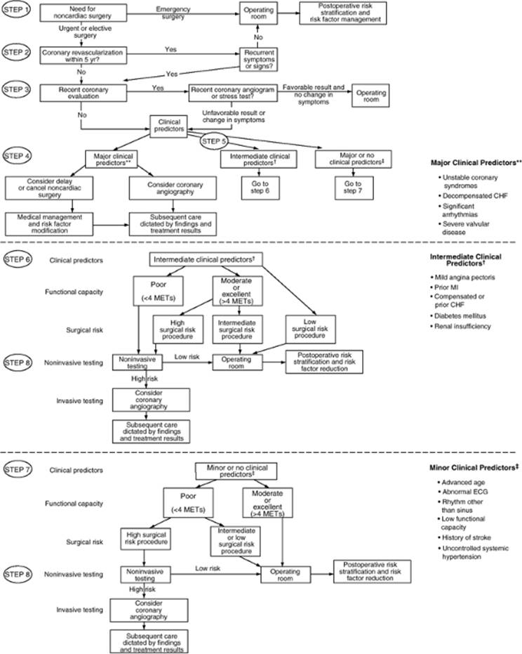
The ACC/AHA guideline is an eight-step algorithm that incorporates clinical predictors based on the patient's history and physical examination, surgery-specific risk, and the functional capacity or exercise tolerance (Fig. 70.1). In the event of an emergency procedure, patients should be taken to surgery with risk stratification performed after the surgical procedure is completed. No preoperative testing is warranted under these circumstances. If an elective or urgent procedure is planned, the decision making proceeds down steps 2 and 3 of the algorithm. If the patient has had a coronary revascularization procedure, either coronary artery bypass grafting (CABG) or percutaneous coronary intervention (PCI) performed within the last 5 years with no recurrent symptoms or signs, then no further workup is necessary. A cardiac evaluation performed within the last 2 years with no deterioration in cardiac status also negates the need for further workup.
Steps 5 to 8 of the algorithm integrate the clinical predictor, surgery-specific risk, and the functional capacity to determine whether the patient warrants further cardiac workup.
The presence of major clinical predictors demands intense further workup and may result in delay or cancellation of elective surgery. Intermediate clinical predictors increase the risk of perioperative cardiovascular complications, whereas minor clinical predictors have not been proven to independently increase cardiac risk (Table 70.2).
The nature and duration of the surgical procedure is a strong predictor of cardiovascular morbidity and mortality. Aortic, major vascular, and prolonged procedures associated with significant fluid shifts have a greater than 5% cardiac risk. Intermediate-risk procedures, which include intrathoracic, major orthopedic, intraperitoneal, head and neck, and prostate surgery, have a cardiac risk that is less than 5%. Endoscopic procedures, and cataract, breast, and superficial procedures are associated with minimal risk (less than 1%), and further workup is necessary only if the patient has major clinical predictors (Table 70.3).
It is important to factor institutional and surgical expertise when evaluating the surgery-specific risk. Pronovost et al. (12) analyzed outcomes from abdominal aortic surgery in nonfederal acute care hospitals in Maryland. Mortality varied among hospitals from 0% to 66%, based on several factors including hospital and surgeon volume. Postoperatively, the absence of daily rounds by an ICU physician increased the risk of cardiac arrest, acute renal failure, sepsis, and reintubation (12).
Finally, the functional capacity of the patient must be evaluated, as it is a strong predictor of perioperative outcome (13). Functional capacity is expressed in metabolic equivalents (METs), where one MET is 3.5 mL/kg/min of oxygen consumption in a 70 kg, 40-year-old man at rest. Increasing levels of activity correlate with increasing METs, with strenuous sports requiring greater than 10 METs, whereas activities of daily living require between 1 and 3 METs (Table 70.4). According to the ACC/AHA guidelines, the inability to perform at least 4 METs is associated with increased perioperative cardiac risk.
Based on the aforementioned triad of clinical predictors, functional capacity, and surgery-specific risk, a decision is made whether the patient can proceed to surgery or whether additional investigations to delineate the ischemic burden are required (Table 70.5) (14).
Delineation of the ischemic burden can be broadly achieved by two methods. The first method involves coronary vasodilatation and induction of a “steal” phenomenon by pharmacologic agents, followed by a nuclear imaging technique to determine the degree of myocardial ischemia. The second method involves increasing myocardial oxygen demand and evaluating electrocardiographic or echocardiographic data for evidence of ischemia. Myocardial oxygen demand can be increased either by exercise stress testing or pharmacologically with dobutamine or atropine.
In light of their limited functional capacity, vascular patients can rarely complete exercise stress testing. Therefore, dipyridamole-thallium nuclear imaging or dobutamine stress echocardiography remains the mainstay of noninvasive testing for this cohort of patients. The negative-predictive value of both tests is high. However, the positive-predictive value of dobutamine stress echocardiography is higher (14). To increase the predictive value of nuclear imaging, several criteria have been proposed that help to differentiate the low-risk scan from the high-risk scan. These include the size of the defect, increased lung uptake, and the presence of left ventricular cavity dilation (15).
Once the degree of myocardial ischemia is quantified, patients can either undergo perioperative medical optimization or revascularization by either percutaneous coronary intervention or surgery. It is important to note that to obtain benefit from a preoperative coronary intervention, the risk of noncardiac surgery must supersede the combined risk of both coronary catheterization and subsequent revascularization procedure. Eagle et al. (16) evaluated the Coronary Artery Surgery Study (CASS) database for patients requiring noncardiac surgery. CASS registry enrollees had coronary artery disease and were randomized to either optimal medical therapy or CABG. In patients undergoing high-risk surgery, prior CABG was associated with fewer postoperative deaths (1.7% versus 3.3%, p = 0.03) and MIs (0.8% versus 2.7%, p = 0.002) compared to medical management. In patients undergoing vascular surgery, the mortality benefit was similar to the high-risk cohort; however, there was a 7.9% reduction in the perioperative MI rate. Therefore, among high-risk patients with multivessel CAD and evidence of significant myocardial ischemic burden, preoperative CABG confers a survival benefit and decreases the incidence of perioperative MI. With regard to the coronary intervention, there does not appear to be any difference in mortality or MIs in patients with multivessel disease—randomized to either CABG or percutaneous coronary angioplasty (PTCA)—presenting for noncardiac surgery (17).
|
|
||
|
Figure 70.1. Stepwise approach to preoperative cardiac assessment. (Reproduced with permission from Eagle KA, Berger PB, Calkins H, et al. ACC/AHA Guideline Update for Perioperative Cardiovascular Evaluation for Noncardiac Surgery: a report of the American Heart Association/American College of Cardiology Task Force on Practice Guidelines (Committee to Update the 1996 Guidelines on Perioperative Cardiovascular Evaluation for Noncardiac Surgery). Circulation. 2002;105:1257–1267. |
||
|
Table 70.2 Clinical Predictors of Increased Perioperative Cardiovascular Risk (Myocardial Infarction, Congestive Heart Failure, Death) |
||
|
Currently, angioplasty is followed by placement of either a bare-metal or a drug-eluting stent; stents reduce both the acute risk of major complications and long-term restenosis rate. Following placement of a stent, patients require antiplatelet therapy to prevent in-stent thrombosis. Antiplatelet therapy is maintained for 1 to 12 months, depending on whether a bare-metal or a drug-eluting stent is placed. The presence of antiplatelet therapy adds a new dimension of complexity to the patient presenting for noncardiac surgery following PCI. The risk–benefit ratio of preventing thrombosis of the stent versus the risk of catastrophic perioperative bleeding must be carefully weighed. Kaluza et al. (18) reported 7 myocardial infarctions, 11 major bleeding episodes, and 8 deaths in 40 consecutive patients presenting for noncardiac surgery following placement of a stent. All deaths and MIs, as well as 8 of the 11 bleeding episodes, occurred within 2 weeks of coronary stent placement. Wilson et al. (19) reported a 4% incidence of death, MI, or stent thrombosis among 207 patients at the Mayo Clinic. Furthermore, they documented no adverse events in the 39 patients undergoing surgery 7 weeks after stent placement. It appears from these two important studies that the greatest risk of adverse cardiovascular events and bleeding complications occur within 2 weeks of stent placement. Elective surgery should be delayed for greater than 6 weeks to allow for endothelialization of the stent and discontinuation of antiplatelet therapy. In the event of urgent surgery and severe CAD, angioplasty alone with no stent placement can be performed. This obviates the need for prolonged antiplatelet therapy and the risk of perioperative bleeding.
Perioperative β-Blockade Therapy
Of the pharmacologic agents that have been used during the perioperative period, β-blockade therapy remains the most studied. β-Blockers have several salutary effects that decrease the risk for cardiovascular morbidity and mortality in a select cohort of patients.
|
Table 70.3 Cardiac Riska Stratification for Noncardiac Surgical Procedures |
||||
|
β-Blockers help to correct the imbalance between myocardial oxygen demand and supply. They have additional plaque-stabilizing, antiarrhythmic, anti-inflammatory, and altered gene expression effects (20). Their beneficial effects on perioperative mortality has been assessed in two much-discussed studies (21,22). Although both studies have design flaws, they demonstrate both short-term and potentially long-term beneficial effects of perioperative β-blockade. However, two recent trials have failed to demonstrate a beneficial effect with β-blockade therapy. The Diabetic Postoperative Mortality and Morbidity (DIPOM) trial, involving 921 diabetic patients undergoing noncardiac surgery, did not demonstrate a significantly decreased risk of death and cardiac complications with metoprolol use (23). In the Metoprolol after Vascular Surgery (MaVS) trial, vascular patients scheduled for abdominal aortic aneurysm reconstruction or infrainguinal or extra-anatomic revascularization were randomized to either metoprolol or placebo 2 hours prior to surgery (24). The study drug was continued until hospital discharge or a maximum of 5 days. This trial demonstrated no difference in cardiac mortality, nonfatal MI, or new congestive heart failure between the two groups.
Although the aforementioned trials demonstrated no benefit in certain cohorts of patients, is there potential harm in initiating perioperative β-blockade? Lindenauer et al. (25) conducted a retrospective cohort study of patients 18 years of age or older undergoing noncardiac surgery at 329 hospitals. Propensity score matching was used to adjust for differences between patients who received perioperative. β-blockade and those who did not receive such therapy. In-hospital mortality was compared using multivariable logistic modelling. The Revised Cardiac Risk Index (RCRI) score was used to assess the association between β-blocker therapy and the risk of in-hospital death. In patients with a RCRI of 0 or 1, perioperative β-blockade was associated with no benefit and possible harm (RCRI 0: odds ratio [OR] 1.43; 95% confidence interval [CI] 1.29–1.58). The beneficial effects of β-blockade were seen only in patients with a RCRI of 2 or more (OR 0.9; 95% CI 0.75–1.08). In this study, β-blockade therapy appears to have a beneficial effect only in a select high-risk patient group.
|
Table 70.4 Estimated Energy Requirements for Various Activities |
||||||||||||||||||||||||
|
||||||||||||||||||||||||
Currently, the best evaluation of the level of evidence for perioperative β-blockade is the ACC/AHA 2006 Guideline Update on Perioperative Cardiovascular Evaluation for Noncardiac Surgery: Focused Update on Perioperative Beta-Blocker Therapy (26). Based on the patient's cardiac risk and the nature of the surgery, three classes of recommendations are made from the current evidence. Insufficient data is available regarding the use of β-blockade therapy in low cardiac risk patients undergoing intermediate or high-risk surgery. In addition, the role of β-blockade therapy in low-risk surgery has not been defined.
|
Table 70.5 Summary of American College of Cardiology and American Heart Association guidelines for cardiac evaluation before nonemergent, noncardiac surgery |
||||||||||||||||||||||||||||||||||||||||||
|
||||||||||||||||||||||||||||||||||||||||||
In an attempt to clarify the role of β-blockade therapy in these groups of patients, two randomized controlled trials are in their recruitment phase. The PeriOperative ISchemic Evaluation trial (POISE) is designed to evaluate the efficacy of 30 days of controlled-release metoprolol to prevent major perioperative cardiovascular events in patients undergoing all types of noncardiac surgery (27); a total recruitment of 10,000 patients is planned. The DECREASE IV trial is designed to evaluate the efficacy of combination therapy with fluvastatin and bisoprolol in 6,000 patients scheduled to undergo noncardiac, nonvascular surgery (28). It is hoped that these trials with large study samples will clarify the role of β-blockade therapy in the low- and intermediate-risk patient.
Cardiac Surgery
Perioperative and long-term risk evaluation in cardiac surgery is complicated by several factors. These include procedural factors, patient factors, and data collection.
Cardiac surgery, with its many confounding variables, requires large patient numbers for studies to be statistically relevant. Appropriate and meaningful data collection is a relatively recent phenomenon (29,30). However, this collection effort has been hampered by the reluctance to publish data on high-risk subgroups and the inclusion of data from low-output centers that are not part of a larger data collection network. Risk factors for cardiac surgery are identified by examining multiple databases and large case series. In one large database, 19 independent variables have been identified (31). However, there are no standardized definitions for risk thresholds; many of the assessments of statistical risk are based on odds ratios. In addition, multiple risk factors frequently coexist, making risk profiling for the individual patient difficult.
Patients are presenting cumulatively with more risk factors; however, the impact of the individual risk factor appears to be decreasing. Data accrued over the last two decades suggest a steady improvement in cardiac surgical outcomes (32). This improvement is attributed to improving surgical technique, perioperative care, and patient selection. There is still, however, large variation in surgical technique across the spectrum of cardiac surgical procedures, which may explain the significant interunit variation in outcomes (32,33,34).
Preoperative Evaluation
Cardiac risk profiling begins with a thorough clinical examination and a review of the completed special investigations. Additional investigations will be guided by the presence and severity of other organ dysfunction.
Cardiac risk evaluation can be performed by risk assessment tools. These tools are based on large databases, such as the EuroSCORE and the Society for Thoracic Surgeons (STS) database (29,31). The value of these databases is that standardized definitions are used to classify patients. The risk assessment tools have two important objectives:
1. Identifying independent risk factors for morbidity and mortality in valvular, coronary, and thoracic aortic surgery.
2. Risk prediction modelling through multivariate logistic regression analysis with a view to assessing individual patient risk, comparing and auditing individual units, and appropriate resource allocation.
The STS database working group has recently published their analysis of independent risk factors in valvular surgery (31). Table 70.6 represents the information submitted by North American centers for 409,904 cardiac valvular procedures for the decade starting in 1994 and ending 2003. The risk factors are stratified according to procedural risk and patient-related factors.
Statistical analysis techniques have been used to generate scoring systems. The Parsonnet score, developed in the late 1980s, predicts risk for CABG and valvular surgery based on an additive score of weighted risk factors (35). This score tends to overestimate mortality in modern clinical practice (32). In 1999, Nashef et al. (29) published an additive-weighted scoring system called EuroSCORE based on and validated using the EuroSCORE database. This system has an improved correlation with modern practice but still overestimates the mortality in higher-risk patients. The Parsonnet and EuroSCORE have modified versions applicable at the bedside.
|
Table 70.6 Perioperative risk factors for valvular cardiac surgery |
|||||||||||||||||||||||||||||||||||||||||||||||||||||||||||||||||||||||||||||||||||||||||||||||||||||||||
|
|||||||||||||||||||||||||||||||||||||||||||||||||||||||||||||||||||||||||||||||||||||||||||||||||||||||||
Recently, a score based on Bayesian modeling has been advocated by the United Kingdom's Society of Cardiothoracic Surgeons (32). This system uses only nine weighted variables and has the best correlation and receiver operator curve of all three systems tested.
Scoring systems have a role in predicting both perioperative and long-term mortality and intensive care unit (ICU) resource use (30,32,36,37,38). Furthermore, they provide a framework to direct clinical examination and special investigations, thereby facilitating the process of identifying and modifying preoperative risk. Patients with a prohibitive perioperative risk can be better identified by these scoring systems. EuroSCORE is a compilation of risk factors as weighted by the Parsonnet, EuroSCORE, and Society of Cardiothoracic Surgeons databases. The percentages quoted in Table 70.7 are for individual risk factors in each section. Risk factors may be additive and/or synergistic.
Preoperative Risk Modification
Preoperative risk modification involves optimization of comorbidity and limiting cardiopulmonary bypass-related myocardial injury.
Comorbidity
Cardiac patients have multiple comorbidities; the most common are renal dysfunction, diabetes mellitus, and congestive cardiac failure. Renal dysfunction and failure are significant risk factors in both valvular and CABG surgery (39). The severity of renal dysfunction preoperatively correlates well with mortality postoperatively. Kuitunen et al. (40) reported that patients with an increase in plasma creatinine of one and half times from baseline with short periods of oliguria had a 90-day mortality of 8%. However, in the anuric patient with a threefold increase in creatinine, mortality increased to 32%.
|
Table 70.7 Perioperative risk factors for CABG according to scoring system |
|||||||||||||||||||||||||||||||||||||||||||||||||||||||||||||||||||||||||||||||||||||||||||||||||||||||||||||||
|
|||||||||||||||||||||||||||||||||||||||||||||||||||||||||||||||||||||||||||||||||||||||||||||||||||||||||||||||
Patients with cardiogenic shock or an emergent indication for surgery often have acute renal failure; these patients have a high risk of death perioperatively. All scoring systems categorize their mortality above 40%, irrespective of the reason for the cardiogenic shock or the surgery required (30,31,32). These poor outcomes make it difficult to ascertain the beneficial effects that preoperative dialysis will confer.
Patients presenting for coronary revascularization with dialysis-dependent end-stage kidney disease have been extensively studied (41,42). These patients appear to have a major survival benefit when coronary revascularization is performed under cardiopulmonary bypass. This survival advantage is lost when either off-pump CABG or percutaneous coronary intervention is performed (42). Survival rates at 8 years in dialysis-dependent patients were 45.9% for CABG, 32.7% for PCI, and 29.7% for no surgical intervention. Patients with non–dialysis-dependent renal insufficiency have a significant incidence of postbypass renal failure (43). The STS CABG database suggests that they have higher perioperative mortality than patients with dialysis-dependent kidney failure (44). Prophylactic dialysis in nondialysis-dependent renal insufficiency may decrease the incidence of postbypass acute renal failure (45). In addition, these patients are fluid restricted and are often on diuretic therapy. Marathias et al. (46) reported that preoperative rehydration, with 1 mL/kg/hour of 0.45% saline, nearly halved the incidence of acute renal failure and decreased the need for postoperative dialysis.
Diabetics undergoing cardiac surgery are at increased risk of prolonged ventilation, postoperative sepsis, renal failure, and cognitive dysfunction (47). The perioperative management of diabetes and hyperglycemia in cardiac surgery is controversial. Insulin has been used in two strategies: tight glycemic control and as part of glucose-insulin-potassium regimens (GIK). The studies evaluating these strategies have concentrated on the intraoperative and postoperative periods.
The implementation of tight glycemic control in the postoperative setting improves mortality significantly: 8.0% versus 4.6% for tight glycemic control (48). This study, by Van den Berghe et al., was performed on predominantly postoperative cardiac patients and revealed significant reductions in length of stay, renal failure, and nosocomial sepsis. Intraoperative insulin infusion has also been shown to reduce postoperative complications in diabetic CABG patients (49,50). The use of a GIK infusion in the setting of ongoing myocardial ischemia and infarction results in significant improvements in myocardial preservation and contractile function (51). The technique has been applied to both coronary and valvular surgery with mixed results. The trials showing modest benefit initiated GIK preoperatively, used high doses of insulin, and continued the infusion through cardiopulmonary bypass and reperfusion (51). Meta-analysis of GIK therapy indicates that trials using tight glycemic control gave the best results. This observation needs validation by other randomized trials.
Clinical experience indicates that ongoing myocardial ischemia and poor diabetic control frequently occur preoperatively. Evidence from the intraoperative and postoperative periods suggests that preoperative initiation of GIK, combined with tight glycemic control, may significantly decrease postoperative complications.
Congestive cardiac failure represents a complex neurohumoral syndrome that develops in response to altered cardiac function. The stages of this condition have been classified by the ACC/AHA (Table 70.8) (52). In the perioperative period, decompensated stage C or D heart failure represents an independent risk factor for cardiac complications (31). The syndrome covers a spectrum of patients: from those with cardiogenic shock and an ejection fraction less than 30% to those with stable but inotrope-dependent cardiac function. These patients are at high risk for postoperative complications (29,30,31,32,35).
Decompensated cardiac failure in valvular or coronary heart disease may represent a progression of the primary disease process. Under these circumstances, surgery offers the only chance to improve the biomechanical cardiac dysfunction and attenuate the maladaptive myocardial response. Mortality in medically managed decompensated cardiac failure can be improved by using new classes of drugs such as β-type natriuretic peptide and the calcium channel sensitizers (53,54,55,56). These agents have an inotrope-sparing effect, shorten hospital stay, and improve medium-term survival. The role of these agents in the perioperative setting has not been assessed, although small studies show promising results. The role of nonsurgical therapy is in the long-term prevention of progression of structural heart disease, prevention of remodeling, and modification of underlying risk factors (57).
|
Table 70.8 ACC/AHA Classification of Heart Failure |
||||||||
|
||||||||
|
Table 70.9 Indications for Intra-aortic Balloon Counterpulsation |
||||||||
|
Myocardial Preservation Interventions
Several nonpharmacologic and pharmacologic interventions can be initiated preoperatively to improve intraoperative and postoperative outcomes. Intra-aortic balloon counter pulsation (IABP) is a nonpharmacologic intervention that can be commenced preoperatively in the appropriate group of patients (Table 70.9). Pharmacologic therapies include preoperative statins, β-type natriuretic peptide, calcium channel sensitizers, and antioxidant therapy.
Intra-aortic balloon counterpulsation (IABP)
The optimal use of the IABP in cardiac surgery is controversial. Preoperative IABP may offer improved myocardial perfusion and stability during induction and maintenance of anesthesia, prior to cardiopulmonary bypass. Good evidence exists for IABP in CABG patients with ischemia or with an ejection fraction less than 25% undergoing nonelective operation or reoperation, or who have New York Heart Association (NYHA) class III to IV symptoms. The evidence is less clear for patients without ongoing ischemia but who are undergoing reoperation, have isolated left main disease or a low ejection fraction, or who are undergoing procedures other than an isolated CABG (58). The efficacy of IABP for valvular surgery is poor and is associated with a twofold increase in mortality regardless of timing of use.
This probably reflects the fact that the ventricular dysfunction is either nonreversible or only partially reversible (59).
IABP improves cardiac output by approximately 20% if set to maximal efficiency. However, the insertion and use of an IABP is not a benign procedure. Limb ischemia occurs in 8% to 42% of patients, and 30% of those will require a surgical intervention. Limb ischemia is more common when a Seldinger technique is used for insertion as opposed to surgical exposure of the femoral vessels and placement under direct vision (59).
In conclusion, the best 5-year results for IABP are in patients undergoing isolated CABG (51%), whereas those undergoing CABG with aortic valve replacement have the lowest actuarial survival (34%). Further research is required to better define the role of this intervention in cardiac surgery (59).
HMG CoA reductase inhibitors
HMG CoA reductase inhibitors, commonly known as statins, have several beneficial effects on arteriosclerosis and vascular graft disease. Their mechanism of action is via a lipid-dependent and a lipid-independent pathway. Inhibition of atherogenesis, thrombosis, and inflammation and maintenance of endothelial integrity are all attributed to this class of drug (60,61). The efficacy of statins in the reduction of graft stenosis and progression of atheroma in native vessels post-CABG is well documented (62). However, in many of these trials, statin therapy was initiated after surgery. The early beneficial effects of statins are on endothelium recovery and in inflammation in coronary vessels (63). In a large prospective longitudinal study, Collard et al. (64) evaluated the effect of preoperative statin therapy on cardiac mortality following CABG. Preoperative statin therapy was associated with a 1.1% absolute reduction in mortality (OR 0.25; CI 0.07–0.87). Interestingly, cessation of statin therapy after surgery was associated with an increased in-hospital and late cardiac mortality. This suggests that preoperative statin therapy must be considered prior to CABG and may become a standard of care in the future.
Brain natriuretic peptide
Nesiritide is a recombinant form of brain natriuretic peptide that decreases pulmonary artery pressures and myocardial oxygen consumption, and increases coronary blood flow and urine output. Nesiritide is used in two clinical settings: inotrope-resistant cardiac failure and postcardiac surgery patients with high pulmonary pressures and low cardiac output syndrome. Salzberg et al. (53) published a case series of 14 patients with severe mitral regurgitation and pulmonary pressures above 60 mm Hg undergoing cardiac surgery. Their predicted mortality based on EuroSCORE was 26%. These patients received a nesiritide infusion preoperatively, with the goal of reducing pulmonary pressures by 25%. The infusion was discontinued intraoperatively and restarted on return to the ICU. There was no reported mortality among the patients receiving nesiritide (53). These results need to be confirmed in a properly powered study, but the evidence for preoperative use in heart failure patients with pulmonary hypertension is promising.
Calcium sensitizers
Levosimendan is a calcium-sensitizing inodilator that improves myocardial contractility without increasing oxygen demand. It also decreases pulmonary vascular resistance in patients with heart failure. The LIDO trial showed it to be more effective than dobutamine in the management of severe congestive heart failure insofar as hemodynamic and mortality benefit (54). These findings have been verified by other large double-blind randomized trials (55).
Experience with levosimendan in cardiac surgery is currently limited to small studies. These studies looked at the physiologic effects of levosimendan in patients with good left ventricular function, poor ventricular function, or acute myocardial ischemia with hemodynamic compromise. In all three groups, low-dose levosimendan infusions improved cardiac output and decreased systemic vascular resistance, myocardial oxygen demand, and inotropic requirements coming off bypass (54). This drug offers enormous promise in the preoperative period in patients with severe congestive cardiac failure and poor cardiac output.
Antioxidants
Reactive oxygen species (ROS), both within myocardial cells and those derived from the systemic circulation, are thought to overwhelm local endogenous antioxidant systems during bypass. They initiate cellular damage, necrosis, and apoptosis during cardiopulmonary bypass and reperfusion. There have been many attempts to provide external sources of antioxidants or to improve endogenous antioxidant systems, and these approaches are supported by a large body of animal studies (65).
Allopurinol, which inhibits xanthine oxidase, a significant source of ROS outside the myocardium, has been studied in ten human CABG trials. Eight of these trials showed improved hemodynamic markers and less cardiac enzyme release (65). However, despite these encouraging data, allopurinol has not received widespread support. Superoxide dismutase, desferrioxamine, mannitol, vitamins C and E, and N-acetylcysteine are additional antioxidants with encouraging results in small human trials. They demonstrate decreased surrogate markers of tissue damage, although no outcome improvements have been shown. More research is required to define the role of antioxidant therapy in cardiac surgery.
There has been an improvement in the ability to identify and categorize the high-risk cardiac patient presenting for cardiac surgery. As more data are accrued, risk profiling is becoming more accurate. This will allow for cost-effective implementation of promising preoperative interventions in the appropriate patient.
Pulmonary System
The risk of postoperative pulmonary complications varies widely and according to the definitions applied. The risk evaluation process also differs between cardiothoracic and noncardiothoracic surgery. In elective noncardiothoracic surgery, postoperative pulmonary complications vary between 1.7% and 2.6%, whereas in valvular heart surgery, the Society of Thoracic Surgeons database reports a pulmonary complication rate of 8.9% (66,67,68). The definition of pulmonary complications in these studies included respiratory failure, atelectasis, pneumonia, and pulmonary edema. Pulmonary thromboembolic disease has been specifically excluded in these studies.
The contribution of postoperative pulmonary complications to morbidity, mortality, and length of stay is not dissimilar to the cardiac complication profile (66,67,69,70,71). However, in the subgroup of patients older than 70 years of age, pulmonary complication is a better predictor of long-term mortality than cardiac risk factors (72). Predicting the likelihood of postoperative pulmonary complications requires preoperative pulmonary risk stratification. Smetana et al. (3) in a systematic review identified and categorized preoperative risk factors that predicted postoperative pulmonary complications following noncardiothoracic surgery.
Preoperative Evaluation
The initial evaluation for risk factors requires a thorough history and clinical examination. Following the history and clinical examination, patients can be classified into two groups: those with known pulmonary disease and those with suspected pulmonary disease. Both groups require an assessment of their functional classification and the degree of pulmonary reversibility.
Following the history and physical examination, laboratory and special investigations are guided by the database established from the clinical evaluation. Laboratory investigations with a good predictive value for postoperative pulmonary complications include blood urea nitrogen greater than 7.5 mMol/L (21 mg/dL) and creatinine level greater than 133 µmol/L (1.5mg/dL) (73,74,75). However, the most powerful predictor of pulmonary outcome is a serum albumin level. Albumin less than 3.0 g/dL correlates with an increased 30-day perioperative morbidity and mortality (76).
The utility and cost effectiveness of routine preoperative chest radiography has been extensively debated. An abnormal chest radiograph does predict postoperative complications; however, only 4.9% of radiographs in patients younger than 50 years of age will be abnormal. Among routine preoperative chest radiographs ordered, only 0.1% to 3% will alter management (77,78). A focused history and physical examination should identify the patient who is likely to have an abnormal preoperative chest radiograph; this is supported by a recent practice guideline issued by the American College of Physicians suggesting that (3):
1. Only patients with known cardiopulmonary disease should have a routine preoperative chest radiograph.
2. Patients older than 50 years undergoing procedures with high pulmonary risk should have a preoperative chest radiograph. These procedures include aortic surgery (thoracic or abdominal), neurosurgery, abdominal surgery, and prolonged surgery.
Spirometry has been evaluated as a predictive tool for pulmonary disorders in noncardiothoracic surgery. There are, unfortunately, no studies to guide spirometry evaluation in the perioperative period for restrictive pulmonary disorders. In obstructive pulmonary disorders, there are conflicting data on the utility of spirometry; however, it may identify patients at higher risk for postoperative pulmonary complications (3). No threshold or prohibitive value has been defined for spirometry indices in obstructive pulmonary disease, probably related to the evidence that long-term prognosis for chronic obstructive pulmonary disease (COPD) is better predicted by the BODE severity scoring system (79). This system uses a holistic approach assessing the body mass index, degree of airway obstruction, symptom scoring, and exercise testing to assess severity and predict outcome.
|
|
|
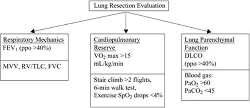
Figure 70.2. The best validated test is shown in the first box. Alternative tests are shown below. DLCO, total diffusing capacity for carbon monoxide; FEV1, forced expiratory volume at 1 second; FVC, forced vital capacity; MVV, maximal voluntary ventilation; PaO2, arterial partial pressure of oxygen; PaCO2, arterial partial pressure of carbon dioxide; ppo, predicted postoperative value based on the number of lung segments remaining after resection; RV/TLC, residual volume divided by total lung capacity; SpO2, pulse oximetric oxygen saturation; VO2, oxygen uptake/consumption. |
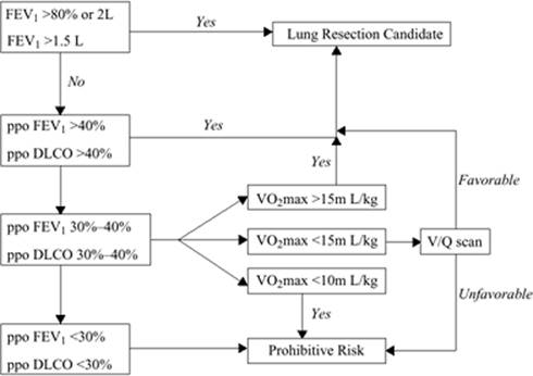
In lung resection surgery, however, spirometry forms the cornerstone of the evaluation process in both Europe and North America (80,81). Here, the perioperative risk of morbidity and mortality is directly related to a three-legged physiologic testing algorithm. Spirometry data are an integral part of the respiratory mechanics evaluation process. Other parameters evaluated are the cardiopulmonary reserve and the lung parenchymal function (Fig. 70.2). A simplified algorithm integrating these parameters assists in the preoperative workup for lung resection surgery (Fig. 70.3).
A forced expiratory volume (FEV1) greater than 80% of predicted or greater than 2 L allows pneumonectomy without further investigation. An FEV1 greater than 1.5 liters allows lobectomy without further investigation. If any of these criteria are not met, then a predicted postoperative FEV1 (ppoFEV1) and carbon monoxide diffusion capacity (ppoDLCO) need to be calculated. This allows for further risk stratification:
1. Patients with ppoFEV1 and ppoDLCO greater than 40% can be resected.
2. If the ppoFEV1 or ppoDLCO is less than 40% and >30%, then a VO2max needs to be assessed. If the VO2max is greater than 15 mL/kg/min, then resection can continue.
3. If the VO2max is less than 15 mL/kg/min, then the risk is prohibitive unless V:Q scanning indicates that the lung pathology is predominantly involving the area to be resected.
4. A VO2max less than 10 mL/kg/min or ppoFEV1 less than 30%, and ppoDLCO less than 30% are all prohibitive risks (79,80).
When the information gathered from history, physical examination, and special investigations is examined, a risk profile can be constructed from the guidelines published by the American College of Physicians (Table 70.10) (3,82).
Important points that need to be highlighted from Table 70.10 are as follows:
|
Table 70.10 Risk Factors for Respiratory Complications |
||||||||||||||||||||||||||||||||||||||||||||||||||||||||||||||||||||||||||||||||||||||||||||||||||||||||||||||||||||||||||||||||||||||||||||||||||||||||||||||||||||||||||||||||||||||||||||||||||||||||||||||||||||||||||||||||||||
|
||||||||||||||||||||||||||||||||||||||||||||||||||||||||||||||||||||||||||||||||||||||||||||||||||||||||||||||||||||||||||||||||||||||||||||||||||||||||||||||||||||||||||||||||||||||||||||||||||||||||||||||||||||||||||||||||||||
|
|
||||||||||||||||||||||||||||||||||||||||||||||||||||||||||||||||||||||||||||||||||||||||||||||||||||||||||||||||||||||||||||||||||||||||||||||||||||||||||||||||||||||||||||||||||||||||||||||||||||||||||||||||||||||||||||||||||||
|
Figure 70.3. Simplified algorithm for the preoperative evaluation for lung resection surgery. DLCO, total diffusing capacity for carbon monoxide; FEV1, forced expiratory volume at 1 second; ppo, predicted postoperative value based on the number of lung segments remaining after resection; VO2, oxygen uptake/consumption; V/Q, ventilation/perfusion ratio. |
||||||||||||||||||||||||||||||||||||||||||||||||||||||||||||||||||||||||||||||||||||||||||||||||||||||||||||||||||||||||||||||||||||||||||||||||||||||||||||||||||||||||||||||||||||||||||||||||||||||||||||||||||||||||||||||||||||
· Advanced age is reported with differing definitions. The multivariate analyses shown on the table represent odds ratios for ages 60 to 69 and 70 to 79 years.
· Functional dependence refers to both total dependence for activities of daily living, and partial dependence when equipment is required to perform activities of daily living. This is distinct from exercise capacity, which has not been evaluated as an independent risk factor in the setting of noncardiothoracic surgery.
· COPD has been poorly investigated as a risk factor, but evidence consistently suggests an independent risk.
· Impaired sensorium refers to patients with mental status changes, delirium, or both who are able to respond to verbal cues or light tactile stimulation.
· Cigarette use refers to ongoing smoking. Recent cessation, within an 8-week period, carries an increased risk of perioperative pulmonary complications over and above that associated with continued smoking (83).
· When eight multivariate trials were examined, only one trial suggested that obesity was an independent pulmonary predictor. This is indirectly verified by evidence that increasing levels of morbid obesity is not associated with increased pulmonary complications following surgery (84).
· Obstructive sleep apnea is associated with airway management difficulty and an increased all-cause admission to the intensive care. An increased pulmonary complication rate has not been elucidated.
· Asthmatic patients have a pulmonary complication rate of only 3%, which is similar to the general surgical population. This is probably related to pulmonary optimization before elective and emergency surgery (3).
· Studies of diabetes mellitus are of poor quality and are unadjusted univariate analyses, which makes them difficult to interpret.
The relationships between individual risk factors in each risk category have not been fully elucidated; they may be either additive or synergistic. Once a risk profile is formulated, risk modification strategies should be implemented.
Preoperative Management
The management of pulmonary disorders in the preoperative period fall into two categories, which include restrictive and obstructive disorders. Characteristics of restrictive disorders include the presence of mechanical volume limitations, the occasional presence of bronchial hyperreactivity, and the static nature of the disease. Obstructive pulmonary disorders are recognized by fixed airway obstruction, the presence of bronchial hyperreactivity, and a predisposition to infection.
Restrictive Pulmonary Disorders
Restrictive pulmonary disorders are substantially less common than obstructive disorders. Furthermore, they are an uncommon cause of complication or death postoperatively (3). These disorders are static in nature unless bronchial hyperreactivity coexists (e.g., hypersensitivity pneumonitis). The restriction can be either pulmonary or extrapulmonary (Table 70.11). In the extrapulmonary group, management of the mechanical volume effects can improve the perioperative pulmonary status of the patient. Drainage of a pleural effusion and re-expansion of a collapsed lung are interventions that optimize ventilation-perfusion matching. An improvement in oxygenation and ventilation can be expected. Another strategy associated with an improvement in outcome is lung protective ventilation using positive end-expiratory pressures (PEEP) and low tidal volumes of 6 to 8 mL/kg (85). This ventilator strategy has become the standard of care for acute respiratory distress syndrome (ARDS) and other disorders of static compliance found in this group of conditions. It must be emphasized that the ideal low tidal volume and appropriate PEEP strategy have still not been resolved.
|
Table 70.11 Causes of Restrictive Pulmonary Disease |
||||||
|
Obstructive Airways Disease
Chronic obstructive pulmonary disease (COPD) is differentiated from asthma on the basis of having a chronic fixed component of airway obstruction. In nonoptimized patients with COPD, a component of reversible airway obstruction or bronchial hyperreactivity, similar to asthma, is present. Furthermore, both asthmatics and COPD patients are prone to acute exacerbations of airway obstruction triggered by upper and lower airway infections (86).
Obstructive pulmonary disorders are associated with increased risk of postoperative pulmonary complications (3). When reversible airway obstruction is present, this risk is amplified (87,88). However, Milledge and Nunn (89) demonstrated that even patients with severe airway obstruction—defined as an FEV1 less than 1 L—can safely be operated on without an increase in postoperative complications. The key element in the management of patients with COPD is the identification and appropriate treatment of the reversible component of the airway disease.
β2-Agonists have a salutary effect on airway hyperreactivity in obstructive airway disease. When symptom-free mild asthmatic volunteers were intubated under local anesthesia, FEV1 decreased by 50%. In the group pretreated with a β2-agonist, the FEV1 decreased by only 20% (90). However, it is important to note that the incidence of postintubation bronchospasm is still significant, even when β2-agonists are used as monotherapy (91,92).
Preoperative steroid therapy, even of short duration, has been shown to decrease the incidence of wheezing post intubation (91,93,94). The concern for negative effects on wound healing and increased infection rates have not been borne out in the literature (95). With regard to the use of methylxanthines, a Cochrane review in 2001 showed that neither theophylline nor aminophylline offered any advantage over β2-agonists in the setting of acute bronchospasm (96).
|
|
|
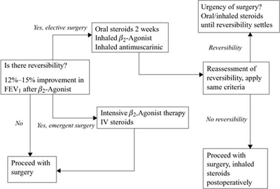
Figure 70.4. Preoperative optimization of obstructive airways disease. FEV1, forced expiratory volume at 1 second; IV, intravenous. |
Finally, there is good evidence in adults that upper and lower airway infections increase airway reactivity and result in demonstrable spirometry abnormalities for 6 to 8 weeks post infection (97). Elective surgery should ideally be delayed in patients with underlying bronchial hyperreactivity. The American Thoracic Society and the Global Initiative for Chronic Obstructive Lung Disease (GOLD) have issued guidelines for the assessment and management of obstructive lung disorders and their acute exacerbations (86,98). This approach can be modified for the patient presenting for surgery (Fig. 70.4). The most important intervention on a global scale is smoking cessation and prevention of exposure to second-hand smoke. Epidemiology studies have shown that a patient who successfully stops smoking will have a life table mortality rate that comes to parallel that of someone who has never smoked (86).
Another intervention that can be started in the preoperative period is nutritional support, which can be administered either enterally or parenterally. Preoperative enteral nutrition with an immune-repleting diet improves outcomes in malnourished elective gastroenterology oncology patients; they experience a significant decrease in nosocomial sepsis and hospital length of stay (99,100). Enthusiasm for total parenteral nutrition has waned due to the increased rates of infection associated with long-term central venous access and hyperglycemia (99).
In conclusion, the Practice Guideline published by the American College of Physicians helps generate a risk profile for noncardiothoracic surgery. Risk modification strategies can then be applied to optimize the patient.
Renal System
Acute and chronic renal failure are important medical problems worldwide. The incidence of ICU-associated acute renal failure (ARF) varies between 15% and 35%, whereas the incidence of ARF requiring renal replacement therapy approximates 1%. In the United States, the incidence of end-stage kidney disease (ESKD) varies between 331 and 343 cases per million population, whereas more than 104,000 new patients began therapy for ESKD in 2004. The economic burden of ESKD continues to rise and currently exceeds 20 billion dollars, approximately 6.7% of the Medicare budget (101).
The preoperative evaluation and management of the patient with renal disease is complicated by the coexistence of multiple medical and surgical problems. Therefore a stepwise logical approach to these patients is required to ensure that important data are not omitted.
Chronic Renal Failure
Risk Evaluation and Stratification
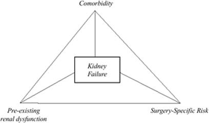
Preoperative renal risk evaluation and stratification are based on the comorbid medical condition of the patient, pre-existing renal function, and the procedure-specific renal risk (Fig. 70.5).
Comorbid Status
Comorbid conditions that increase the risk of chronic renal insufficiency include a spectrum of cardiovascular, endocrine, hepatic, autoimmune, and congenital disorders (Table 70.12). The severity, duration, and appropriate management of the conditions determine the degree of renal dysfunction a patient will develop.
|
|
|
|
Figure 70.5. Triad of factors that collectively increases the risk of perioperative kidney failure |
|
|
Table 70.12 Causes of Chronic Kidney Disease |
|
|
Diabetes mellitus (DM) is the leading cause of ESKD, with hypertension, glomerulonephritis, and polycystic kidney disease being the other major diagnoses (101). The incidence of diabetes mellitus–related ESKD has risen over the past three decades from 28% in 1980 to approximately 50% currently (101). Diabetes affects the kidney by several mechanisms, resulting in proteinuria, the nephrotic syndrome, and progressive renal failure. Patients with diabetes mellitus who have microalbuminuria (30–300 mg/24 hours) have not yet begun to lose glomerular filtration but they are at high risk of renal complications. The introduction of ACE inhibitors in this group of patients has a powerful renoprotective effect and slows the progression to overt renal failure (102).
Coronary artery disease is common in patients with diabetes mellitus and may be asymptomatic due to an associated autonomic neuropathy. A high index of suspicion for untreated CAD should be maintained for patients with diabetic nephropathy. Danaei et al. (103) reported that 21% of deaths from ischemic heart disease and 13% from stroke worldwide are attributable to higher-than-optimum blood glucose concentrations. The ACC/AHA guidelines for the preoperative evaluation of the cardiac patient presenting for noncardiac surgery list DM as an intermediate clinical predictor for perioperative cardiovascular events (11). Therefore, the preoperative workup of the diabetic patient with renal dysfunction should be done within the framework of the ACC/AHA algorithm discussed previously (11).
Hypertension is the second leading cause of ESKD in the United States. Rates of ESKD caused by hypertension show dramatic variation when comparing African American patients with those of other races and ethnicities (101). The 2004 rate of hypertensive ESKD in African American patients aged 30 to 39 years is 149 cases per million population, approximately 15 times higher than in Caucasian counterparts. Hypertension is both a cause and a consequence of ESKD. Aggressive control of the blood pressure to approximately 125/75 mm Hg in patients with diabetic renal disease is recommended by the National Kidney Foundation. The Modification of Diet in Renal Disease study provided convincing evidence that lower blood pressure reduces the rate of loss of renal function in patients with proteinuric renal disease (104). Polycystic kidney disease is an inherited disorder that is characterized by multiple cysts in the kidney as well as the liver. It is responsible for 4% to 5% of ESKD in the United States (101). Important extrarenal manifestations of polycystic kidney disease include intracranial aneurysm in approximately 10% of patients, whereas 26% of patients have evidence of mitral valve prolapse.
|
Table 70.13 Stages of Chronic Kidney Disease |
||||||||||||||
|
||||||||||||||
Once comorbid conditions and their related complications are identified, an attempt must be made to quantify the degree of renal dysfunction.
Pre-existing Renal Dysfunction
The National Kidney Foundation-Kidney Disease Outcomes Quality Initiative recently proposed a standardized classification system to assess the severity of pre-existing chronic renal disease. It is based on an estimation of the glomerular filtration rate (GFR) and the documentation of renal injury (Table 70.13) (105). The GFR can be estimated using either mathematical models or by determining the clearance of inulin or other filtration markers. To estimate the GFR, either the Cockroft-Gault equation or the formula derived from the Modification of Diet in Renal Disease Study can be used. The latter formula, although mathematically more complex, uses readily available data to provide a more accurate estimate of the GFR than the Cockroft-Gault equation (106):
GFR (mL/min/1.73 m2) = 170 × [PCr]-0.999 × [age]-0.176 × [0.762 if patient is female] × [1.18 if patient is black] × [SUN]-0.17 × [Alb]+0.318
where SUN is serum urea nitrogen and Alb is albumen. The presence of pre-existing renal dysfunction increases the incidence of perioperative morbidity and mortality in high-risk surgery. Safi et al. (107) analyzed factors responsible for developing acute renal failure following thoracoabdominal repair. On multivariate analysis, preoperative creatinine (PCr) greater than or equal to 2.8 mg/dL was strongly associated with postoperative ARF (OR 10.3; 95% CI 12–411, p <0.0001). Of the patients who developed ARF, the mortality rate was 49%. This elevated operative mortality risk associated with renal failure is reflected by Kashyap et al. (108) in their study of thoracoabdominal aortic surgery (OR 9.2; 95% CI 2.6–33; p < 0.005).
Procedure-related Risk
Procedure-related renal risk is an important determinant of perioperative renal failure. Cardiac and major vascular procedures are associated with a high incidence of ARF. For example, cardiopulmonary bypass (CPB) has several negative effects on renal function. The use of nonpulsatile flow, inadequate renal perfusion pressure, and the induction of an inflammatory response all contribute to renal dysfunction. The incidence of ARF following cardiac surgery varies between 1% and 30% (109). The development of renal failure is associated with increased mortality, hospital length of stay, and cost. Conlon et al. (109) studied 2,672 consecutive patients undergoing CABG and reported a 7.9% incidence of ARF and 0.7% incidence of ARF requiring renal replacement therapy. The mortality for patients who developed ARF was 14% compared with 1% among those who did not develop ARF.
Off-pump CABG, by avoiding cardiopulmonary bypass, has been shown to have a protective effect on renal function post surgery. Ascione et al. (110) randomized patients with normal renal function prior to cardiac surgery to either an on-pump or off-pump group. Postoperatively, the off-pump group had better preservation of renal function as evidenced by the creatinine clearance, albumin-creatinine ratio, and the N-acetyl-β-glucosamine levels. The same group evaluated 253 patients with preoperative renal insufficiency undergoing CABG. ARF occurred in 15.8% of patients who underwent on-pump CABG compared to 5.9% of those who had off-pump CABG (111). It appears that off-pump CABG has a renoprotective effect in patients with both normal and impaired renal function. Furthermore, it highlights the positive influence that modification of a surgical technique can have on organ protection.
Renal insufficiency or failure exists in many patients presenting for major vascular surgery. Swaminathan and Stafford-Smith (112) analyzed data from eight studies and reported an incidence between 4% and 24% of pre-existing renal insufficiency in patients with aortic disease. The broad range of reported incidence is attributed to the lack of consensus for the definition of renal insufficiency.
In vascular surgery, the location of the arterial reconstruction, the duration of the aortic cross-clamp, and the emergent nature of the procedure are all strong predictors of postoperative renal complications. Reported incidence of acute postoperative renal dysfunction for thoracoabdominal reconstruction varies between 13% and 25%, whereas renal failure rates for abdominal aortic reconstruction are much lower at approximately 1.5% to 2% (113). The hypothesized mechanisms of postoperative renal injury are ischemia-reperfusion of the kidneys, atheroembolic injury, nephrotoxin, and inflammatory damage.
|
Table 70.14 The RIFLE Classification Scheme for Acute Renal Failure |
||||||||||||||||||||||||||||
|
||||||||||||||||||||||||||||
Measures aimed at modifying surgical technique to reduce the incidence of postoperative renal dysfunction in aortic surgery have been met with mixed success. The most significant advance recently has been the widespread adoption of endovascular techniques for aortic aneurysm repair. Several small studies have suggested that the reduced aortic manipulation and renal ischemia that accompany the endovascular technique is associated with reduced inflammation and postoperative renal injury (114,115). However, a recent large study comparing endovascular to open repair for abdominal aortic aneurysm (AAA) demonstrated no difference in renal outcome between the two groups. Renal complications were 1.1% and 1.2% in the open and endovascular group, respectively (116). In patients with baseline chronic renal insufficiency, endovascular aortic repair does not confer a renal benefit compared to open AAA surgery. Parmer et al. (117) reviewed 98 patients undergoing endovascular and open aortic repair at a single institution with baseline renal insufficiency. Postoperative renal failure rates were 28% and 29% for the open and endovascular group, respectively. Further research will be required to establish whether endovascular surgery is truly renoprotective.
Acute Renal Failure
Definition
Acute renal failure (ARF) is a common complication of critical illness and is associated with significant morbidity and mortality. There is no consensus definition of ARF in critically ill patients. More than 30 definitions have been used in the literature, making it difficult to compare data from different studies. In order to standardize the definition of ARF, the Acute Dialysis Quality Initiative (ADQI) group proposed the RIFLE classification of ARF (Table 70.14) (118). The classification system includes criteria for serum creatinine, GFR, and urine output. The RIFLE classification is a valuable tool in determining the extent of acute renal dysfunction and helps to prognosticate outcome in the face of ARF. Kuitunen et al. (40) used the RIFLE classification to categorize postoperative renal impairment following cardiac surgery. Patients with RIFLE-F (failure) had a 90-day mortality of 32.5% compared with 8% for those in the RIFLE-R (risk) and 21.4% for RIFLE-I (injury) patients (Table 70.14). Multivariate logistic regression analysis demonstrates that the RIFLE classification is an independent risk factor assessment for 90-day mortality.
Classification of ARF
ARF is divided into three categories based on its pathophysiology. They are prerenal, postrenal, and intrarenal ARF.
1. Prerenal ARF: Prerenal ARF is reversible renal insufficiency due to renal hypoperfusion. If the renal hypoperfusion is left untreated, acute tubular necrosis (ATN) secondary to ischemia will develop. Prerenal ARF is characterized by clinical evidence of hypovolemia such as systemic hypotension, low central venous pressures, low cardiac output, or systolic pulse variation on positive pressure ventilation. Laboratory data that indicate prerenal ARF include low urine Na+, fractional excretion of Na+ less than 1%, and bland urine sediment.
2. Postrenal ARF: Postrenal ARF is due to obstruction of urine flow at any level of the urine collecting system. Postrenal ARF can be diagnosed promptly by either ultrasound or CT scan, and relief of the obstruction usually results in prompt reversal of the renal insufficiency. Postrenal ARF must be excluded in every patient presenting with ARF.
3. Intrarenal ARF: Intrarenal ARF is divided into five groups based on the underlying pathology. Acute tubular necrosis (ATN) is injury and subsequent death of the tubular epithelium. ATN is caused by either ischemia or nephrotoxic agents. Acute interstitial nephritis is an inflammation of the renal interstitium and the tubules. It may occur secondary to infections or drugs, such as the penicillins or cephalosporins. Other causes of intrarenal ARF include acute glomerulonephritis, acute vascular syndromes, and intratubular obstruction. The differential diagnosis of intrarenal ARF should be guided by the clinical history and physical examination. Examination of the urine sediment helps to narrow the differential diagnosis. The presence of tubular epithelial cells and granular cast is suggestive of ATN, whereas the presence of red cell cast indicates glomerulonephritis. Eosinophiluria suggests the presence of interstitial nephritis; however, it is not diagnostic.
ARF is characterized by retention of nitrogenous waste products, fluid and electrolyte abnormalities, acid-base disorders, and impairment of the hematologic and coagulation systems. The preoperative evaluation of these patients must therefore take into account these specific changes and the increased risk they pose during the perioperative period.
Preoperative Evaluation of Renal Failure
As highlighted previously, both acute and chronic renal failure affect multiple organ systems. The history and physical examination should be directed toward evaluating the severity of the comorbid conditions and the complications related to the acute renal dysfunction. Signs and symptoms of uncompensated cardiac failure and pericarditis should be elicited. Uremic patients are at risk for the development of large pericardial effusions, which can be hemodynamically compromising. The presence of an elevated jugular venous pressure and pulsus paradoxus of greater than 10 mm Hg should alert the clinician to the presence of a pericardial effusion.
Uremia is associated with nausea, vomiting, and recurrent episodes of hiccoughing. Severe nausea and vomiting may result in dehydration, and a thorough evaluation of the patient's volume status must be performed. These patients may either be on intermittent hemodialysis or peritoneal dialysis. Records of the last dialysis, fluid balance, and body weight must be obtained to help with the assessment of the fluid status.
Anemia in renal failure is multifactorial in nature. Bleeding from platelet dysfunction, malnutrition, and decreased erythropoietin production all contribute toward the low red cell mass. Electrolyte abnormalities are common in renal failure. Hyperkalemia, hyperphosphatemia, and hypocalcemia is the typical electrolyte profile seen in renal failure. Hypocalcemia may manifest as cramps, paraesthesia, and, in severe cases, with mental status changes.
Diagnostic Testing
The diagnostic studies in patients with renal dysfunction are determined by the findings on the history and physical examination. Complete blood count helps to assess the severity of the anemia, morphology of the red blood cells, and the platelet count. Although uremic patients may have normal platelet numbers, they develop an acquired platelet dysfunction that results in an increased risk of bleeding. The pathogenesis of this hemostatic dysfunction is multifactorial and includes the effects of circulating toxins, alteration of the vessel wall, and anemia. To assess the degree of platelet function, either a bleeding time, or more accurately, a platelet function assay can be performed.
The basic metabolic panel helps to determine the electrolyte profile and allows the anion gap to be calculated. ARF is characterized by an increased anion gap metabolic acidosis. The BUN and creatinine can be tracked to assess the efficacy of renal replacement therapy.
An electrocardiogram must be performed to determine whether ischemia, ventricular hypertrophy, or strain pattern is present. Hyperkalemia is characterized by tall peaked T waves, widened QRS complex, and shortened QT interval. The ECG of patients with pericardial effusion may demonstrate small QRS complexes and the presence of electrical alternans (change in QRS amplitude with each heartbeat). A chest radiograph may reveal signs of pulmonary edema, cardiomegaly, or a large pericardial effusion.
Examination of the urine and determination of the urine indices provides invaluable data in helping to differentiate prerenal from intrarenal ARF (Table 70.15). The fractional excretion of urea is a useful index to differentiate prerenal from intrarenal failure if diuretic therapy has been initiated:
|
Table 70.15 Criteria to Differentiate Prerenal from Intrarenal failure |
|||||||||||||||||||||
|
FEUN = [(urine nitrogen/blood urea nitrogen)/(urine creatinine/plasma creatinine)] × 100
Fractional excretion of urea nitrogen is primarily dependent on passive forces and is therefore less influenced by diuretic therapy. In contrast, diuretic therapy will falsely raise the FENa+ under prerenal conditions.
Primary and Secondary Prevention of ARF
Primary prevention of ARF refers to clinical strategies that reduce the occurrence of ARF in patients with or without chronic renal disease. Secondary injury is additional renal injury developing in the face of a primary insult to the renal system. Various nonpharmacologic and pharmacologic strategies have been developed to reduce both primary and secondary renal injury. However, these have been met with mixed success, and no pharmacologic agent has been approved in the United States as a sole renoprotective agent.
The principles of management of patients with ARF are to maintain an adequate mean arterial pressure, maintain an appropriate cardiac output, ensure euvolemia, and avoid nephrotoxic agents. The mean arterial pressure required to maintain adequate renal perfusion pressure, however, may vary according to the patient's underlying medical condition. Patients with hypertension have an autoregulatory curve that is shifted to the right; therefore, they may require a higher mean arterial pressure. Low cardiac output states induce renal ischemia and reduce the glomerular filtration rate. It is important to restore cardiac output to normal levels to prevent secondary injury to the kidney. This may require placement of monitors such as a pulmonary artery catheter or a transesophageal echocardiography probe to determine cardiac output.
Volume expansion has been shown to prevent contrast-induced nephropathy and attenuate the tubular injury associated with rhabdomyolysis (119,120). Furthermore, Mertens et al. (121) compared the incidence of contrast-induced nephropathy with either isotonic saline or bicarbonate hydration. Patients received an intravenous bolus of 3 mL/kg of the study solution over an hour, before radiocontrast injection. This was followed by a continuous infusion of 1 mL/kg/hour during the procedure and for 6 hours after the procedure. The incidence of contrast-induced nephropathy was 13.6% in the saline group but only 1.7% in the bicarbonate arm. These are low-cost, high-yield interventions that can significantly affect the outcome of patients with marginal renal function.
In conclusion, the best evidence to date suggests that nonpharmacologic therapy strategies are more effective than drugs in reducing the risk of ARF. High-risk patients should be identified early and secondary renal injury aggressively prevented.
Neurologic System
Ischemic Cerebrovascular Disease
The incidence of perioperative stroke in patients undergoing nonvascular surgery under general anesthesia is less than 0.5%. However, the mortality associated with a perioperative stroke may be as high as 26% (122).
Carotid Stenosis
The risk of a perioperative stroke increases in the presence of carotid stenosis or a history of transient ischemic attack (TIA). In a retrospective study, Evans et al. (123) studied 284 patients with ultrasound-documented evidence of carotid stenosis undergoing general surgical procedures. The presence of carotid stenosis of at least 50% was associated with a perioperative stroke rate of approximately 3.6%. Although higher than the general population stroke rate, this risk does not appear sufficient to mandate prophylactic carotid endarterectomy (CEA). The cumulative risk for stroke in asymptomatic patients is the sum of the perioperative stroke risk for CEA and the residual perioperative stroke risk for the general surgical procedure. This cumulative risk must be significantly lower than 3.6% to justify preemptive CEA. Therefore, asymptomatic carotid stenosis discovered during general surgical procedure workup does not require carotid endarterectomy. Patients with carotid disease who present with TIAs have a 10% risk of stroke during the subsequent year. Of the patients who develop a stroke, 20% will have their stroke within the first month and about 50% within 1 year of the TIA. After the first year, the stroke rate decreases to about 5% per year (124). Carotid stenosis can be managed both surgically and pharmacologically.
Two large randomized studies have defined the role of CEA in symptomatic patients with carotid stenosis. The North American Symptomatic Carotid Endarterectomy Trial (NASCET) and the European Carotid Surgery Trial (ESCT) validated the role of CEA among symptomatic patients with severe (70%–99%) and moderate (50%–69%) carotid stenosis (125). Two criteria must be met for CEA to have a beneficial effect. First, surgical skills with a low complication rate are essential; second, the surgical benefit must persist for several years to justify the perioperative risk.
The benefit of antiplatelet therapy in reducing perioperative stroke during CEA is unresolved. However, based on the current level of evidence, patients should receive aspirin prior to surgery unless there are obvious contraindications. The optimal dose of aspirin is uncertain; however, a dose range of 50 to 1,300 mg/day has been used in various studies (126).
Surgery-specific Risk
Perioperative stroke rates vary depending on the surgery-specific risk. Currently, the noncarotid procedure associated with the highest risk of perioperative neurologic injury is cardiac surgery. Contemporary prospective studies report a 3% incidence of stroke in CABG procedures, 8% in isolated valve surgery, and 11% in combined CABG-valve surgery. Advanced age and female gender are additional risk factors for perioperative neurologic injury (127). The single most important factor for cerebral injury during cardiac surgery is macroembolization of atheromatous debris during aortic manipulation. Every attempt should be made to identify the high-risk patient with a large atheromatous burden by using epiaortic echocardiography and minimizing manipulation of the aorta (128).
Preoperative Evaluation
The history and physical examination of the patient with cerebrovascular disease requires a thorough assessment of the cardiovascular and neurologic system. Patients with carotid artery stenosis are at an increased risk of coronary artery disease. Severe correctable CAD is evident in approximately 26% of patients with cerebrovascular disease, whereas only 9% of patients have normal coronary anatomy (10). Despite the increased incidence of CAD in patients with carotid stenosis, the rate of medical complications in patients undergoing CEA is low. Paciaroni et al. (129) recorded medical complications that occurred within 30 days after CEA in 1,415 patients enrolled in the NASCET trial. Perioperative medical complications occurred in less than 10% of patients who underwent CEA, and only 0.4% had severe complications. Of note, perioperative nonfatal and fatal MI occurred in only 1% of the patients and was associated with a mortality rate of approximately 0.2%. Therefore, recommending a CABG procedure to a patient with symptomatic carotid disease and asymptomatic CAD is not justified, as the combined mortality and stroke rate after CABG is higher than an expertly performed CEA.
The ACC/AHA guidelines for noncardiac surgery list CEA as an intermediate-risk procedure where the perioperative cardiac risk is less than 5% (11). The cardiac workup of patients presenting for CEA can be performed within the framework of the ACC/AHA algorithm.
Hypertension is another chronic condition that needs to be evaluated during the history and physical examination. Hypertension is a prevalent and treatable risk factor for stroke. Treatment of systolic and diastolic hypertension results in a 36% and 42% stroke reduction, respectively (126). The preoperative blood pressure is important in determining the hemodynamic management strategies intraoperatively. Patients with long-standing hypertension have their cerebral autoregulatory curve shifted to the right; therefore, a higher mean arterial pressure may be required during periods of cerebral ischemia to ensure adequate cerebral perfusion. Poor control of blood pressure following CEA increases the risk of cerebral hyperperfusion syndrome. This complication occurs due to impairment of cerebral autoregulation and can result in intracerebral hemorrhage and white matter edema. Patients with severe preoperative carotid stenosis and chronic hypertension are at greatest risk for this complication. Blood pressure should be carefully monitored and aggressively treated if symptoms of hyperperfusion syndrome develop (126).
|
Table 70.16 WFNS, Hunt-Hess, and Fischer Grading System for Subarachnoid Hemorrhage |
||||||||||||||||||||||||||||||||
|
||||||||||||||||||||||||||||||||
Hemorrhagic Cerebrovascular Disease
Aneurysmal Subarachnoid Hemorrhage
The prevalence of cerebral aneurysm in the general population ranges between 0.2% and 7.9% (130). Subarachnoid hemorrhage (SAH) accounts for 2% to 5% of all new strokes in the United States (131). The incidence increases with age, with a higher preponderance in females and the African American population. The vast majority of deaths occur within 2 weeks of the rupture, with 10% occurring in the prehospital area. The risk of re-rupture is greatest within 24 hours, and thereafter decreases to approximately 1% to 1.5% per day for the initial 14 days. The cumulative re-rupture risk during the first 14 days is about 25% (132).
Preoperative Evaluation
The preoperative evaluation of the patient with an intracranial bleed must begin with a thorough history and physical examination. Patients with an aneurysmal SAH will complain of a severe sudden headache following the ictus. Features of meningeal irritation including neck stiffness and photophobia can be elicited. An altered level of consciousness, seizures, and coma are found in the higher grades of SAH. Aneurysmal subarachnoid hemorrhage is graded according to the Hunt-Hess classification or the World Federation of Neurologic Surgeons Scale. The amount of subarachnoid blood is evaluated using the Fischer grading system. The quantity of blood in the subarachnoid space determines the risk of developing delayed cerebral ischemia or vasospasm (Table 70.16).
The initial diagnostic modality of choice is a CT scan of the brain, which can detect SAH in approximately 95% of cases. Once the diagnosis of a SAH is made, either a CT angiogram or a four-vessel cerebral angiogram is performed to delineate the cause of the SAH. A four-vessel cerebral angiogram remains the gold standard for diagnosing an intracranial aneurysm; however, it has the disadvantage of being highly invasive and time consuming. Furthermore, the incidence of microembolism during this procedure is high and the risk of rebleeding is increased (133).
The sensitivity and specificity of CT angiogram for aneurysm detection are 87% and 100%, respectively (134).
The advantages of a CT angiogram compared with four-vessel angiogram include its rapidity, decreased invasiveness, and substantially lower cost. The disadvantages include its difficulty in detecting small and unusually located aneurysms and the use of an iodinated contrast medium. However, in the era of cost-consciousness, CT angiogram remains a viable option for the diagnostic workup for aneurysmal SAH.
Magnetic resonance (MR) angiography conversely does not require radiation exposure, and its contrast material has a substantially lower allergic and renal complication rate. MR angiography has a 74% to 98% detection rate for aneurysms greater than 3 mm. However, in aneurysms smaller than 3 mm, the detection rate is generally low (133). An advantage that MR angiography has over CT angiography is its ability to locate aneurysms close to the cranial base. With continual improvement in technology, the role of noninvasive cerebral angiography is rapidly being defined and may eventually supersede four-vessel cerebral angiography.
Complications of Aneurysmal SAH
Central nervous system
Vasospasm or delayed cerebral ischemia occurs in 60% to 70% of patients following SAH, half of whom will develop symptomatic ischemia (130). The exact cause of vasospasm is not fully understood, but the breakdown products of subarachnoid blood are probably responsible for initiating the ischemia. Vasospasm begins within 4 days following the ictus and may last for as long as 14 to 16 days. Vasospasm is diagnosed clinically and confirmed by transcranial Doppler or angiography. The treatment of delayed cerebral ischemia is broadly classified into preventative measures, triple-H therapy (hypertension, hemodilution, hydration), and endovascular intervention. The only useful pharmacologic agent available for vasospasm is the calcium channel blocker nimodipine. It is given orally or by nasogastric tube. A Cochrane database review of eight trials demonstrated a reduction in poor neurologic outcome, secondary ischemia, and mortality with oral nimodipine (135).
With aggressive multimodal therapy, morbidity from vasospasm can be reduced to 5% (130). Triple-H therapy involves inducing hypertension to systolic blood pressures between 180 and 200 mm Hg. This is achieved initially with fluids and with pressors such as phenylephrine when needed. Rheologic benefits are achieved by keeping the hematocrit at approximately 30%. Care must be taken not to induce severe anemia, which can decrease cerebral oxygen delivery and worsen ischemia. Hydration is optimized with either a colloid or an isotonic crystalloid. Given that the central venous pressure is a poor indicator of intravascular volume status (136), these patients may require placement of a pulmonary artery catheter to better manage their fluid status (137).
Endovascular therapy for vasospasm consists of balloon angioplasty and intra-arterial infusions of nimodipine or verapamil; papaverine has fallen out of favor. Balloon angioplasty is best used for large vessel spasm whereas vasodilator therapy is useful for distal branch vasospasm.
Cardiovascular
Rupture of an intracranial aneurysm is associated with substantial cardiovascular and hemodynamic changes. The myocardial injury following SAH is related to the massive sympathetic discharge and is characterized by subendocardial contraction band necrosis. ECG abnormalities are detected in 50% to 80% of patients following aneurysmal SAH. The spectrum of ECG changes includes repolarization abnormalities, ST-T wave changes, and conduction alterations. Myocardial injury and enzyme leaks are found in 20% to 50% of patients with abnormal postictal ECGs, whereas echocardiographic evidence of myocardial dysfunction is found in two thirds of patients with elevated levels of troponin I (138).
The cardiac workup of patients with aneurysmal SAH is determined by their premorbid cardiovascular status and the degree of hemodynamic compromise following rupture of the aneurysm. An ECG, cardiac enzymes, and echocardiography should be considered for patients presenting with hemodynamic instability. In our experience, patients with mild-to-moderate cardiac dysfunction often require no specific cardiac intervention, and tolerate surgery and neurointerventional procedures well. The patient with severe cardiac dysfunction may require placement of invasive monitors such as a pulmonary artery catheter to optimize cardiac output and cerebral perfusion pressure. In a retrospective study of 453 patients, Kim et al. (137) showed that pulmonary artery catheter-guided hemodynamic management reduced the incidence of pulmonary complications and sepsis by 8%.
Pharmacologic and mechanical intervention, such as intra-aortic balloon counterpulsation, may also be required to support the severely dysfunctional myocardium (139). Intra-aortic balloon counterpulsation has the added advantage of possibly improving cerebral blood flow in patients with severe vasospasm that is refractory to traditional triple-H and neurointerventional therapy (140).
Pulmonary
Pulmonary complications occur in approximately 22% of aneurysmal SAHs and are associated with a substantial risk of mortality (141,142). The most frequent pulmonary complications are nosocomial pneumonia and pulmonary edema. Patients at risk for nosocomial pneumonia are those with an altered level of consciousness and with aspiration of gastric contents. Neurogenic pulmonary edema occurs from the massive sympathetic discharge following the ictus and is characterized by disruption of the pulmonary epithelial-endothelial barrier. Protein-rich fluid leaks into the alveoli and results in alveoli instability and atelectasis. Pulmonary edema can also be triggered secondary to aggressive triple-H therapy. Friedman et al. (141), using logistic regression analysis, showed a strong and independent association between pulmonary complications and the development of symptomatic vasospasm. The likely explanation for this phenomenon is that patients with pulmonary compromise are treated less aggressively with triple-H therapy than their counterparts with normal pulmonary function.
Evaluation of the pulmonary system begins with an assessment of the neurologic status of the patient and the determination of whether he or she is able to protect the airway. Patients with a GCS score of less than or equal to 8 require intubation to protect the lower respiratory tract from aspiration. A chest radiograph and arterial blood gas assessment help to assess the degree of pulmonary dysfunction and the need for intubation and ventilation. Ventilation strategies that should be used include a lung-protective ventilation strategy and the appropriate use of positive end expiratory pressure.
Electrolytes
The most common electrolyte abnormality following SAH is hyponatremia, occurring in 30% to 40% of patients (142). The two major mechanisms of hyponatremia are either the syndrome of inappropriate antidiuretic hormone (SIADH) or cerebral salt-wasting syndrome (CSW). It is important to differentiate these two conditions, as the management strategies are markedly different. Table 70.17 shows the clinical and biochemical features of SIADH and CSW. The most important feature between the two syndromes is the extracellular volume, which is elevated in SIADH and decreased in CSW. Either clinical or hemodynamic data such as pulmonary artery occlusion pressure and ventricular end-diastolic volume must be assessed to determine the volume status of the hyponatremic patient. SIADH is treated with fluid restriction whereas cerebral salt-wasting syndrome is treated with hypertonic fluid and salt replacement.
|
Table 70.17 Clinical and biochemical features of SIADH and CSW |
||||||||||||||||||||||||
|
||||||||||||||||||||||||
Other electrolyte abnormalities commonly encountered in patients with SAH include hypomagnesemia and hypokalemia in 37% and 27% of patients, respectively (142). Magnesium has been shown to have a neuroprotective effect in numerous stroke models and reverses vasospasm and infarct volume in rat models of SAH (143,144). Van den Bergh et al. (145) conducted a prospective human trial on the use of magnesium prior to the onset of vasospasm following SAH. The study demonstrated that magnesium infusion during the vasospasm period decreased the incidence of “poor outcome” by 23%. Magnesium levels were kept between 1 and 2 mMol/L by means of a continuous magnesium infusion. Although based on a small cohort, this low-cost, high-yield intervention holds great promise for the future.
Hyperglycemia
The incidence of hyperglycemia following SAH is 30% (142). This is due to the activation of the sympathoadrenal system and the increase in stress hormones following the ictus. Hyperglycemia is associated with a poor prognosis in the face of cerebral ischemia (146). Blood glucose levels must be monitored regularly, and an insulin infusion should be initiated if blood glucose exceeds 110 mg/dL. The salutary effects of “tight” glucose control are not limited to only the central nervous system. Van den Berghe et al. (48) randomized 1,548 postcardiac surgery patients to either an intensive insulin (80–110 mg/dL) or conventional therapy (180–200 mg/dL). Intensive insulin therapy was associated with a lower mortality and a decreased incidence of acute renal failure requiring renal replacement therapy. Critical illness polyneuropathy and documented bacteremia rates were also lower in the intensive insulin therapy arm. Tight glucose control should therefore be an integral part of the management algorithm for the patient with a SAH.
The proportion of deaths directly attributed to medical complications following SAH is 23% (142). This is comparable to the death rate following vasospasm and rebleeding. Therefore, medical complications following SAH should be aggressively sought and treated. A stepwise systematic approach to the various organ systems will help identify and treat complications related to SAH.
Summary
High-risk surgical patients present a unique challenge to the perioperative physician. Due to their multiple comorbidities and the increasing complexity of surgery being performed, their perioperative risk is disproportionately higher than the general surgical population. To appropriately manage these patients, risk factors must be identified and stratified following completion of the clinical, laboratory, and special investigations. Risk-modification strategies may be implemented preoperatively if they are likely to have a beneficial effect during the operative course. Otherwise, they can be initiated postoperatively as part of the long-term care plan for the patient.
Pearls
· Assessment of the high-risk patient begins with preoperative identification, stratification, and modification of risk factors.
· No preoperative test should be performed unless it is likely to influence patient treatment.
· In noncardiac surgery, the ACC/AHA algorithm provides a structured and cost-effective evaluation strategy.
· Dobutamine stress echocardiography is a useful preoperative test to detect ischemia in patients with a limited ability to perform exercise testing.
· Scoring systems in cardiac surgery assist with risk profiling, predicting mortality, and resource use.
· Promising interventions exist for the optimization of the severely dysfunctional ventricle.
· Systematic reviews have now defined patient and surgery-specific risk for postoperative pulmonary complications.
· It is very important to assess and treat reversible airway obstruction in chronic obstructive airway disease.
· The risk of acute renal failure in patients with chronic renal insufficiency is determined by comorbidities, baseline renal function, and procedure-specific risk.
· The RIFLE classification categorizes the severity and prognosticates the outcome of acute renal failure.
· Nondialysis-dependent chronic renal insufficiency patients are at a high risk for perioperative renal complications; aggressive management of secondary renal injury is advised.
· The prevention of secondary neuronal injury must be the focus of perioperative intervention.
· Medical complications are a significant contributor to perioperative morbidity and mortality in aneurysmal SAH. Aggressive investigation and treatment of these complications improves outcome.
References
1. Brown DL, Heard SO, Stevens DS, et al. Preoperative Evaluation of High-Risk Surgical Patients. In: Civetta JM, ed. Critical Care. 3rd ed. Philadelphia, PA: Lippincott-Raven; 1997:999.
2. Browner WS, Li J, Mangano DT. In-hospital and long term mortality in male veterans following noncardiac surgery. The Study of Perioperative Ischemia Research Group. JAMA. 1992;268:252.
3. Smetana GW, Lawrence VA, Comell JE. Preoperative pulmonary risk stratification for noncardiothoracic surgery: systematic review for the American College of Chest Physicians. Ann Intern Med. 2006;144:581.
4. Holt NF, Silverman DG. Modeling perioperative risk: can numbers speak louder than words? Anesthesiology Clin. 2006;24:427.
5. Almanaseer Y, Mukherjee D, Kline-Rogers EM, et al. Implementation of the ACC/AHA Guidelines for Preoperative Cardiac Risk Assessment in a general medicine preoperative clinic: improving efficiency and preserving outcomes. Cardiology. 2005;103:24.
6. Froehlich JB, Karavite D, Russman PL, et al. American College of Cardiology/American Heart Association preoperative assessment guidelines reduce resource utilization before aortic surgery. J Vasc Surg. 2002;36:758.
7. Mangano DT, Browner WS, Hollenberg M, et al. Association of perioperative myocardial ischemia with cardiac morbidity and mortality in men undergoing noncardiac surgery. The Study of Perioperative Ischemia Research Group. N Engl J Med. 1990;323:1781.
8. Goldman L, Caldera DL, Southwick FS, et al. Cardiac risk factors and complications in non-cardiac surgery. Medicine (Baltimore). 1978;57:357.
9. Lee TH, Marcantonio ER, Mangione CM, et al. Derivation and prospective validation of a simple index for prediction of cardiac risk of major noncardiac surgery. Circulation. 1999;100:1043.
10. Hertzer NR, Beven EG, Young JR, et al. Coronary artery disease in peripheral vascular patients. A classification of 1000 coronary angiograms and results of surgical management. Ann Surg. 1984;2:223.
11. Eagle KA, Berger PB, Calkins H, et al. ACC/AHA Guideline Update for Perioperative Cardiovascular Evaluation of Noncardiac Surgery: Executive Summary. A report of the American College of Cardiology/American Heart Association Task Force on Practice Guidelines (Committee to Update the 1996 Guidelines on Perioperative Cardiovascular Evaluation for Noncardiac Surgery). Anesth Analg. 2002;94:1052.
12. Pronovost PJ, Jenckes MW, Dorman T, et al. Organizational characteristics of intensive care units related to outcomes of abdominal aortic surgery. JAMA. 1999;281:1310.
13. Morris CK, Ueshima K, Kawaguchi T, et al. The prognostic value of exercise capacity: a review of the literature. Am Heart J. 1991;122:1423.
14. Akhtar S, Silverman DG. Assessment and management of patients with ischemic heart disease. Crit Care Med. 2004;32(Suppl):S126.
15. Fleisher LA. Preoperative cardiac evaluation. Anesthesiology Clin N Am. 2004;22:59.
16. Eagle KA, Rihal CS, Mickel MC, et al. Cardiac risk of noncardiac surgery: influence of coronary disease and type of surgery in 3368 operations. Circulation. 1997;96:1882.
17. Hassan SA, Hlatky MA, Boothroyd DB, et al. Outcomes of noncardiac surgery after coronary bypass surgery or coronary angioplasty in the Bypass Angioplasty Revascularization Investigation (BARI). Am J Med. 2001;110:260.
18. Kaluza GL, Joseph J, Lee JR, et al. Catastrophic outcomes of noncardiac surgery soon after coronary stenting. J Am Coll Cardiol. 2000;35:1288.
19. Wilson SH, Fasseas P, Orford JL, et al. Clinical outcome of patients undergoing non-cardiac surgery in the two months following coronary stenting. J Am Coll Cardiol. 2003;42:234.
20. Preibe HJ. Perioperative myocardial infarction-aetiology and prevention. Br J Anaesth. 2005;95:3.
21. Poldermans D, Boersma E, Bax JJ, et al. The effect of bisoprolol on perioperative mortality and myocardial infarction in high-risk patients undergoing vascular surgery. Dutch Echocardiographic Cardiac Risk Evaluation Applying Stress Echocardiography Study Group. N Engl J Med. 1999;341:1789.
22. Mangano DT, Layug EL, Wallace A, et al. Effect of atenolol on mortality and cardiovascular morbidity after noncardiac surgery. Multicenter Study of Perioperative Ischemia Research Group. N Engl J Med. 1996;335:1713.
23. Juul AB, Wetterslev J, Gluud C, et al. Effect of perioperative beta blockade in patients with diabetes undergoing major non-cardiac surgery: randomised placebo controlled, blinded multicentre trial. BMJ. 2006;332:1482.
24. Yang H, Raymer K, Butler R, et al. Metoprolol after vascular surgery (MaVS) (Abstract). Can J Anaesth. 2004;51.
25. Lindenauer PK, Pekow P, Wang K, et al. Perioperative beta-blocker therapy and mortality after major noncardiac surgery. N Engl J Med. 2005;353:349.
26. Fleisher LA, Beckman JA, Brown KA, et al. ACC/AHA 2006 Guideline Update on Perioperative Cardiovascular Evaluation for Noncardiac Surgery: Focused Update on Perioperative Beta-blocker Therapy. A Report of the American College of Cardiology/American Heart Association Task Force on Practice Guidelines (Writing Committee to Update the 2002 Guidelines on Perioperative Cardiovascular Evaluation for Noncardiac Surgery). J Am Coll Cardiol. 2006;47:2343.
27. Devereaux PJ, Yusuf S, Yang H, et al. Are the recommendations to use perioperative beta-blocker therapy in patients undergoing noncardiac surgery based on reliable evidence? CMAJ. 2004;171:245–247.
28. Schouten O, Poldermans D, Visser L, et al. Fluvastatin and bisoprolol for the reduction of perioperative cardiac mortality and morbidity in high-risk patients undergoing non-cardiac surgery: rationale and design of the DECREASE-IV study. Am Heart J. 2004;148:1047.
29. Nashef SA, Roques F, Michel P, et al. European system for cardiac operative risk valuation (EuroSCORE). Eur J Cardiothorac Surg. 1999;16:9.
30. Roques F, Nashef SA, Michel P, et al. Risk factors and outcome in European cardiac surgery: analysis of the EuroSCORE multinational database of 19030 patients. Eur J Cardiothorac Surg. 1999;15:816.
31. Rankin J, Hammill B, Ferguson T, et al. Determinants of operative mortality in valvular heart surgery. J Thorac Cardiovasc Surg. 2006;131:547.
32. National Adult Cardiac Surgical Database Report 2000–2001. Available at: www.ctsnet.org. Accessed November 30, 2006.
33. Edwards FH, Grover FL, Shroyer AL, et al. The Society of Thoracic Surgeons national cardiac surgery database: current risk assessment. Ann Thorac Surg. 1997;63:903.
34. Shroyer AL, Grover FL, Edwards FH. 1995 Coronary artery bypass risk model: The Society of Thoracic Surgeons adult cardiac national database. Ann Thorac Surg. 1998;65:879.
35. Parsonnet V, Dean D, Bernstein AD. A method of uniform stratification of risk for evaluating the results of surgery in acquired heart disease. Circulation. 1989;79;I3.
36. Parsonnet V, Bernstein A. Bedside estimation of risk as an aid for decision-making in cardiac surgery. Ann Thorac Surg. 2000;69:823.
37. Biancari F, Kangasniemi OP, Luukkonen J, et al. EuroSCORE predicts immediate and late outcome after coronary artery bypass surgery. Ann Thorac Surg. 2006;82:57.
38. Nilsson J, Algotsson L, Hoglund P, et al. EuroSCORE predicts intensive care unit stay and costs of open heart surgery. Ann Thorac Surg. 2004;78:1528.
39. Chertow GM, Levy EM, Hammermeister KE, et al. Independent association between acute renal failure and mortality following cardiac surgery. Am J Med. 1998;104:343.
40. Kuitunen A, Vento A, Suojaranta-Ylinen R, et al. Acute renal failure after cardiac surgery: evaluation of the RIFLE classification. Ann Thorac Surg. 2006;81:542.
41. Dewey TM, Herbert MA, Prince SL, et al. Does coronary artery bypass graft surgery improve survival among patients with end-stage renal disease? Ann Thorac Surg. 2006;81:591.
42. Hemmelgarn BR, Southern D, Culleton BF, et al. Survival after coronary revascularization among patients with kidney disease. Circulation. 2004;110:1890.
43. Rao V, Weisel RD, Buth KJ, et al. Coronary artery bypass grafting in patients with non-dialysis-dependent renal insufficiency. Circulation. 1997;96(Suppl 2):38.
44. Cooper WA, O'Brien SM, Thourani VH, et al. Impact of renal dysfunction on outcomes of coronary artery bypass surgery. Circulation. 2006;113:1063.
45. Durmaz I, Yagdi T, Calkavur T, et al. Prophylactic dialysis in patients with renal dysfunction undergoing on-pump coronary artery bypass surgery. Ann Thorac Surg. 2003;75:859.
46. Marathias K, Vassli M, Robola A, et al. Preoperative intravenous hydration confers renoprotection in patients with chronic kidney disease undergoing cardiac surgery. Artif Organs. 2006;30:615.
47. Ouattara A, Lecomte P, Le Manach Y, et al. Poor intraoperative blood glucose control is associated with a worsened hospital outcome after cardiac surgery in diabetic patients. Anesthesiology. 2005;103:687.
48. Van den Berghe G, Wouters P, Weekers F, et al. Intensive insulin therapy in critically ill patients. N Eng J Med. 2001;345:1359.
49. Furnary AP, Gao G, Grunkemeier GL, et al. A continuous insulin infusion reduces mortality in patients with diabetes undergoing coronary artery bypass grafting. J Thorac Cardiovasc Surg. 2003;125:985.
50. Lazar HL, Chipkin SR, Fitzgerald CA, et al. Tight glycemic control in diabetic coronary artery bypass graft patients improves perioperative outcomes and decreases recurrent ischemic events. Circulation. 2004;109:1497.
51. Doenst T, Bothe W, Beyersdorf F. Therapy with insulin in cardiac surgery: controversies and possible solutions. Ann Thorac Surg. 2003;75:S721.
52. Hunt SA, Baker DW, Chin MH, et al. ACC/AHA guidelines for the evaluation and management of chronic heart failure in the adult: executive summary: a report of the American College of Cardiology/American Heart Association Task Force on Practice Guidelines (Committee to revise the 1995 Guidelines for the Evaluation and Management of Heart Failure). J Am Coll Cardiol. 2001;38:2101.
53. Salzberg SP, Filsoufi F, Anyanwu A, et al. High-risk mitral valve surgery: perioperative hemodynamic optimization with Nesiritide (BNP). Ann Thorac Surg. 2005;80:502.
54. Follath F, Cleland JG, Just H, et al. Efficacy and safety of intravenous levosimendan compared with dobutamine in severe low-output heart failure (the LIDO study): a randomised double-blind trial. Lancet. 2002;360:196.
55. Raja SG, Rayen BS. Levosimendan in cardiac surgery: current best available evidence. Ann Thorac Surg. 2006;81:1536.
56. Mills RM, Hobbs RE, Young JB. “BNP” for heart failure: role of nesiritide in cardiovascular therapeutics. Congest Heart Fail. 2002;8:270.
57. Jessup M, Brozena S. Heart failure. N Engl J Med. 2003;348:2007.
58. McCarthy P, Golding L. Temporary mechanical circulatory support. In: Edmunds L, ed. Cardiac Surgery in the Adult. New York, NY: McGraw-Hill; 1997:319.
59. Baskett RJF, Ghali WA, Maitland A, et al. The intraaortic balloon pump in cardiac surgery. Ann Thorac Surg. 2002;74:1276.
60. Corsini A, Raiteri M, Soma MR, et al. Pathogenesis of atherosclerosis and the role of drug intervention: focus on HMG-CoA reductase inhibitors. Am J Cardiol. 1995;76:21A.
61. Bellosta S, Ferri N, Arnaboldi L, et al. Pleiotropic effects of statins in atherosclerosis and diabetes. Diabetes Care. 2000;23(Suppl 2):B72.
62. Hindler K, Shaw AD, Samuels J, et al. Improved postoperative outcomes associated with preoperative statin therapy. Anesthesiology. 2006;105:1260.
63. Werba JP, Tremoli E, Massironi P, et al. Statins in coronary bypass surgery: rationale and clinical use. Ann Thorac Surg. 2003;76:2132.
64. Collard CD, Body SC, Shernan SK, et al. Preoperative statin therapy is associated with reduced cardiac mortality after coronary artery bypass graft surgery. J Thorac Cardiovasc Surg. 2006;132:392.
65. Kevin LG, Novalija E, Stowe DF. Reactive oxygen species as mediators of cardiac injury and protection: the relevance to anesthesia practice. Anesth Analg. 2005;101:1275.
66. Thomas EJ, Goldman L, Mangione CM, et al. Body mass index as a correlate of postoperative complications and resource utilization. Am J Med. 1997;102:277.
67. Rosen AK, Geraci JM, Ash AS, et al. Postoperative adverse events of common surgical procedures in the Medicare population. Med Care. 1992;30:753.
68. Rankin J, Hammill B, Ferguson T, et al. Determinants of operative mortality in valvular heart surgery. J Thorac Cardiovasc Surg. 2006;131:3:547.
69. Lawrence VA, Hilsenbeck SG, Noveck H, et al. Medical complications and outcomes after hip fracture repair. Arch Intern Med. 2002;162:2053.
70. Escarce JJ, Shea JA, Chen W, et al. Outcomes of open cholecystectomy in the elderly: a longitudinal analysis of 21,000 cases in the prelaparoscopic era. Surgery. 1995;117:156.
71. Pedersen T. Complications and death following anaesthesia. A prospective study with special reference to the influence of patient-, anaesthesia-, and surgery-related risk factors. Dan Med Bull. 1994;41:319.
72. Manku K, Bacchetti P, Leung JM. Prognostic significance of postoperative in-hospital complications in elderly patients, I: long-term survival. Anesth Analg. 2003;96:583.
73. Arozullah AM, Daley J, Henderson WG, et al. Multifactorial risk index for predicting postoperative respiratory failure in men after major noncardiac surgery. The National Veterans Administration Surgical Quality Improvement Program. Ann Surg. 2000;232:242.
74. Arozullah AM, Khuri SF, Henderson WG, et al. Development and validation of a multifactorial risk index for predicting postoperative pneumonia after major noncardiac surgery. Ann Intern Med. 2001;135:847.
75. O'Brien MM, Gonzales R, Shroyer AL, et al. Modest serum creatinine elevation affects adverse outcome after general surgery. Kidney Int, 2002;62:585.
76. Gibbs J, Cull W, Henderson W, et al. Preoperative serum albumin level as a predictor of operative mortality and morbidity: results from the National VA Surgical Risk Study. Arch Surg. 1999;134:36.
77. Smetana GW, Macpherson DS. The case against routine preoperative laboratory testing. Med Clin North Am. 2003;87:7.
78. Archer C, Levy AR, McGregor M. Value of routine preoperative chest x-rays: a meta-analysis. Can J Anaesth. 1993;40:1022.
79. Celli B, Cote C, Marin J, et al. The body-mass index, airflow obstruction, dyspnoea and exercise capacity index in chronic obstructive pulmonary disease. N Eng J Med. 2004;350:1005.
80. Mazzone P, Aroliga A. Preoperative pulmonary evaluation of the lung resection candidate. Am J Med. 2005;118:578.
81. Slinger P, Johnston M. Preoperative assessment for pulmonary resection. J Cardiothorac Vasc Anesth. 2000;14:202.
82. Qaseem A, Snow V, Fitterman N, et al. Risk assessment for and strategies to reduce perioperative pulmonary complications for patients undergoing noncardiothoracic surgery: a Guideline from the American College of Physicians. Ann Int Med. 2006;144:575.
83. Bluman LG, Mosca L, Newman N, et al. Preoperative smoking habits and postoperative pulmonary complications. Chest. 1998;113:883.
84. Blouw E, Rudolph A, Narr B, et al. The frequency of respiratory failure in patients with morbid obesity undergoing gastric bypass. AANA J. 2003;71:45.
85. Acute Respiratory Distress Syndrome Network. Ventilation with lower tidal volumes as compared with traditional tidal volumes for acute lung injury and the acute respiratory distress syndrome. N Eng J Med. 2000;342:1301.
86. Pauwels RA, Buist AS, Calverley PM, et al. Global strategy for the diagnosis, management, and prevention of chronic obstructive pulmonary disease. NHLBI/WHO Global Initiative for Chronic Obstructive Lung Disease (GOLD) Workshop summary. Am J Resp Crit Care Med. 2001;163:1256.
87. Warner DO, Warner MA, Offord KP, et al. Airway obstruction and perioperative complications in smokers undergoing abdominal surgery. Anesthesiology. 1999;90:372.
88. Warner DO, Warner MA, Barnes RD, et al. Perioperative respiratory complications in patients with asthma. Anesthesiology. 1996;85:460.
89. Milledge JS, Nunn JF. Criteria of fitness for anaesthesia in patients with chronic obstructive lung disease. BMJ. 1975;3:670.
90. Groeben H, Schlicht M, Stieglitz S, et al. Both local anesthetics and salbutamol pretreatment affect reflex bronchoconstriction in volunteers with asthma undergoing awake fiberoptic intubation. Anesthesiology. 2002;97:1445.
91. Silvanus MT, Groeben H, Peters J. Corticosteroids and inhaled salbutamol in patients with reversible airway obstruction markedly decrease the incidence of bronchospasm after tracheal intubation. Anesthesiology. 2004;100:1052.
92. Maslow AD, Regan MM, Israel E, et al. Inhaled albuterol, but not intravenous lidocaine, protect against intubation-induced bronchoconstriction in asthma. Anesthesiology. 2000;93:1198.
93. Sauder RA, Lenox WC, Tobias JD, et al. Methylprednisolone increases sensitivity to β-adrenergic agonists within 48 hours in Basenji greyhounds. Anesthesiology. 1993;79:1278.
94. Barnes PJ. Mechanisms of action of glucocorticoids in asthma. Am J Resp Crit Care Med. 1996;154:S21.
95. Pien LC, Grammer LC, Patterson R. Minimal complications in a surgical population with severe asthma receiving prophylactic corticosteroids. J Allergy Clin Immunol. 1988;82:696.
96. Belda J, Parameswaran K, Rowe BH. Addition of intravenous aminophylline to beta 2-agonists in adults with acute asthma. The Cochrane Library 2001;2:1.
97. Tait A, Malviya S. Anesthesia for the child with an upper respiratory tract infection: still a Dilemma? Anesth Analg. 2005;100:59.
98. Ferguson GT. Recommendations for the management of COPD. Chest. 2000;117:23S.
99. Braunschweig CL, Levy P, Sheean PM, et al. Enteral compared with parenteral nutrition: a meta-analysis. Am J Clin Nutr. 2001;74:534.
100. Sacks GS, Genton L, Kudsk KA. Controversy of immunonutrition for surgical critical-illness patients. Curr Opin Crit Care. 2003;9:300.
101. U.S. Renal Data System, USRDS 2006 Annual Data Report: Atlas of End-Stage Renal Disease in the United States. Bethesda, MD: National Institutes of Health, National Institute of Diabetes and Digestive and Kidney Diseases; 2006.
102. Ritz E, Orth SR. Nephropathy in patients with type 2 diabetes mellitus. N Engl J Med. 1999;341:1127.
103. Danaei G, Lawes CMM, Vander Hoorn S, et al. Global and regional mortality from ischaemic heart disease and stroke attributable to higher-than-optimum blood glucose concentration: comparative risk assessment. Lancet. 2006;368:1651.
104. Peterson JC, Adler S, Burkart JM, et al. Blood pressure control, proteinuria, and the progression of renal disease. The Modification of Diet in Renal Disease Study. Ann Intern Med. 1995;123:754.
105. National Kidney Foundation. K/DOQI clinical practice guidelines for chronic renal disease: evaluation, classification and stratification. Am J Kidney Dis. 2002;39:S1.
106. Levey AS, Bosch JP, Lewis JB, et al. A more accurate method to estimate glomerular filtration rate from serum creatinine: a new prediction equation. Ann Intern Med. 1999;130:461.
107. Safi HJ, Harlin SA, Miller CC, et al. Predictive factors for acute renal failure in thoracic and thoracoabdominal aortic aneurysm surgery. J Vasc Surg. 1996;24:338.
108. Kashyap VS, Cambria RP, Davison K, et al. Renal failure after thoracoabdominal aortic surgery. J Vasc Surg. 1997;26:949.
109. Conlon PJ, Stafford-Smith M, White WD, et al. Acute renal failure following cardiac surgery. Nephrol Dial Transplant. 1999;14:1158.
110. Ascione R, Lloyd CT, Underwood MJ, et al. On-pump versus off-pump coronary revascularization: evaluation of renal function. Ann Thorac Surg. 1999;68:493.
111. Ascione R, Nason G, Al-Ruzzeh S, et al. Coronary revascularization with or without cardiopulmonary bypass in patients with preoperative non-dialysis-dependent renal insufficiency. Ann Thorac Surg. 2001;72:2020.
112. Swaminathan M, Stafford-Smith M. Renal dysfunction after vascular surgery. Curr Opin Anaesthesiol. 2003;16:45.
113. Hertzer N, Mascha EJ, Karafa MT, et al. Open infrarenal abdominal aortic aneurysm repair: the Cleveland Clinic experience from 1989 to 1998. J Vasc Surg. 2002;35:1145.
114. Sweeney KJ, Evoy D, Sultan S, et al. Endovascular approach to abdominal aortic aneurysms limits the postoperative systemic immune response. Eur J Vasc Endovasc Surg. 2002;23:303.
115. Wijnen MHWA, Cuypers P, Buth J, et al. Differences in renal response between endovascular and open repair of abdominal aortic aneurysms. Eur J Vasc Endovasc Surg. 2001;21:171.
116. Prinssen M, Verhoeven ELG, Buth J, et al. A randomized trial comparing conventional and endovascular repair of abdominal aortic aneurysms. N Engl J Med. 2004;351:1607.
117. Parmer SS, Fairman RM, Karmacharya J, et al. A comparison of renal function between open and endovascular aneurysm repair in patients with baseline chronic renal insufficiency. J Vasc Surg. 2006;44:706.
118. Bellomo R, Ronco C, Kellum JA, et al. Acute renal failure-definition, outcome measures, animal models, fluid therapy and information technology needs: the second international consensus conference of the Acute Dialysis Quality Initiative (ADQI) group. Crit Care. 2004;8:R204.
119. Trivedi HS, Moore H, Nasr S, et al. A randomized prospective trial to assess the role of saline hydration on the development of contrast nephrotoxicity. Nephron Clin. 2003;93:C29.
120. Gunal AL, Celiker H, Dogukan A, et al. Early and vigorous resuscitation prevents acute renal failure in the crush victims of catastrophic earthquakes. J Am Soc Nephrol. 2004;15:1862.
121. Mertens GJ, Burgess WP, Gray LV, et al. Prevention of contrast-induced nephropathy with sodium bicarbonate. JAMA. 2004;291:2328.
122. Parikh S, Cohen JR. Perioperative stroke after general surgical procedures. N Y State J Med. 1993;93:162.
123. Evans BA, Wijdicks EFM. High-grade carotid stenosis detected before general surgery: is endarterectomy indicated? Neurology. 2001;57:1328.
124. Ellis JE, Roizen MF, Mantha S, et al. Anesthesia for vascular surgery. In: Barash PG, ed. Clinical Anesthesia. 3rd ed. Philadelphia, PA: Lippincott Williams & Wilkins; 1997:880.
125. Naylor AR, Rothwell PM, Bell PRF. Overview of the principal results and secondary analyses from the European and North American randomized trials of endarterectomy for symptomatic carotid stenosis. Eur J Vasc Endovasc Surg. 2003;26:115.
126. Biller J, Feinberg WM, Castaldo JE, et al. Guidelines for carotid endarterectomy. A statement for healthcare professionals from a special writing group of the Stroke Council, American Heart Association. Stroke. 1998;29:554.
127. Taggart DP, Westaby S. Neurological and cognitive disorders after coronary artery bypass grafting. Curr Opin Cardiol. 2001;16:271.
128. Djaiani GN. Aortic arch atheroma: stroke reduction in cardiac surgical patients. Semin Cardiothorac Vasc Anesth. 2006;10:143.
129. Paciaroni M, Eliasziw M, Kappelle J, et al. Medical complications associated with carotid endarterectomy. Stroke. 1999;30:1759.
130. Jabbour PM, Awad IA, Huddle D. Hemorrhagic cerebrovascular disease. In: Layon AJ, ed. Textbook of Neurointensive Care. 1st ed. Philadelphia, PA: WB Saunders; 2004:155.
131. Suarez JI, Tarr RW, Selman WR. Aneurysmal subarachnoid hemorrhage. N Engl J Med. 2006;354:387.
132. Berkow LC, Mirski M, Kirsch JR. Subarachnoid hemorrhage and cerebrovascular accident. In: Murray MJ, ed. Critical Care Medicine. Perioperative Management. 2nd ed. Philadelphia, PA: Lippincott Williams & Wilkins; 2002:264.
133. Sato M, Nakano M, Sasanuma J, et al. Preoperative cerebral aneurysm assessment by three-dimensional magnetic resonance angiography: feasibility of surgery without conventional catheter angiography. Neurosurgery. 2005;56:903.
134. Karamessini MT, Kagadis GC, Petsas T, et al. CT angiography with three-dimensional techniques for the early diagnosis of intracranial aneurysm. Comparison with intra-arterial DSA and the surgical findings. Eur J Radiol. 2004;49:212.
135. Rinkel GJ, Feigin VL, Algra A, et al. Calcium antagonist for aneurysmal subarachnoid hemorrhage. Cochrane Database Syst Rev. 2005;1:CD000277.
136. Kuntscher MV, Germann G, Hartmann B. Correlation between cardiac output, stroke volume, central venous pressure, intra-abdominal pressure and total circulating blood volume in resuscitation of major burns. Resuscitation. 2006;70:37.
137. Kim DH, Haney CL, Van Ginhoven G. Reduction of pulmonary edema after SAH with pulmonary artery catheter-guided hemodynamic management protocol. Neurocrit Care. 2005;3:11.
138. Lobato EB, Sulek CA. Cardiac care in neurosurgery. In: Layon AJ, ed. Textbook of Neurointensive Care. 1st ed. Philadelphia, PA: WB Saunders; 2004:533.
139. Apostolides PJ, Greene KA, Zambramski JM, et al. Intraaortic balloon pump counterpulsation in the management of concomitant cerebral vasospasm and cardiac failure after subarachnoid hemorrhage. Technical case report. Neurosurgery. 1996;38:1056.
140. Nussbaum ES, Sebring LA, Ganz WF, et al. Intra-aortic balloon counterpulsation augments cerebral blood flow in the patient with cerebral vasospasm: a xenon-enhanced computed tomography study. Neurosurgery. 1998;42:206.
141. Friedman JA, Pichelmann MA, Piepgras DG, et al. Pulmonary complications of aneurysmal subarachnoid hemorrhage. Neurosurgery. 2003;52:1025.
142. Wartenberg KE, Mayer SA. Medical complications after subarachnoid hemorrhage: new strategies for prevention and management. Curr Opin Crit Care. 2006;12:78.
143. van Den Bergh WM, Zuur JK, Kamerling NA, et al. Role of magnesium in the reduction of ischemic depolarization and lesion volume after experimental subarachnoid hemorrhage. J Neurosurg. 2002;97:416.
144. Ram Z, Sadeh M, Shacked I, et al. Magnesium sulfate reverses experimental delayed cerebral vasospasm after subarachnoid hemorrhage in rats. Stroke. 1991;22:922.
145. van den Bergh WM, Algra A, van Kooten F, et al. Magnesium sulfate in aneurysmal subarachnoid hemorrhage. Stroke. 2005;36:1011.
146. Bruno A, Biller J, Adams HP, et al. Acute blood glucose level and outcome from ischemic stroke. Neurology. 1999;52:280.