David A. Decker
Brian L. Hoh
Michael F. Waters
Immediate Concerns
Major Problems
The first concern is to establish the diagnosis of stroke and determine if the patient is a candidate for thrombolytic therapy.
Stress Points
1. Strokes may be ischemic, resulting from the occlusion of small or large arteries, or hemorrhagic, resulting from the rupture of a conducting artery or an intraparenchymal arteriole.
2. An abrupt focal lateralizing neurologic deficit attributable to a cerebrovascular distribution is the hallmark of ischemic stroke.
3. A depressed level of consciousness is rarely the presenting symptom of ischemic stroke and much more commonly occurs in the setting of a hemorrhagic event.
4. Patients with acute ischemic stroke may be candidates for thrombolytic therapy, but the therapeutic window is extremely narrow, so timely diagnosis and evaluation is of the utmost importance.
Essential Diagnostic Tests and Procedures
1. A computed tomography (CT) scan of the brain is critical for the initial evaluation and management of the stroke patient. Additionally, when available, CT angiography and perfusion studies may aid in diagnosis and management.
2. Magnetic resonance imaging (MRI) is more sensitive than CT, but it is usually less available urgently, and patients must remain still for a much longer period of time.
3. Vascular ultrasound allows rapid bedside assessment of abnormal flow within the major intracranial and extracranial arteries and can provide valuable immediate information about the vascular physiology to supplement the anatomic information provided by the CT scan.
4. A transthoracic or transesophageal echocardiogram may identify potential cardiac sources of cerebral emboli.
5. An electrocardiogram (ECG) followed by continuous cardiac telemetry monitoring is often necessary to identify arrhythmias associated with stroke.
6. A lumbar puncture may be necessary to rule out subarachnoid hemorrhage (SAH) in patients in whom the diagnosis is strongly suspected but CT is unrevealing.
Initial Therapy
1. Thrombolytics are the mainstay of treatment of acute ischemic stroke in eligible patients.
2. Careful attention to blood pressure may reduce complications such as hemorrhage.
3. Supportive care, with special attention paid to prevention of aspiration pneumonia and deep vein thromboses, is essential to reduce mortality associated with stroke.
4. Rapid initiations of secondary preventive therapies are effective in reducing risk for recurrent stroke.
5. Certain patients, such as those with large ischemic or hemorrhagic strokes, will require monitoring of intracranial pressure and, potentially, decompressive surgery.
Differential Diagnosis
Most patients who suddenly develop a lateralized focal neurologic problem have, in fact, had a stroke. It is by far the most common acute, focal, nontraumatic brain disease. When the presentation differs from this definition, further investigation and supportive evidence should be sought before establishing the diagnosis. As shown in Table 84.1, there are many stroke symptoms that may occur alone, unaccompanied by other evidence of neurologic damage, that are not an expression of vascular disease. Most of the errors in the diagnosis of stroke occur in patients with altered mental status. Beware of attributing such nonfocal symptoms to strokes without corroborating historic or diagnostic evidence.
|
Table 84.1 Symptoms Seldom Resulting From Cerebrovascular Disease |
||||||||||||||
|
Table 84.2 lists the diseases most commonly mistaken for stroke. Epilepsy mimics stroke more often than any other condition. In one study of 821 consecutive patients admitted to a stroke unit, only 13% had a disease other than stroke, but almost 40% of these misdiagnosed patients had seizures (1). Focal onset seizures may leave a portion of the ictal brain dysfunctional for a prolonged period (hours or more), and the deficits may be indistinguishable from those of ischemic stroke. Often, the only clues will be a history of seizures or absence of diagnostic evidence of ischemia.
Intracranial hemorrhage, encephalitis, or other structural brain lesions, such as tumors, may produce focal deficits identical to ischemic stroke. However, headache and altered or depressed level of consciousness are more likely to be the primary complaint in these cases. Findings consistent with infection or demonstration of a hemorrhage on CT are usually all that is required to differentiate these disorders from ischemic stroke. The next largest group of mistaken diagnoses occurs in patients suffering confusion and neurologic deficits from drug intoxication, alcohol, or metabolic abnormalities. Extreme electrolyte or serum glucose derangements can produce temporary focal deficits.
Migraine may produce several transient neurologic symptoms that may be misinterpreted as stroke. Visual phenomena, such as bright lines, and blurriness or loss of vision are commonly described. Sensory disturbances, and particularly well-demarcated regions on the upper extremity and periorally often occur. These symptoms may occur with or without the associated head pain but do not respect laterality or vascular territories as ischemic strokes do. People who suffer migraines, however, are at increased risk for ischemic stroke, so a thorough evaluation is warranted before dismissing the symptoms, particularly if it is the first occurrence. Motor deficits only very rarely result from migraine, so they should be attributed to stroke until proven otherwise.
|
Table 84.2 Conditions Most Frequently Mistaken for Stroke |
||||||||||||||
|
Occasionally, peripheral nerve lesions, such as Bell palsy, may appear suddenly and mimic an ischemic stroke. Careful differentiation between upper and lower motor signs will most often clarify the diagnosis, but incomplete presentations can be confusing. In general, dense paralysis in the absence of other neurologic signs or complaints is more likely the result of a peripheral lesion. Although uncommon, stroke may present with bizarre or otherwise unbelievable symptoms, so the diagnosis of a psychogenic disorder should remain one of exclusion.
Establishing a correct diagnosis of these stroke mimics usually depends heavily on the patient's history, and the physician must specifically probe for characteristics of these diseases. A thorough history and physical examination, combined with appropriate laboratory testing and brain imaging such as MRI or CT scan, can usually exclude most conditions that mimic a stroke.
Ischemic Stroke
Pathogenesis
Ischemic stroke occurs when the supply of blood to brain tissue is acutely interrupted. Normal cerebral blood flow in gray matter is about 80 mL/100 g tissue per minute, whereas white matter is about 20 mL/100 g tissue per minute. A global average in cortical mantle (assuming a 50:50 mix of gray and white matter) is about 50 mL/100 g tissue per minute. Modest perturbations of the amount of cerebral blood flow can be accommodated by the autoregulatory capacity of the cerebral vasculature. When systemic blood pressure drops, resistance vessels in the brain dilate to increase flow. Once these vessels are maximally dilated, further drops in systemic pressure will reduce cerebral blood flow. If the average cerebral blood flow drops below 35 mL/100 g tissue per minute, protein synthesis stops; below 20 mL/100 g tissue per minute, synaptic failure occurs and neurons cease to function. As this threshold is crossed, patients become suddenly symptomatic. When cerebral blood flow drops below 10 mL/100 g tissue per minute, metabolic failure and irreversible cell death occur.
Disruption of blood flow to the brain does not affect all tissues within the vascular distribution equally. Most often, there is a region with markedly reduced flow that quickly undergoes infarction; surrounding that area are regions with diminished flow that will survive longer, but not indefinitely. This potentially salvageable area is referred to as the penumbra. The purpose of acute stroke therapies is to salvage the penumbra.
In the general adult population, the causes of disruptions of arterial flow can be separated into three major categories based on cause: (i) large vessel atherothrombotic disease, (ii) small vessel (lacunar) infarction, and (iii) cardiogenic embolism. This categorization will focus the diagnostic evaluation and therapy and has prognostic implications. Determining the cause will also guide the clinician in administering the appropriate level of care based on the possibility of progression and anticipated complications. For example, subtle speech changes and right hand weakness could be the presenting symptoms of any of the three stroke subtypes outlined above. If a lacunar infarction is the cause, symptoms will most likely be maximal at onset and rarely will the patient experience complications compromising respiration or circulation. The volume of brain tissue involved is by definition small, and the risk for progression or recurrence in the acute period is quite small. A cardiogenic embolus would also be expected to cause symptoms maximal at onset, but the infarct volume could be large enough to cause mass effect and, depending on the underlying cardiac pathology, the risk for complications could be substantial.
Large Vessel Atherothrombosis
Large vessel atherothrombosis encompasses approximately 15% of all strokes; of these, two thirds are of extracranial internal carotid artery (ICA) origin, and one third are due to intracranial atheromatous disease (2). Atheromatous disease of large vessels is a slowly degenerative process, but as the disease progresses, the chance for lesion instability increases. Vascular plaques may fragment, exposing an ulcerated surface that is highly thrombogenic and leading to local occlusion or creation of emboli material. Thus, in large vessel disease, thrombosis or artery-to-artery embolism are often parts of the same underlying pathologic process. Atherosclerotic lesions tend to occur at bifurcations or sharp turns in the vessel, both of which are associated with increased blood flow turbulence. The prototypic example of this is carotid stenosis at the origin of the ICA. Other common extracranial sites include the origin of the vertebral artery and the other great vessels. Intracranially, common sites include the distal vertebral artery, the midbasilar artery, the siphon of the ICA, and the proximal middle cerebral artery (MCA).
Lacunar Strokes
Lacunar strokes represent approximately one quarter of all such events. They are caused by occlusion of a single perforating arteriole, such as those that supply deep brain structures like the thalamus, pons, or basal ganglia (Fig. 84.1). The result is a small infarction—by most definitions, less than 1 cm3—that undergoes liquefaction necrosis with time, leaving a tiny fluid-filled space for which they are named. The primary risk factor for lacunar infarction is hypertension, which results in lipohyalinosis of the vessel wall with progressive concentric stenosis and eventual thrombosis (3).
Cardiogenic Embolism
Approximately 60% of all ischemic strokes are caused by cerebral embolism, of which only one third have a definitively known clinical source (4). Cerebral emboli may be composed of atherosclerotic plaque material, clotted blood, or, in rare cases, air or fat. Once free in the arterial circulation, the emboli will tend to follow the straightest path formed by the most blood. Therefore, most emboli will affect distal branches of the MCA, although other locations are possible; the larger the embolus, the more proximal it will lodge.
Cerebral emboli may originate from atheromatous disease of more proximal large vessels, such as the aortic arch, as outlined above, or the heart (5). Atrial fibrillation is the most common cardiac cause, but others include valvular heart disease, intracardiac thrombus, atrial myxoma, dilated cardiomyopathy, patent foramen ovale (PFO), especially when accompanied by an atrial septal defect, and endocarditis. Air emboli are usually iatrogenic and result when a large amount of air enters the venous circulation (e.g., through a central venous catheter) and bypasses the lungs through a PFO, thereby entering the arterial circulation. Fat emboli are generally the result of long-bone fractures in severe trauma. It is important to seek out the definitive source whenever possible, as it may have a profound impact on the management of secondary stroke prevention. For example, although most sources of cardiogenic emboli are treated with oral anticoagulation, several conditions may contraindicate it, such as bacterial endocarditis (6) or atrial myxoma (7).
Arterial Dissections
Although arterial dissections may occur at any age, they are probably the most common cause of stroke in young patients (younger than 50 years old) who are unlikely to have typical risk factors. Arterial dissections may arise spontaneously or following a traumatic head or neck injury (8). These lesions typically arise at the petrous portion of the ICA or at the cervical 1–2 level in the vertebral artery. Thrombus may form at the site of intimal tear, extending into the media, with subsequent artery-to-artery embolism. If a large intimal flap or intramural hematoma forms, occlusion of the affected vessel may occur. Dissection may also lead to subarachnoid hemorrhage when a pseudoaneurysm forms after the artery passes intradurally (9).
Clinical Evaluation
History
The history, when available, is the key instrument in diagnosing neurologic disease. If the patient is unable to provide a reliable history, which is often the case in acute brain dysfunction, then historic details should be sought from witnesses, family, EMS records, or whatever sources are available; no other diagnostic tool will so quickly narrow the differential diagnosis.
The key historic element in ischemic cerebrovascular disease is sudden onset of symptoms, which are typically maximal at onset. Under certain circumstances, the symptoms may follow a stuttering or stepwise progression, but in each instance there is a sudden change. Contrast this with the waxing and waning character of delirium or the symptoms that may develop over days to weeks from a brain tumor. There is an unfortunate tendency to dismiss symptoms of short duration, but this is a serious mistake, as the duration of the symptoms speaks little to the underlying pathogenesis. For example, a patient with occult atrial fibrillation may suffer transient neurologic deficits from an embolus that happens to spontaneously lyse before infarction has completed. Without addressing the underlying cause, the patient is unlikely to continue to be so fortunate, and an opportunity to prevent a devastating neurologic injury will have been lost.
If the ictal event is consistent with a stroke, the remainder of the initial history should be focused on determining if the patient is a candidate for thrombolytic therapy and identifying possible risk factors. Whereas other details not elucidated in the initial history can be revisited at a later time, it is of the utmost importance to obtain the exact time of symptom onset, as current acute stroke treatment protocols depend on this for inclusion. If the onset was not witnessed, then the time when last known to be normal is used. For example, if the patient awakes with symptoms, then the time when the patient retired is used (assuming he or she was asymptomatic then). The reason time of onset is so important is that it is used as a surrogate marker for the likelihood that salvageable tissue remains. Additionally, the risk of intracerebral hemorrhage associated with thrombolytics increases with time after onset of symptoms. Current research is focused on using multimodal imaging to generate physiologic data that may be used in lieu of time to identify patients with a salvageable penumbra, but these techniques have yet to mature.
|
|
||
|
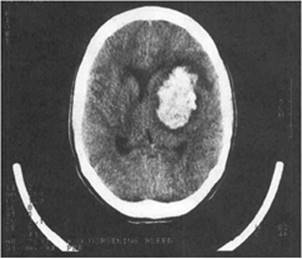
Figure 84.1. Intracerebral hemorrhage of the left basal ganglia. Hyperdense appearance of blood on the CT scan easily defines the extent of hemorrhage. |
||
|
Table 84.3 Inclusion Criteria for tPA USE |
||
|
Aside from time of onset, factors that may place the patient at increased risk for bleeding must be sought. Tables 84.3 through 84.5 list common inclusion and exclusion criteria for intravenous (IV)-tPA (tissue plasminogen activator), which are based on those used in the pivotal trials. Most institutions have their own protocols, which may vary somewhat from those used in the trials.
|
Table 84.4 Absolute Contraindications to tPA USE |
||
|
Once a decision regarding acute treatment has been made, attention can be directed to identifying conditions that may have caused the patient's stroke. Cerebrovascular and cardiovascular diseases share many of the same risk factors, including hypertension, hyperlipidemia, diabetes mellitus, cigarette smoking, and obesity. Other risk factors that may be important include obstructive sleep apnea, migraine headaches with an aura, and drug abuse. Family history may provide insight into heritable causes of stroke such as cerebral autosomal dominant arteriopathy with subcortical infarcts and leukoencephalopathy (CADASIL) or Fabry disease.
|
Table 84.5 Relative Contraindications to tPA USE |
||
|
Neurologic Examination
The neurologic examination will allow the clinician to quickly determine which brain areas are dysfunctional and further narrow the differential diagnosis. The neurologic deficits caused by ischemic cerebrovascular disease are expected to be lateralizing and confined to a vascular distribution. For example, the triad of language disturbance and right face and arm motor deficits is typical for occlusion of the left MCA. However, sudden-onset motor and sensory deficit of both arms is not lateralizing, nor readily explained by a cerebrovascular occlusion, and thus is more likely due to spinal cord pathology.
The first step in localization of vascular lesions is determining, based on the signs and symptoms, whether they arise from the anterior circulation (carotid artery and its main branches, the anterior and middle cerebral arteries) or the posterior circulation (vertebral, basilar, and posterior cerebral arteries). This finding will guide the remainder of the diagnostic evaluation, therapy, and prognosis. Ideally, these two separate circulations would be robustly connected such that a failure in one could be compensated by the other, but this is rarely the case.
The two symptoms that most accurately reflect carotid circulation disease are aphasia and monocular blindness. Aphasia is a deficit in either the expression or comprehension of language and may involve both in the acute period. Aphasia must be distinguished from dysarthria, which is the inability to correctly produce words due to motor impairment of facial, lingual, or pharyngeal muscles; dysarthria may result from either anterior or posterior circulation infarcts. The areas responsible for language reside in the dominant (nearly always left) hemispheric cortex, within the territory of the MCA; a stroke causing aphasia must, therefore, involve this circulation. Similarly, the blood supply of the eye arises largely from the ophthalmic artery, a direct branch from the carotid artery, and monocular ischemia therefore implicates the carotid circulation. The prototypic example of this process is amaurosis fugax, or transient monocular blindness in which vision is lost in one eye for minutes. This must be contrasted with a visual field deficit, which affects one field of both eyes, as this is more likely the result of posterior circulation ischemia. Pain is rarely a significant complaint, but when present, especially if following the course of a major blood vessel, arterial dissection should be considered. Involvement of the carotid artery may cause a Horner syndrome.
Because of the density of discrete populations of neurons supplied by the posterior circulation, the clinical syndromes that result from strokes in this area are usually more complex than those in the cerebral hemispheres. The medulla, pons, midbrain, cerebellum, parts of the thalami, and the visual cortices are the major structures involved. Strokes involving the brainstem often manifest with cranial nerve dysfunction (dysarthria, dysphagia, diplopia). Crossed signs, with motor or sensory deficits affecting one side of the face and the opposite side of the body, may occur as major decussations in these pathways and occur in the pons and medulla. The unique vascular anatomy of the basilar artery, with a single midline vessel supplying both sides of the pons and the posterior cerebral arteries, may lead to bilateral neurologic deficits.
Lacunar infarctions also have a set of clinical features that may be used to differentiate them from other stroke subtypes. Classic lacunar syndromes include pure motor hemiparesis (caused by infarction in the internal capsule or basis pontis), pure hemisensory symptoms (caused by infarction in the ventral posterolateral [VPL] thalamic nucleus), dysarthria–clumsy hand syndrome (with pontine or internal capsule infarcts), and ataxia-hemiparesis (pontine infarct).
Vascular System Examination
Physical examination of the vascular system itself is usually surprisingly unrewarding. Atherosclerosis may present few outward signs. Although carotid bruits were classically emphasized, modern ultrasound techniques have proven them to be of low sensitivity and specificity for predicting vascular disease. No characteristic feature, including the volume, pitch, or duration of the bruit, reliably indicates the degree or the nature of constriction of the vascular lumen. Many bruits reflect benign conditions. The clinical significance of carotid bruits is minimized because they are audible in many asymptomatic persons without atherosclerosis who never suffer from cerebrovascular disease, but may be absent in severely diseased vessels. Therefore, even if a carotid bruit is detected, it may be difficult to decide whether it is relevant to the patient's symptoms, and it should not be given undue emphasis in the overall evaluation. Clinical decisions should be based on definitive assessment of blood vessels using ultrasound or angiography.
Examination of the heart should focus on detecting thrombogenic diseases, including myocardial infarction, congestive heart failure, arrhythmias, prosthetic valves, and bacterial endocarditis. Heart disease is a key risk factor for stroke and may complicate the acute period. Patients with stroke may also present concurrently with a myocardial infarction (MI) and, without careful examination, the less dramatic of the two may go undetected. Elevation of serum troponin levels is a very sensitive and specific indicator of an MI. Isolated creatine kinase (CK)-MB elevations should be interpreted with caution, however, as the MB fraction is expressed in brain tissue, and small elevations are not uncommon in stroke (10).
Laboratory Studies
Laboratory studies will also help narrow the differential diagnosis and may reveal relevant comorbid conditions. Some studies need to be obtained immediately to determine a patient's candidacy for thrombolytic therapy, including an electrolyte battery, glucose, platelet count, cardiac enzymes, beta-HCG (human chorionic gonadotropin), and coagulation parameters. Severe electrolyte (specifically hyponatremia or hypernatremia) or glucose disturbances can cause neurologic dysfunction that may mimic stroke. Cardiac enzymes will determine if cardiac ischemia is part of the current presentation, and a beta-HCG will reveal occult pregnancy, both of which may be contraindications to systemic thrombolytics. Coagulation parameters and a platelet count will identify patients who may be at greater risk for bleeding.
Other laboratory studies will help determine the cause and identify risk factors but do not need to be obtained immediately. A fasting lipid profile should be obtained for potential vascular risk factor modification. In patients older than 50 years of age, an erythrocyte sedimentation rate and C-reactive protein are essential if giant cell arteritis is suspected. In patients for whom an unusual cause of stroke is suspected (young patients or minimal vascular risk factors), laboratory investigation of prothrombotic states could be considered. The interpretation of these tests is very complex and should be performed in consultation with a hematologist (11). Toxicology screening should be performed on hospital admission, with attention directed to amphetamines, phencyclidine, ephedrine, and cocaine.
|
|
|
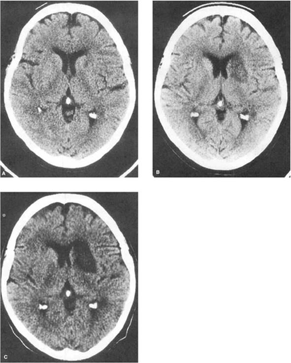
Figure 84.2. Evolving radiographic evidence of cerebral infarction. CT scan at 3 hours (A) shows little evidence of acute ischemia. At 3 days, damaged brain is indicated by hypodensity in the left subcortical region (B). At 8 days, frank infarction is now clearly demonstrated by CT (C). |
Imaging Studies
CT Scan
Several imaging studies are used in the evaluation of acute stroke. A noncontrasted CT scan of the brain is the standard initial evaluation for stroke. CT is very sensitive to the presence of hemorrhage, which is the primary reason for its use in the acute setting, and is the only radiologic test necessary to determine eligibility for IV-tPA. CT is not sensitive for detection of an acute cerebral infarction (Fig. 84.2), and the lack of abnormality within the first 24 hours should be expected. The view of the posterior fossa is also quite limited, and any changes (with the exception of hemorrhage) seen within the brainstem or cerebellum should be confirmed with MRI. When available, CT angiography and perfusion studies can be obtained with very little additional time and provide valuable additional information regarding the patency of blood vessels and blood flow to individual large vessel territories. Pathologic changes on these studies are visible immediately, but they require iodinated contrast, which may be a limiting factor for some patients. The angiographic results from CTA are closest to the traditional gold standard exam: digital subtraction catheter angiography (CTA). When examining the carotid arteries, the results from CTA are often sufficient to differentiate a high-grade stenosis (60%–99%) that is treatable from complete occlusion that is not.
MRI
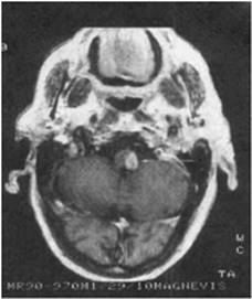
MRI is far more sensitive for acute infarction than CT. Diffusion MR sequences can detect ischemia within, perhaps, minutes of onset. MRI is of special value in brainstem and posterior circulation strokes, since the images it produces are not obscured by bony artifacts as with CT (Fig. 84.3). The combination of multiple MRI sequences allows for much more specific differentiation between ischemic brain and other structural abnormalities. In addition, MRI can display flow-related enhancement of the vasculature, resulting in a magnetic resonance angiogram (MRA). The resulting image can be manipulated in three dimensions, allowing for more accurate interpretation of small abnormalities. However, MRA tends to slightly overestimate the degree of stenosis, and thus is not usually sufficient to differentiate high-grade stenosis from occlusion (12). MRA uses gadolinium as a contrast agent instead of an iodinated material, making the study available to more patients. Perfusion studies, very similar to those performed with CT, may also be obtained if the right equipment is available. Disadvantages of MRI and MRA include reduced availability and substantially longer scanning times, which place some limitations on their use.
Digital Subtraction Catheter Angiography
Digital subtraction catheter angiography is an invasive imaging technique in which the artery of interest is selectively catheterized under fluoroscopy and dye injection enables a high resolution view of the vessel that can be obtained in multiple planes. As the quality of noninvasive imaging techniques has improved and the associated morbidity has decreased to approximately 1% (11), catheter angiography is no longer used as a routine screening test. Indications now include precisely delimiting critical vascular stenoses and examination of arterial dissections, arteriovenous malformations, and aneurysms.
Carotid Ultrasound
Carotid ultrasound provides a rapid noninvasive assessment of carotid artery disease, based on abnormalities of either flow (Doppler) or morphology (B-mode). As with any ultrasound technique, sensitivity is to some degree operator dependent, but with experienced technicians and interpreters, duplex scanning provides a reproducible, accurate screening examination for carotid disease. However, this technique suffers from the same limitation as MRA in differentiating high-grade stenosis and occlusion. Lesions of the more distal internal carotid artery may also be difficult to visualize in some patients.
Transcranial Doppler
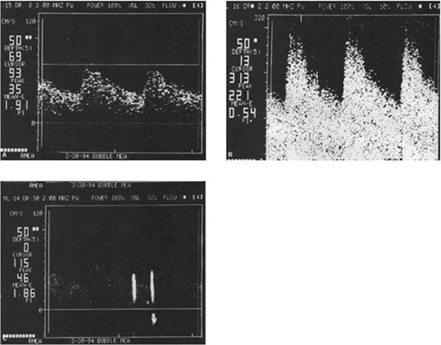
Transcranial Doppler (TCD) allows rapid bedside assessment of abnormal flow within the distal ICA and major intracranial arteries. It is primarily a functional study that provides information about blood flow velocity and vascular resistance rather than structural features. The 2-MHz ultrasonic signal can penetrate various bony “windows” in most patients, and its gated character allows identification of arteries by “depth” of the reflected signal. TCD can examine proximal portions of all major branches of the circle of Willis, but is insensitive for pathology beyond the A1, M1, or P1 segments. Newer applications include detection of microemboli and online monitoring of arterial flow during invasive procedures, such as carotid endarterectomy (Fig. 84.4). TCD may also be useful as an adjunctive therapy to tPA, as continuous insonation may enhance thrombolysis (13). Disadvantages include major dependency on operator skill and the prevalence of acoustically inadequate bony windows.
Transthoracic Echocardiography
Transthoracic echocardiography (TTE) is essential to evaluate cardiac function. Physicians must attend not only to a visualized thrombus, but also to other pathologic states associated with systemic embolization, including left ventricular wall motion abnormalities, chamber dilatation, valvular disease, ejection fraction, and septal defects. TTE can be routinely performed and is a superior study for the detection of ventricular apex pathology, left ventricle thrombus, and views of prosthetic valves. Transesophageal echocardiography (TEE) provides much greater resolution and is more sensitive for pathology of the left atrial appendage, intra-atrial septum, atrial aspect of mitral-tricuspid valves, and the ascending aorta. TEE is an invasive procedure that requires sedation but can be performed safely on most patients (14).
|
|
|
Figure 84.3. MRI demonstrates enhancing left lateral medullary infarction in a patient with Wallenberg syndrome. |
Management
Acute Therapy
As with any acutely ill patient, attention should initially be focused on the evaluation of airway, breathing, and circulation. A secure airway should be established for patients with a depressed consciousness. Supplemental oxygen or mechanical ventilation should be used as needed to treat any degree of hypoxia. Circulation assessment includes evaluation of blood pressure and cardiac electrical activity with an ECG, as coexistent MI is not uncommon. Patients with acute stroke are often markedly hypertensive, and one should be cautious in aggressively treating elevated blood pressure before a more complete assessment of the patient has been completed (15). Blood pressure goals are determined by type of stroke, cause, and the presence of comorbid conditions, such as coronary artery disease; this will be discussed in the following sections.
|
|
|
Figure 84.4. Transcranial Doppler. A: The normal flow-velocity profile through the middle cerebral artery (velocity plotted over time during three cardiac cycles) is demonstrated. B: Elevated flow velocities as a result of the arterial spasm associated with subarachnoid hemorrhage is demonstrated. C: Two microemboli are detected through the middle cerebral artery as transient high-intensity signals. |
The immediate goal will be to determine if the patient is a candidate for thrombolytic therapy; thus attention should be focused on obtaining the relevant history and performing a neurologic examination. The care of patients who will ultimately not receive thrombolytic therapy will be discussed below (see Supportive Care). Crucial for therapy is the proper determination of the exact time of onset of the stroke. The patient must be witnessed to have had an abrupt change in neurologic status by a reliable observer; otherwise the time of onset, by default, must be the last time the patient was seen at his or her baseline level of neurologic function. All patients should be evaluated with the National Institutes of Health Stroke Scale (NIHSS), which can help to exclude a patient from potentially harmful therapy on the basis of the stroke being too small or too severe.
Thrombolytic Therapy
Currently, thrombolytic treatment with recombinant tissue plasminogen activator (tPA) in eligible stroke patients is the standard of care based on the results of four large trials: the National Institute of Neurological Disorders and Stroke (NINDS) recombinant t-PA study (16,17,18), the European Cooperative Acute Stroke Study (ECASS)-I (19), ECASS-II (20), and the ATLANTIS tPA (Alteplase) Acute Stroke Trial (parts A and B) (21,22). The collective results indicate that patients treated with tPA within 3 hours of onset were ≥30% more likely to have minimal or no disability at 3 months compared with placebo-treated patients (23). The average disability across all groups was, additionally, decreased in the treatment group. The benefit seen by the tPA-treated group existed regardless of patient age or stroke subtype.
To maximize the possible benefit and minimize the risk for hemorrhage, the NINDS study helped to establish strict inclusion criteria for the administration of thrombolytic therapy in acute stroke patients, which are outlined in Table 84.3. Absolute exclusion criteria have been established as well (Table 84.4). In addition, there is a long list of relative contraindications to thrombolytic therapy (Table 84.5), which often vary slightly in institutional protocols. These can be summarized as risks for bleeding from a noncompressible site, the presence of potential stroke mimics, or uncontrollable hypertension. The rationale for excluding patients with improving symptoms is to avoid giving a potentially harmful treatment to patients with epileptic postictal presentations or spontaneous recovery.
Tissue Plasminogen Activator (tPA)
When the protocol is followed, the administration of tPA is safe relative to other commonly accepted treatments. Several studies have attempted to examine the risk, but perhaps the most clinically relevant is the number needed to harm. For every 100 patients treated with tPA who match the NINDS trials populations across all levels of final global disability, approximately 32 will receive benefit, and approximately 3 will be harmed (24). The risk–benefit ratio, thus, strongly favors treatment. Many of the hemorrhages that occur are asymptomatic, and of those that are symptomatic, not all symptoms persist to discharge. Most patients who experience intercerebral hemorrhage (ICH) after tPA therapy have severe baseline insults and were already destined for a poor outcome; thus the hemorrhage does little to alter the final outcome (25). One should keep in mind that, although excluding a patient from treatment will mitigate the risk for hemorrhage, it also denies the patient a significantly better chance of recovery.
The dose of tPA is 0.9 mg/kg, with a maximum dose of 90 mg; 10% is given as a bolus over 1 minute, and the remaining 90% is infused over 60 minutes. Following treatment, patients should be monitored in an ICU for more than 24 hours. While many post-tPA patients will not need ventilatory or pressor support, as do many other patients in the ICU, blood pressure monitoring and frequent neurologic exams are critical for a favorable outcome, as hypertension dramatically increases the risk for hemorrhage. Blood pressure must initially be monitored noninvasively as arterial puncture is contraindicated for 24 hours after administration of tPA. All other invasive procedures, such as placement of nasogastric tubes, urinary bladder catheters, central venous lines, intramuscular injections, and rectal temperature must be avoided for 24 hours as well. Also, any drug that impairs hemostasis such as heparin, or antiplatelet or nonsteroidal anti-inflammatory agents, are contraindicated during this 24-hour period.
Vital Signs.
Vital signs should be checked every 15 minutes for the first 2 hours, then every 30 minutes for 6 hours, and then every hour for 16 hours. Blood pressure should be strictly controlled for 24 hours, keeping the systolic blood pressure less than 180 mm Hg and the diastolic blood pressure less than 105 mm Hg (Table 84.6). Labetalol is recommended for control of hypertension; 10 mg should be given intravenously over 1 to 2 minutes, and the dose repeated or doubled every 10 to 20 minutes, up to a total of 150 mg. If the blood pressure remains refractory despite these measures, consideration can be given to a continuous infusion of nicardipine or sodium nitroprusside. Neurologic evaluation should be performed every hour. Oxygenation should be checked by continuous pulse oximetry and oxygen provided to keep saturation >95%. The benefit of therapeutic hypothermia is yet to be confirmed, but euthermia is clearly associated with better outcome (26). Acetaminophen, 650 mg every 4 hours orally or rectally, should be given for any temperature higher than 37.4°C, and a cooling blanket used for temperatures over 38.9°C.
STAT Head CT
A STAT head CT should be performed for any worsening neurologic status. Should an intracerebral hemorrhage develop following thrombolysis, several steps must be taken emergently. Neurosurgery should be contacted for possible hematoma evacuation, and anticoagulation should be reversed according to the protocol outlined in Table 84.7.
Intra-arterial Approach for Thrombolytic Therapy
The use of thrombolytic interventions outside of the 3-hour time window is controversial. Trials that have extended the therapeutic window beyond 3 hours for intravenous therapy have failed to show convincing benefit (19,20,22), as the risk of hemorrhage rapidly increases with time. However, there have been several attempts to prove the benefit of catheter-directed therapy via an intra-arterial approach for focal clot lysis. The most studied agent was prourokinase (27), but it was unfortunately removed from the market in 1999 due to concerns with its preparation. The agent most commonly used today is tPA.
|
Table 84.6 Blood Pressure Control after tPA |
||
|
Theoretically, there are several potential benefits to treatment of stroke via an intra-arterial approach. First, angiographic confirmation of vessel occlusion can be obtained at the time of treatment. Second, high concentrations of thrombolytic agents can be given directly at the site of thrombosis, thereby minimizing systemic exposure. Third, the response to lysis can be monitored by direct visualization. Fourth, mechanical disruption of the clot (e.g., via balloon angioplasty, MERCI device) may accelerate thrombolysis (28).
According to the American Stroke Association guidelines (29), intra-arterial thrombolysis is an option for treatment of selected patients who have major stroke of <6 hours' duration due to occlusion of the MCA and who are not otherwise candidates for intravenous tPA. The availability of intra-arterial thrombolysis should not preclude the administration of IV-tPA in otherwise eligible patients. Treatment requires immediate access to cerebral angiography and qualified interventionalists.
Supportive Care
Supportive care lacks the excitement and drama of acute therapy, but nonetheless is critical to patient outcomes. Since the 1970s, mortality from stroke has markedly diminished from a rate of 156 to 56/100,000 cases (30), with only 3% to 10% of stroke patients receiving thrombolytic therapy; this trend cannot be explained by tPA (31,32). Rather, it is the advancements made in the prevention of medical complications that has reduced mortality.
|
Table 84.7 Management of post-tPA Hemorrhage |
||
|
||
|
Table 84.8 Suggested Recommended Guidelines for Treating Elevated Blood Pressure in Spontaneous ICH |
||
|
Motor Deficits
Dysphagia is common after stroke, whether it be from upper or lower motor neuron deficits. All patients with stroke should be screened for dysphagia before being allowed to take anything by mouth, as aspiration pneumonia is a substantial contributor to mortality after stroke (33). Any impairment in level of consciousness or motor function of mouth, tongue, palate, or muscles of facial expression should alert the physician to a high risk for dysphagia. Coarse breath sounds may indicate that aspiration has already occurred. A preserved gag reflex may not indicate safety with swallowing (34). If any of these signs are present, a formal swallowing evaluation is indicated. Enteral access can be achieved by placement of a nasogastric or Dobhoff tube. Feeding and hydration should be initiated immediately (35). Dysphagia often improves rapidly, but placement of a percutaneous endoscopic gastrostomy (PEG) tube may be necessary for prolonged feeding.
Before the advent of routine prophylaxis, deep vein thromboses and resulting pulmonary emboli were common in stroke patients who frequently have profound lower extremity immobility; pulmonary embolism presently accounts for approximately 10% of deaths in stroke patients (36). Recent trials, including PREVAIL (37), have shown low-molecular-weight heparin to be safe and more effective than unfractionated heparin. Early mobilization not only prevents deep vein thromboses (DVTs), but also speeds rehabilitation.
Blood Pressure Maintenance
Blood pressure goals in the acute period after ischemic stroke are often a source of confusion for many practitioners. Ischemic stroke patients are often quite hypertensive, and, while the reflex may be to aggressively lower the blood pressure, this may cause more harm than benefit. When blood flow is impaired, the resulting change in pressure gradients will allow circulation through alternative or collateral paths. Through autoregulation, these collateral vessels maximally dilate, and thus flow becomes entirely dependent on cerebral perfusion pressure. Some even advocate the induction of therapeutic hypertension with pressors, but insufficient evidence exists to recommend this practice. Therefore, lowering blood pressure potentially reduces blood flow to the potentially salvageable penumbra. Determining the exact pressure required for adequate blood flow is not readily accomplished, so one must err on the side of hypertension. For most stroke patients with acute hypertension, the general practice is to refrain from intervening until the pressure exceeds an arbitrary limit of 220 mm Hg systolic or 120 mm Hg diastolic (29). Patients with coronary artery disease or those with another comorbidity that may preclude tolerance of such pressures may require a careful decrement in blood pressure. In these cases, it is recommended that blood pressure be lowered slowly, using frequent smaller doses of drug rather than larger ones that may cause rapid changes; patients should be carefully observed for acute worsening as pressure is lowered. In patients having received thrombolytics, the tolerable limit is lower, as the risk for intracerebral hemorrhage increases with increasing blood pressure. In this case, pressures greater than 180 mm Hg systolic or greater than 105 mm Hg diastolic require treatment according to the protocol (Table 84.8).
Hyperglycemia
Hyperglycemia will be detected on admission in approximately one third of patients with stroke (38). Predictions for patients with persistent hyperglycemia (blood glucose level >200 mg/dL) during the first 24 hours after stroke are expansion of the volume of ischemic stroke and poor neurologic outcomes (39). Our practice is to aggressively control glucose—keeping it between 80 and 140 mg/dL—but care must be taken to avoid hypoglycemia, as the morbidity from that may abolish the benefit obtained from treating hyperglycemia.
Although most patients eventually improve substantially after a stroke, early clinical deterioration is not uncommon. Neurologic causes of clinical deterioration include progressive or recurrent stroke, hemorrhagic transformation of the infarct, and local cerebral edema. The latter is the most common cause of deterioration, and may well cause fatal herniation in large MCA infarctions, especially in the young, women, and in patients with involvement of additional vascular territories (40). Brain swelling typically appears about 4 days after the stroke onset (41). Dramatic early swelling has been described; the term malignant MCA infarction is used to delineate a group of patients with large territorial infarcts that swell within 24 hours (42).
Ischemia-related edema is cytotoxic and unresponsive to treatments useful for vasogenic edema. Corticosteroids, in particular, do not appear helpful, and hyperglycemia associated with their use may worsen clinical outcome (29,43,44). Although no evidence exists that ultimate outcome is improved, certain treatments are often used to reduce intracranial pressure: mannitol (1 g/kg bolus, then 0.3 g/kg every 6 hours) dehydrates viable brain tissue (45) to create more space for swelling tissue, and is primarily useful as a temporizing measure for patients destined for decompressive surgery, as the effects of this medication are transient and associated with eventual rebound. Other measures such as mechanical hyperventilation (to a PaCO2 of 25–30 mm Hg), or use of albumin and furosemide to raise colloid oncotic pressure (to 25–30 mm Hg) are used, but, again, their efficacy in improving outcome remains to be demonstrated.
Decompressive Surgery
Decompressive surgery, including hemicraniectomy and durotomy with temporal lobe resection, for treatment of brain edema after stroke has been a controversial topic. Many studies in the past have shown conflicting results, but these trials enrolled mixed age groups, and surgery was often not performed until symptomatic herniation occurred (46). Three large European trials (HAMLET, DESTINY, and DECIMAL) (47,48,49) that enrolled patients younger than 60 years of age and treated within 48 hours of onset were prematurely terminated after it became clear that decompressive craniotomy was associated with a dramatic survival and outcome benefit. In elderly patients, the results have generally shown that, while mortality may be decreased, outcomes remained poor (50).
The likelihood of hemorrhagic transformation of a stroke increases as stroke volume increases. Often, this transformation may be limited to petechial transudation of blood products into the ischemic tissue bed. Generally, this phenomenon occurs in a delayed fashion with no associated clinical deterioration. Specific therapy is not usually required, although any ongoing anticoagulation is generally held for 1 to 2 weeks. If the hemorrhage is associated with clinical deterioration, management should follow those principles outlined in the intracerebral hemorrhage section below.
Early initiation of physical, occupational, and speech therapy services hastens functional recovery from stroke. Each patient requires individualized assessment for potential benefit from these services. Speech therapists are also commonly involved in formal assessment of aspiration risk. A video fluoroscopy swallowing study is the most sensitive measure and should be a consideration for most patients with a stroke. At the least, bedside swallowing function should be observed by a trained technician, nurse, or physician before oral intake is resumed.
Intracerebral Hemorrhage
Pathogenesis
In contrast to ischemic stroke, primary intracerebral hemorrhage (ICH) involves bleeding, usually of arterial origin, into normally perfused brain, and thus must be distinguished from hemorrhagic transformation of an initially ischemic stroke. The expanding hematoma causes direct injury to local brain tissue and dysfunction in surrounding regions. The onset is typically very sudden, although continued bleeding often progresses over minutes or hours. Very often, there is either depression or loss of consciousness due to an abrupt increase in intracranial pressure (ICP) from the sudden outpouring of blood into the brain. In addition to the initial cerebral insult caused by the hemorrhage, secondary injury can occur by various means, including seizures, hydrocephalus, and edema, all of which can lead to a further increase in ICP.
In younger patients, hypertension is by far the more common cause, and, as such, ICH tends to occur in the same brain areas where other hypertensive pathologies occur, specifically brainstem, cerebellum, and deep supratentorial structures (51). In contrast, lobar ICHs occur more commonly in the elderly population and are often associated with cerebral amyloid angiopathy in the absence of hypertension (52). ICH may also occur in the setting of trauma, use of illicit drugs (e.g., cocaine) or over-the-counter medications (e.g., phenylpropanolamine) (53), excessive alcohol consumption (54), an underlying vascular abnormality (e.g., arteriovenous malformation, cerebral aneurysm), brain tumor (primary or secondary), or a bleeding diathesis.
ICH causes approximately 10% of first-time strokes. The 30-day mortality rate is high at 35% to 50%, with half of the deaths occurring within the first 2 days (12). Outcome in ICH is dependent on several factors, including the location and size of the hemorrhage (55), the age of the patient, the Glasgow Coma Scale (GCS) on presentation (56), and the cause of the hemorrhage. When intraventricular blood is present, the mortality substantially increases (57) and worsens further with increasing volume (58). The presence of hydrocephalus also confers a poor prognosis (59).
Clinical Evaluation
Rapid diagnosis of ICH is essential, as progression during the first several hours is the norm. The hallmark is sudden onset focal neurologic deficit, which progresses over minutes to hours. Steady symptomatic progression of a focal deficit is rare in either ischemic stroke or subarachnoid hemorrhage. Headache, increased blood pressure, and impaired level of consciousness are common features that complete the presentation. History gathering should be directed at elucidating the presence of risk factors as outlined above. Other considerations include the use of antithrombotic medications (e.g., aspirin or warfarin) or hematologic disorders that predispose to bleeding, such as severe liver disease. The initial physical examination is similar to that of patients with ischemic stroke, focusing on airway, breathing, and circulation before assessing the level of consciousness and neurologic deficits. The patient's coagulation parameters should be checked immediately and corrected if abnormal.
Once stabilized, the patient should undergo a noncontrast head CT immediately to verify brain hemorrhage. CT angiography may also be helpful in detecting aneurysms, arteriovenous malformations (AVMs), underlying tumors, or abscesses. Contrast extravasation into the hematoma is thought to represent ongoing bleeding (60). MRI will also provide information about the hemorrhage but is time consuming and potentially dangerous for an unstable patient. When it can be obtained safely, MRI is most useful for dating the time course of the hemorrhage if the history is in doubt, detecting areas of prior hemorrhage (with the use of gradient-echo imaging) (61), and diagnosing cavernous malformations. Catheter angiography should be considered in a young patient with an ICH with no history of hypertension, as an occult AVM or aneurysm may be responsible.
|
Table 84.9 Intravenous Medications That May Be Considered for Control of Elevated Blood Pressure in Patients With ICH |
|||||||||||||||||||||||||||
|
|||||||||||||||||||||||||||
Cardiac arrhythmias represent another potentially catastrophic secondary complication of ICH, especially those that occur in the right hemisphere insular region. Dysfunction of this area has a propensity for causing abnormal cardiac electrical activity and “cerebrogenic sudden death” (62). Patients with such lesions must have close cardiac monitoring in the intensive care unit during their first several days after hemorrhage.
Management
The mainstays of medical treatment of acute ICH are correction of any coagulopathy and avoidance of hypertension. Most studies suggest that hematoma expansion occurs within the first several hours of onset, and therefore treatment must begin as soon as possible (63). If the patient recently received heparin, protamine sulfate (1 mg/100 units of heparin) should be administered; this drug is given carefully to avoid hypotension. Patients anticoagulated with warfarin with an elevated international normalized ratio (INR) should be reversed with vitamin K (10 mg intravenously administered over 15–20 minutes to prevent anaphylactoid reaction), as well as fresh frozen plasma to normalize the prothrombin time (PT).
Beyond the first few hours after onset, aggressive lowering of blood pressure may be potentially harmful. Large hemorrhages will lead to increased ICP, and attention must be focused on maintaining the cerebral perfusion pressure (mean arterial pressure–intracranial pressure [MAP-ICP]) >70 mm Hg to avoid secondary ischemia (64). Despite several clinical trials, a definitive guideline has yet to emerge. The American Stroke Association guidelines regarding blood pressure, presented in Table 84.8, are based on the best available, albeit incomplete, evidence. Table 84.9 lists recommended antihypertensives.
Seizures—occasionally nonconvulsive—occur commonly after ICH. The published incidence rates vary from 4% in unmonitored populations (65) to 28% in patients with continuous electrophysiologic monitoring in a neurocritical care unit (66). Prophylactic anticonvulsant medications may be considered for patients, especially for those with cortical involvement. If seizures are confirmed, they should be treated aggressively. Phenytoin remains the preferred first-line agent, as it is nonsedating, and loading doses can be given intravenously. Loading doses are 15 to 20 mg/kg and should be given as fosphenytoin if needed quickly to avoid hypotension and potential toxic infusion reactions. Maintenance doses are often 4 to 5 mg/kg and may be given slowly. Total serum levels of phenytoin should be followed daily, at least initially. Free levels may be necessary, as phenytoin is protein bound, and critically ill patients are very frequently protein depleted. Additional antiepileptics should be added as necessary. The duration of therapy with anticonvulsants is unclear, but in seizurefree nonepileptic patients, antiepileptic medications are often arbitrarily withdrawn after 4 to 6 weeks of therapy.
Other general supportive care measures are similar to those described above for ischemic stroke, including attention to DVT prophylaxis, and treatment of hyperthermia and hyperglycemia. Patients with increased intracranial pressure often develop disturbances of free water homeostasis in the form of either hyponatremia or hypernatremia. As with ischemic stroke, corticosteroids for treatment of edema are of no benefit and actually increase morbidity (67).
Treatment of increased ICP should initially focus on more conservative noninvasive measures, such as keeping the head of the bed at 30 degrees, hyperventilation if intubated, and osmolar therapy with hypertonic saline or mannitol. An implanted ICP monitor should be considered for those patients with large hematomas. This will inform the decision to place an intraventricular drain or perform a surgical evacuation.
In general, patients with a GCS of 4 or more have a uniformly poor outcome, whether or not surgery is performed, and thus these patients should be treated medically. Patients with cerebellar hemorrhages >3 cm in diameter should be considered for emergency decompression, especially if there are signs of brainstem compression, hydrocephalus, or neurologic deterioration (64); whether surgery is indicated in most other patients is not clear. Clinicians treating patients who deteriorate despite maximal medical therapy may turn to surgical decompression, but results from clinical trials have been mixed (68,69). Patients with lobar hemorrhages secondary to amyloid angiopathy have exceptionally friable cortical blood vessels and are poor surgical candidates.
|
Table 84.10 Severity Grade of Subarachnoid Hemorrhage |
||
|
Subarachnoid Hemorrhage
Subarachnoid hemorrhage (SAH) is a relatively uncommon but often devastating type of stroke. Incidence is estimated at 30,000 patients per year in the United States, with a mortality that exceeds 50%. Whereas head trauma is the most frequent cause of subarachnoid hemorrhage, aneurysmal rupture results in the greatest morbidity and mortality. Clinically, this is an apoplectic disorder. Most commonly, patients perceive a sudden severe headache with rapid impairment of consciousness, both symptoms related to the sudden release of irritating blood products into the meningeal spaces surrounding the brain. Focal neurologic symptoms such as hemiparesis, sensory loss, or diplopia may occur if loculation of subarachnoid blood or intraparenchymal extension of the hemorrhage develops. The most important features of the neurologic examination are the assessment of level of consciousness, cranial nerve function, and motor function. Clinical severity of SAH is graded on these findings (70) (grades I to V, Table 84.10) and can be rough prognostic indicators.
Diagnosis
Diagnosis of SAH is based on neuroimaging or cerebrospinal fluid (CSF) analysis. Brain CT scan is a very sensitive indicator of the presence of subarachnoid blood, although close examination must be paid to the subarachnoid spaces surrounding the brainstem and over the cerebral convexities (Fig. 84.5). Brain parenchyma itself most commonly displays no acute abnormalities. Erythrocyte concentration in CSF below approximately 30,000 cells/µL may not result in the diagnostic increased density within CSF on CT scans. In approximately 10% of patients, diagnosis therefore requires CSF analysis through lumbar puncture. In addition to elevated erythrocyte count, CSF xanthochromia and elevation of CSF D-dimer can often be detected in true subarachnoid hemorrhage. The latter two findings may help distinguish bloody CSF from a “traumatic tap,” as these serve as markers of the breakdown of thrombosis or blood products. Serial cell counts should always be obtained, however, whenever SAH is suspected. Cell counts in SAH should be roughly equivalent in all tubes, whereas a declining count is usual in traumatic punctures. It should be stressed that lumbar puncture should be avoided in any patient with a depressed level of consciousness until CT scan excludes a focal mass (such as intraparenchymal or subdural hemorrhage). If bacterial meningitis is a concern, blood cultures should be obtained and antibiotics started while awaiting results of the CT scan.
|
|
||||||||||||||||||||||||||||||||||||
|
Figure 84.5. Subarachnoid hemorrhage. Blood is imaged as hyperdense fluid within the cisterns surrounding the brainstem and within bilateral sylvian fissures. |
||||||||||||||||||||||||||||||||||||
|
Table 84.11 Complications of Subarachnoid Hemorrhage |
||||||||||||||||||||||||||||||||||||
|
||||||||||||||||||||||||||||||||||||
Management
Patients with acute SAH are at high risk for a multitude of complications (71) (Table 84.11) that usually mandate admission to an intensive care facility. All patients should be placed on strict bed rest, with appropriate precautions for deep venous thrombosis and aspiration. Patients with progressive lethargy may require intubation for airway protection and mechanical ventilation. Until the aneurysm has been ablated, blood pressure should be kept in the normotensive range, and isotonic intravenous fluids should be used to maintain normovolemia. All patients should be started on nimodipine at 60 mg every 4 hours (duration 21 days), either orally or through a nasogastric tube, for prevention of vasospasm (see below).
Historically, patients were placed on prophylactic anticonvulsants; this practice has recently come into question, as patients enrolled in the international tirilazad trials who were on prophylactic anticonvulsants had significantly more in-hospital complications and worse clinical outcomes (72).
ECG changes and elevations in cardiac enzymes, troponin, and CK-MB are commonly seen in SAH patients and may represent the phenomenon of stunned myocardium, in which case management should be aimed at optimizing left ventricular function to support cardiovascular and cerebrovascular perfusion (73,74,75,76). Serum electrolytes are closely monitored, as hyponatremia may be seen in more than 30% of patients after SAH; however, hypernatremia can occur as well and is significantly associated with clinical outcome (77). The cause of hyponatremia after SAH is most commonly reported to be due to syndrome of inappropriate antidiuretic hormone (SIADH) but can also be due to cerebral salt-wasting (CSW) syndrome and other causes (78). One theory links SAH-induced hyponatremia to levels of serum brain natriuretic peptide, which is thought to be associated with delayed ischemic neurologic deficits (79). It is intuitive that SIADH and CSW are not treated in the same manner.
Serum glucose levels should also be closely monitored, as hyperglycemia has been significantly associated with mortality and poor functional outcome in SAH patients (80). Because fever in SAH patients has been associated with mortality and poor clinical outcome (80), and has even been linked to vasospasm (81), patients should be kept normothermic. Platelet levels should be monitored, as a relatively significant incidence of heparin-induced thrombocytopenia has been reported in SAH patients (82).
Rebleeding
In those patients surviving the initial hemorrhage, the leading factor associated with mortality is rebleeding from the aneurysm. A second bleed from an aneurysm is associated with a 74% mortality rate (83). Untreated aneurysms rebleed at a rate of 4% on day 1, then 1% to 2% a day for the next 4 weeks. Thus, early treatment to secure a ruptured aneurysm is critical. This can be done either via a craniotomy and clipping, or by endovascular coiling. The findings of the International Cooperative Study on the Timing of Aneurysm Surgery concluded that aneurysmal clipping was best performed either early (0 to 3 days) or late (11 to 14 days), but outcome was worse when performed at 7 to 10 days after the onset of SAH (84). Because of the concern for rebleeding, we have adopted a protocol of treating aneurysms in the ultra-early period (less than 24 hours after presentation). The advent of endovascular therapy to treat ruptured aneurysms may bypass the risks associated with open clipping during the 7- to 10-day period; thus we recommend treatment for the ruptured aneurysm as soon as it is found, and not waiting until the late 11- to 14-day period. Early treatment of the ruptured aneurysm also allows aggressive management of vasospasm, manipulations that would increase the risk of rebleeding from an unsecured aneurysm.
|
|
|
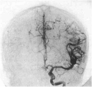
Figure 84.6. Aneurysm at the bifurcation of the left middle cerebral artery demonstrated by angiography. (Courtesy of R. Nick Bryan, MD, Baylor College of Medicine, Houston, TX.) |
Vasospasm
After rebleeding, vasospasm is the next leading cause of mortality and morbidity from a SAH (85,86). The exact cause of arterial vasospasm following SAH is unknown, but its incidence does appear to be correlated with the density of blood products seen on CT scan, the basis for the Fisher score to predict vasospasm (87,88). Severe vasospasm may result in cerebral infarction within the vascular distribution of the involved artery. The risk for vasospasm begins about 3 days after the bleed and may persist for 3 weeks. Transcranial Doppler is a sensitive, noninvasive indicator of the presence and degree of vasospasm within proximal arteries, although it may not detect vasospasm restricted to smaller peripheral vessels. This technique may be used daily to guide and monitor management strategies. Modern techniques, such as CT angiography and CT perfusion studies, have been reported to be successful in diagnosing vasospasm (89) (Fig. 84.6).
The calcium channel blockers nimodipine and nicardipine can reduce the incidence of vasospasm as well as associated cerebral infarction. Trials with magnesium sulfate have yielded promising results in reducing vasospasm (90,91,92) or achieving better clinical outcomes in patients (93). Studies with statin therapy (HMG Co-A inhibitors), such as pravastatin and simvastatin, have demonstrated promising results with reduced rates of vasospasm and better clinical outcomes (94,95,96,97).
Once the aneurysm is secured, vasospasm can be managed aggressively. Triple-H therapy—hypertension, hypervolemia, hemodilution—is the first-line therapy against vasospasm. Once vasospasm occurs, IV fluids should be pushed to the tolerance of the patient (below 400 mL/hour). Since congestive heart failure is a potential complication, central pressure monitoring—at a minimum—should be used in patients at risk; optimal central venous pressure is 8 to 12 mm Hg. Other modalities of monitoring, from pulse-waveform variability (FlowTrak, PICCO, LiDCO) to placement of a pulmonary artery catheter, may be necessary to properly care for these patients. If hypervolemic therapy is not adequate to control vasospasm, hypertension can be initiated by cessation of antihypertensives or use of vasopressors (e.g., phenylephrine, 0.1–5 µg/kg per minute, or vasopressin, 0.01–0.04 units per minute), targeting mean arterial pressures of 120 to 140 mm Hg (systolic blood pressure [BP] 180–200 mm Hg). Triple-H therapy does not increase the risk of rupture of other, incidentally found, aneurysms in patients with multiple aneurysms (98).
For symptomatic vasospasm refractory to these therapies, endovascular interventions can be performed, such as percutaneous transluminal balloon angioplasty, and/or intra-arterial administration of calcium channel blockers or other vasodilating agents (99) may be considered in experienced hands. Even using the most aggressive management strategies, vasospasm remains a leading cause of morbidity and mortality after subarachnoid hemorrhage.
Acute Hydrocephalus
Acute hydrocephalus occurs in approximately 20% of survivors of SAH, either as a result of direct obstruction of CSF channels or by impeding CSF absorption at arachnoid granulations. The likelihood of hydrocephalus increases with worsening grade of hemorrhage. Ventriculostomy drainage is recommended for patients with acute hydrocephalus and decreased level of consciousness; improvement can be expected in >50% of patients.
References
1. Norris JW, Hachinski VC. Misdiagnosis of stroke. Lancet. 1982;1(8267): 328–331.
2. Inzitari D, et al. The causes and risk of stroke in patients with asymptomatic internal-carotid-artery stenosis. North American Symptomatic Carotid Endarterectomy Trial Collaborators. N Engl J Med. 2000;342(23):1693–1700.
3. Fisher CM. The arterial lesions underlying lacunes. Acta Neuropathol. 1968; 12(1):1–15.
4. Bogousslavsky J, Van Melle G, Regli F. The Lausanne Stroke Registry: analysis of 1,000 consecutive patients with first stroke. Stroke. 1988;19(9):1083–1092.
5. Mohr JP, et al. The Harvard Cooperative Stroke Registry: a prospective registry. Neurology. 1978;28(8):754–762.
6. Pruitt AA, et al. Neurologic complications of bacterial endocarditis. Medicine (Baltimore). 1978;57(4):329–343.
7. Roeltgen DP, Weimer GR, Patterson LF. Delayed neurologic complications of left atrial myxoma. Neurology. 1981;31(1):8–13.
8. Bogousslavsky J, Pierre P. Ischemic stroke in patients under age 45. Neurol Clin. 1992;10(1):113–124.
9. Kalb R. Spontaneous dissection of the carotid and vertebral arteries. N Engl J Med. 2001;345(6):467.
10. Ay H, Arsava EM, Saribas O. Creatine kinase-MB elevation after stroke is not cardiac in origin: comparison with troponin T levels. Stroke. 2002; 33(1):286–289.
11. Hankey GJ, et al. Inherited thrombophilia in ischemic stroke and its pathogenic subtypes. Stroke. 2001;32(8):1793–1799.
12. Anderson CS, et al. Spectrum of primary intracerebral haemorrhage in Perth, Western Australia, 1989–90: incidence and outcome. J Neurol Neurosurg Psychiatry. 1994;57(8):936–940.
13. Alexandrov AV, et al. Ultrasound-enhanced systemic thrombolysis for acute ischemic stroke. N Engl J Med. 2004;351(21):2170–2178.
14. DeRook FA, et al. Transesophageal echocardiography in the evaluation of stroke. Ann Intern Med. 1992;117(11):922–932.
15. Johannessen KA, et al. Risk factors for embolisation in patients with left ventricular thrombi and acute myocardial infarction. Br Heart J. 1988;60(2): 104–110.
16. Tissue plasminogen activator for acute ischemic stroke. The National Institute of Neurological Disorders and Stroke rt-PA Stroke Study Group. N Engl J Med. 1995;333(24):1581–1587.
17. Generalized efficacy of t-PA for acute stroke. Subgroup analysis of the NINDS t-PA Stroke Trial. Stroke. 1997;28(11):2119–2125.
18. Intracerebral hemorrhage after intravenous t-PA therapy for ischemic stroke. The NINDS t-PA Stroke Study Group. Stroke. 1997;28(11):2109–2118.
19. Hacke W, et al. Intravenous thrombolysis with recombinant tissue plasminogen activator for acute hemispheric stroke. The European Cooperative Acute Stroke Study (ECASS). JAMA. 1995;274(13):1017–1025.
20. Hacke W, et al. Randomised double-blind placebo-controlled trial of thrombolytic therapy with intravenous alteplase in acute ischaemic stroke (ECASS II). Second European-Australasian Acute Stroke Study Investigators. Lancet. 1998;352(9136):1245–1251.
21. Clark WM, et al. The rtPA (alteplase) 0- to 6-hour acute stroke trial, part A (A0276g): results of a double-blind, placebo-controlled, multicenter study. Thrombolytic therapy in acute ischemic stroke study investigators. Stroke. 2000;31(4):811–816.
22. Clark WM, et al. Recombinant tissue-type plasminogen activator (Alteplase) for ischemic stroke 3 to 5 hours after symptom onset. The ATLANTIS Study: a randomized controlled trial. Alteplase Thrombolysis for Acute Noninterventional Therapy in Ischemic Stroke. JAMA. 1999;282(21):2019– 2026.
23. Kwiatkowski TG, et al. Effects of tissue plasminogen activator for acute ischemic stroke at one year. National Institute of Neurological Disorders and Stroke Recombinant Tissue Plasminogen Activator Stroke Study Group. N Engl J Med. 1999;340(23):1781–1787.
24. Saver JL. Number needed to treat estimates incorporating effects over the entire range of clinical outcomes: novel derivation method and application to thrombolytic therapy for acute stroke. Arch Neurol. 2004;61(7):1066–1070.
25. Saver JL. Hemorrhage after thrombolytic therapy for stroke: the clinically relevant number needed to harm. Stroke. 2007;38(8):2279–2283.
26. Olsen TS, Weber UJ, Kammersgaard LP. Therapeutic hypothermia for acute stroke. Lancet Neurol. 2003;2(7):410–416.
27. del Zoppo GJ, et al. PROACT: a phase II randomized trial of recombinant pro-urokinase by direct arterial delivery in acute middle cerebral artery stroke. PROACT Investigators. Prolyse in Acute Cerebral Thromboembolism. Stroke. 1998;29(1):4–11.
28. Smith WS, et al. Safety and efficacy of mechanical embolectomy in acute ischemic stroke: results of the MERCI trial. Stroke. 2005;36(7):1432– 1438.
29. Adams HP Jr., et al. Guidelines for the early management of adults with ischemic stroke: a guideline from the American Heart Association/American Stroke Association Stroke Council, Clinical Cardiology Council, Cardiovascular Radiology and Intervention Council, and the Atherosclerotic Peripheral Vascular Disease and Quality of Care Outcomes in Research Interdisciplinary Working Groups: the American Academy of Neurology affirms the value of this guideline as an educational tool for neurologists. Stroke. 2007; 38(5):1655–1711.
30. Jemal A, et al. Trends in the Leading Causes of Death in the United States, 1970–2002. JAMA. 2005;294(10):1255–1259.
31. Heuschmann PU, et al. Frequency of Thrombolytic Therapy in Patients With Acute Ischemic Stroke and the Risk of In-Hospital Mortality: The German Stroke Registers Study Group. Stroke. 2003;34(5):1106–1112.
32. Koennecke H-C, et al. Intravenous tPA for Ischemic Stroke Team Performance Over Time, Safety, and Efficacy in a Single-Center, 2-Year Experience. Stroke. 2001;32(5):1074–1078.
33. Aslanyan S, et al. Pneumonia and urinary tract infection after acute ischaemic stroke: a tertiary analysis of the GAIN International trial. Eur J Neurol. 2004;11(1):49–53.
34. Addington WR, Stephens RE, Gilliland KA. Assessing the laryngeal cough reflex and the risk of developing pneumonia after stroke: an interhospital comparison. Stroke. 1999;30(6):1203–1207.
35. Choi-Kwon S, et al. Nutritional status in acute stroke: undernutrition versus overnutrition in different stroke subtypes. Acta Neurol Scand. 1998;98(3): 187–192.
36. Wijdicks EF, Scott JP. Pulmonary embolism associated with acute stroke. Mayo Clin Proc. 1997;72(4):297–300.
37. Sherman DG, et al. The efficacy and safety of enoxaparin versus unfractionated heparin for the prevention of venous thromboembolism after acute ischaemic stroke (PREVAIL Study): an open-label randomised comparison. Lancet. 2007;369(9570):1347–1355.
38. Williams LS, et al. Effects of admission hyperglycemia on mortality and costs in acute ischemic stroke. Neurology. 2002;59(1):67–71.
39. Baird TA, et al. Persistent poststroke hyperglycemia is independently associated with infarct expansion and worse clinical outcome. Stroke. 2003;34(9): 2208–2214.
40. Maramattom BV, Bahn MM, Wijdicks EF. Which patient fares worse after early deterioration due to swelling from hemispheric stroke? Neurology. 2004;63(11):2142–2145.
41. Ropper AH, Shafran B. Brain edema after stroke. Clinical syndrome and intracranial pressure. Arch Neurol. 1984;41(1):26–29.
42. Qureshi AI, et al. Timing of neurologic deterioration in massive middle cerebral artery infarction: a multicenter review. Crit Care Med. 2003;31(1):272–277.
43. Tellez H, Bauer RB. Dexamethasone as treatment in cerebrovascular disease. 1. A controlled study in intracerebral hemorrhage. Stroke. 1973;4(4):541–546.
44. Bauer RB, Tellez H. Dexamethasone as treatment in cerebrovascular disease. 2. A controlled study in acute cerebral infarction. Stroke. 1973;4(4):547–555.
45. Videen TO, et al. Mannitol bolus preferentially shrinks non-infarcted brain in patients with ischemic stroke. Neurology. 2001;57(11):2120–2122.
46. Gupta R, et al. Hemicraniectomy for massive middle cerebral artery territory infarction: a systematic review. Stroke. 2004;35(2):539–543.
47. Hofmeijer J, et al. Hemicraniectomy after middle cerebral artery infarction with life-threatening edema trial (HAMLET). Protocol for a randomised controlled trial of decompressive surgery in space-occupying hemispheric infarction. Trials. 2006;7:29.
48. Vahedi K, et al. Sequential-design, multicenter, randomized, controlled trial of early decompressive craniectomy in malignant middle cerebral artery infarction (DECIMAL Trial). Stroke. 2007;38(9):2506–2517.
49. Juttler E, et al. Decompressive surgery for the treatment of malignant infarction of the middle cerebral artery (DESTINY): a randomized, controlled trial. Stroke. 2007;38(9):2518–2525.
50. Holtkamp M, et al. Hemicraniectomy in elderly patients with space occupying media infarction: improved survival but poor functional outcome. J Neurol Neurosurg Psychiatry. 2001;70(2):226–228.
51. Flaherty ML, et al. Racial variations in location and risk of intracerebral hemorrhage. Stroke. 2005;36(5):934–937.
52. Tonk M, Haan J. A review of genetic causes of ischemic and hemorrhagic stroke. J Neurol Sci. 2007;257(1–2):273–279.
53. Yoon BW, et al. Phenylpropanolamine contained in cold remedies and risk of hemorrhagic stroke. Neurology. 2007;68(2):146–149.
54. Monforte R, et al. High ethanol consumption as risk factor for intracerebral hemorrhage in young and middle-aged people. Stroke. 1990;21(11):1529–1532.
55. Broderick JP, et al. Volume of intracerebral hemorrhage. A powerful and easy-to-use predictor of 30-day mortality. Stroke. 1993;24(7):987–993.
56. Tuhrim S, et al. Prediction of intracerebral hemorrhage survival. Ann Neurol. 1988;24(2):258–263.
57. Tuhrim S, et al. Validation and comparison of models predicting survival following intracerebral hemorrhage. Crit Care Med. 1995;23(5):950–954.
58. Tuhrim S, et al. Volume of ventricular blood is an important determinant of outcome in supratentorial intracerebral hemorrhage. Crit Care Med. 1999; 27(3):617–621.
59. Diringer MN, Edwards DF, Zazulia AR. Hydrocephalus: a previously unrecognized predictor of poor outcome from supratentorial intracerebral hemorrhage. Stroke. 1998;29(7):1352–1357.
60. Becker KJ, et al. Withdrawal of support in intracerebral hemorrhage may lead to self-fulfilling prophecies. Neurology. 2001;56(6):766–772.
61. Greenberg SM, Finklestein SP, Schaefer PW. Petechial hemorrhages accompanying lobar hemorrhage: detection by gradient-echo MRI. Neurology. 1996;46(6):1751–1754.
62. Cheung RT, Hachinski V. The insula and cerebrogenic sudden death. Arch Neurol. 2000;57(12):1685–1688.
63. Kazui S, et al. Enlargement of spontaneous intracerebral hemorrhage. Incidence and time course. Stroke. 1996;27(10):1783–1787.
64. Broderick J, et al. Guidelines for the management of spontaneous intracerebral hemorrhage in adults: 2007 update: a guideline from the American Heart Association/American Stroke Association Stroke Council, High Blood Pressure Research Council, and the Quality of Care and Outcomes in Research Interdisciplinary Working Group. Circulation. 2007;116(16):e391–e413.
65. Passero S, et al. Seizures after spontaneous supratentorial intracerebral hemorrhage. Epilepsia. 2002;43(10):1175–1180.
66. Vespa PM, et al. Acute seizures after intracerebral hemorrhage: a factor in progressive midline shift and outcome. Neurology. 2003;60(9):1441–1446.
67. Poungvarin N, et al. Effects of dexamethasone in primary supratentorial intracerebral hemorrhage. N Engl J Med. 1987;316(20):1229–1233.
68. Morgenstern LB, et al. Surgical treatment for intracerebral hemorrhage (STICH): a single-center, randomized clinical trial. Neurology. 1998;51(5): 1359–1363.
69. Auer LM, et al. Endoscopic surgery versus medical treatment for spontaneous intracerebral hematoma: a randomized study. J Neurosurg. 1989;70(4):530–535.
70. Hunt WE, Hess RM. Surgical risk as related to the time of intervention in the repair of intracranial aneurysms. J Neurosurg. 1968;28:14.
71. Mayberg MR, Batjer HH, Dacey R, et al. Guidelines for the management of aneurysmal subarachnoid hemorrhage. Circulation. 1994;90:2592.
72. Rosengart AJ, Huo JD, Tolentino J, et al. Outcome in patients with subarachnoid hemorrhage treated with antiepileptic drugs. J Neurosurg. 2007; 107:253–260.
73. Urbaniak K, Merchant AI, Amin-Hanjani S, Roitberg B. Cardiac complications after aneurysmal subarachnoid hemorrhage. Surg Neurology. 2007; 67:21–28.
74. Banki NM, Kopelnik A, Dae MW, et al. Acute neurocardiogenic injury after subarachnoid hemorrhage. Circulation. 2005;112:3314–3319.
75. Naidech AM, Kreiter KT, Janjua N, et al. Cardiac troponin elevation, cardiovascular morbidity, and outcome after subarachnoid hemorrhage. Circulation. 2005;112:2851–2856.
76. Deibert E, Barzilai B, Braverman AC, et al. Clinical significance of elevated troponin I levels in patients with nontraumatic subarachnoid hemorrhage. J Neurosurg. 2003;98:741–746.
77. Qureshi AI, Suri MF, Sung GY, et al. Prognostic significance of hypernatremia and hyponatremia among patients with aneurysmal subarachnoid hemorrhage. Neurosurgery. 2002;50:749–755.
78. Sherlock M, O'Sullivan E, Agha A, et al. The incidence and pathophysiology of hyponatremia after subarachnoid haemorrhage. Clin Endocrinol (Oxf). 2006;64:250–254.
79. McGirt MJ, Blessing R, Nimjee SM, et al. Correlation of serum brain natriuretic peptide with hyponatremia and delayed ischemic neurologic deficits after subarachnoid hemorrhage. Neurosurgery. 2004;54:1369–1373.
80. Wartenberg KE, Schmidt JM, Claassen J, et al. Impact of medical complication on outcome after subarachnoid hemorrhage. Crit Care Med. 2006; 34:617–623.
81. Oliveira-Filho J, Ezzeddine MA, Segal AZ, et al. Fever in subarachnoid hemorrhage—relationship to vasospasm and outcome. Neurology. 2001; 56:1299–1304.
82. Hoh BL, Aghi M, Pryor JC, et al. Heparin-induced thrombocytopenia Type II in subarachnoid hemorrhage patients—incidence and complications. Neurosurgery. 2005;57:243–248.
83. Juvela S. Rebleeding from ruptured intracranial aneurysms. Surgical Neurol. 1989;32:323–326.
84. Kassell NF, Torner JC, Jane JA, et al. The international cooperative study on the timing of aneurysm surgery Part 2—Surgical results. J Neurosurg. 1990;73:37–47.
85. Hoh BL, Topcuoglu MA, Singhal AB, et al. Effect of clipping, craniotomy, or intravascular coiling on cerebral vasospasm and patient outcome after aneurysmal subarachnoid hemorrhage. Neurosurgery. 2004;55:779–786.
86. Hoh BL, Rabinov JD, Pryor JC, et al. A modified technique for using elastase to create saccular aneurysms in animals that histologically and hemodynamically resemble aneurysms in human. Acta Neurochir (Wien). 2004;146:1177–1183.
87. Fisher CM, Kistler JP, Davis JM. Relation of cerebral vasospasm to subarachnoid hemorrhage visualized by computer tomographic scanning. Neurosurgery. 1980;6:1–9.
88. Kistler JP, Crowell RM, Davis KR, et al. The relation of cerebral vasospasm to the extent and location of subarachnoid blood visualized by CT scan—a prospective study. Neurology. 1983;33:424–436.
89. Binaghi S, Colleoni ML, Maeder P, et al. CT angiography and perfusion CT in cerebral vasospasm after subarachnoid hemorrhage. AJNR Am J Neuroradiol. 2007;28:750–758.
90. van den Bergh WM, Algra A, van Kooten F, et al. Magnesium sulfate in aneurysmal subarachnoid hemorrhage—a randomized controlled trial. Stroke. 2005;36:1011–1015.
91. Stippler M, Crago E, Levi EI, et al. Magnesium infusion for vasospasm prophylaxis after subarachnoid hemorrhage. J Neurosurg. 2006;105:723– 729.
92. Schmid-Elsaesser R, Kunz M, Zausinger S, et al. Intravenous magnesium versus nimodipine in the treatment of patients with aneurysmal subarachnoid hemorrhage—a randomized study. Neurosurgery. 2006;58:1054–1065.
93. Muroi C, Terzic A, Fortunati M, et al. Magnesium sulfate in the management of patients with aneurysmal subarachnoid hemorrhage—a randomized, placebo-controlled, dose-adapted trial. Surg Neurol. 2008;69:33– 39.
94. McGirt MJ, Blessing R, Alexander MJ, et al. Risk of cerebral vasospasm after subarachnoid hemorrhage reduced by statin therapy. A multivariate analysis of an institutional experience. J Neurosurg. 2006;105:671–674.
95. Lynch JR, Wang H, McGirt MJ, et al. Simvastatin reduces vasospasm after aneurysmal subarachnoid hemorrhage—results of a pilot randomized clinical trial. Stroke. 2005;36:2024–2026.
96. Tseng MY, Czosnyka M, Richards H, et al. Effects of acute treatment with pravastatin on cerebral vasospasm, autoregulation and delayed ischemic deficits after aneurysmal subarachnoid hemorrhage—a phase II randomized, placebo controlled trial. Stroke. 2005;36:1627–1632.
97. Tseng MY, Hutchinson PJ, Czosnyka M, et al. Effects of acute pravastatin treatment on intensity of rescue therapy, length of inpatient stay, and 6 month outcome in patients after aneurysmal subarachnoid hemorrhage. Stroke. 2007;38:1545–1550.
98. Hoh BL, Carter BS, Ogilvy CS. Risk of hemorrhage from unsecured, unruptured aneurysms during and after hypertensive, hypervolemic therapy. Neurosurgery. 2002;50:1207–1211.
99. Hoh BL, Ogilvy CS. Endovascular treatment of cerebral vasospasm—transluminal balloon angioplasty, intra-arterial papaverine, and intra-arterial nicardipine. Neurosurg Clin North Am. 2005;16:501–516.