Deborah Stern
Andrew Pollak
Thomas M. Scalea
Bony and soft tissue injuries often complicate trauma. Optimal treatment of these injuries is essential for good overall patient care. Patients treated suboptimally are at risk for myriad complications such as fat embolism syndrome, pulmonary embolus, soft tissue infection, and multiple organ failure. There is little prospective randomized data to guide treatment decisions in patients with bony injuries. However, certain treatment principles have emerged. These must, of course, be interpreted in the context of the total patient.
Fracture stabilization is an operative procedure of significance. Some patients may be too critically ill or injured at the time of initial presentation to tolerate definitive fracture fixation. The critical care staff must be well versed in the management principles that govern musculoskeletal injuries as they will often have important roles to play in decision making. Orthopedic surgeons and/or anesthesiologists may not have sufficient information on total patient physiology in order to make these decisions without input from the critical care staff.
Diagnosing Musculoskeletal Injury
Patients presenting after injury should typically have either signs or symptoms to guide diagnosis of their injuries. Pain, swelling, and soft tissue discoloration often accompany musculoskeletal injury, particularly in badly displaced or comminuted fractures. However, the initial presentation of many fractures may be far more subtle. Patients who have fractures that are not badly displaced may actually have only modest soft tissue swelling and discoloration or none at all at the time of presentation. Patients who are multiply injured often undergo endotracheal intubation at the time of emergency department presentation and are unable to complain of pain. Multiply injured patients may have other injuries that distract them from the pain of a minor fracture. Patients with brain injury and/or intoxication may not complain of pain. The authors are constantly impressed how the signs and symptoms of fractures can be subtle or completely absent initially. Fractures missed at the time of initial presentation can cause significant morbidity or mortality. While these data on missed injuries are sparse, estimates are that fractures are missed up to 10% of the time in multiply injured patients (1). Several options exist to try to reduce that number.
Certainly any patient with any signs, symptoms, or suspicion of fractures should undergo evaluation both by physical exam and plain radiographs. While the yield is low, obtaining radiographs on every area of soft tissue contusions will yield unsuspecting fractures in a small number of cases. Thus, screening algorithms should be broad to avoid missing fractures. Technologic advances have helped. The Lodox Corporation has produced the Statscan, a total body screening x-ray machine (Fig. 85.1). Using low-dose radiation developed in the diamond mine industry, it is possible to obtain both anteroposterior (AP) and lateral total body images of the skeleton (Fig. 85.2). These can be obtained in 13 seconds and the images available for viewing 15 seconds later. This allows the clinician to obtain an entire skeletal survey rapidly (2).
Another option is to perform a “tertiary survey” somewhere between 12 and 24 hours postadmission (1). Patients with fractures that have been missed generally develop some signs or symptoms in the ensuing hours after admission. The tertiary survey is a careful head-to-toe physical examination attempting to identify areas of missed injuries. Diagnostic imaging can be obtained to diagnose injuries that had been initially missed.
As with any injury, fractures are dynamic. It is not simply that a bone is broken. X-ray images are static and will not allow the clinician to appreciate the degree of energy transfer at the time of impact. Bones may be substantially displaced at the time of impact and then be spontaneously reduced or be reduced by actions of the local muscle and appear relatively undisplaced in the emergency department. Fractures are a source of significant bleeding, either from the fracture fragments themselves, muscle bleeding, or accompanied major vascular injury.
Physical examination should be able to identify blood loss in the extremity. The generally accepted rule is that closed long bone fractures each bleed up to 2 units of blood. Thus, bilateral femur fractures and a tibia fracture in a 70-kg person will result in a 3-liter blood loss, which produces class III hemorrhage.
Neurovascular injury can be diagnosed relatively simply in patients who are awake and alert. Careful neurovascular exam directed by patient complaints should pick up virtually every injury. The same is not true for patients who are multiply injured, intubated, and in the intensive care unit (ICU). In addition, neurovascular injury may evolve over time. For instance, nerve contusion can progress to neurapraxia or paralysis. Swelling around the fracture site can progress and compress the nerve, producing neurologic dysfunction. Compartment syndrome often occurs 6 to 24 hours postinjury as a result of muscular swelling within the noncompliant fascial envelope following crush injuries and/or fractures. Partial thickness vascular injuries can go on to vascular thrombosis hours or even days after injury, producing limb-threatening ischemia.
Critical care practitioners must be cognizant of all of these. Careful ongoing assessment is critical in order to make these diagnoses as early as possible. A delayed diagnosis can produce substantial disability that is lifelong. Even worse, unrecognized ischemia and/or compartment syndrome can place a limb at jeopardy, requiring amputation later. Unrecognized ischemia can also produce life-threatening electrolyte abnormalities such as hyperkalemia and/or be the source of multiple organ failure, either one of which can prove to be fatal.
|
|
|
Figure 85.1. Statscan in trauma bay. |
|
|
|
Figure 85.2. Normal anterior total body and lateral spine. |
Treatment Options for Fractures
Clearly, fractures that are not associated with life-threatening bleeding are not the highest priority following initial patient presentation. However, all long bone fractures should be splinted as part of the initial management. Every time the patient is transferred or moved in any way, unsplinted fractures are displaced, producing additional blood loss and pain and risking secondary nerve or vascular injury. Splinting can be accomplished simply by utilizing several IV boards and some rolled gauze to fashion a makeshift splint. There are commercially available molded metal splints that can also be used short term. These two methods can lead to skin breakdown if applied for long periods of time. Once time permits, more definitive, lightweight plaster splints can be made to restore stability and decrease the risk of skin breakdown.
It is also important for the critical care practitioner to understand the various options for more definitive fracture fixation. Rigid fracture fixation is generally obtained via open reduction/internal fixation (ORIF) or closed intramedullary (IM) nail fixation. ORIF involves directly exposing the fracture fragments. They are then manually reduced and rigid fixation is achieved utilizing a number of techniques such as plates and screws or rods. Overlying muscles must often be divided in order to obtain adequate exposure. This also allows for debridement of nonviable and/or contused muscle and irrigation of the fracture hematoma. Depending on the particular fracture, ORIF can be a substantial operative procedure with significant soft tissue injury and blood loss from the procedure itself. Fractures often treated with ORIF include proximal and distal tibial fractures, acetabular fractures, periarticular humerus, elbow fractures, and distal radius fractures (Fig. 85.3).
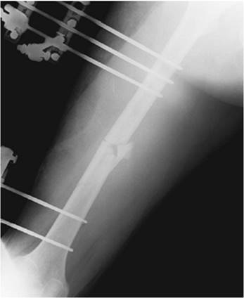
Approximately 70 years ago, the technique of closed IM nailing was developed. This technique involves making a small incision proximally or distally in the extremity. The fracture fragments are then reduced manually under fluoroscopic visualization. The medullary canal is accessed and a guidewire is placed across the fracture. A nail is then driven up the medullary canal over the guidewire to sterilize the bone. If desired, the canal can be reamed before the nail is placed. The nail can be locked in place by placing several screws in the very proximal or distal portion of the nail through a secondary small incision. These screws are placed under fluoroscopic control (Fig. 85.4).
|
|
|
Figure 85.3. This 17-year-old female sustained a high-energy distal tibial fracture as a result of a motorcycle crash. Open reduction with plate fixation offers the best chance of achieving anatomic restoration of articular congruity and reduction of the risk of posttraumatic arthritis. |
IM nailing does not typically require direct exposure of the fracture fragment. Theoretically, there is less soft tissue injury and less blood loss associated with IM nailing as compared to ORIF of fractures. Passage of a nail through the medullary canal is, however, associated with liberation of a number of mediators and/or fat globules into the systemic circulation. Either of these can produce systemic inflammatory response syndrome (SIRS) and/or acute lung injury (ALI). In addition, passage of the nail can produce significant bleeding around the fracture itself, which may not be appreciated as it is not directly visualized in the operative field. IM nailing is generally used for most midshaft femur and tibia fractures. It is necessary to have adequate bone on either side of the fracture in order to achieve sufficient fixation to restore stability.
External fixation involves placing an external frame that spans the fracture, limiting fracture movement (Fig. 85.5). External fixation achieves relatively good fracture stabilization, though it may not produce optimal fracture reduction, particularly with periarticular injuries. It can be rapid and noninvasive and result in substantially less blood loss than ORIF or IM nailing. Pins are placed into the bone on either side of the fracture connected to a frame. This can be used as definitive fixation for some fractures if acceptable restoration of length, alignment, and stability have been achieved.
|
|
|
Figure 85.4. The fracture was treated with early intramedullary nail fixation. With early fixation of femoral fractures in multiply injured patients, many pulmonary problems can be avoided. Stable fixation of even complex, comminuted injuries can allow early weight bearing and facilitate functional rehabilitation despite polytrauma. |
|
|
|
Figure 85.5. This patient's pelvic ring injury was treated with posterior screw fixation and an anterior external fixator. |
Open fractures require an initial debridement and irrigation of the fracture fragments as a minimum. This is done to reduce the rate of early soft tissue infection and late osteomyelitis. While ideal therapy involves irrigation, debridement, and definitive fracture fixation, this is not always the wisest course. Options include irrigation, and debridement can be followed by temporary external fixation. In addition, irrigation and debridement can be followed by simple splinting of the fractures if there are more immediate patient care priorities. This technique is ideally performed in the operating room and involves debridement followed by pulse lavage. Patients can be temporized with a bedside procedure if that is all they are able to tolerate initially or if there are other more urgent priorities. While the data supporting any treatment algorithm are not definitive, most studies suggest that rates of infection are similar provided operative debridement is accomplished within 24 hours of injury (3).
Optimal Timing of Fracture Fixation
There are a significant number of advantages to early definitive fracture fixation. Ideally, this should take place as soon as possible following patient presentation. While the definition of early fracture fixation has varied over the years, most would agree that fracture fixation within 24 hours is early. These advantages may be most pronounced in patients with multiple system injury. Obviously, these are the patients most at risk for systemic complications from a significant operative procedure. Thus, judgment must be key in weighing the various risks and benefits to early fracture fixation.
Patients who are treated nonoperatively initially are often treated in some type of balanced traction, particularly for femoral and acetabular fractures. This requires that the patient remain supine in bed. Patients are at risk for developing dependent atelectasis resulting in worsening respiratory failure. Patients with brain injury and intracranial hypertension may have worsening of their intracranial pressures as they cannot have the head of the bed elevated as easily. These patients may also be at increased risk for aspiration. Even though the fracture is somewhat reduced by the traction apparatus or by a splint, as with any fracture, motion at the fracture site worsens bleeding and produces pain and such motion is only reduced by traction and splinting, not eliminated. Either bleeding or pain can increase the systemic inflammatory response. Any immobilized patient is at risk for thromboembolic complications such as deep vein thrombosis or pulmonary embolus. Those in traction may be at particular risk. Patients kept at bed rest in traction cannot be rolled from side to side, producing pressure ulcerations on areas in contact with the bed. Finally, the incidence of fat embolism syndrome may be higher in patients who undergo delayed fracture repair.
In 1985, Seibel et al. published a prospective nonrandomized cohort study of patients with femoral shaft fractures or acetabulum fractures who also had additional injury (4). All aspects of care of the patients were reportedly controlled except fracture management. Their patient population was divided into four groups. The first group underwent immediate operative fixation of their femoral or acetabular fracture (defined as less than 24 hours after injury). The second group underwent an average of 10 days of femoral traction. The third group underwent up to 30 days of femoral traction. The fourth group had special circumstances that were predicted to potentially contribute to their pulmonary failure septic state. Ventilator days and intensive care days were both substantially greater in the groups that underwent prolonged traction than in the immediate fixation group, supporting the conclusion that femoral traction was detrimental in the setting of blunt multiple trauma.
This led the authors to propose a number of principles governing the care of patients with multisystem trauma and fractures. They proposed that the fracture hematoma was itself a metabolically active organ, capable of producing multiple organ failure. Early fracture fixation allowed for irrigation of the fracture hematoma. They also included the orthopedic surgeon as one of the frontline members of the trauma team, emphasizing the need for early consultation. In addition, they strongly advocated for fracture repair on the night of admission including using two teams to simultaneously repair fractures if that was available and prudent. They argued that all efforts must be made to support the patient through early fracture fixation as they had demonstrated far better results with this technique as opposed to delaying repair for some days to allow patient stabilization.
Following this report, others also demonstrated better results with early fracture fixation (5,6,7). Fracture fixation within 24 hours became the standard of care. However, in the early 1990s, data began being published that suggested that not every patient benefited from early fracture fixation. In fact, these studies strongly suggested that patient outcome was much more a function of underlying injury and not so much the timing of fracture fixation. Poole et al. published a series of patients with long bone fracture and brain injury (8). They were unable to demonstrate any advantage to early fracture fixation, and instead argued that the severity of the brain injury was the ultimate determinate of final outcome. Rogers et al. addressed the cost of performing early fracture fixation, particularly on off hours in hospitals that did not have in-house operating room resources (9). Outcomes were the same when femur fractures were repaired in the middle of the night as compared to a group of patients who were placed on the operating room schedule for the next day. The cost, however, was significantly higher in patients who underwent emergency fracture fixation.
It is unclear as to why these authors were unable to substantiate the findings that seemed to show clear benefit to early operative fixation. One possibility is that the care during the critical care phase of these multiply injured patients improved over the late 1980s and early 1990s. End points of resuscitation were defined (10). The role of invasive monitoring was made more clear (11). Specific needs for patient populations such as geriatric trauma patients were defined (12). Thus, it is possible that the time taken to stabilize the patient in the ICU preoperatively was now, in fact, time well spent.
In the mid- to late 1990s, several papers were published that strongly suggested that early fracture fixation was, in fact, dangerous, particularly in patients who sustained traumatic brain injury. The group from Yale reported on 32 patients treated over a 5-year period with brain injury and long bone fractures (13). Those who underwent early fracture fixation, defined as within the first 24 hours of admission, had statistically significant worse neurologic outcome at the time of discharge. This was thought to be secondary to the increased rate of intraoperative hypotension and hypoxia seen in patients with early fracture fixation. Townsend et al. observed similar findings (14). In their study, episodes of intracranial hypertension were more common in patients who underwent early fracture fixation. They also noted an increased rate of intraoperative hypotension and hypoxia. This risk seemed to persist for approximately 24 hours.
Scalea et al., however, reported different findings (15). They studied over 180 patients with long bone fracture and traumatic brain injury over the same 5-year period as studied by the group at Yale. They found no difference in neurologic outcome in the group of patients treated with early fracture fixation.
How, then, should a critical care physician decide when a patient ought to have fracture fixation? It would seem reasonable to address a number of issues. The first would be to assess the risk of anesthesia and the magnitude of the operative procedure. There certainly are scoring systems that can be applied to assess perioperative risk. Careful clinical assessment may be equally good. The second issue would be to examine operative options. For instance, does the patient have a reasonable nonoperative option? Would a lesser procedure, while perhaps not ideal, at least be adequate? This obviously would require a discussion between the critical care service and the orthopedic surgeons, trauma team, and anesthesiologist. Finally, one must ask whether the patient is in optimal condition. Can we reduce perioperative risk with a reasonable period of preoperative optimization? Can cardiac performance be improved with volume and/or inotropes? Will 1 to 2 days allow ventilatory requirements to be reduced? Would a delay improve renal function? Only when all of these questions are answered can an intelligent decision about the optimal time and fracture fixation be made.
Damage Control Orthopedics
Damage control was a technique developed in the late 1980s and early 1990s in early urban American trauma centers. This shift in approach coincided with a period of time when penetrating trauma became much more common. Not only was penetrating trauma more common, but also multiple high-velocity missiles became the norm, rather than the exception. Traditional teaching was to repair all injuries at the time of initial surgical procedure. Unfortunately, this often required prolonged operative care. Patients developed what was termed the lethal triad of acidosis, coagulopathy, and hypothermia, only to arrive in the ICU and die of acute organ failure, usually 6 to 24 hours postoperatively.
Many trauma centers began using a staged approach with encouraging results. In 1993, Rotundo et al. published the series that named the technique damage control, and demonstrated statistically significant better outcomes in a group of patients treated with the new techniques (16).
The principles that govern damage control are listed in Table 85.1 when damage control is used for abdominal injuries. In essence, only life-saving procedures are performed at the time of the initial procedure. Important but non–life-threatening injuries such as gastrointestinal injuries are simply temporized. The patient is then packed and the fascia left open to avoid the potential of developing abdominal compartment syndrome. The patient is then admitted to the ICU and resuscitated. Between 1 and 3 days later, the patient can be brought back to the operating room for re-exploration, gastrointestinal reconstruction, and an attempt at fascial closure.
|
Table 85.1 Principles of Damage Control |
|
|
The same principles can be applied to bony injuries. Fractures are common and occur in over 75% of multiply injured patients. There are clear advantages to early fracture fixation. Yet, some patients may not be best served by such technique. A technique that would achieve many of the advantages of fracture fixation without the disadvantages of definitive surgery would be advantageous for some patients.
It is important to recognize that remote organ injury occurs not only in association with, but also as a direct consequence of long bone fractures. In addition to the fracture itself, soft tissue injury, compartment syndrome, infection, and extremity ischemia-reperfusion injury can all be associated with release of toxic mediators that can cause remote endothelial cell damage. The primary target of this remote organ injury appears to be the lungs, but secondary targets include the gut, kidney, and brain. The resultant injury is a progressive one and can lead to multiple organ dysfunction syndrome. This understanding led to the development of the concept of damage control orthopedics (DCO). DCO is the process by which temporary stabilization of long bone fractures is accomplished using techniques not associated with secondary systemic injury. Secondary definitive stabilization is delayed until after physiologic stabilization has been achieved (Figs. 85.6 through 85.8). The goal of damage control therefore is to provide the patient with the benefits of early stabilization of long bones without exposing him or her to the risks associated with definitive stabilization procedures.
For that subset of patients whose physiologic condition precludes primary definitive operative stabilization of fractures (early total care), external fixation can be a useful technique for temporary stabilization of long bone fractures. Such external fixation is advantageous because it can be applied rapidly, with minimal blood loss, and with sufficient restoration of stability to prevent ongoing soft tissue injury and to prevent ongoing release of inflammatory mediators. Effective limitation of secondary effects of shock has been demonstrated with this type of initial DCO approach.
The disadvantages to employing this type of technique in all patients with even questionable polytrauma are at least twofold. First, employing temporizing external fixation techniques necessitates a return to the operating room for delayed definitive fixation, thus exposing the patient to two separate operative interventions. This is obviously warranted if the benefit is prevention of a serious secondary consequence such as acute respiratory distress syndrome (ARDS). In the absence of a clear decrease in the risk of ARDS, it is harder to justify exposing polytrauma patients to a need for secondary surgical procedures.
|
|
|
Figure 85.6. Damage control orthopedics case example. This 27-year-old motorcyclist sustained multiple injuries including an open right femoral shaft fracture. He presented in physiologic extremis. Laparotomy demonstrated a grade 5 hepatic injury and a retrohepatic caval injury. Right hepatic lobectomy and repair of a vena caval laceration were performed as part of a damage control laparotomy procedure. |
A second major disadvantage to external fixation (a temporary device) is the risk of complications with conversion from external fixation to intramedullary nailing or other definitive treatment. Experience in the tibia has suggested that if external fixation is in place for less than 2 weeks, the risk of infection with definitive fixation is lower, particularly if there is no history of pin tract infection at any point (17). In the femur, conversion from external fixation to definitive nailing appears safe within the first 4 weeks following application of the initial frame (18).
Advantages of external fixation include rapid application with minimal blood loss and simultaneous stabilization of multiple limbs expeditiously.
The issues that led to the use of DCO have been debated for some time. In 1989, Bone et al. published a prospective randomized trial of early (less than 24 hours after injury) versus late (greater than 72 hours after injury) nail fixation of femoral shaft fractures (19). They found that in patients with an overall Injury Severity Score (ISS) of greater than 18 (indicative of polytrauma), intensive care days and pulmonary complications were reduced with early fixation as compared to late fixation. They concluded that stabilization of femoral fractures within 24 hours of injury in multiply injured patients decreases pulmonary morbidity as compared to stabilization after 72 hours. The study was flawed in that it was not blinded, that parameters of resuscitation were not defined, and that very few patients with severe pulmonary injury were included in the early fixation group.
|
|
|
Figure 85.7. Damage control orthopaedics consisted of rapid debridement of the open femoral fracture plus temporizing external fixation to achieve restoration of femoral length and alignment and decrease ongoing injury secondary to release of inflammatory mediators. |
In 1993, Pape et al. published a worrisome report implicating primary nail fixation of femoral shaft fractures (reamed) as a potential cause of posttraumatic ARDS (20). They reviewed patients admitted over a period of 10 years with an ISS greater than 18 and midshaft femoral fractures. Those with and without chest injury and those who underwent femoral nailing less than 24 hours and greater than 24 hours after injury were compared. They found a higher incidence of ARDS and death in patients with severe chest trauma who were treated with early reamed nailing as compared to those with severe chest trauma treated with delayed reamed nailing. They concluded that early reamed nail stabilization of femoral shaft fractures in “borderline” patients may increase the risk of secondary pulmonary injury and development of ARDS. They recommended that definitive fracture fixation be delayed in these patients and that temporizing techniques such as external fixation be employed.
The problem with Pape's study was that the early nailing group included eight patients with pulmonary contusions as compared to only two in the late nailing group. Both Pape et al. (20) and Bone et al. (19) failed to report on any parameters of resuscitation within their patient populations.
What was becoming obvious as trauma surgeons became more aggressive with early nailing of femoral (and to a certain extent tibial) shaft fractures in polytrauma patients was that the operative procedure for intramedullary stabilization of long bone fractures was associated with some component of pulmonary injury (21,22). While fracture fixation–associated pulmonary injury appeared to be well tolerated in the majority of patients including polytrauma patients, certain susceptible polytrauma patients seemed to be at risk for worsening lung injury. Paradoxically, these are likely the same patients who are at greatest risk for developing secondary pulmonary injury from a nonoperative management of their long bone fractures and the most in need of early fracture stabilization.
|
|
|
Figure 85.8. On postinjury day 5, after adequate restoration of physiologic stability, the patient underwent removal of the temporary external fixator and definitive fixation of the femoral fracture using a reamed intramedullary nail. He was discharged to a rehabilitation facility 19 days after injury. |
Several groups of surgeons have engaged in the process of temporary stabilization of long bone fractures in polytrauma patients using techniques associated with minimal systemic consequences. Scalea et al. published their experience with external fixation as a temporary tool for stabilization of long bone fractures in polytrauma patients in 2000 (23). They reported that as part of DCO, external fixation was an alternative mechanism of achieving temporary stabilization in multiply injured patients and that it was rapid, associated with minimal blood loss, and safely convertible to better definitive fixation after appropriate delayed physiologic stabilization of the patient. Other authors have expanded on the DCO concept and reported more specifically on its efficacy (24).
The physiologic rationale for DCO is based upon the timing and extent of the initial inflammatory response that follows a major injury. In most individuals, that initial inflammatory response is followed by a counterregulatory anti-inflammatory response that leads to spontaneous recovery. In situations where the initial inflammatory response is excessive, secondary remote organ injury such as acute lung injury related to increased pulmonary capillary membrane permeability can occur. The likelihood of this secondary remote organ injury developing may be increased in situations associated with substantial ischemia-reperfusion injury (such as prolonged shock), continued loss of body temperature, tourniquet-mediated ischemia-reperfusion injury, a surgical blood loss with failure of resuscitation, and embolization of fat and marrow contents secondary to instrumentation of the femoral or tibial canal. This secondary remote organ injury is characterized by increased capillary membrane permeability, acute lung injury, gut bacterial translocation, and acute tubular necrosis. It can be mediated by cytokines, complement, activated neutrophils, eicosanoids, and reactive oxygen products.
Thus, both early total care and ongoing long bone instability can serve as second hits that lead to the development of severe secondary remote organ injury. In the “at-risk” patient population, early DCO can limit ongoing release of inflammatory mediators and allow for further physiologic stabilization prior to proceeding with definitive stabilization (25).
Hildebrand et al. evaluated the association between the timing of secondary definitive surgical intervention, inflammatory changes, and systemic outcome (26). They reviewed a prospective cohort of patients treated with DCO. They compared those treated with early secondary surgery (2–4 days after initial injury) to those treated with late secondary surgery (5–8 days after initial injury). They found that early secondary surgery was associated with a higher incidence of organ dysfunction and concluded that there was no particular advantage to early secondary surgery. They recommended that if DCO is selected, secondary surgery should be delayed more than 5 days after the initial DCO procedure.
In a prospective randomized trial, Pape et al. compared nailing to DCO with regard to immunoinflammatory parameters in patients with tibial or femoral shaft fractures, and ISS greater than 16 (27). Patients with a thoracic abbreviated injury scale score (AIS) greater than 3, ongoing shock, or elevated intracranial pressure were excluded. They found that primary femoral nailing was associated with significantly higher systemic interleukin (IL)-6 and IL-8 levels for 48 hours postoperatively compared to either initial DCO procedures or secondary femoral nailing after initial damage control. This suggested that the secondary release of inflammatory mediators associated with femoral nailing can be effectively mitigated by initial DCO with delayed femoral nailing.
O'Toole et al. looked at the effect of resuscitation prior to femoral nailing on the development of ARDS in multiply injured patients (28). They found that a practice of resuscitation protocol that included normalization of lactate prior to IM nailing resulted in rare need of damage control and development of ARDS.
DCO, the practice of utilizing temporary external fixation to limit ongoing injury secondary to long bone fractures in situations where definitive operative stabilization is contraindicated, may be effective in patients with polytrauma and lung or brain injury. For the majority of patients with femoral shaft fractures, including most polytrauma patients, primary reamed femoral nailing within 24 hours of injury remains the gold standard of treatment (21). Defining the “at-risk” patients who are unable to tolerate early reamed nailing is difficult, but if adequate resuscitation and normalization of serum lactate is achieved, damage control should rarely be necessary (28). In borderline patients who are physiologically unstable because of severe chest or head injury or inadequate resuscitation, temporizing external fixation and damage control orthopedics may be advantageous.
Compartment Syndrome
Compartment syndrome occurs when there is increased tissue pressure within a confined osseofascial space. This increased pressure compromises blood flow and subsequently results in tissue damage if left untreated. The fascia of muscle compartments is stiff and does not expand to accommodate significant swelling. Increases in compartment volume and subsequent pressure can be caused by fractures, soft tissue crush injury, hemorrhage, reperfusion following arterial revascularization, and increased capillary permeability as may occur in the setting of burns or shock states. Compartment syndrome was first recognized as a causative factor in ischemia of the hand by Richard von Volkmann in 1881 (29). Other investigators subsequently described and confirmed that ischemia of muscle in a fascial compartment was caused by an increase in pressure from compromised venous outflow and edema secondary to reperfusion (30,31). Although most typically found in the leg, compartment syndrome of the arm and hand is well recognized as is compartment syndrome of the buttock, thigh, and foot (32,33,34,35). The reported incidence of compartment syndrome varies widely. Rates of 6% in patients with open tibial fractures and 1% in patients with closed fractures have been reported, but much higher percentages have been published in association with concomitant vascular injury (36,37). In children, rates of up to 20% have been reported with open forearm fractures (32).
Any increase in compartment volume elevates compartment pressures, which, in turn, compromises lymphatic and venous outflow. This leads to additional edema and ultimately arterial insufficiency. Compromised arterial inflow causes tissue ischemia and cellular edema, which only serves to increase compartment pressures further (38,39). Experimental studies have shown that the longer pressure remains elevated, the more severe the muscle and nerve damage (39). Additionally, episodes of hypotension increase the extent of muscle ischemia.
External restriction of an extremity may also lead to compartment syndrome. Compartment syndrome has been described as a result of constrictive casting or dressings and with the use of pneumatic antishock garments (military antishock trousers) (40). Regardless of the underlying cause, if left untreated, the end result of compartment syndrome is muscle and nerve injury from infarction and necrosis.
The clinical diagnosis of compartment syndrome can be elusive. Symptoms may often be ascribed to other causes (41,42). In the alert patient, pain is typically the first symptom. Pain is typically worsened with palpation of the affected compartment and ameliorated with passive stretch of the muscle group. Pain or physical findings are frequently attributed to the associated fracture, however. Sensory deficits may occur late and are typically in the distribution of the sensory nerve traversing the affected compartment. The classic description of sensory loss is in the first web space of the foot secondary to elevated anterior leg compartment pressures and the effect on the deep peroneal nerve. Motor weakness is a late and ominous sign. Loss of palpable pulses is typically associated with vascular injury and is a very late and rare finding in the extremity with compartment syndrome.
In the patient who is not awake and alert, the diagnosis of compartment syndrome requires extreme vigilance and a high degree of clinical suspicion. The diagnosis in these patients requires frequent physical examination and tissue pressure measurements. Tissue pressure measurements are typically accomplished via accessing the affected compartment with a needle or catheter and recording pressure measurements via manometry or pressure transduction. Many techniques have been described, but the simplest and most widely used is the STIC Device (Stryker Corporation, Kalamazoo, MI). The handheld STIC catheter utilizes a disposable syringe and needle and after zeroing the monitor, insertion of the needle through the fascia of the compartments. It allows for rapid and reliable assessment of compartment pressures (43). Alternatively, a 16-gauge needle attached to a transduction system also can produce reliable measurements (44). Some have advocated continuous pressure monitoring using systems designed specifically for that purpose, although recent data do not support the use of continuous monitoring in the alert patient (45,46). Several researchers have attempted to develop noninvasive methods of compartment pressure measurements with limited success in the setting of acute compartment syndrome (47,48).
There is some debate in the literature concerning the measured pressure at which the diagnosis of compartment syndrome should be made and fasciotomy performed. Many advocate the use of absolute values of greater than 30 to 35 mm Hg as an indication for fasciotomy (45,49). Others have stressed the importance of the difference between systemic diastolic pressure and measured compartment pressure (50,51). This difference in pressures has been shown to be a more reliable indicator of impending compartment syndrome. A Δp of less than 30 mm Hg is used by many as an indication for fasciotomy. This highlights the concern that patients who are hypotensive are at significantly greater risk of compartment syndrome than normotensive patients with comparable absolute compartment pressure measurements. Therefore, intraoperative Δp should be calculated based on the preoperative diastolic blood pressure and compartment pressures should be measured early in the postoperative period (51).
In the patient who is at risk of compartment syndrome, there are some steps that can be taken that may minimize the development of compartment syndrome. First, tight casts, splints, and dressings should be avoided and promptly removed if the patient complains of pain out of proportion to the underlying injury or fracture. There is some controversy about the optimal extremity position in patients at risk for compartment syndrome. Elevation of the extremity may lead to a reduction of arterial perfusion pressure and subsequent blood flow (52). Alternatively, placing the limb in a dependent position may exacerbate edema and reduce venous outflow. The optimal position for the patient at risk of compartment syndrome is to place the affected extremity at the level of the heart to optimize both arterial and venous flow.
Surgical decompression is the treatment of compartment syndrome. Care must be taken to ensure that all affected osseofascial compartments are widely opened. The fasciotomy wounds should be left open, as any attempt to provide tissue coverage over the at-risk muscle may lead to additional muscle injury. A variety of techniques have been described for fasciotomy of the various compartments of the extremities.
In the patient at high risk for the development of compartment syndrome, prophylactic fasciotomy may be appropriate. If the patient will be unexaminable, unable to complain of symptoms, or unavailable for serial or continuous measurements of compartment pressures, prophylactic fasciotomy may be indicated to prevent the morbid sequelae of untreated compartment syndrome. In patients with prolonged hypotension or arterial vascular repairs, prophylactic fasciotomy should be performed if the affected extremity was poorly vascularized or unvascularized for more than a few hours. In the setting of concomitant venous injury, fasciotomy should not be delayed because venous insufficiency may exaggerate edema and increase the risk of the development of compartment syndrome.
Once fasciotomies are performed, sterile dressings or a closed vacuum suction device should be applied and the wounds re-examined in about 48 hours. If fasciotomy was done late in the course of compartment syndrome, frankly necrotic muscle should be debrided at the time of decompression. Muscle with borderline or questionable viability should be left and re-examined in 24 to 48 hours. There are a number of techniques available for delayed muscle coverage following fasciotomy that have been described. Closure by secondary intention is an option, as is split-thickness skin grafting. Other techniques of delayed coverage include the “Op-Site roller,” the “shoelace technique,” the STAR (suture tension adjustment reel) method, and vacuum-assisted closure (53,54,55,56).
Complications of untreated compartment syndrome include muscle necrosis, irreparable nerve damage, and limb loss (57,58). Delay in therapy may also result in these morbid sequelae (50,57,58). When irreversible ischemia in an affected extremity occurs, there is a depreciable decrease in functional recovery (59,60). Additionally, delay in fasciotomy causes an increase in infection rates, which may lead to sepsis and multiple organ system failure (57,60). When treatment is delayed more than 12 hours, amputation rates may be over 20%.
Massive necrosis of muscle also has significant systemic effects. Myonecrosis can result in the release of large amounts of myoglobin. Myoglobin is released from damaged muscle, particularly during the reperfusion phase following fasciotomy, causing rhabdomyolysis. Myoglobin can precipitate renal failure via three mechanisms: decreased renal perfusion, renal tubular obstruction due to cast formation, and direct toxic effects of myoglobin on the kidney (61,62). The incidence of renal failure in the setting of rhabdomyolysis is estimated at 4% to 33% and carries a mortality of 3% to 50% (62,63). Early diagnosis is critical to prevention of renal failure, and all patients who have compartment syndrome should be monitored with serial creatine kinase (CK) levels. Generally, CK levels of greater than 5,000 U/L are diagnostic of rhabdomyolysis, although renal failure is usually seen at higher levels (63). The mainstay of treatment of rhabdomyolysis is aggressive hydration and maintenance of high-volume urine output. Some have advocated the use of mannitol and urine alkalinization with bicarbonate-containing solutions, although others have demonstrated no benefit of these therapies over high-volume normal saline hydration alone. Continuous renal replacement therapies can also be used to clear myoglobin from the blood and are advantageous if renal failure occurs due to greater hemodynamic stability and tolerance in critically ill patients.
In addition to poor long-term functional recovery following late or untreated compartment syndrome, a Volkmann contracture may occur. A Volkmann contracture is the chronic limb deformity arising from untreated compartment syndrome causing muscular ischemia and subsequent fibroblastic proliferation, contraction, and adhesion formation. Additionally, nerve ischemia leads to loss of denervation of the muscle, muscle paresis, and paralysis. A Volkmann contracture causes significant long-term morbidity and may lead to a need for amputation or extensive reconstruction to regain function of the affected extremity.
Fat Embolism Syndrome
Fat embolism syndrome (FES) has been reported to occur in bone marrow transplant, pancreatitis, fatty liver, and liposuction (64,65). However, FES is most commonly associated with long bone fractures. Although fat embolism may occur in up to 90% of trauma patients, FES occurs in only 2% to 5% of patients with long bone fractures (66,67). FES is characterized by both pulmonary and systemic fat embolism and includes a spectrum from subclinical to mild and fulminate presentations (65,68,69). Clinical FES typically involves multiple organ systems; however, involvement of the pulmonary, neurologic, hematologic, and dermatologic systems is the most common.
Fat embolization can occur at the time of fracture. Long bone fixation may result in additional embolization and FES. Elevated pressures during reaming of the intramedullary canal appear to be temporally associated with embolization to the pulmonary circulation when studied with echocardiography (70). Once fat is liberated into the circulation and embolizes, the pulmonary microvasculature becomes occluded.
Depending on the size of fat globules, smaller globules may traverse the pulmonary microvasculature and reach the systemic circulation, leading to the common neurologic manifestation of FES. Although the pulmonary, cerebral, retinal, and skin microcirculations are typical clinical manifestations of FES, fat embolization can affect any microcirculatory bed.
Acute lung injury (ALI) and ARDS may result from fat emboli occluding pulmonary capillaries, and biochemical alterations directly damage the pulmonary capillary endothelium (65,71,72,73).
Although many patients with long bone fractures develop fat embolism, far fewer develop FES, suggesting that additional factors may be necessary in the development of lung injury. Biochemical fat embolization is associated with the release of free fatty acids (FFAs) (74). FFAs in the lung are locally hydrolyzed in pulmonary circulation by lipoprotein lipase, which releases toxic substances that injure the capillary endothelium. The release of FFAs increases vascular permeability, producing alveolar hemorrhage, edema, and inactivation of the surfactant molecules (75,76). Ultimately, these pulmonary alterations lead to ALI and ARDS. As fat accumulates in the pulmonary microcirculation and lipoprotein lipase liberates FFAs, disseminated intravascular coagulation (DIC) and platelet aggregation further compound capillary disruption and systemic inflammation.
Fat emboli that pass through the pulmonary vasculature result in systemic embolization, most commonly in the brain and kidneys (77). Cerebral FES is a rare, yet potentially lethal, complication of long bone fractures. Neurologic symptoms vary from confusion to encephalopathy with coma and seizures. A clinical diagnosis may be difficult as cerebral FES may be masked by other clinical scenarios (78). Diffuse encephalopathy, petechial hemorrhages, localized cerebral edema, and white matter changes have also been seen in patients diagnosed with FES. Magnetic resonance imaging (MRI) may be necessary to show the characteristic cerebral lesions of the acute state of FES as opposed to a computed tomography (CT) scan, which often may appear normal (79).
A specific treatment for FES does not currently exist. Treatments with heparin, dextran, and corticosteroids have not been shown to reduce the morbidity or mortality (64,80). However, when given prophylactically, corticosteroids (methylprednisolone) may have beneficial effects (81,82). The mainstay of treatment for FES is supportive; therefore, prevention, early diagnosis, and adequate symptom management are paramount. Although long bone fracture fixation is the main cause of fat embolism and FES, early fracture fixation may be critical in reducing recurrent liberation of fat into the circulation as a result of fracture movement and decreases the incidence of FES (83).
Patients with polytrauma are at risk of other forms of respiratory failure (atelectasis, pneumonia) and multiple system organ failure (MSOF). Early fixation and patient mobilization may reduce those complications (84). Methods to reduce intramedullary pressure and embolization during reaming have been developed, which include venting or applying a vacuum during reaming to limit the elevation of intramedullary pressure and thus reduce the incidence of fat embolization (85,86).
Respiratory failure from FES is characterized as permeability edema with decreased compliance similar to oleic acid lung injury. Gas exchange abnormalities include shunt and increased dead space from atelectasis and alveolar flooding comparable to ALI and ARDS from other causes (87,88). The general goals of ALI and ARDS management focus on maintaining acceptable gas exchange while limiting ventilator-associated lung injury (VALI).
Patients with FES may develop cerebral edema, leading to rapid deterioration (89). In such cases, intracranial pressure (ICP) monitoring may be beneficial (90). In general, trauma patients should not have their neurologic examination obscured by excessive sedation or neuromuscular blocking agents in order to allow them to tolerate mechanical ventilation (91).
The outcome in patients with FES who receive supportive care is generally favorable, with mortality rates of less than 10% (92). Pulmonary, neurologic, and retinal abnormalities generally resolve completely. General management is supportive in nature and focuses on early fixation and mobilization. Organ support includes shock resuscitation and gas exchange support, which balances lung recruitment and limits the potential for VALI. Ideally, neurologic support would include the ability to conduct a clinical neurologic examination.
Pelvic Fractures
Pelvic fractures occur from high-impact trauma and are associated with mortality rates ranging from 10% to 50% (93). The principles of pelvic fracture management after the initial assessment of “airway, breathing, and circulation” include detection of associated injuries (especially intraperitoneal bleeding source), stabilization of pelvic fracture in patients who demonstrate signs of hemorrhage (external compression devices), early embolization of pelvic arterial bleed, and avoiding coagulopathy and hypothermia by vigilant monitoring and treatment. Older patients (≥60 years) have a higher likelihood of arterial bleeding with more transfusion requirement (94). After control of life-threatening issues such as bleeding, careful assessment of other injuries that may cause significant morbidity such as rectal and vaginal tears and bladder and urethral injuries need to be assessed. Missed open pelvic fractures will lead to sepsis. Adequate hydration, monitoring for abdominal compartment syndrome, and vigilant secondary and tertiary survey are necessary. Some patients require more than one embolization to control hemorrhage (95). A newer concept adopted from the European trauma groups is the method of preperitoneal packing, which can be done expeditiously and save time in transporting patients to the radiology suite and waiting for angiographers (96). Due to the high-energy impact required to cause pelvic fractures, these patients have multiple sites of injury and remain a challenge.
References
1. Enderson BL, Reath DB, Meadors J. The tertiary trauma survey: a prospective study of missed injury. J Trauma. 1990;30:666–670.
2. Miller LA, Mirvis SE, Harris L, et al. Total-body digital radiography for trauma screening: Initial experience. Appl Radiol. 2004;33:8–14.
3. Pollak A. Timing of debridement of open fractures. J Am Acad Orthopaed Surg. 2006;14:548–551.
4. Seibel R, LaDuca J, Hassett JM, et al. Blunt multiple trauma (ISS 36), femur traction, and the pulmonary failure-septic state. Ann Surg. 1985;202:283–295.
5. Charash WE, Fabian TC, Croce MA. Delayed surgical fixation of femur fractures is a risk factor for pulmonary failure independent of thoracic trauma. J Trauma. 1994;37:663–672.
6. Behrman SW, Fabian TC, Kudsk KA, et al. Improved outcome with femur fractures: early versus delayed fixation. J Trauma. 1994;37:667–672.
7. Johnson KD, Cadambi A, Seibert B. Incidence of adult respiratory distress syndrome in patients with multiple musculoskeletal injuries: effect of early operative stabilization of fractures. J Trauma. 1985;25:375–384.
8. Poole GV, Miller JD, Agnew SG, et al. Lower extremity fracture fixation in head-injured patients. J Trauma. 1992;32:654–659.
9. Rogers FB, Shackford SR, Vane DW, et al. Prompt fixation of isolated femur fractures in a rural trauma center: a study examining the timing of fixation and resource allocation. J Trauma. 1994;36:774–777.
10. Abrahamson D, Scalea T, Hitchcock R, et al. Lactate clearance and its effects on survival. J Trauma. 1992;32:951.
11. Abou-Khalil B, Scalea T, Trooskin S. Hemodynamic responses to shock in young trauma patients. The need for invasive monitoring. J Trauma. 1991;31:1713.
12. Scalea TM, Simon HM, Duncan AO, et al. Geriatric blunt trauma: improved survival with early invasive monitoring. J Trauma. 1990;30:129–136.
13. Jaicks RR, Cohn SM, Moller BA. Early fracture fixation may be deleterious after head injury. J Trauma. 1997;42P:1–6.
14. Townsend RN, Lheureau T, Protetch J, et al. Timing fracture repair in patients with severe brain injuries. J Trauma. 1998;44:977–981.
15. Scalea TM, Scott JD, Brumback RJ, et al. Early fracture fixation may be “just fine” after head injury: no difference in central nervous system outcomes. J Trauma. 1999;46:839–846.
16. Rotondo M, Schwab CW, McGonigal M, et al. Damage control: an approach for improved survival in exsanguinating penetrating abdominal injury. J Trauma. 1993;35:375–382.
17. Bhandari M, Zlowodzki M, Tornetta P, et al. Intramedullary nailing following external fixation in femoral and tibial shaft fractures. J Orthopaed Trauma. 2005;19:140–144.
18. Nowotarski PJ, Turen CH, Brumback RJ, et al. Conversion of external fixation to intramedullary nailing for fractures of the shaft of the femur in multiply injured patients. J Bone Joint Surg. 2000;6:781–787.
19. Bone LB, Johnson KD, Weigelt J, et al. Early versus delayed stabilization of femoral fractures—a prospective randomized study. J Bone Joint Surg. 1989;3:336–340.
20. Pape HC, Auf m'Koolk M, Paffrath T, et al. Primary intramedullary femur fixation in multiple trauma patients with associated lung contusion—a cause of posttraumatic ARDS? J Trauma. 1993;34:540–548.
21. Bosse MJ, MacKenzie EJ, Riemer BL, et al. Adult respiratory distress syndrome, pneumonia and mortality following thoracic injury and a femoral fracture treated either with intramedullary nailing with reaming or with a plate. J Bone Joint Surg. 1997;6:799–809.
22. Pape HC, Regel G, Dwenger A, et al. The risk of early intramedullary nailing of long bone fractures in multiply traumatized patients. Complications Orthop. 1995;15–23.
23. Scalea TM, Boswell SA, Scott JD, et al. External fixation as a bridge to intramedullary nailing for patients with multiple injuries and with femur fractures: damage control orthopaedics. J Trauma. 2000;48:613–623.
24. Pape HC, Hildebrand F, Pertschy S, et al. Changes in the management of femoral shaft fractures in polytrauma patients. From early total care to damage control orthopedic surgery. J Trauma. 2002;53:452–462.
25. Roberts CS, Pape HC, Jones AL, et al. Damage control orthopaedics—evolving concepts in the treatment of patients who have sustained orthopaedic trauma. J Bone Joint Surg. 2005;2:434–449.
26. Hildebrand F, Giannoudis PV, Griensven M, et al. Management of polytraumatized patients with associated blunt chest trauma: a comparison of two European countries. Injury. 2005;36:293–302.
27. Pape HC, Grimme K, Van Griensven M, et al.; EPOFF study group. Impact of intramedullary instrumentation versus damage control for femoral fractures on immunoinflammatory parameters: prospective randomized analysis by the EPOFF study group. J Trauma. 2003;55:7–13.
28. O'Toole R, O'Brien M, Habashi N, et al. Resuscitation prior to stabilization of femoral shaft fractures limits ARDS in polytrauma patients despite low utilization of damage control orthopaedics. Presented at the Annual Meeting of the Orthopaedic Trauma Association, Phoenix, AZ, October 5, 2006.
29. Volkmann R. Die ischaemischen muskellahmungen und kontrakturen. Zentrabl Chir. 1881;8:801.
30. Murphy JB. Myositis. JAMA. 1914;63:1249.
31. Brooks B, Johnson JS, Kirtley JA. Simultaneous vein ligation. Surg Gynecol Obstet. 1934;59:496.
32. Haasbeek JF, Cole WG. Open fractures of the arm in children. J Bone Joint Surg. 1995;77:576–581.
33. Hayden G, Leung M, Leong J. Gluteal compartment syndrome. ANZ J Surg. 2006;76:668–670.
34. Schwartz JT, Brumback RJ, Lakatos R, et al. Acute compartment syndrome of the thigh. J Bone Joint Surg. 1989;71:392.
35. Meyerson M. Acute compartment syndromes of the foot. Bull Hosp J Dis Orthop. 1987;47:251.
36. DeLee JC, Stiehl JB. Open tibia fracture with compartment syndrome. Clin Orthop. 1981;160:175–184.
37. Rorabeck CH, Clarke KM. The pathophysiology of the anterior tibial compartment syndrome: an experimental investigation. J Trauma. 1978;18:299.
38. Heppenstall RB, Scott R, Sapiga A, et al. A comparative study of the tolerance of skeletal muscle to ischemia. J Bone Joint Surg. 1986;68:820.
39. Geary N. Late surgical decompression for compartment syndrome of the forearm. J Bone Joint Surg. 1984;66:745.
40. Kunkel JM. Thigh and leg compartment syndrome in the absence of lower extremity trauma following MAST application. Am J Emerg Med. 1987;5:118.
41. Rorabeck CH. The treatment of compartment syndromes of the leg. J Bone Joint Surg. 1984;66:93.
42. Boody AR, Wongworawat MD. Accuracy in the measurement of compartment pressures: a comparison of three commonly used devices. J Bone Joint Surg. 2005;87:2415–2422.
43. Wilson SC, Vrahas MS, Berson L, et al. A simple method to measure compartment pressures using an intravenous catheter. Orthopedics. 1997;20:403.
44. Rorabeck CH, Castle GSP, Hardie R, et al. Compartment pressure measurements: an experimental investigation using the slit catheter. J Trauma. 1981;21:446.
45. Harris IA, Kadir A, Donald G. Continuous compartment pressure monitoring for tibia fractures: does it influence outcome? J Trauma. 2006;60:1330–1335.
46. Abraham P, Leftheriotis G, Saumet JL. Laser Doppler flowmetry in the diagnosis of chronic compartment syndrome. J Bone Joint Surg. 1998;80:365.
47. Weimann JM, Ueno T, Leek BT, et al. Noninvasive measurements of intramuscular pressure using pulsed phase-locked loop ultrasound for detecting compartment syndrome. J Orthop Trauma. 2006;20:458–463.
48. Amendola A, Twaddle BC. Compartment syndrome. In: Browner: Skeletal Trauma: Basic Science, Management and Reconstruction. 3rd ed. WB Saunders St. Louis, MO; 2003.
49. McQueen JMM, Court-Brown CM. Compartment monitoring in tibial fractures. J Bone Joint Surg. 1995;78:99.
50. Whitesides TE, Haney TC, Morimoto K, et al. Tissue perfusion measurements as a determinant for the need for fasciotomy. Clin Orthop. 1975;113:43.
51. Kakar S, Firoozabadi R, McKean J, et al. Diastolic blood pressure in patients with tibia fractures under anaesthesia: implications for the diagnosis of compartment syndrome. J Orthop Trauma. 2007;21:99–103.
52. Matsen FA, Wyss CR, Krugmire RB, et al. The effects of limb elevation and dependency on local arteriovenous gradients in normal human limbs with particular reference to limbs with increased tissue pressure. Clin Orthop. 1980;150:187.
53. Bulstrode CK, King JB, Worpole R, et al. A simple method for closing fasciotomies. Ann R Coll Surg. 1985;67:119.
54. Bermann SS, Schnilling JD, McIntyre KE, et al. Shoelace technique for delayed primary closure of fasciotomies. Am J Surg. 1994;167:435–436.
55. McKenney MG, Nir I, Fee T, et al. A simple device for closure of fasciotomy wounds. Am J Surg. 1995;172:275.
56. Yang CC, Chang DS, Webb LX. Vacuum-assisted closure of fasciotomy following compartment syndrome of the leg. J Surg Orthop Adv. 2006;15:19–23.
57. Frink M, Klaus AK, Kuther G, et al. Long term results of compartment syndrome of lower limb in polytraumatized patients. Injury Int J Care Injured. 2007;38:607–613.
58. Mithoefer K, Lhowe DW, Vrahas MS, et al. Functional outcome after acute compartment syndrome of the thigh. J Bone Joint Surg. 2006;88:729–737.
59. Bradley EL. The anterior tibial compartment syndrome. Surg Gynecol Obstet. 1973;136:289–297.
60. Sheridan GW, Matsen FA. Fasciotomy in the treatment of the acute compartment syndrome. J Bone Joint Surg. 1976;58:112–115.
61. Slater M, Mullins R. Rhabdomyolysis and myoglobinuric renal failure in trauma and surgical patients. A review. J Am Coll Surg. 1998;186:693–716.
62. Ward MM. Factors predictive of acute renal failure in rhabdomyolysis. Arch Intern Med. 1998;148:1553–1557.
63. Homsi E, Barreiro MF, Orlando JM, et al. Prophylaxis of acute renal failure in patients with rhabdomyolysis. Ren Fail. 1997;19:283–288.
64. Dudney TM, Elliott CG. Pulmonary embolism from amniotic fluid, fat and air. Prog Cardiovasc Dis. 1994;36:447–474.
65. Levy D. The fat embolism syndrome. A review. Clin Orthop Relat Res. 1990;261:281–286.
66. Riska EB, Myllynen P. Fat embolism in patients with multiple injuries. J Trauma. 1982;22:891–894.
67. Glover P, Worthley L. Fat embolism. Crit Care Resus. 1999;1:276–284.
68. Peltier LF. Fat embolism. A perspective. Clin Orthop Relat Res. 1988;232:263–207.
69. Fabian TC, Hoots AV, Stanford DS, et al. Fat embolism syndrome: prospective evaluation in 92 fracture patients. Crit Care Med. 1990;18:42–26.
70. Mellor A, Soni N. Fat embolism. Anesthesia. 2001;56:145–154.
71. Riseborough EJ, Herndon JH. Alterations in pulmonary function, coagulation and fat metabolism in patients with fractures of the lower limbs. Clin Orthop Relat Res. 1976;115:248–267.
72. Fonte DA, Hausberger FX. Pulmonary free fatty acids in experimental fat embolism. J Trauma. 1971;11:668–672.
73. Hofmann S, Huemer G, Slazer M. Pathophysiology and management of the fat embolism syndrome. Anaesthesia. 1998;2:35–37.
74. Nakata Y, Tanaka H, Kuwagata Y, et al. Triolein-induced pulmonary embolization and increased microvascular permeability in isolated perfused rat lungs. J Trauma. 1999;47:111–119.
75. Brow PJ, Toung T, Margolis S, et al. Pulmonary injury caused by free fatty acid: evaluation of steroid and albumin therapy. Surgery. 1981;89:582–587.
76. Gemer M, Dunegan LJ, Lehr JL, et al. Pulmonary insufficiency induced by oleic acid in the sheep: a model for investigation of extracorporeal oxygenation. J Thorac Cardiovasc Surg. 1975;69:793–799.
77. Richards RR. Fat embolism syndrome. Can J Surg. 1997;40:334–339.
78. Parizel PM, Demey HE, Veeckmans G, et al. Early diagnosis of cerebral fat embolism syndrome by diffusion-weighted MRI (starfield pattern). Stroke. 2001;32:2942–2944.
79. Satoh H, Kurisu K, Ohtani M, et al. Cerebral fat embolism studied by magnetic resonance imaging, transcranial Doppler sonography, and single photon emission computed tomography: case report. J Trauma. 1997;43:345–348.
80. Worthley LI, Fisher MM. The fat embolism syndrome treated with oxygen, diuretics, sodium restriction and spontaneous ventilation. Anaesth Intensive Care. 1979;7:136–142.
81. Alho A, Saikku K, Eerola P, et al. Corticosteroids in patients with a high risk of fat embolism syndrome. Surg Gynecol Obstet. 1978;147:358–362.
82. Shier MR, Wilson RF, James RE, et al. Fat embolism prophylaxis: a study of four treatment modalities. J Trauma. 1977;17:621–629.
83. Gossling HR, Donohue TA. The fat embolism syndrome. JAMA. 1979;241:2740–2742.
84. Brundage SI, McGhan R, Jurkovich GJ, et al. Timing of femur fracture fixation: effect on outcome in patients with thoracic and head injuries. J Trauma. 2002;52:299–307.
85. Pitto RP, Schramm M, Hohmann D, et al. Relevance of the drainage along the linea aspera for the reduction of fat embolism during cemented total hip arthroplasty. A prospective, randomized clinical trial. Arch Orthop Trauma Surg. 1999;119:146–150.
86. Pitto RP, Koessler M, Kuehle JW. Comparison of fixation of the femoral component without cement and fixation with use of a bone-vacuum cementing technique for the prevention of fat embolism during total hip arthroplasty. A prospective, randomized clinical trial. J Bone Joint Surg Am. 1999;81:831–843.
87. Peltier LF. Fat embolism. III. The toxic properties of neutral fat and free fatty acids. Surgery. 1956;40:665–670.
88. Gossling Hr, Pellegrini VD Jr. Fat embolism syndrome: a review of the pathophysiology and physiological basis of treatment. Clin Orthop Relat Res. 1982;165:68–82.
89. Meeke RI, Fitzpatrick GJ, Phelan DM. Cerebral edema and the fat embolism syndrome. Intensive Care Med. 1987;15:147–148.
90. Sie MY, Toh KW, Rajeev K. Cerebral fat embolism: an indication for ICP monitor? J Trauma. 2003;55:1185–1186.
91. Habashi N. Other approaches to open-lung ventilation: airway pressure release ventilation. Crit Care Med. 2005;33:228–240.
92. Fulde GW, Harrison P. Fat embolism—a review. Arch Emerg Med. 1991;8:233–239.
93. Durkin A, Sagi HC, Durham R, et al. Contemporary management of pelvic fractures. Am J Surg. 2006;192:211.
94. Kimbrell BJ, Velhamos GC, Chan LS et al. Angiographic embolization for pelvic fractures in older patients. Arch Surg. 2004;139:728.
95. Gourlay D, Hoffer E, Routt M et al. Pelvic angiography for recurrent traumatic pelvic arterial hemorrhage. J Trauma. 2005;59:1168.
96. Cothren CC, Osborn PM, Moore EE et al. Preperitoneal pelvic packing for hemodynamically unstable pelvic fractures: a paradigm shift. J Trauma. 2007;62:834.