
Elisabetta Costantini1 , Franca Natale2 , Antonio Carbone3, Antonio Luigi Pastore3 and Giovanni Palleschi3
(1)
Urology and Andrology Clinic, Department of Surgical and Biomedical Science, University of Perugia, Perugia, Italy
(2)
Department of Urogynecology, San Carlo-IDI Hospital, Rome, Italy
(3)
Department of Sciences and Medico-Surgical Biotechnologies, Sapienza, University of Rome, Rome, Italy
Elisabetta Costantini (Corresponding author)
Email: elisabetta.costantini@unipg.it
Franca Natale
Email: francanatale@libero.it
Pelvic Organ Prolapse Quantification
Elisabetta Costantini4
(4)
Urology and Andrology Clinic, Department of Surgical and Biomedical Science, University of Perugia, Rome, Italy
Elisabetta Costantini
Email: ecostant@unipg.it
Pelvic organ prolapse (POP) is one of the most common problems faced by every gynecologist. Understandably, terminology is very important when describing vaginal or uterine descent, for two reasons. First, the physician’s notes must represent the clinical problem and be able to communicate it to another colleague exactly as it has been seen. The second reason is quality assurance in research, where exact terminology is needed to accurately describe changes in pelvic organ prolapse and to allow comparisons between institutions and studies.
Urogenital prolapse has traditionally been classified according to the degree of anatomical descent, the site of the defect and the presumably involved pelvic viscera. The large number of different grading systems that have been used reflects the difficulty in designing an objective, reproducible system for grading prolapse. Intra- and interobserver variability is often of importance and may lead to confusion. This makes difficult comparing successive examinations over time in the same woman or between different women.
The Baden–Walker halfway system is one of the most used for POP classification. It consists of a simple 0–4 grading scale which classifies prolapse according to the defect site (urethra, bladder, cervix, or cuff, pouch of Douglas, rectum, and perineum) and descent above or below the vaginal introitus. For its simplicity, the Baden–Walker system has maintained considerable popularity among gynecologists in their daily clinical practice.
In 1996 Bump et al. published a new classification: the POP-Q system that has been widely accepted and has become the de facto standard in clinical medicine and research.
It has been adopted by major organizations including the International Continence Society (ICS), the Society of Gynecologic Surgeons, the American Urogynecologic Society, and the National Institute of Health (NIH) as an accepted method of describing pelvic support and comparing exams over time or before and after interventions. The POP-Q has been shown to have reproducibility in several centers when the exam is conducted in a standardized fashion. The clinical description of pelvic floor anatomy is determined during the physical examination of the external genitalia and vaginal canal. It is critical that the examiner sees and describes the maximum protrusion noted by the individual during her daily activities and during physical examination in gynecological position (better on 45° angle) at maximum effort or Valsalva maneuver. The system relies on specific measurements of six defined points in the midline of the vaginal wall: Aa, Ba, C, D, Ap, Bp, and three other landmarks: GH (genital hiatus), TVL (total vaginal length), and PB (perineal body). Each of the six points is measured in centimeters above or proximal to the hymen (negative number) or in centimeters below or distal to the hymen (positive number) with the plane of the hymen being defined as zero (0). The hymen was selected as the reference point rather the introitus because it is more precisely identified. Each reference point is measured and recorded.
Measurements may be recorded as a simple line of numbers (e.g., −3, −3, −7, −9, −3, −3, 9, 2, 2 for points Aa, Ba, C, D, Bp, Ap, TVL, GH, PB, respectively). Note that the last three numbers have no + or – sign attached to them because they denote lengths, not positions relative to the hymen. Alternatively, a three-by-three “tic-tac-toe” grid can be used to concisely organize the measurements (Fig. 7.1).
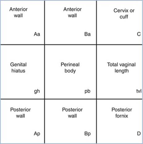
Fig. 7.1
Three by three grid for recording quantitative description of pelvic organ support
Points and Landmarks for POP-Q System Examination
Table 7.1 illustrates the reference points: three reference points are anteriorly (Aa, Ba, and C) and three posteriorly (Ap, Bp, and D). Points Aa and Ap are 3 cm proximal to or above the hymenal ring anteriorly and posteriorly, respectively. Points Ba and Bp are defined as the lowest points of the prolapse between Aa anteriorly or Ap posteriorly and the vaginal apex. Anteriorly, the apex is point C (cervix), and posteriorly is point D (pouch of Douglas). In women after hysterectomy, point C is the vaginal cuff and point D is omitted. Three other measurements are taken: the total vaginal length (TVL) at rest, the genital hiatus (GH) from the middle of the urethral meatus to the posterior hymenal ring, and the perineal body (PB) from the posterior aspect of the genital hiatus to the mid-anal opening. Normal position of the reference points is illustrated in Fig. 7.2. Once the measurements are taken, the patients are assigned to a corresponding stage (Fig. 7.3).
Table 7.1
Description of the points and their range of values
|
Points |
Description |
Range of values |
|
Aa |
Anterior vaginal wall 3 cm proximal to the hymen |
−3 cm to + 3 cm |
|
Ba |
Most distal position of the remaining upper anterior vaginal wall |
−3 cm to + tvl |
|
C |
Most distal edge of cervix or vaginal cuff scar |
−10 cm to + 10 cm |
|
D |
Posterior fornix (N/A if post hysterectomy) |
|
|
Ap |
Posterior vaginal wall 3 cm proximal to the hymen |
−3 cm to + 3 cm |
|
Bp |
Most distal position of the remaining upper posterior vaginal wall |
−3 cm to + tvl |
Additional measurements:
Genital hiatus (gh) measured from middle of external urethral meatus to posterior midline hymen
Perineal body (pb) measured from posterior margin of gh to middle of anal opening
Total vaginal length (tvl) depth of vagina when point D or C is reduced to normal position

Fig. 7.2
Normal position of the reference points

Fig. 7.3
POP-Q staging
The POP-Q system has been criticized for being cumbersome and difficult to learn, although some instructional videotapes are available through the websites and facilitate the learning process. Some tips and tricks can be used to improve the system measurements. Different kinds of devices facilitate the examiner in making the measurements. Figures 7.4 and 7.5 show a home-made system in which a uterine dilatator is used to measure the point Aa (Fig. 7.4) and the point PB (Fig. 7.5). An alternative useful method in the clinical practice mark centimeters on the examiner’s finger and using it as a ruler (Fig. 7.6).
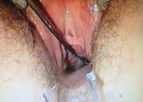
Fig. 7.4
Uterine dilatator is used to measure the point Aa
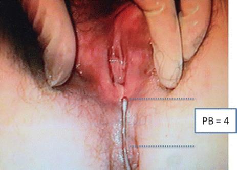
Fig. 7.5
Uterine dilatator is used to measure the point PB

Fig. 7.6
Finger with centimetric scale marked
It has been shown that the routine use of the POP-Q system decreases significantly the amount of time needed to collect the desired data. Experienced examiners averaged 2.05 min per examination while new examiners averaged 3.73 min. There is also a high correlation between the POP-Q findings in left lateral and lithotomy position.
Figures 7.7, 7.8, 7.9, 7.10, 7.11, 7.12, 7.13, and 7.14 illustrate some examples of the different stages of prolapsed anterior and posterior compartments, vault and uterus prolapse.
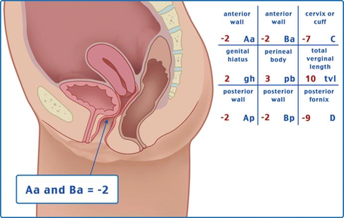
Fig. 7.7
Anterior prolapse stage 1 (with uterus)
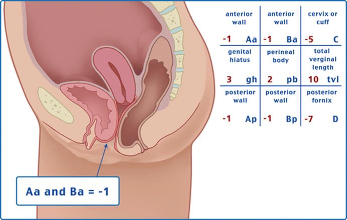
Fig. 7.8
Anterior prolapse stage 2 (with uterus)
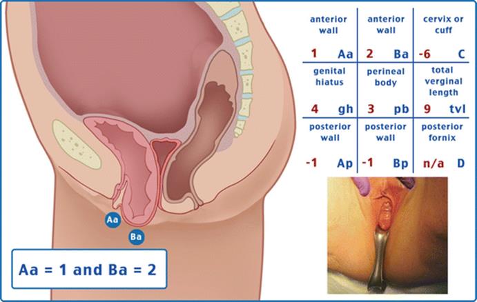
Fig. 7.9
Anterior prolapse stage 3 (without uterus)
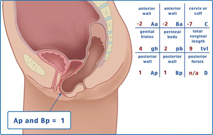
Fig. 7.10
Posterior prolapse stage 2 (without uterus)
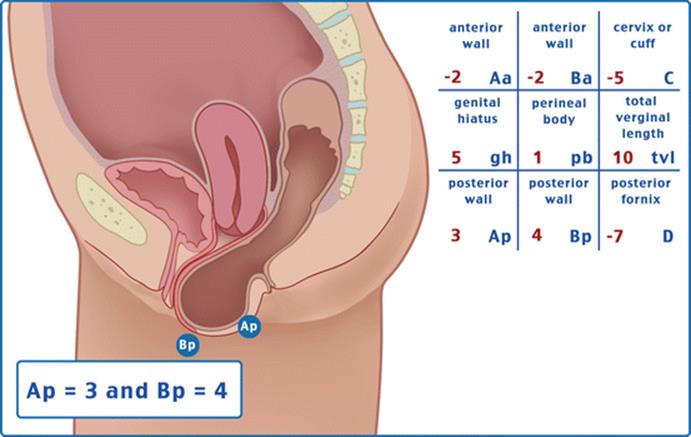
Fig. 7.11
Posterior prolapse stage 3 (with uterus)
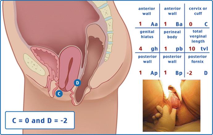
Fig. 7.12
Uterine prolapse stage 2

Fig. 7.13
Vaginal vault prolapse stage 4
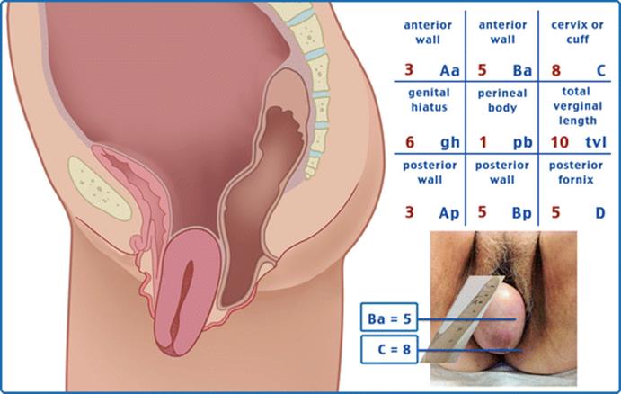
Fig. 7.14
Uterine prolapse stage 4
The POP-Q system does not include findings that some investigators believe to be essential for complete patient description, such as vaginal caliber, status of paravaginal sulci, pelvic muscle strength, or the presence of symptoms, although all of these information should be collected during POP evaluation.
Pelvic organ prolapse quantification (POP-Q) system is an objective, site-specific system for describing, quantifying, and staging pelvic support in women. It provides a standardized tool for documenting, comparing, and communicating clinical findings with proven interobserver and intraobserver reliability. It has been used for longitudinal follow-up of a population of women with prolapse and extensively for outcome reporting after prolapse repairs.
Recently a POP-Q simplified system has been developed, based on POP-Q with similar ordinal staging but with only four points measured instead of nine (Aa, Ba, C, D). Evaluation of the interobserver reproductibility and intersystems reliability (in comparison with the standard POP-Q system) showed good correlation.
Pessaries
Elisabetta Costantini5
(5)
Urology and Andrology Clinic, Department of Surgical and Biomedical Science, University of Perugia, Rome, Italy
Elisabetta Costantini
Email: ecostant@unipg.it
Pelvic organ prolapse (POP) is common and is seen in up to 50 % of parous women in a clinic setting. In the general population, an estimated 30 % of women will have signs of prolapse although the majority are asymptomatic. The etiology of pelvic organ prolapse is complex and multifactorial. Risk factors include pregnancy, childbirth, congenital or acquired connective tissue abnormalities, denervation or weakness of the pelvic floor, aging, menopause, and factors associated with chronically raised intra-abdominal pressure such as obesity, cough, and heavy lifting. Women with prolapse may have a variety of pelvic floor symptoms. Only some of the symptoms are directly related to the prolapse, including pelvic heaviness, a dragging sensation in the vagina, a bulge, lump or protrusion coming down from the vagina, and backache. Symptoms of bladder, bowel, or sexual dysfunction are frequently present. Symptoms may negatively affect body image, quality of life, and a woman’s ability to perform everyday activities. Prolapse treatment may be dependent on a number of factors including the severity of prolapse, the bothersomeness of the associated symptoms, the woman’s general health, and the woman’s treatment preference. Options available for treatment are conservative (pelvic floor muscle training), mechanical support (such as vaginal pessaries), estrogens, and surgery. Before the nineteenth century, the primary POP treatment was the vaginal pessary and Fig. 7.15 shows some of spiral-, oval-, and doughnut-shaped pessaries used for prolapse during the eighteenth century. Despite numerous technological breakthroughs in the medical field over recent decades, pessaries have remained essentially unchanged throughout the twentieth century. The use of pessaries has become commonplace over many years without full evaluation of their efficacy in comparison to other modes of treatment such as surgery, estrogens, or pelvic floor muscle training. Eighty-seven to 98 % of clinicians report using pessaries in their clinical practice and 77 % of gynecologist report using pessaries as the first-line treatment for prolapse.

Fig. 7.15
Pessaries in the nineteenth century from Stromayr C: Die Handschrift des Schnitt-und Augenarztes Caspar Stromayr. Berlin, 1925
In 2013 the Cochrane review demonstrated a lack of consensus on the use of different types of the device, the indications, and the pattern of replacement and follow-up care. However pessaries are commonly used when conservative treatment, like physiotherapy, and surgery have either failed or are not suitable. They present a good option for patients who have not completed childbearing, do not desire surgery, or are poor surgical candidates.
The pessary is inserted into the vagina in order to physically support the vaginal walls, holding the prolapsed organs inside the vagina, supporting the pelvic structures and relieving pressure on the bladder and bowel. In this way it should be able to prevent the prolapse becoming worse and in some cases it is used to avert or delay the need for surgery.
Pessaries are now generally made of an inert plastic or silicone material to prevent odors and absorption of vaginal secretions. Silicone pessaries can be autoclaved and latex allergy is not a contraindication. There are currently many shapes and sizes of pessaries available to suit individual needs, all with their own advantages and disadvantages.
The clinicians choose the type of pessary based on severity of prolapse, presence or absence of the uterus, sexual activity, and concomitant stress urinary incontinence. A cystocele is best treated with a ring with support (filled-in center), uterine prolapse with a ring without support (hollow), and stress incontinence with a ring with a knob. Two main groups of pessaries are available: support pessaries and space-filling pessaries (Fig. 7.16). Generally the first-line pessary for clinicians is the ring pessary, due to ease of insertion and removal. The ring is easy to insert by folding the ring in half and placing a small amount of lubricant on the tip of the pessary to aid in insertion (Fig. 7.17). It is then placed into the vagina where it unfolds once above the pubic symphysis. For removal, the pessary is gently pulled and folded in half. A string can be attached to the ring to aid in insertion and removal. Patients can easily be taught to do this by themselves.
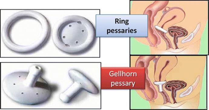
Fig. 7.16
Different pessaries and how they fit
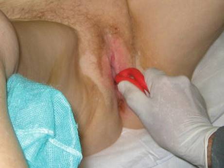
Fig. 7.17
Insertion of a ring pessary
Another kind of pessary is the Gellhorn, generally used for more advanced-stage prolapse or in a patient who is no longer sexually active. It is a space-occupying pessary; its removal and insertion is more difficult and therefore cannot be done by the patient. This pessary has a concave portion attached to a stem that faces into the vagina (Fig. 7.16). To insert the Gellhorn, the pessary is folded in half with the use of lubricant on the leading edge to ease insertion. Once the pessary is behind the pubic symphysis, it will expand and rest against the leading edge of prolapse creating suction. To remove the Gellhorn, the knob is grasped, generally with the help of a ring forceps, while the concave end of the pessary is rotated to release the suction and the pessary is pulled downward, folded, and removed. Another space-filling pessary is the cube pessary. It is made of flexible silicone and is an option in cases of stage III and IV prolapse. The pessary has a string on one end for ease of removal. To insert, the cube pessary is compressed and inserted into the vagina. The cube applies suction to the leading edge of prolapse and often vaginal secretions are trapped in the crevices of the pessary, leading to malodorous discharge; it is usually the pessary of last resort. This pessary should be removed on a nightly basis when possible.
For pessary placement the patient should empty her bladder before pessary fitting. To begin fitting, the clinician can estimate the width of the mid-vagina and use this information to select the appropriate size pessary. The patient should be fitted with the largest size pessary that fits comfortably. The patient is examined in supine and standing position with and without Valsalva. The examiner should be able to comfortably fit a finger on either side of the pessary. If atrophy is present, estrogen should also be prescribed, generally in cream, ring, or tablet form. The patient is then instructed to ambulate, sit on the toilet, and strain to further assess comfort and appropriate pessary fitting. Once the correct pessary type and size are chosen and successfully fitted in place, the patient should attempt to remove and reinsert the pessary on her own. This is commonly possible with the ring type. It is always reassuring if the patient can void with the pessary in place before she leaves the office. Patients should be warned that urine leakage may increase with prolapse reduction. Pessaries can be removed daily, weekly, or monthly, at patients’ discretion, for washing with regular soap and water. Ring pessaries can be removed or left in place for intercourse. If patients are unable to care for the devices on their own, they will require health-care providers to remove, wash, and reinsert them every 3–6 months. To prevent infections and odors, an acidifier (usually supplied with the pessary) or estrogen must be applied vaginally 2 or 3 times a week. Oral or transdermal estrogen and an estradiol-17 ring (placed behind the pessary) are also options. All patients wearing pessaries should be examined by health-care professionals every 3–6 months to check for vaginal erosions or ulcers. If lesions are found, pessaries should be removed until the lesions have healed, and affected areas should be treated with topical estrogen.
There are very few contraindications to pessary use, which allows clinicians to offer pessaries to almost all patients presenting with prolapse and incontinence. Pessaries should not be placed in patients with evidence of an active pelvic infection or severe ulceration or allergy to silicone or those patients who are noncompliant and unlikely to comply with follow up. Risks of wearing a pessary include vaginal infection, erosions, discharge, odor, pain, bleeding, failure to reduce the prolapse, and expulsion. Serious complications from pessaries are rare; however, vesicovaginal fistula, rectovaginal fistula, erosion, and subsequent impaction have all been reported.
In conclusion the use of pessary is one possible option for conservative POP treatment. It is cheap and complications are reported to be rare; however the efficacy of pessary use in the management of prolapse still requires to be clearly established. In the 2013 Cochrane review, one randomized controlled trial compared ring and Gellhorn pessaries and the results showed that both pessaries were effective for approximately 60 % of women who completed the study with no significant differences identified between the two types of pessary. So far there is little evidence and no consensus on the indications, choice of device, or follow-up for pessaries. Although serious side effects are infrequent, insertion and removal of most pessary types still pose a challenge for many patients. Pessary design should continue to improve, making its use a more attractive option.
Anterior Repair
Elisabetta Costantini6 and Franca Natale7
(6)
Urology and Andrology Clinic, Department of Surgical and Biomedical Science, University of Perugia, Perugia, Italy
(7)
Department of Urogynecology, San Carlo-IDI Hospital, Rome, Italy
Elisabetta Costantini (Corresponding author)
Email: ecostant@unipg.it
Franca Natale
Email: francanatale@libero.it
Background
Anterior compartment support depends on the connection of the vagina and periurethral tissues to the muscles and fascia of the pelvic wall via the arcus tendineus fascia pelvis. On both sides of the pelvis, the arcus tendineus fascia pelvis is a band of connective tissue attached at one end to the lower sixth of the pubic bone, 1 cm lateral to the midline, and at the other end to the ischium, just above the spine (Fig. 7.18).

Fig. 7.18
Scheme of anterior compartment support
Anterior vaginal prolapse can result from defects in different areas of pelvic support including:
1.
2.
3.
4.
The goal of surgery is to repair these defects to recreate a strong anatomical support of the anterior vaginal wall.
Clinical Practice
Surgical correction of anterior vaginal prolapse should be proposed in case of symptomatic ≥2 stage cystocele.
Patients could complain of symptoms directly related to the prolapse: vaginal bulging and heaviness in the suprapubic area and/or pelvis; vaginal bleeding, discharge, or infection related to ulceration of the prolapse; need of digitally replace the prolapse to assist voiding; and low backache. Often patients refer also symptoms of bladder dysfunction and in particular storage symptoms (increased daytime frequency, urgency, nocturia), voiding symptoms (hesitancy, slow stream, feeling of incomplete emptying), urinary incontinence (stress, urgency or mixed incontinence), and sexual difficulty.
Diagnostic Evaluation
After a detailed history of bladder, bowel, and sexual function, a pelvic examination should be performed with patient in lithotomy position. A detailed assessment of the support of all segments of the vagina should be made using the pelvic organ prolapse quantification (POP-Q) system and an assessment for central, lateral, and superior anterior vaginal support. If physical findings do not correspond to symptoms or if the maximum extent of the prolapse cannot confirmed, the patient should be examined in standing position.
Urodynamics is useful to assess symptoms of urinary incontinence and voiding dysfunction that are common in women with advanced vaginal prolapse. Additionally, because significant anterior vaginal prolapse often results in kinking of the urethra that may mask underlying stress incontinence, a preoperative urodynamics with vaginal packing or pessary is recommended, to evaluate the lower urinary tract in these patients.
Magnetic Resonance Imaging (MRI) can be useful to further define the nature of the prolapse and evaluate for potential enterocele, rectocele, or uterine prolapse.
Preoperative cystoscopy is useful in the evaluation of the prolapse in patients with lower urinary tract symptoms such as urinary urgency, hematuria, and obstructed voiding to rule out concurrent bladder pathology.
Surgical Technique
Anterior vaginal prolapse resulting from a central defect is traditionally corrected with anterior colporrhaphy. Anterior colporrhaphy was popularized by Howard Kelly in 1911 and still remains the most common technique for transvaginal correction of the anterior vaginal prolapse. Recently to improve the outcomes and avoid the recurrences, the anterior colporrhaphy with use of graft has been proposed. On July 13, 2011, the FDA issued a statement that serious complications are not rare with the use of surgical mesh in transvaginal repair of pelvic organ prolapse. For this reason and on the basis of the Cochrane review conclusions that the transvaginal repair of pelvic organ prolapse with mesh does not improve symptoms or quality of life more than non-mesh repair, the use of mesh during transvaginal surgery is recommended only in the context of RCTs.
Although many variations of anterior repair technique have been described in the last century, the basic approach is still similar to that originally described by Kelly.
A longitudinal mucosal incision is made near the apex of the vagina (Figs. 7.19 and 7.20) and, through this, the vesicovaginal space entered with the tip of a pair of Metzenbaum scissors. Dissection is then performed bluntly by opening and closing the scissors just beneath the vaginal mucosa. The mucosa is incised and the dissection continues along the length of the vaginal wall to within 1 cm of the urethral meatus. Sharp and blunt dissection are used to mobilize connective tissue (vesicovaginal fascia), bladder, and urethra away from the overlying epithelium, just beneath the mucosa. Thompson speculated that the success of the procedure depends on the integrity of the fascial layer left attached to the bladder. Later dissection into the space beneath the inferior pubic ramus is necessary bilaterally (Fig. 7.21). Kelly plication is then performed: a tissue bite is taken laterally in the periurethral tissue just under the symphysis pubis, with the suture crossing under the bladder neck, before a similar bite is taken on the other side. Then, three to five delayed absorbable sutures are placed to reduce the cystocele (Figs. 7.22, 7.23, and 7.24). Care must be taken to avoid deep tissue bites, as such bites may result in ureteral kinking. Then excess vaginal mucosa is trimmed bilaterally (Fig. 7.23) and the anterior vaginal wall is closed with interrupted vertical mattress sutures that include the underlying connective tissue. A vaginal pack can be placed for 24 h to minimize the chance of postoperative hematoma formation. Cystoscopy should then be performed to ensure bladder and ureteral integrity.

Fig. 7.19
Anterior midline incision

Fig. 7.20
The vaginal mucosa is incised to enter the vesicovaginal space

Fig. 7.21
Surgical view: the bladder is isolated and dissected from the vaginal wall mucosa

Fig. 7.22
Delayed absorbable sutures are placed on the lateral edges of the fascia to reduce the cystocele
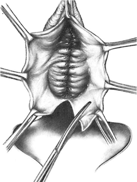
Fig. 7.23
Colporrhaphy is completed and the excess vaginal mucosa is trimmed of bilaterally
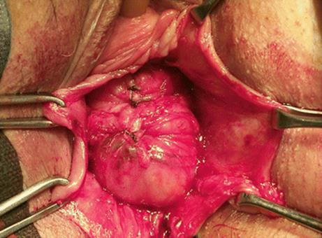
Fig. 7.24
Surgical view of Anterior repair sutures
When anterior vaginal prolapse results from a lateral detachment of the anterior vaginal wall at the pelvic side wall, the goal of surgery is to reattach the lateral vaginal sulcus to its normal lateral attachment. The lateral vaginal attaches to the levator ani (LA) muscle along a line from the anterior pubic rami to the ischial spine known as the “white line” or arcus tendineus fascia pelvis (ATFP) (Fig. 7.18). The paravaginal defect can be repaired retropubically or vaginally. The transvaginal approach (vaginal paravaginal repair) can be more challenging than the retropubic approach but has the advantage of avoiding an abdominal incision and facilitating a concurrent central defect. In case of transvaginal approach, the vaginal epithelium is sharply dissected from the underlying vaginal muscularis and the dissection is continued laterally to the ATFP. Four to six interrupted nonabsorbable sutures are placed through the ATFP and the aponeurosis of the LA muscle at 1 cm intervals. Each stitch is then placed through the lateral edge of the pubocervical fascia and then tightened.
Complications
· Accidental cystotomy, particularly in women with previous vaginal surgery. In this case, the defect is closed in two layer using delayed absorbable sutures, and the bladder is drained for 5–7 days to allow for adequate healing.
· Ureteric injury, particularly during placement of the Kelly plication sutures. To avoid this complication, the distance between the sutures and the ureteric orifices must be greater than 0.9 mm.
· Voiding symptoms (greater than 3 months), which occurs more often in women with some degree of preoperative voiding dysfunction as suggested by preoperative urodynamic parameters (elevated post-void residual >100 ml, decreased peak voiding flow rate <15 ml/s, and low voiding detrusor pressure >15 cmH2O). Long-term voiding difficulty is best managed with clean intermittent self-catheterization.
· Detrusor overactivity, which could be due to the surgical dissection around the urethra and bladder neck that damages the parasympathetic and sympathetic nerves leading to denervation of the urethra and detrusor muscle.
· De novo stress incontinence, which was attributed to a hypercorrection of the cystocele with a loss in the normal posterior urethrovesical angle between the bladder base and the bladder neck. Currently it is believed that de novo stress incontinence could be due to the denervation of the urethral sphincter or to a condition of latent stress incontinence that is unmasked when urethral kinking is surgically corrected.
· Decreased mobility and capacity of the vagina with subsequent dyspareunia and sexual dysfunctions (avoid to remove the vaginal walls in excess).
Regarding vaginal paravaginal repair, significant complications have been reported such as ureteric obstruction, retropubic hematoma, vaginal abscesses, and abnormal bleeding.
What Do the Guidelines Say
Outcomes of the traditional surgery for the correction of the anterior vaginal prolapse are largely limited to retrospective reviews and case series. Moreover, reporting of outcomes has been extremely subjective and, before the advent of POP-Q system, pre- and postoperative staging have been quite variable between surgeons.
Considering these limitations, the success rates of anterior colporrhaphy ranged from 80 to 100 % in retrospective series, while the success rates of the vaginal paravaginal repair for cystoceles vary from 67 to 100 % in case series. Studies that differentiated lateral from central recurrences have revealed that central recurrence (22–25 %) is more common than a lateral recurrence (2–8 %).
Comparing anterior colporrhaphy alone to anterior repair with graft or mesh reinforcement, it was demonstrated that absorbable polyglactin mesh (Vicryl) might reduce objective prolapse recurrence and this is true also using absorbable porcine dermis or polypropylene mesh. However improved outcomes including patient satisfaction, quality of life, and reduced operations for recurrences have not yet been demonstrated. Furthermore, anterior polypropylene mesh alone demonstrated an improved subjective outcome as compared to native tissue anterior repair, but there was no difference between the groups in the rate of dyspareunia. The operating time, blood loss, rate of apical or posterior compartment prolapse, and de novo stress urinary incontinence were greater in the polypropylene mesh group, which was associated with an 11.4 % rate of mesh erosion and 6.8 % requiring surgical reintervention.
Sacrospinous Fixation
Franca Natale8 and Elisabetta Costantini9
(8)
Urogynecology Unit, Department of Gynecology, San Carlo-IDI Hospital, Rome, Italy
(9)
Urology and Andrology Clinic, Department of Surgical and Biomedical Sciences, University of Perugia, Perugia, Italy
Franca Natale (Corresponding author)
Email: francanatale@libero.it
Elisabetta Costantini
Email: elisabetta.costantini@unipg.it
Background
Post-hysterectomy vault prolapse and uterovaginal prolapse are challenging problems for the pelvic reconstructive surgeon. The main cause of these apical prolapses is the weakness of the uterosacral/cardinal ligament complex, and the restoration of this support is the “cornerstone” of the reconstructive surgery. Various approaches are available to achieve this: vaginal and open, or laparoscopic techniques. The vaginal approach includes: the iliococcygeus fascia fixation, the uterosacral ligament suspension, the Mayo/McCall culdoplasty, and the sacrospinous fixation (SSF).
Clinical Practice
The SSF gained popularity in the second half of the twentieth century, and its most common indication is the resuspension of a symptomatic, post-hysterectomy prolapsed vaginal apex.
Sacrospinous fixation is also indicated when women desire to preserve fertility or the uterus. SSF may also be used to decrease the operative time and morbidity in elderly women who have no evidence of uterine pathologies.
Diagnostic Evaluation
After a detailed history, a pelvic examination should be performed with the patient in the lithotomy position using the pelvic organ prolapse quantification (POP-Q) system. In cases of post-hysterectomy vaginal vault prolapse, a simultaneous rectal and vaginal examination may aid in detecting the presence of an enterocele and in differentiating an enterocele from a high rectocele. Many radiologic modalities have been used in the diagnosis, including defecography, fluoroscopy, dynamic cystodefecography, and MRI. In cases of severe uterovaginal prolapse of the anterior and apical vaginal wall, the upper urinary tract must be evaluated because severe prolapse can cause angulation of the ureters resulting in ureteral obstruction.
Surgical Technique
Several modifications of the SSF have been described. The original technique described by Nichols involves suspension of the vaginal vault to the sacrospinous ligament (SSL) (Fig. 7.25) through the posterior compartment (Table 7.2). A modification was introduced by Winkler et al., suspending the vault to the SSL through the anterior compartment, with the aim to reduce the postoperative narrowing and lateral deviation of the upper vaginal wall. In 1993 Kovac and Cruikshank proposed the sacrospinous hysteropexy, in which the uterus was suspended bilaterally to the SSLs.

Fig. 7.25
The sacrospinous ligament and its relation with pudendal vessels and nerves and ischial spine
Table 7.2
The technique step-by-step
|
Identification of the pararectal space |
|
Visualization of the sacrospinous ligament |
|
Avoid injury of pudendal vessel and nerve (behind the ligament and the ischial spine) |
|
Sutures >2 cm medial to the spine |
|
Nonabsorbable sutures |
SSF is typically performed unilaterally on the right side (Fig. 7.26), which offers the anatomical advantage of absence of the sigmoid colon on this side. In cases of severe genital prolapse, bilateral suspension may be considered. In this latter case the surgeon must ensure that there is adequate vaginal space, so that the bilateral suspension does not cause stricture in the rectum.
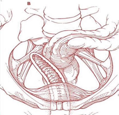
Fig. 7.26
Schematic view of monolateral sacrospinous ligament fixation
The posterior approach involves a posterior vaginal incision and a posterior colporrhaphy dissection, with subsequent perforation of the rectal pillar near the ischial spine. With blunt dissection of the pararectal space medial to the ligament, the coccygeus muscle and the sacrospinous ligament are exposed (Fig. 7.27). The anterior approach involves an anterior vaginal wall incision and a dissection of the ipsilateral paravesical and paravaginal area from the level of the bladder neck to the ischial spine along the arcus tendineus fasciae pelvis (Fig. 7.28). This dissection opens a large space for the vaginal apex and avoids the narrowing often caused by the posterior approach. Then the ligament is exposed with posterolateral dissection within the retroperitoneal place using two Breisky-Navratil retractors (one placed medially to sweep the rectum medially off the ligament and the other laterally) and a Haney retractor posteriorly, just in front of the coccygeus muscle at the 7 o’clock position. Various devices (Fig. 7.29) have been used to place sutures into the sacrospinous ligament, including the Deschamps ligature carrier, Miya hook, and Capio in-line “push and catch” suturing devices. The lateral suspension suture is placed through the ligament 1–2 cm medially to the ischial spine to avoid injury to the pudendal vessels. Fixation of the vaginal apex is most commonly performed with permanent monofilament sutures (Fig. 7.30), but some surgeons prefer absorbable sutures to reduce the risk of suture erosion and granuloma formation. The relative advantages of permanent versus absorbable sutures have not been evaluated.

Fig. 7.27
Exposure of sacrospinous ligament

Fig. 7.28
Sacrospinous ligament fixation – anterior access: (a) anterior vaginal wall incision, (b) dissection of paravesical and paravaginal area, (c) placement of suture into the sacrospinous ligament
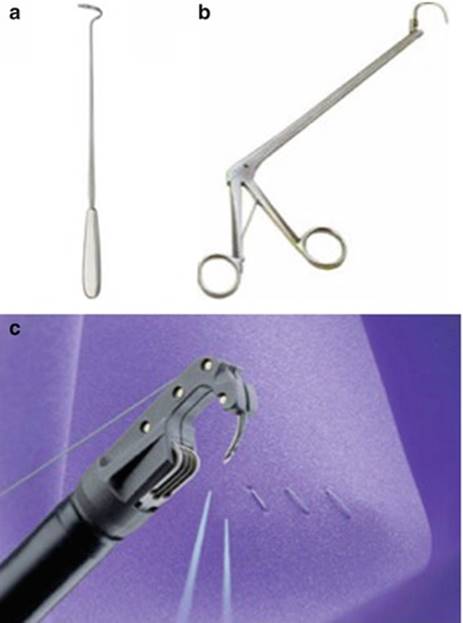
Fig. 7.29
Devices used to place sutures into the sacrospinous ligament – (a) Deschamps ligature carrier, (b) Miya hook, (c) Capio™ suturing device
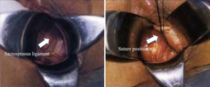
Fig. 7.30
Sacrospinous ligament fixation: intraoperative images
In cases of uterine hysteropexy, the dissection and suture placement are similar to those of vault suspension. The difference is that, after suture placement in the sacrospinous ligament, the end of the suture is brought through the uterosacral ligament and then through the vaginal epithelium.
Complications
Local neurologic complications may include pudendal, sacral, or sciatic neuropathy. A direct injury to major nerves is rare. In this case a re-operation is mandatory to remove the sutures. More frequently pudendal nerve entrapment occurs and results in pain localized in the buttock or perineum. The pain may improve after replacement of lateral fixation sutures more medially. Moreover, gluteal pain or paresthesias occurs after SSF, possibly due to peripheral nerve trauma. Usually these symptoms are transient and self-limited; sometimes they persist for weeks or months postoperatively.
Serious vascular injury is a rare complication, but it can be life-threatening. It usually results from injury of small vessels along the medial aspect of the rectum. Less commonly, the medial retractor can disrupt deeper vessels in the presacral area. For most cases, sutures or surgical clips to the involved vessels can be placed under direct vision after exposure with simple retraction. If the vessels cannot be visualized, the use of prolonged pressure packing may become necessary. Rarely a selective arterial embolization is necessary.
More rare complications of SSF include: infections, suture abscesses, bowel injury, and bladder lacerations.
Point of Interest
The anatomical cure rate of the apical vaginal segment after SSF ranges from 89 to 90 %, but the anterior vaginal segment represents a site particularly vulnerable to recurrence. Recurrent cystoceles have been reported in 7.6–92 % of patients after SSF, depending on the other concomitant procedures performed. It has been proposed that posterior deflection of the vaginal wall after SSF leads to increased exposure of the anterior compartment to intraperitoneal forces. It has also been suggested that underestimation of the severity of anterior prolapse on preoperative examination may play a role. A preoperative identification of all areas of prolapse and a subsequent modification of the pelvic reconstruction technique according to intraoperative findings are important key elements to reduce this kind of complication.
Sexual dysfunction has been reported after SSF. The retroversion of the vaginal apex and deviation after unilateral SSF appear to have no adverse effects on coital function, but the postoperative vaginal narrowing is likely correlated with this kind of dysfunction. Some papers underlined that vaginal narrowing after SSF was more likely the result of repair of other concurrent vaginal defects (mainly anterior colporrhaphy), rather than of SSF itself.
What Do the Guidelines Say
Abdominal Sacral Colpopexy Versus Vaginal Sacrospinous Colpopexy
SSF showed a higher rate of recurrent vault prolapse, more dyspareunia, shorter operating time and recovery time, and lower cost. Data on the subjective success rate, patient satisfaction, and impact of the surgery on quality of life were too few for reliable conclusions.
Comparing Different Vaginal Approaches: One Type of Upper Vaginal Prolapse (Uterine and Vaginal Vault) Repair Versus Another
The last Cochrane review on POP surgery (2013) concluded that all vaginal approaches to correct upper vaginal prolapse, i.e., uterosacral ligament suspension, McCall culdoplasty, iliococcygeus fixation, and colpocleisis, are relatively safe and effective interventions (level 3 evidence, grade C), but there is no evidence that one technique is better than another.
Vaginal Sacrospinous Uterine Suspension Versus Vaginal Hysterectomy
Women undergoing sacrospinous hysteropexy had a median hospital stay that was 1 day shorter than that in the hysterectomy group, and the mean number of days to return to work was 23 days less compared to the hysterectomy group. No differences were reported in domain scores for quality of life and urogenital symptoms between the two procedures. There were more adverse symptoms in the SSF group, mostly due to buttock pain. No differences were found between the two groups in terms of apical, anterior, and posterior prolapse recurrences.
Total Vaginal Polypropylene Mesh (TVM) Versus Sacrospinous Colpopexy
The SSF group had a higher objective recurrence rate. No differences were identified between the groups in terms of de novo SUI, bladder overactivity, dyspareunia, and pelvic pain or in functional outcomes measured with the Pelvic Organ Prolapse/Urinary Incontinence Sexual Questionnaire (PISQ-12), the Urinary Impact Questionnaire (UIQ), the Colo-Recto-Anal Impact Questionnaire (CRAIQ), or the Pelvic Organ Prolapse Impact Questionnaire (POPIQ).
Meshes
Franca Natale10 and Elisabetta Costantini11
(10)
Urogynecology Unit, San Carlo-IDI Hospital, Rome, Italy
(11)
Urology and Andrology Clinic, Department of Surgical and Biomedical Science, University of Perugia, Rome, Italy
Franca Natale
Email: francanatale@libero.it
Elisabetta Costantini (Corresponding author)
Email: ecostant@unipg.it
Background
Despite modifications and evolutions of the technique, traditional surgery for pelvic organ prolapse (POP) has a failure or recurrence rate of 30–50 % and a prevalence of reoperation of 29–40 % within 3 years.
The highest rates of reoperation occur for apical defects (33 %), combined anterior and apical defects (15 %), or posterior and apical defects (12 %).
In an effort to improve outcomes in POP repair, especially of the anterior compartment, multiple biological and synthetic graft materials have been introduced.
Prosthetic materials have at least four main purposes in pelvic reconstructive surgery:
· Substitution, where lacking supportive tissue is replaced by mesh
· Reinforcement, where inadequate tissue is augmented to improve its performance
· Generation, which involves the induction of new supportive tissue
· Consolidation, where mesh is used to complement surgery
On the basis of these premises, the rationale for the use of mesh is:
· To replace the damaged visceral fascia
· To restore cohesion between visceral and parietal fascia to rebuild the hammock connected to the arcus tendineus fascia pelvis
· To stabilize the bladder hammock
Unfortunately, because of secondary complications associated with the use of mesh, all the guidelines and reviews recommend their use under strict control. In particular the evidence is not sufficient to support the use of permanent meshes or grafts at the time of vaginal apical or anterior compartment repair surgery except in the context of randomized controlled trials.
Type of Meshes
The ideal graft/mesh should be:
· Clinically and physically inert
· Noncarcinogenic
· Mechanically strong
· Without allergic or inflammatory reaction
· Sterile
· Not physically modified by body tissue
· Readily available
· Inexpensive
· With minimal risk of infection and rejection
The optimal implant, once healed, would restore normal anatomy and function to the vagina and the surrounding pelvic organs and be more durable or equally durable to autologous tissue. It would be biocompatible, and, if biodegradable, it should persist long enough for incorporation of the surrounding native tissue. It should resist to mechanical stress and shrinkage, be pliable and easily manipulated during surgery, and not result, once implanted, in adhesion formation at the visceral surfaces.
Currently, there are no biologic or synthetic implants that meet all the above criteria.
Surgical mesh materials can be divided into three general categories:
· Synthetic
· Biologic
· Composite (i.e., a combination of any of the previous two categories)
Synthetic Meshes
Synthetic mesh can be absorbable or nonabsorbable. Once absorbable synthetics are implanted, macrophage activation leads to mesh absorption and subsequent recycling of by-products into new collagen fibers. The commercially available absorbable synthetic mesh implants are polyglycolic acid and polyglactin 910. Although both products are considered absorbable, their properties are distinct. Polyglactin 910 starts to hydrolyze during the third week after implantation and loses the majority of its mechanical value after 30 days, whereas polyglycolic acid requires 90 days for absorption.
The main characteristics of nonabsorbable synthetic meshes are pore size, fiber type, and stiffness, and on the basis of pore size and fiber type, they have been classified in four types (Fig. 7.31):

Fig. 7.31
Different synthetic meshes used in urogynecology
· Type I meshes (Prolene®, Marlex®) contain large pores (>75 μm) that allow the admission of macrophages to prevent infection and in-growth of fibroblasts, blood vessels (angiogenesis), and collagen, to form fibrous connections to the surrounding tissue.
· Type II meshes (Gore-Tex®) have a pore size <10 μm in at least one of the three dimensions (microporous).
· Type III meshes (Mersilene®) are macroporous but have microporous components that often include braided and/or multifilament materials.
· Type IV meshes have submicronic pore size.
Flexibility is an important property that appears to be related to pore size: the greater the pore size and the more the mesh flexibility, the lower the erosion rate. Currently the type I meshes are considered the best choice in urogynecology.
Recently other kinds of meshes have been introduced: the titanium-coated mesh, the ultralightweight mesh and the polyvinylidene fluoride (PVDF) monofilament mesh (Fig. 7.31). Some studies found that coating the mesh with titanium causes less severe inflammatory reactions than non-coated polypropylene. Ti-mesh® is a titanium-coated low-weight mesh and combines the advantages of light material with a superior biocompatibility due to the titanium coating, at least in terms of chronic inflammatory reactions.
Another mesh today available is the lightweight mesh, to add more favorable biocompatible characteristics without losing any critical tensile strength mesh. The main characteristics of lightweight meshes are:
· Reduced overall “mesh load,” high hydrophilic
· Low memory and easily intraoperative handling while maintaining durability
· Generally not palpated during postoperative examinations
Different types are available in different shapes including Artisyn® Y-Shaped Mesh, Alyte® Y-Mesh Graft, and Restorelle™ Y.
The Y-meshes, particularly useful in colposacropexy, have a vaginal section with anterior and posterior flaps and a sacral section. The anterior and posterior flaps are used for anterior and posterior vaginal attachment, and the sacral flap for attachment to the sacral promontory (Fig. 7.32). Finally the polyvinylidene fluoride (PVDF) (DynaMesh®) mesh, widely used in abdominal hernia repair, has been recently introduced in the POP surgical treatment. It is highly biocompatible, it seems to be more resistant to hydrolysis and degradation in comparison to polyester and polypropylene. It also shows a reduced inflammatory and fibrotic reaction compared to polypropylene.

Fig. 7.32
Y-mesh used in colposacropexy. The anterior and posterior flaps are used for anterior and posterior vaginal attachment and the sacral flap for attachment to the sacral promontory
Up to now it is not clear whether the features of these new meshes would translate into any direct clinical benefit. Such questions can only be answered through clinical research.
Biologic Grafts
The most commonly used biologic materials available for POP repair include:
· Autograft (rectus fascia)
· Allograft (cadaveric fascia)
· Xenograft
Xenografts may be either nonabsorbable (i.e., Pelvicol®) or absorbable (small intestine submucosa [SIS]) (Fig. 7.33). Pelvicol® is derived from porcine dermis: a patented process removes all fats and cellular materials through a series of organic and enzymatic extraction that leaves the material without any DNA. Then the matrix is stabilized using a cross-linking agent to maintain strength and provide permanence. Gamma sterilization of the tissue ensures sterility. It has been demonstrated that perforating the porcine dermis improves the graft take and decreases wound infections by allowing for increased tissue in-growth and revascularization of the vaginal epithelium overlying the graft. Tensile strength and suture pullout strength were maintained in the perforated graft.

Fig. 7.33
Biological grafts
SIS is harvested from porcine jejunum and is composed of mucosa, muscularis mucosa, and submucosa. Processing leaves the extracellular collagen matrix intact, thus allowing the presence of collagen, growth factors, glycosaminoglycans, proteoglycans, and glycoproteins to promote host cells proliferation through SIS layers. Theoretically, the SIS scaffold should be entirely remodeled and replaced by the host’s connective tissue in 90 days.
Surgical Techniques Using Meshes
Historically the first mesh used in POP repair refers to abdominal colposacropexy, the gold standard for the surgical correction of vaginal vault prolapse, a technique which demonstrated a long-term durability and precise anatomical restoration. The goal of this procedure is to elevate the vaginal apex to the sacral promontory using a mesh bridge: the resultant vaginal axis is the most physiologic of all reconstructive procedures, and the vaginal length is maintained.
Colposacropexy can be also performed by laparoscopic- or robotic-assisted procedure with the aim to improve visualization of anatomy of the peritoneal cavity because of laparoscopic and robotic magnification; decrease postoperative pain; shorten hospitalization, resulting in potential cost reduction; and allow more rapid return to work.
In the last two decades, the meshes started to be used also in the vaginal approach to correct cystocele (anterior repair) and rectocele (posterior repair) and also the top of the vagina to correct uterine prolapse or vaginal apical prolapse (apical repair).
Vaginal Anterior Repair
Hand-Tailored Mesh
In the 1990s, urogynecologists began using surgical mesh for transvaginal POP repair and, to do so, surgeons cut the mesh to the desired shape, placing it through a corresponding incision. A variety of permanent polypropylene mesh overlays have been evaluated in case series for the management of anterior wall prolapse with an anatomical success rate ranging from 76 to 100 % (Fig. 7.34a).
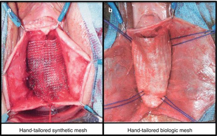
Fig. 7.34
Tailored synthetic (a) and biological grafts (b)
With the aim to reduce the complications associated with the synthetic meshes, biologic materials (cadaveric fascia lata, porcine dermis, porcine small intestine submucosa, bovine pericardium collagen) have been used (Fig. 7.34b). The reported success rate widely ranges from 52 %, using porcine dermis, to 100 % using cadaveric fascia lata.
Trans-obturator Kits
Over the next years, surgical meshes for transvaginal POP repair became incorporated into “kits” that included tools to aid in the delivery and insertion of the mesh. The surgical mesh kits continue to evolve, adding new insertion tools, tissue fixation anchors, and surgical techniques, using absorbable and biologic materials. The trans-obturator kits consist of four helical needles and a polypropylene mesh with four arms, two for each side. After the dissection of the bladder laterally toward the ischiopubic ramus, the superior needle is inserted lateral to the level of the clitoris, with an angle of 45° from the patient’s midline (Fig. 7.35a, b). Then the inferior needle is inserted 3 cm below and 2 cm lateral to the superior needle (Fig. 7.35c, d). The tip of the needle should be pointed directly at the ischial spine. The procedure is repeated on the contralateral side. The endoscopy is then performed to ensure the integrity of bladder and urethra. The mesh should now site underneath the cystocele in a tension-free manner, the tail is attached at the vault and under urethra, and the redundant tail is trimmed off. The vaginal incision is closed, the plastic sheaths are removed, and the mesh arms are cut at skin incisions.
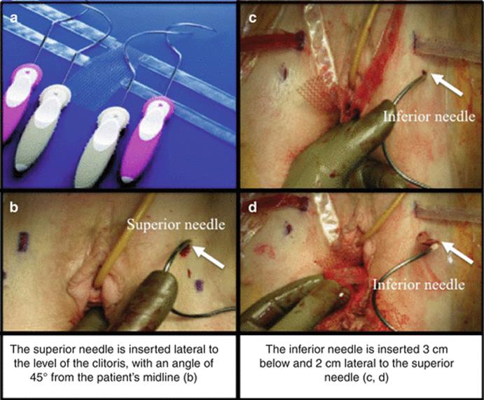
Fig. 7.35
Trans-obturator kits for anterior vaginal POP repair: (a) the elicoidal needles and the polypropylene mesh; (b) position and insertion of the superior needle; (c, d) insertion of the inferior needle
Single-Incision Kits
Recently new single-incision kits were introduced in the market. The devices allow a minimally invasive approach to treat anterior and apical defects. The mesh is inserted through a small vaginal incision and secured in their position using self-fixating tips attached to the mesh (Fig. 7.36); they are inserted into the sacrospinous ligament (SSL) and the obturator internus muscle without trocars to secure the correct positioning of the mesh until natural tissue ingrowth occurs.
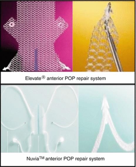
Fig. 7.36
Single-incision kits
Currently, there are not long-term data to recommend the use of these kits.
Vaginal Posterior Repair
As traditional posterior repair is associated with high success rates (76–90 %), the need for mesh augmentation in the posterior compartment is questioned, and the use of the prosthetic surgery is limited. Furthermore physicians have been hesitant to place mesh in the posterior compartment, secondary to its proximity to the rectum and potential infection issues with foreign body placement. Moreover the mesh avoids the expansion during evacuation or coitus, leading to difficult defecation and dyspareunia, the most frequent complications of this kind of surgery.
Vaginal Posterior and Apical Repair
For the simultaneous repair of an apical and posterior defect, it is possible to use kits consisting of two curved needles and a central polypropylene mesh (Fig. 7.37). The needle is inserted 3 cm lateral and posterior to the anal orifice, crossing the ischiorectal fossa underneath the iliococcygeus muscle. At 2 cm posterior and 3 cm medial to the ischial spine, the trocar pierces the sacrospinous ligament and passes through the pararectal space emerging in the vaginal incision. In this way, the vaginal apex is suspended at the level of the ischial spine, replicating a more normal vaginal axis. Locking connectors on the ends of the arms of the mesh attach to each needle tip and are used to hold the mesh secure to the needle during passage of the mesh through the body. Once snapped onto the needle tip, the connectors cannot be removed.
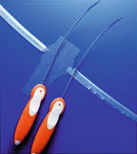
Fig. 7.37
Anterior and apical support system (Apogee system, AMS)
The most important complication associated with this kind of surgery and generally in the sacrospinous ligament suspensions is the vessel injury, and the inferior gluteal artery is the most susceptible vessel. To avoid complications, it is important to insert the needle into the sacrospinous ligament 3 cm medial and 2 cm lower than the level of the ischial spine to avoid the pudendal and inferior gluteal bundles.
Complications
The most common complication of transvaginal POP repair with mesh is vaginal mesh erosion – also called exposure, extrusion, or protrusion – which occurs in approximately 10 % of cases but reported in higher percentages in some series. More than half of the women who experienced erosion from nonabsorbable synthetic mesh required surgical excision in the operating room and some women required two or three additional surgeries. Another new complication, increasingly reported in the literature, is mesh contraction – which causes vaginal shortening, tightening, and pain.
New-onset stress urinary incontinence has been reported to occur more frequently after mesh-augmented anterior repair than after traditional anterior repair without mesh. Other reported complications are pain (including dyspareunia), infection, urinary problems, bleeding, organ perforation, recurrent prolapse, neuromuscular problems, vaginal scarring or shrinkage, and emotional problems. Many of these complications need additional intervention, including medical or surgical treatment and hospitalization, and some sequelae (e.g., pain) may continue despite mesh removal.
Research continues to look for reducing these complications through less invasive surgical techniques, expertise, and new type of mesh.
The FDA Warning
In order to better understand the use of surgical mesh for POP and SUI, the Food and Drug Administration (FDA) in 2011 conducted a systematic review of the published scientific literature from 1996 to 2011 to evaluate its safety and effectiveness. The literature review revealed that:
· Mesh used in transvaginal POP repair introduces risks not present in traditional non-mesh surgery for POP repair.
· Mesh placed abdominally for POP repair appears to result in lower rates of mesh complications compared to transvaginal POP surgery with mesh.
· There is no evidence that transvaginal repair to support the top of the vagina (apical repair) or the back wall of the vagina (posterior repair) with mesh provides any added benefit compared to traditional surgery without mesh.
· While transvaginal surgical repair to correct weakened tissue between the bladder and vagina (anterior repair) with mesh augmentation may provide an anatomical benefit compared to traditional POP repair without mesh, this anatomical benefit may not result in better symptomatic results.
Basing on these data, FDA recommends that health-care providers should:
· Obtain specialized training for each mesh placement technique and be aware of the risks of surgical mesh.
· Be vigilant for potential adverse events from the mesh, especially erosion and infection.
· Watch for complications associated with the tools used in transvaginal placement, especially bowel, bladder, and blood vessel perforations.
· Inform patients that implantation of surgical mesh is permanent and that some complications associated with the implanted mesh may require additional surgery that may or may not correct the complication.
· Inform patients about the potential for serious complications and their effect on quality of life, including pain during sexual intercourse, scarring, and narrowing of the vaginal wall in POP repair using surgical mesh.
· Provide patients with a copy of the patient labeling from the surgical mesh manufacturer if available.
In addition, the FDA also recommends that health-care providers should choose mesh surgery only after weighing the risks and benefits of surgery with mesh versus all surgical and nonsurgical alternatives.
What the Guidelines Say
The International Consultation on Urological Diseases (ICUD) is a non-governmental organization registered with the World Health Organization (WHO). In the last 10 years ICUD has held consultations on several urological pathologies, including urinary incontinence and POP. This branch of the consultations is known as the International Consultation on Incontinence (ICI).
ICUD issues guidelines and subsequent recommendations are based on the published evidence and graded as follows:
· Grade A: highly recommended
· Grade B: recommended
· Grade C: optional
· Grade D: not recommended
ICI 2013
On the basis of the latest ICI evaluation, we summarize the most important conclusions reached:
· Absorbable mesh augmentation of native tissue repair improves the anatomical outcome as compared to native tissue repair alone with no increased complication rate (Grade B).
· Biological grafts in meta-analysis have improved anatomical outcomes with no change in subjective outcomes as compared to native tissue repairs (Grade B).
· Consistent data supports a superior anatomical outcome for polypropylene mesh as compared to biological graft (Pelvicol) in the anterior compartment. Mesh exposure rate was significantly higher in the polypropylene mesh group (Grade A).
· Consistent evidences demonstrate improved anatomical and subjective outcomes for polypropylene mesh as compared to anterior colporrhaphy (Grade A). These outcomes did not translate into improved functional outcomes using validated questionnaires or a lower reoperation rate for prolapse. The mesh group was also associated with longer operating time, greater blood loss, and a not significant tendency toward higher cystotomy, de novo dyspareunia, and de novo stress urinary incontinence rate as compared to anterior colporrhaphy without mesh. Apical or posterior compartment prolapse was significantly more common following anterior repair using polypropylene mesh and mesh extrusion rate was 10.4 % with 6.3 % undergoing surgical correction (Grade B).
· To date no study has shown any benefit to mesh overlay or augmentation of a suture repair for posterior vaginal wall prolapse (Grade B).
All these data were confirmed by the last Cochrane review on POP surgery (2013) which showed that the use of absorbable polyglactin mesh overlay, absorbable porcine dermis, or polypropylene mesh at the time of anterior vaginal wall repair reduces the risk of recurrent cystocele on examination; however improved outcomes including patient satisfaction, quality of life, and reduced operations for recurrences have not yet been demonstrated. Furthermore, anterior polypropylene mesh alone demonstrated an improved subjective outcome as compared to native tissue anterior repair without any difference between the groups in the rate of dyspareunia. The operating time, blood loss, rate of apical or posterior compartment prolapse, and de novo stress urinary incontinence were greater in the polypropylene mesh group, which was associated with an 11.4 % mesh erosion rate and 6.8 % requiring surgical reintervention.
Sacrocolpopexy
Elisabetta Costantini12
(12)
Urology and Andrology Clinic, Department of Surgical and Biomedical Science, University of Perugia, Rome, Italy
Elisabetta Costantini
Email: ecostant@unipg.it
Pelvic organ prolapse (POP) is estimated to affect nearly half of all females over 50 years of age and has a negative impact on the patient’s quality of life. Women have an 11 % lifetime risk of undergoing pelvic reconstructive surgery for POP or urinary incontinence and the costs of POP surgery, as recorded in the North American population, might be in excess of $1 billion dollars. The etiology of POP is complex and multifactorial. Treatment depends on factors such as the severity or grade of POP, symptoms, the patient’s general condition and expectations, and the surgeon’s experience. Diverse surgical approaches are available for POP repair, the goals of which are to restore normal pelvic anatomy, restore or maintain normal urinary, bowel and sexual function, reduce the impact of symptoms, and improve quality of life.
Sacrocolpopexy (SP) is a procedure used to treat apical pelvic organ prolapse (POP) including uterine prolapse and posthysterectomy vaginal vault prolapse. Vaginal vault prolapse is an increasing problem in the United States, accounting for more than 30,000 surgical procedures in 1999. The risk of post-hysterectomy vaginal vault prolapse is estimated to be 1 % at 3 years and 5 % at 17 years. Although defects in the supporting structures of pelvic organs have been thought to be the cause of pelvic organ prolapse, the increase in surgical procedures over one decade suggests that other factors exist, including problems with surgical technique or improvements in POP diagnosis. Vaginal vault prolapse remains a challenging condition for pelvic surgeons. Clinical presentation varies and is associated with other compartment defects (cystocele, rectocele, or enterocele) in nearly 75 % of affected patients. Successful therapy for vaginal vault prolapse depends on a comprehensive evaluation including an assessment of all potential sites, specific vaginal support defects, and an assessment of urinary, fecal, and sexual function. These factors must be considered when selecting therapy for each patient. In addition, associated comorbidities will greatly influence the route of treatment. Thus, effective management of POP hinges on an individualized, comprehensive evaluation of the pelvic floor tailored to fit each patient.
Restoration of apical support can be carried out through vaginal or abdominal approaches. The transvaginal approach avoids the morbidity of an abdominal incision and affords access to and fixation of other concomitant vaginal defects. Transvaginal techniques require fixation of the apex to a number of pelvic supporting structures (sacrospinous ligament, iliococcygeus fascia, uterosacral ligaments). Some of these structures may be weakened or they can often be difficult to identify, particularly in the posthysterectomy patient. Depending on the method of apical support, the vaginal apex may also be anatomically displaced. Most commonly, the vagina is displaced posteriorly after a sacrospinous ligament fixation placing the anterior compartment at risk for recurrent cystocele. With some of the vaginal approaches, one must be concerned about reducing vaginal length. Reduction in vaginal length is a significant concern for younger, sexually active women. Despite the potential limitations, transvaginal sacrospinous fixation has been shown to preserve vaginal length and achieve an up to a 79 % success rate for correction of vault prolapse. The benefits of transabdominal approaches include consistent, reliable restoration of anatomy. The abdominal sacral colpopexy offers consistent fixation of the vaginal apex, by using a synthetic mesh and attaching it to the sacrum (Fig. 7.38). Thus, there is less dependence on weakened points of attachment. In addition, there is little anterior or posterior displacement of the vaginal axis. The major benefits of this approach, therefore, include a more reliable fixation of the vaginal apex with minimal changes to the vaginal anatomy. In addition, the transabdominal approach allows for concomitant enterocele repair via culdoplasty, concomitant cystocele, and rectocele repair and finally in some cases urinary incontinence can be treated, with the option of additional concomitant anti-incontinence surgical techniques, although the last point remains controversial.

Fig. 7.38
Schematic SP draw: two meshes fix the vaginal apex to the sacrum
Nowadays SP is considered the gold standard treatment and the most durable technique for repairing vaginal vault prolapse (Fig. 7.39). The technique, described by Huguier and Scalin in 1958 and subsequently by Lane in 1962, has a success rate of more than 95 % at 3 years and 84 % at 5 years. The Cochrane review in 2013 confirmed SP has superior outcomes to a variety of vaginal procedures including sacrospinous colpopexy, uterosacral colpopexy, and transvaginal mesh. These benefits must be balanced against a longer operating time, longer time to return to activities of daily living, and increased cost of the abdominal approach (Fig. 7.40). Although predominantly used in posthysterectomy vaginal vault prolapse, SP can be performed with a uterus in situ either with or without concomitant hysterectomy. Hysterectomy at the time of sacrocolpopexy for management of uterine prolapse, although it allows the same excellent result in terms of functional and anatomical outcomes, may be associated with increased rates of mesh exposure. In recognition of this increased risk, some surgeons have advocated supracervical hysterectomy as an alternative to total hysterectomy before SP. Powered uterine morcellation is required when subtotal hysterectomy is performed at laparoscopic- or robotic-assisted minimally invasive sacrocolpopexy and has become controversial following a recent US Food and Drug Administration safety communication. While subtotal hysterectomy appears to reduce the risk of mesh exposure, further evaluation is required in the light of unanticipated uterine pathology. In case of severe uterine prolapse, another option is the uterus preservation, which recently has gained popularity.
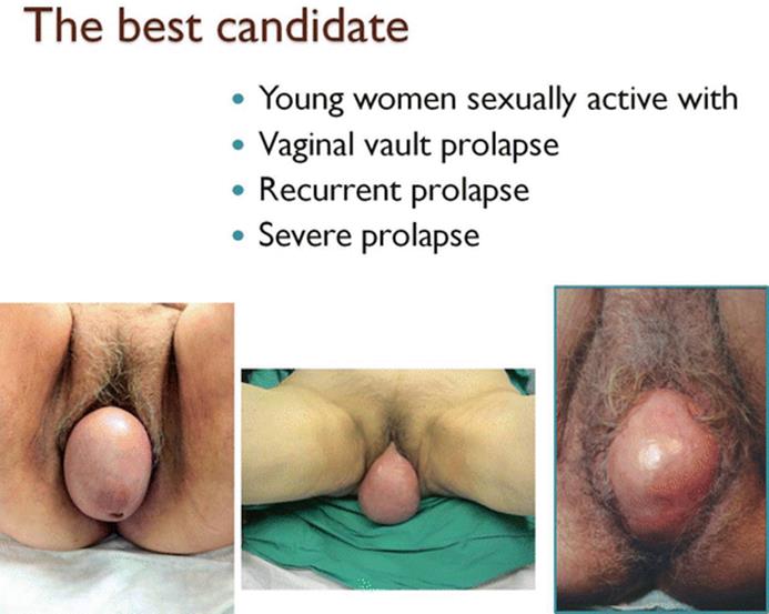
Fig. 7.39
Indications of SP
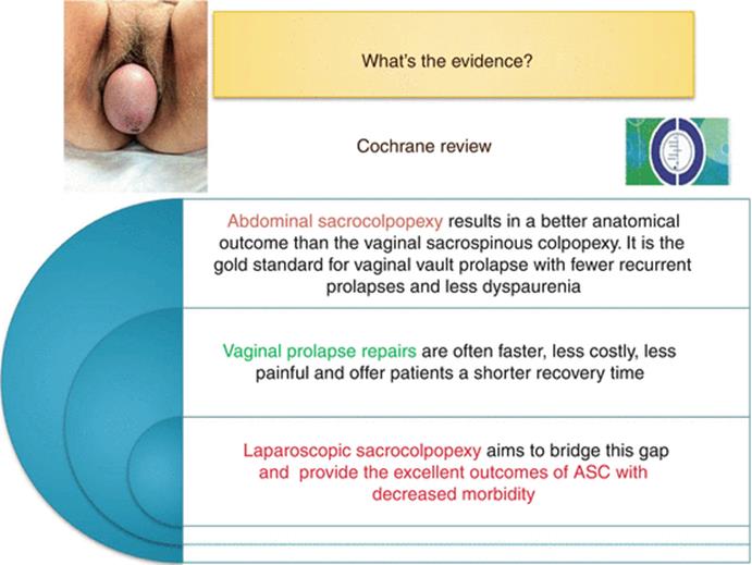
Fig. 7.40
Results from Cochrane review
The feasibility of uterus-sparing POP procedures has been investigated since the late 1990s. There are three major controversies associated with uterus preservation surgery for POP. First, many surgeons believe hysterectomy prevents prolapse recurrence, because the presence of the uterus might subject pelvic reconstruction to undue stress and this is particular true when a mesh is not used. On the other hand, hysterectomy-associated pelvic floor dissection might increase the risk of pelvic neuropathy and disrupt natural support structures. The second argument is that uterus preservation can expose the patient to potential pathologies (such as cancer) in what might be considered an almost useless organ. The risk of potential malignancy is estimated to be different in women with or without symptoms of abnormal vaginal bleeding: 1.09 % versus 0.22 %, respectively. The third controversial point concerns the complications that are associated with hysterectomy apart these considerations, the decision to remove a healthy organ, such as the uterus in POP repair, must take the patient’s preferences, needs, and values into consideration. Moreover, other major factors have come into play such as the profound changes in women’s lifestyles, beliefs, and perspectives, particularly with regard to sexual function and pregnancy. In the last two decades, several studies have described successfull anatomical and functional outcomes after uterus preserving POP repair including the abdominal and the laparoscopic approach, i.e., the hysterocolposacropexy (HSP) (Fig. 7.41). These studies confirm that uterus-sparing surgery is feasible in women who want to preserve integral vaginal function and satisfaction with their body image. The advantages of uterus preservation include the maintenance of pelvic anatomy integrity, reduction of intraoperative blood loss, shortened operating times and hospital stays, and reduced rate of mesh erosion. Finally, uterus preservation appears to contribute positively to the patient’s self-esteem, body image, confidence, and sexuality. Consensus is growing that the uterus can be preserved at the time of pelvic reconstructive surgery in appropriately selected women who desire it. Although the decision will be determined by the patient’s preference and the surgeon’s skills and experience, it is imperative that women fully understand the risks of incurring uterine and cervical pathology over time and the need for continued routine surveillance measures.
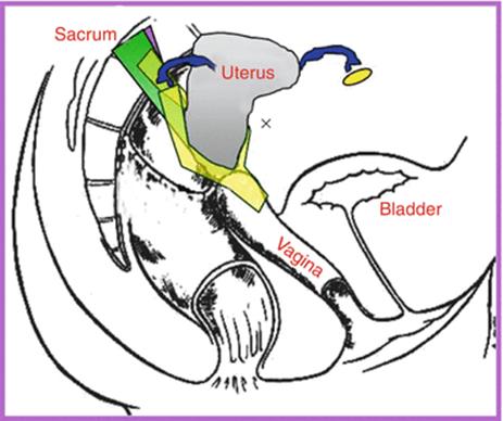
Fig. 7.41
Hysterocolposacropexy
Both SP and HSP can be performed laparoscopically or robotically assisted. The laparoscopic sacrocolpopexy was initially popularized in an attempt to improve pelvic visualization, operative morbidity, and postoperative functional results. The laparoscopic approach provides the excellent outcomes of the abdominal SP with the decreased morbidity of the vaginal approaches. Following the Food and Drug Administration (FDA) transvaginal mesh alerts in 2008 and 2011, there has been a significant increase in uptake of minimally invasive sacral colpopexy performed in the USA. Furthermore the robot-assisted laparoscopic sacral colpopexy was then introduced to shorten the learning curve associated with traditional laparoscopic SP. Currently the preferred route of entry to performing SP is the laparoscopic approach which has been demonstrated to have reduced blood loss, postoperative pain, and recovery time as compared to the open approach and reduced operating time, postoperative pain, and cost as compared to the robotic approach.
Surgical Technique
While the data supporting SP is robust, has been described for over 50 years and nearly 20 years have passed since Dr Benson’s landmark randomized surgical trial, there are still many uncertainties relating to the preferred technical aspects of the procedure. In particular there have been many modifications of the technique in recent years, with a variety of materials (expanded polytetrafluoroethylene, cadaveric fascia, light meshes) and attachments to the sacrum (bone screws, tackles) being advocated. One major modification has been the method of attaching the graft to the vaginal wall, from a simple apical attachment as described by Lane to grafts that extend down to the ventral and dorsal aspects of the vagina. Additional procedures such as paravaginal repair of the anterior vaginal wall or concomitant posterior colporrhaphy have been advocated to increase the durability of abdominal SP, but the success of these modifications and additional procedures has not been definitely studied.
Sacrocolpopexy Technique
The procedure has been performed for many years with several modifications and includes different procedures with different outcomes. The technique most widely used is that described by Timmons with mesh attached to the anterior and posterior aspects of the vault/apex with “extension” to correct prolapse in all three vaginal compartments.
The Procedure Step by Step
· Step 1 – The peritoneal cavity is entered through a lower midline incision or a Pfannestiel incision. Large and small bowel is mobilized as needed to expose the pelvis and posterior peritoneum.
· Step 2 – After identifying the vaginal vault, the peritoneum over the vaginal cuff is incised and dissected away from the vaginal apex. The anterior and posterior vaginal walls are dissected. Some surgeons attach the mesh directly on the vaginal vault without extension on the vaginal walls (Fig. 7.42). This makes the procedure easier and with less complications but the long-term results could be worse due to the higher probability of anterior and posterior compartment recurrences. Furthermore the large contact between mesh and vaginal walls avoids inadequate traction making the risk of mesh erosion lower, reduces the risk of recurrent anterior central defect, and partially corrects urethral hypermobility. So to obtain the best results, dissect the rectum-vaginal septum as far as the levator ani muscle and create a cleavage plane between the anterior vaginal wall and the bladder extending as far as the bladder trigone (the identification of the catheter balloon is the key point) (Fig. 7.43a–c).
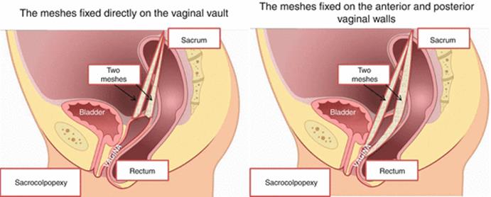
Fig. 7.42
Different attachments of the meshes to the vaginal vault
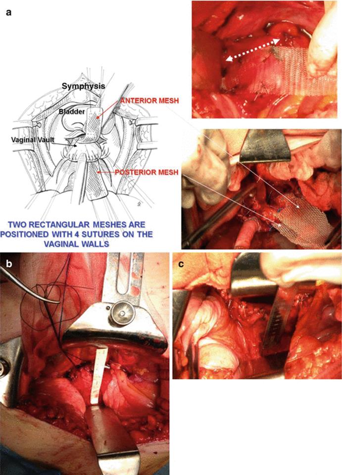
Fig. 7.43
(a) Two meshes are fixed on the anterior vaginal wall after an extended preparation of the anterior space between the vagina and the bladder, double side arrow indicates the anterior and posterior meshes; (b) the anterior vaginal wall preparation reaches the bladder neck level; (c) the posterior vaginal wall is prepared reaching the levator muscle plane
· Step 3 – Fix the mesh/meshes on the vaginal walls. Graft material selection: a variety of different graft materials were have been employed at the surgeon’s discretion. Monofilament polypropylene mesh seems to be the best because the reported vaginal erosion rate seems to be lower than that of other synthetic grafts. What is demonstrated is that absorbable meshes do not work. The design of graft material is variable on the basis of surgeon preference: two rectangular meshes and one single mesh fixed on the anterior vaginal wall and then on the posterior vaginal wall or Y-shaped graft (Fig. 7.44). The Y-mesh is the most used in laparoscopic approach. Suture on the vagina: both permanent and absorbable sutures have been reported. As stitch erosions are reported, some surgeons prefer reabsorbable sutures. Another argument in favor of reabsorbale sutures is that when the mesh is adherent to the vaginal walls, it easily will be incorporated in a short time with no need of permanent sutures. No evidence-based consensus has been reached on the number of sutures necessary to secure the mesh to the vagina. They range from 2 to 3 on the anterior vagina to 6–10 in the posterior vagina (Fig. 7.45). In some reports the meshes are fixed with two longitudinal continue sutures. Whatever the number of sutures is, the recommendation is to avoid folding and wrinkling and the mesh must be well stretched to avoid postoperative dyspareunia and mesh exposure. Controversial point is also the fixation of the posterior mesh. Some surgeons prefer fixing the mesh on the levator ani muscle to avoid mesh erosion and to correct rectocele but this benefit is counteracted a relevant high risk of dyschezia and pelvic pain syndrome. The fixation of the posterior mesh to the levator ani muscle is commonly reported in the laparoscopic series.
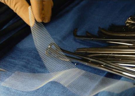
Fig. 7.44
The Y-mesh commonly used in laparoscopic SP
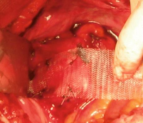
Fig. 7.45
Four sutures fix the mesh on the anterior vaginal wall without folds and well stretched
· Step 4 – The posterior peritoneum over the sacral promontory is incised and the sacral promontory exposed. This is commonly the first step in the laparoscopic approach. The anatomical landmarks must be recognized (vessels, ureter) to avoid complications (Fig. 7.46) and the general recommendation is to secure the meshes at or just below the level of the sacral promontory (Fig. 7.47). One or two nonabsorbable stitches can be inserted remembering to include the periosteum or the anterior longitudinal ligament in the stitches so as to ensure a firm hold. Recently titanium tackers have been introduced on the market, some complications were reported, but there is no evidence they cannot be used.
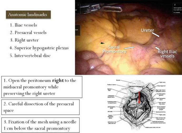
Fig. 7.46
How to prepare the sacrum; red arrow indicates the sacrum
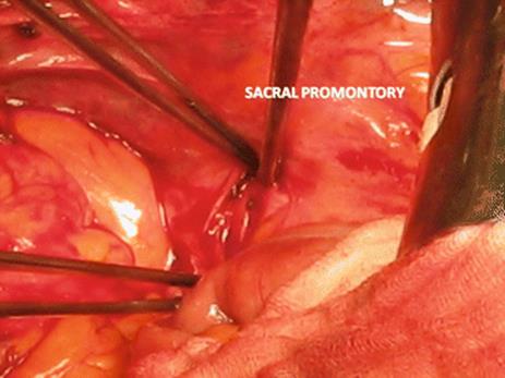
Fig. 7.47
Intraoperative image of the sacral promontory
· Step 5 – A tunnel under the peritoneum is created to pass the meshes through to the sacral promontory (Fig. 7.48a, b). Alternatively the peritoneum can be completely opened from the sacrum to the vaginal vault (commonly performed in the laparoscopic approach). The meshes are then anchored to the sacrum avoiding excessive tension and the redundant mesh is trimmed off. How to determine the right tension is a debatable point; expertise is fundamental.

Fig. 7.48
(a) Schematic subperitoneal tunnel; (b) intraoperative image
· Step 6 – Peritoneal closure over mesh. The majority of the surgeons prefer to close the peritoneum but the decision is left to the surgeon’s discretion. Controversial data on postoperative bowel complications are reported; however closure of the peritoneum at the completion of SP seems to be a good approach to avoid adhesions and subsequent long-term complications. Table 7.3 illustrates the most important surgical tip and tricks.
Table 7.3
Tips and tricks
|
Extended preparation of the vaginal walls |
|
Prefer polypropylene meshes |
|
Use reabsorbable suture on the vagina |
|
Avoid folding and wrinkling; the mesh must be well stretched |
|
Fix the mesh on the posterior vaginal wall and not on the levator ani muscle |
|
Prepare carefully the sacral promontory. All the anatomical landmarks should be recognized (vessels, ureter) |
|
Use nonabsorbable suture to fix the meshes on the sacrum |
|
Avoid excessive tension on the meshes |
|
Close the retroperitoneum over the mesh |
Hysterocolpopexy Technique
The procedure is similar to SP but several modifications are reported regarding the number, the shape, and the fixation of the meshes on the uterus. In step 2 the visceral peritoneum is incised over uterine isthmus. In some reports, a single posterior mesh is fixed on the uterine cervix; in others a sling mesh is positioned around the cervix (Fig. 7.49), or two meshes can be used: a mesh fixed on the anterior vaginal wall passed through the right broad ligament and a second mesh fixed on the posterior vaginal wall or on the levator ani muscle. Figures 7.50 and 7.51 illustrate the technique with the use of two meshes: the anterior Y-shaped mesh whose arms are passed through the broad ligament bilaterally and a posterior rectangular mesh in the rectovaginal space. The other steps are similar to SP technique.
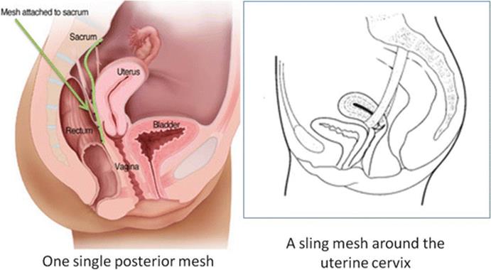
Fig. 7.49
Different meshes to fix the uterus to the sacrum

Fig. 7.50
HSP with anterior mesh Y-shaped. (a) schematic showing the Y-shaped mesh fixed to the anterior vaginal wall (b) the arms are then passed through the broad ligaments bilaterally (c) surgical image showing the passage of one arm of the Y-mesh through the broad ligament on the right (d) both arms are passed
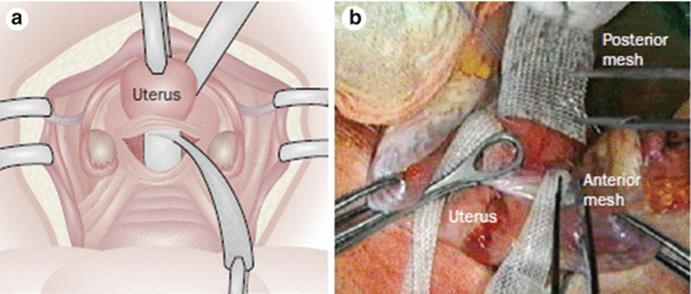
Fig. 7.51
HSP and posterior rectangular mesh. (a) schematic view of the rectangular mesh fixed to the posterior vaginal wall (b) surgical image showing arrangement of anterior and posterior meshes
Laparoscopic/Robotic Approach
Basically the technique of performing an open or LSC is identical. Figure 7.52 illustrates the trocar position. The first surgical step is sacral promontory preparation and opening the peritoneum from the sacrum to the vaginal cuff or the uterus. To make the procedure easier, a single Y-mesh is preferred to the use of two meshes (Fig. 7.44). Technical devices to fix the meshes to the sacrum and to the vagina are still evolving in order to avoid the need of sutures, which generally make the procedure longer although no consensus has been reached on the grade of recommendation. Longer follow-up is necessary to reach a conclusion.
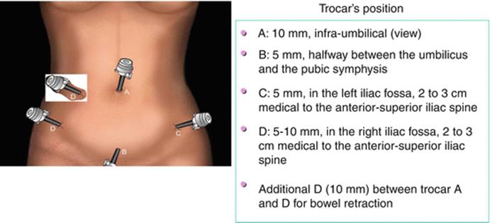
Fig. 7.52
Laparoscopic trocar position
Complications
Complications of the technique are illustrated in Table 7.4. Mesh erosion, reported in 3–9 % of the cases, typically occurs in the first 2 years after surgery, generally located at the level of the vaginal cuff or on the posterior vaginal wall, and the percentage is higher when contemporary hysterectomy is performed at the time of SP. For this reason supracervical hysterectomy is preferred by some surgeons but no consensus has been reached on this topic so far.
Table 7.4
Complications
|
Overall complications |
|
UTIs |
|
Wound infections |
|
Abscesses |
|
Hemorrhage |
|
Dyspareunia |
|
Urethral obstruction |
|
Voiding and/or storage symptoms |
|
Bladder–intestinal–ureteral–pelvic nerve injuries |
|
Sacrocolpopexy (abdominal, laparoscopic-assisted) complications |
|
Ventral hernia |
|
Bowel occlusion, bowel lesions |
|
Bladder lesions, ureteral obstruction or lesions |
|
Vascular injury (sacral promontory!) |
|
Problems at the trocar site |
|
Mesh exposure, erosion in other organs |
The Manchester Fothergill Procedure
Elisabetta Costantini13 and Antonio Carbone14
(13)
Urology and Andrology Clinic, Department of Surgical and Biomedical Science, University of Perugia, Rome, Italy
(14)
Department of Sciences and Medico-Surgical Biotechnologies, Sapienza, University of Rome, Rome, Italy
Elisabetta Costantini
Email: ecostant@unipg.it
Background
The Manchester Fothergill procedure was first proposed in 1888 by Archibald Donald of Manchester, England, and William Fothergill. It was initially described for the treatment of cervical elongation. It was an alternative to vaginal hysterectomy for the management of uterovaginal prolapse in patients with uterine cervical elongation and intact uterosacral-cardinal ligaments. While not commonly performed today, the Manchester procedure and the uterosacral ligament suspension represent the evolution of uterine-sparing techniques.
Clinical Practice
The advantages of the Manchester operation are that the surgeon does not enter the peritoneal cavity, the operating time is reduced, and the operation is not associated with a prolonged or morbid recovery. For all of these reasons, it is ideal for the elderly patient with no other uterine disease. The operation is designed to correct uterine descent associated with cystocele and rectocele where the preservation of the uterus is desirable. So the indications are: (1) preservation of reproductive function and (2) symptomatic vaginal prolapse associated with elongation of the uterine cervix.
Surgical Technique
The procedure consists of transvaginal cervical amputation, colporrhaphy, and fixation of the cervical stump to the cardinal ligaments.
With the patient in gynecological position, the first step of the procedure consists of putting a downward traction on the uterine cervix exposing the junction of the vagina and cervix where a 360° circumcision incision is made (Fig. 7.53). A preliminary uterine dilatation and curettage is generally performed. In this way the surgeon has confirmed the elongation of cervix and the dilatation of the cervical canal facilitates the passage of the sutures through the cervical canal during covering of the amputated cervix by vaginal flaps. It also ensures adequate uterine drainage and prevents cervical stenosis during healing of the external os. The bladder is then sharply and bluntly dissected off the lower uterine segment up to the vesicouterine fold. A right-angle retractor is placed under the bladder to expose the vesicouterine peritoneal fold which is then opened (Fig. 7.54). A retractor is placed in the anterior cul-de-sac, allowing elevation of the bladder and ureter. The cervix is rotated anteriorly, and the posterior cul-de-sac is exposed. The peritoneum of the posterior cul-de-sac is picked up and opened (Fig. 7.55). The cervix is retracted to the contralateral side, exposing the uterosacral and cardinal ligaments which are clamped, incised, and tied with a suture, bilaterally (Fig. 7.56). Depending on the length of the cervix, several bites may be required to remove a long cervix. With two retractors which elevate the bladder and the ureters anteriorly and depress the rectum posteriorly, traction is made on the cervix. The amputation is made with a scalpel at the lower uterine segment (Fig. 7.57). Where future reproduction is required, low amputation is to be done. The right cardinal and uterosacral ligaments are brought across the anterior surface of the cervix and sutured to the lower uterine segment with absorbable sutures (Fig. 7.58). The left cardinal and uterosacral ligaments are sutured overlapping those from the right side creating a firm barrier which holds the lower uterine segment posteriorly, bringing the fundus anteriorly (Fig. 7.59). In most cases of second- and third-degree uterine descensus, there will be significant cystourethrocele. Therefore, a standard anterior repair is performed. Posterior colpoperineorrhaphy is performed in selected cases. Finally the vaginal mucosa is closed with interrupted absorbable suture so that it covers the lower uterine segment leaving the uterine canal opened for drainage of mucus (Fig. 7.60). A Foley catheter is placed in the bladder and left in place for 4–5 days when an anterior repair and Kelly plication have been performed.

Fig. 7.53
Uterine cervix is exposed and a circular circumcision incision is made
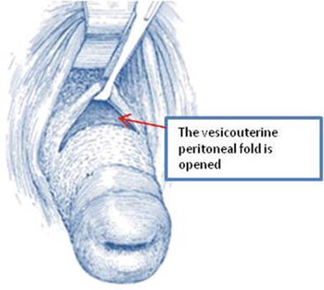
Fig. 7.54
The vesicouterine peritoneal fold is exposed and incised

Fig. 7.55
The peritoneum of the posterior cul-de-sac is picked up and opened

Fig. 7.56
The uterosacral and cardinal ligaments are clamped, incised and tied with a suture

Fig. 7.57
The cervix amputation is made with a scalpel at the lower uterine segment

Fig. 7.58
The right cardinal and uterosacral ligaments are brought across the anterior surface of the cervix and sutured to the lower uterine segment
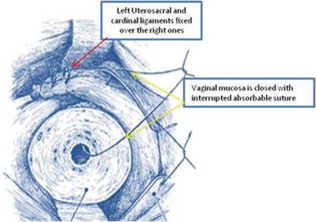
Fig. 7.59
The left cardinal and uterosacral ligaments are sutured overlapping those from the right side
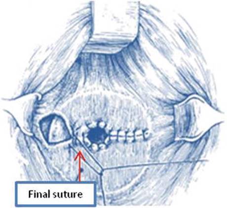
Fig. 7.60
The final suture
Results
Cure rates between 94 and 95.7 % have been reported, and it has been noted to be lower operative time and blood loss as compared to patients undergoing vaginal hysterectomy with anterior and posterior colporrhaphy. Several concerns regarding the Manchester procedure include a reoperation rate of 21 %, including reoperation for prolapse, as well as uterine and cervical disease. Recently a 50 % recurrence rate of the anterior compartment has been reported. Furthermore, because the Manchester procedure requires cervical amputation, it has been associated with infertility, miscarriage, and preterm delivery. For these reasons its role is still controversial; it is used in selected cases and generally by gynecologist.
Mesh Complications
Elisabetta Costantini15 and Franca Natale16
(15)
Urology and Andrology Clinic, Department of Surgical and Biomedical Science, University of Perugia, Rome, Italy
(16)
Urogynecology Unit, San Carlo-IDI Hospital, Rome, Italy
Elisabetta Costantini (Corresponding author)
Email: ecostant@unipg.it
Franca Natale
Email: francanatale@libero.it
As a result of the high rates of recurrence and reoperation with native tissue repairs in the treatment of female pelvic floor problems, the placement of synthetic mesh during pelvic organ prolapse (POP) repair is used to improve durability of surgical results. A significant increase in the use of an ever-widening array of prostheses and grafts has occurred in female pelvic floor surgery over the last 30 years. While the use of such meshes may reduce the recurrence of objective symptoms when compared with repairs made with native tissue only, the occurrence of other complications is increased and the management of these complications is widely controversial because of their heterogeneity.
Mesh exposure through the vagina is the most common and consistently reported mesh-related complication especially after transvaginal POP surgeries using mesh (TVM). According to the 2011 Food and Drug Administration (FDA) report, mesh erosion can require multiple surgeries and can be debilitating for some women. In some cases, even multiple surgeries will not resolve the complication. The synthetic graft that is used in the application of slings for the treatment of stress urinary incontinence has a more predictable and less severe course of complications compared with the synthetic mesh that is used for the management of POP. The 9-year cumulative rate of reoperation/revision after a mid-urethral sling (MUS) is 3.7 % with 60 % of removals/revision caused by mesh exposure. The average rate of mesh exposure in the first year after TVM placement for POP is 10.9 %. More than half of these patients require surgical excision in the operating room; some required multiple operations. Generally complications after TVM tend to be more severe, are more chronic in nature, and can be more difficult to treat. The rate of reoperation for management of complications after sacrocolpopexy (SCP) is 4.8 %.
The International Urogynecological Association (IUGA) and the International Continence Society have attempted to classify terminology and complications related to the use of native tissue or prostheses in female pelvic floor surgery, in order to simplify the decision-making process. These classifications take into account the exact site, the timing relative to the operation, the symptoms, and the general description, which are all necessary information for choosing the most appropriate therapeutical strategy. IUGA/ICS joint report made available a complete revision of the definitions and the terminology and a complete comprehensive coverage of both insertion complications and healing abnormalities. The classification system incorporates (a) category, (b) time, and (c) site divisions into a 6 (or seven)-digit code for any conceivable complication. The classification system includes all the complications after POP surgery without and with the use of grafts (Fig. 7.61). A well-constructed classification is necessary to reach a common language useful for clinical records application, for any database, registry, or surgical audit and finally for academic publications.
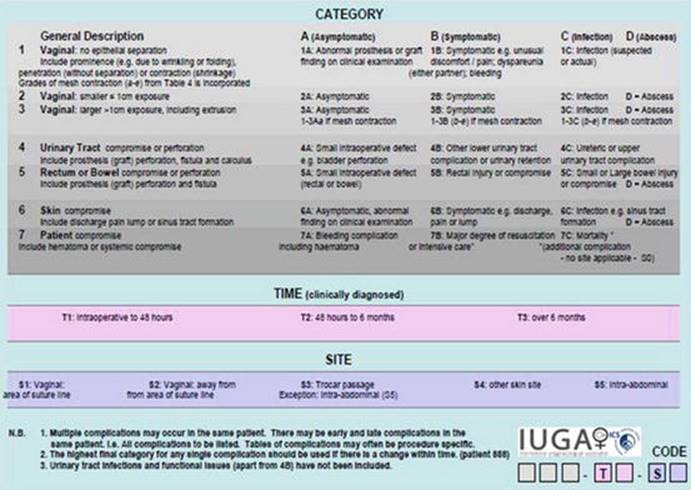
Fig. 7.61
IUGA/ICS classification of complications related directly to the insertion of prostheses (meshes, implants, tapes) or grafts in urogynecological surgery
Vaginal mesh exposure, defined as a condition displaying, revealing, exhibiting, or making the mesh accessible (e.g., vaginal mesh visualized through a separation in the vaginal epithelium), is a complication which is classified as category “2” if it is small (1 cm or less) and as category “3” if it is larger (> 1 cm). Categories such as “2” have been broken down into several divisions, including “A” (asymptomatic), “Bc” (pain during sexual intercourse), and “C” (clinical infection). The time (T) corresponding to a complication is that of its clinical diagnosis (T1, <48 h; T2, 48 h–2 months; T3, 2–12 months; T4, more than 12 months). The selection of the site (S) divisions includes the current sites, where mesh complications have been noted (S1, vaginal area of the suture line; S2, vagina, but remote from the vaginal suture line). Figure 7.62 shows two examples of the classification.
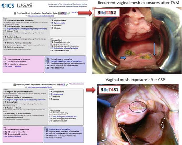
Fig. 7.62
Examples of IUGA/ICS classification (arrows and circle indicate the mesh exposures)
Other classification systems were proposed; one of them, from the American Urological Association, simply classifies the erosion in simple or complex (Fig. 7.63).
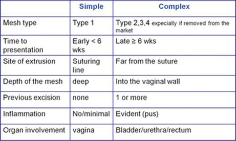
Fig. 7.63
American Urological Association classification
About 35 % of vaginal exposures are asymptomatic and they are discovered only during routine follow-up exams. Data are similar for urethral erosion, but the percentage of asymptomatic urethral erosion is lower (about 11 %). The 66 % of whole exposures is discovered within the first 3 months after surgery. Urologists and gynecologists have to be aware that in a high percentage of cases, signs and symptoms of complication begin early after surgery, and it is a duty of the surgeon to understand and recognize them in order to offer promptly the best treatment. In some cases the patient with a mesh exposure can present a symptomatic vaginal bleeding or malodorous discharge and dyspareunia. Partner’s pain during intercourse is also reported.
Several risk factors have a role in mesh-related complications (Fig. 7.64). The surgical approach is one of the most important discriminators and the risk factors change on the basis of the vaginal or abdominal/laparoscopic approach (Figs. 7.65 and 7.66). The type of incision during vaginal surgery and the use of permanent sutures on the vagina, together with some concomitant surgical procedures such as hysterectomy, are important risk factors. Learning curve is another important element as the erosion rate has been reported to be 2.9 % when surgery was performed by a consultant versus 15.6 % when surgery was performed by a fellow. The main mesh erosion risk factors in the abdominal or laparoscopic approach are concomitant vaginal intervention, concomitant total hysterectomy (5–7 times increased erosion rate), poor preoperative estrogenization, placing sutures at the vaginal apex, and use of permanent braided sutures. In the vaginal approach, the main risk factors are: inverted T colpotomy (erosion rate 20.6 %), excessive colpectomy, inadequate vaginal tissue coverage, permanent braided sutures, and the placement of the mesh in a surgical plane that is too much superficial.
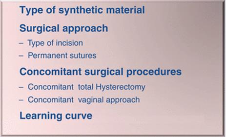
Fig. 7.64
Risk factors for mesh-related complications
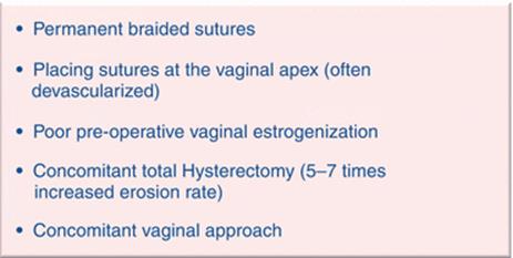
Fig. 7.65
Abdominal/laparoscopic approach: risk factors

Fig. 7.66
Vaginal approach: risk factors
The site of exposure varies with type of surgery. In sacrocolpopexy (both abdominal or laparoscopic) usually exposure occurs at the vaginal apex, sometimes along the distal posterior vaginal wall. In the trans-obturator approach, the main site is the lateral fornix; after an inverted T colpotomy during vaginal approaches, is the midline.
Surgical Treatment
The treatment of mesh exposure is highly individualized, the choice depending on:
· The mesh type (macroporous type I monofilament polypropylene meshes are associated with lower infection rates than multifilament and polyester meshes)
· Time of presentation
· Site of the extrusion and different organ involvement (bladder, urethra, bowel)
· Depth of the mesh
· Presence of signs of inflammation/infection
· Severity of patient’s symptoms
· Recurrency of the exposure/extrusion
· Patient’s expectations. For example, patients who consider the possibility of recurrent prolapse following mesh exposure excision to be an unacceptable risk may prefer an initial conservative treatment.
The treatments for vaginal mesh exposure include conservative measures such as observation, estrogen therapy, topical antiseptics/antibiotics, abstinence from sexual intercourse, and surgical treatments.
Most urogynecologists agree that a symptomatic vaginal mesh exposure must be surgically removed (partial resection or complete explantation). Total ablation of the mesh is recommended in case of mesh infection, local abscess, fistulae, and pelvic cellulitis. However, there is no precise recommendation for patients presenting with a small exposure surface area and/or asymptomatic vaginal mesh exposure and there is not randomized controlled study comparing conservative to surgical treatment. Pros and cons of surgical excision are illustrated in Table 7.5. Conservative management often involves a protracted treatment course, leading to potential frustration and distress. Regular vaginal estrogen use may not be appropriate for patients with known risks for estrogen use, severe arthritis, morbid obesity, or other functional limitations. Even when conservative therapy is chosen for initial management, some of these women ultimately undergo surgery if conservative therapy fails.
Table 7.5
Pros and cons of surgical excision
|
Surgical excision |
|
Cons |
|
Risk of anesthesia |
|
Burden of post-op recovery |
|
Small but possibly catastrophic risk of injury to internal organs |
|
Pros |
|
The potential for earlier and definitive relief of symptoms |
As general suggestion small asymptomatic exposure of type I polypropylene mesh into the vagina [IUGA/ICS category 2A] can be managed conservatively, through abstinence from sexual intercourse (6 weeks) and local estrogen replacement therapy associated with topical antiseptics, with the recommendation for surgeons to remain vigilant with respect to potential infections. In sexually inactive and asymptomatic patients (neither bleeding nor vaginal discharge, infection, or pain), with <3 mm exposure expectant management may be proposed (initially or following unsuccessful conservative medical treatment); though the exposure will often not heal spontaneously, it rarely progresses. Asymptomatic large vaginal mesh exposure (3A) and vaginal mesh exposure associated with infection or pain (2C, 3C, 2Bc, 3Bc) should be treated by surgical resection. Removal of the exposed mesh remains the gold standard for the treatment of symptomatic vaginal mesh exposures.
Surgical treatment includes local excision in the office, local excision in the operating room, and extensive or complete excision of the mesh (Fig. 7.67). A flowchart for the surgical treatment of simple or complex mesh exposure is illustrated in Fig. 7.68.

Fig. 7.67
Local or Total excision of the mesh. The intraoperative image shows a Vaginal mesh exposure after colposacropexy (3BcT4S2)

Fig. 7.68
Flowchart for simple and complex exposure treatment
There is a myriad of clinical problems resulting from transvaginal mesh that clinicians must manage with little data or experience to guide them. The success rate after vaginal mesh removal is around 80–95 % but not all the patients reported complete symptom resolution. The persistence of pain and dyspareunia in 30 % of patients suggests that treatment of persistent pain may be the most difficult to achieve. Pain is related not only to mesh exposure but also to another identified mesh-related complication which is mesh contraction.
Transvaginal mesh contraction or shrinkage refers to situations where part or all of the mesh contracts after being implanted. Mesh contraction can result in severe pelvic pain, painful sexual intercourse, or an inability to engage in sexual intercourse. Symptoms of mesh contraction include vaginal shortening, vaginal tightening, and vaginal pain. Pain is thought to be due to overtensioning of the mesh causing contraction bands or to chronic irritation to the pelvic floor muscles and associated nerves. The indications for treatment depend on the severity of the mesh contraction and the patient’s QoL. The treatment of these cases is complex and can be differentiated on the basis of the presence or absence of concomitant mesh exposure (Figs. 7.69 and 7.70). Generally it starts with oral medication and surgical treatment is reserved to cases associated to mesh exposure or to patients not responding to conservative treatment (physical therapy, trigger point medications, neuropathic pain medications). Unfortunately pain not always resolves after surgical treatment.
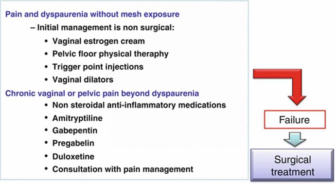
Fig. 7.69
Treatment for pain without mesh exposure
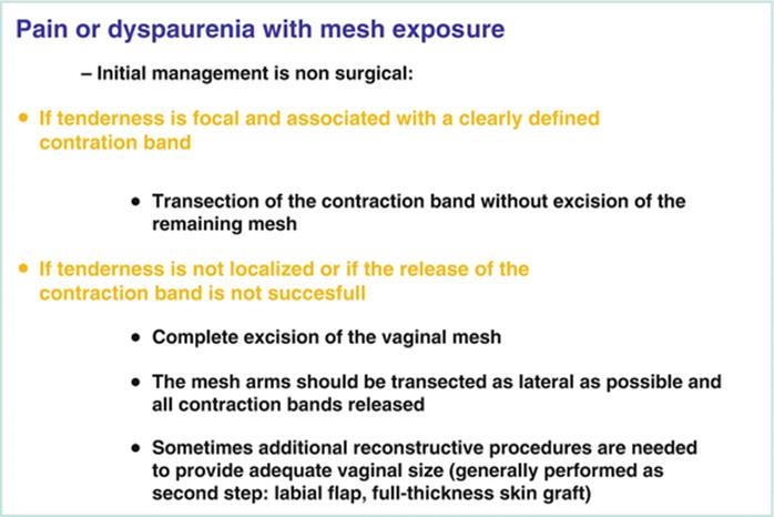
Fig. 7.70
Treatment for pain with mesh exposure
The surgical treatment for vaginal mesh exposure after sacrocolpopexy follows the same guidelines previously indicated. In case of simple vaginal exposure, a partial removal of the mesh is possible by the vaginal route. It’s important to underline that coverage without mesh excision is not recommended because it may lead to exposure recurrence. When a total removal of the mesh is indicated because of infection of the graft or for other organ involvement, an abdominal or laparoscopic route is the best way to reach the sacrum and obtain a complete removal of the graft.
In case of complex erosions such as extrusion in other organs (Fig. 7.71), fistulas, and exposure recurrence, different surgical approaches requiring great surgical skill become necessary. Martius or omental flap, urethral reconstruction, and ureteral reimplantation are some of these procedures which will be tailored to the case. Figures 7.72, 7.73 and 7.74 show a patient with hydroureteronephrosis after shrinkage and mesh erosion into the bladder 2 year after anterior TVM.

Fig. 7.71
Mesh erosion into the bladder after colposacropexy (4B T4S5 IUGA classification)

Figs. 7.72, 7.73 and 7.74
Hydroureteronephrosis after shrinkage and mesh extrusion into the bladder 2 years after anterior TVM (4C T4S5). Arrow indicate the mesh
Some Advice for Prevention of Mesh-Related Complications
The main problem is that all the surgical treatments for mesh-related complications may lead to other complications like fistulas, severe hemorrhage, and prolapse recurrence; for this reason the best treatment is prevention.
Some guidelines could be proposed. In general it is very important to prevent infection by a wide-spectrum antibiotic therapy before and after surgery and use of local estrogen therapy. The use of polypropylene grafts and avoiding concomitant anorectal surgery are recommended. Concomitant hysterectomy should be avoided in particular during TVM surgery. Other recommendations are illustrated in Fig. 7.75. During vaginal surgery, it is better to avoid T incision to prefer a retrograde dissection of the bladder and to limit the use of hemostatic hydrodissection to avoid ischemia. Furthermore it is important to maintain adequate thickness of vaginal epithelium. During sacrocolpopexy the most appropriate approach for preventing mesh exposure is closing the vaginal cuff in two layers or with a continuous introflexing suture, avoiding full-thickness stitches into the vagina, attaching meshes to distal portions of the vaginal walls and not attaching them to vaginal cuff, and finally avoiding excessive tension on the meshes.

Fig. 7.75
Some guidelines to prevent mesh-related complications
Mesh contraction may be prevented by avoiding: (1) direct suspension of mesh with sutures, (2) compression of the bladder and rectum by the mesh and (3) high lateral tension.
In conclusion the treatment of each complication must be carefully individualized; if a surgeon chooses to use a mesh, he must complete a specific training and, most importantly, he must learn how to treat the potential complications.
Sexual Assessment
Elisabetta Costantini17
(17)
Urology and Andrology Clinic, Department of Surgical and Biomedical Science, University of Perugia, Rome, Italy
Elisabetta Costantini
Email: ecostant@unipg.it
Sexuality is a complex process coordinated by the neurological, vascular, and endocrine systems. Sexual satisfaction is a fundamental element of general well-being and physical and mental health has been related to greater levels of sexual activity. Female sexual dysfunction (FSD) is a multi-causal and multidimensional medical problem that adversely affects physical health and emotional well-being. It can have damaging effects on the self-esteem, sense of wholeness, and interpersonal relationships. Epidemiological surveys report a variable prevalence ranging from 19 to 45 %, depending on age, hormonal status, and focus on single or cumulative disorders. Regardless these discrepancies, all studies concluded that FSD is highly prevalent with an increasing incidence in elderly women. Approximately 43 % of American women (depending on age) have sexual complaints and in Europe the prevalence is around 31.1 %. The number of women who have sexual dysfunction ranges from 19 to 50 % in “normal” outpatient population and increases to 68 to 75 % when sexual dissatisfaction or problems (not dysfunctional in nature) are included. While prevalence and risk factors for male sexual dysfunction, in particular erectile dysfunction, have been intensively studied within the past decade, data regarding this issue on women are scant. Investigators identified several possible etiologies of FSD, which include aging, psychological, anatomical and neurological factors, hormonal status, voiding dysfunction, and cultural factors, including race and ethnicity, medications, and drug abuse. Although sexual function (SF) has been extensively studied in several medical specialties dealing with women’s health, it has been largely overlooked in the field of urogynecology. Specifically, anatomical factors relating to SD gained recognition in urogynecology only recently. The female bladder base and urethra are anatomically adjacent to the vagina; in addition to supporting the abdominal and pelvic organs and maintaining urinary and fecal continence, the pelvic floor permits intercourse and parturition and plays a role in sexual response. When voluntarily contracted, pelvic floor muscles can intensify orgasms for both men and women, so pelvic floor dysfunction may be associated with sexual problems.
Female sexual function was found to be negatively influenced or affected by the presence of lower urinary tract symptoms, with urinary incontinence (UI), and detrusor overactivity causing the greatest degree of SD. Several studies reported that 40–50 % of patients with UI report impairment of SF and recent studies demonstrated improvement in SF following treatment for UI. Similarly, multiple studies have found a correlation between pelvic organ prolapse (POP) and SD, with the higher incidence correlating with the severity of prolapse. Despite the clear relationship between pelvic floor disorders and SD, studies assessing SF following surgical repair of pelvic floor disorders have conflicting results. Whereas some authors found an improvement in SF following POP repair, others have found unchanged or increasing incidences of SD and dyspareunia. Although sexual dysfunction is recognized as a highly prevalent condition in women attending urogynecological services, only a minority of urogynecologists screen all patients for FSD. Lack of time, uncertainty about therapeutic options, and older age of the patient have been cited as potential reasons for failing to address sexual complaints as part of routine history.
Although considered as a widespread health problem, FSD continues to be under-recognized and undertreated. It is thus necessary to increase awareness and physicians’ ability to communicate with female patients about sexual problems. Given that assessment by psychometric and paper-and-pencil diagnostic tools is time consuming, medical willingness to explore FSD remains low in the majority of clinical settings. Furthermore, many women may hesitate to share their sexual complaints, also because they are worried about their physicians’ interest in addressing FSD. Direct questioning by physicians about sexual function, therefore, remains critical, as both males and females refrain from referring spontaneously to their sexual symptoms. Finding the correct way to ask delicate questions and to decode answers on sexual health and disease might be difficult and even embarrassing for the inexperienced physician. Standardized, validated sexual inventories might thus become a valuable help.
Definition and Classification
Sexual dysfunction is a disorder that includes manifestations ranging from unresponsiveness to any variation in intensity and quality of desire, arousal, and orgasm. Factors such as general health impairment, chronic diseases, health of relationship, adverse sociodemographic and behavioral conditions including low income, low levels of education, and stress seem to affect sexual capacity significantly. Major risk factors for FSD in women are embedded in a biopsychosocial frame (Table 7.6). Female sexual dysfunction is an overall term to describe problems across the different sexual phases and the definition is a persistent or recurrent inability to engage in satisfying sexual activity leading to marked distress or interpersonal difficulty, excluding general medical conditions, psychiatric issues, substance abuse, and medications. FSDs are classified in four major disorders (Fig. 7.76) hypoactive sexual desire disorder (HSDD), female sexual arousal disorder (FSAD), female orgasmic disorder (FOD), and female sexual pain disorders (FPD). Often women will report difficulties within more than one of the domains, which makes it challenging to identify the treatment target and limits research in clinical trials, where it can be difficult to identify end points in which a change can be measured. Generally the different disorders are interconnected in a multidimensional problem which can be defined as the cycle of sexual dysfunction (Fig. 7.77).
Table 7.6
FSD and risk factors
|
Menopausal transition (hormonal and psychological changes) |
|
Relationship problems |
|
Sexual dysfunction in the partner |
|
Depression |
|
Urinary tract symptoms |
|
Cancer and its treatment |
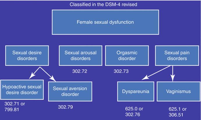
Fig. 7.76
FSD classification on the basis of DSM4 revised

Fig. 7.77
The cycle of SD
Hypoactive Sexual Desire Disorder (HSDD). The hypoactive sexual desire is the lack of sexual thoughts or fantasies and it is different from sexual aversion, a phobic aversion to and avoidance of sexual contact. The percentage of women with low desire ranged from 16% in premenopausal women (aged 20–49) to 46 % in surgically menopausal women aged 50–70. In climacteric syndrome low desire is as common as hot flushes (Fig. 7.78). It has been hypothesized that HSDD can be triggered by an imbalance between CNS inhibitory and excitatory factors, with a reduced excitatory activity, increased inhibitory activity, or a combination of both. Disorders of desire in premenopausal patients may be secondary to lifestyle factors, medications, or another sexual dysfunction (pain or orgasmic disorder); in peri- and postmenopausal women the relationship between hormones and sexuality is unclear. Estrogen replacement therapy has been shown to correlate positively with sexual activity, enjoyment, and desire, although the findings are not universal. The mechanism of estrogen’s effect on desire is probably indirect and occurs through improvement in urogenital atrophy, vasomotor symptoms, and menopausal mood disorders. What is important to underline is however that only a percentage of women consider HSDD as a distressing problem and these women should be carefully evaluated before deciding to treat the sexual problem (Fig. 7.79).

Fig. 7.78
Prevalence of low sexual desire among climacteric symptoms

Fig. 7.79
HSDD and female distress: when the assessment is necessary
Female sexual arousal disorder (FSAD) is the inability to attain or maintain sufficient sexual excitement. It may be expressed as a lack of subjective excitement or genital response and it may be secondary to inadequate stimulation, especially in older women who require more stimulation to reach a level of arousal that was more easily attained at a younger age. Urogenital atrophy is the most common cause of arousal disorders in postmenopausal women.
Female orgasmic disorder (FOD) is the difficulty in attaining orgasm following sufficient sexual stimulation and it includes anorgasmia, a condition often caused by sexual inexperience or by the lack of sufficient stimulation. Orgasmic disorders may also be psychologic (involuntary inhibition of the orgasmic reflex) or caused by medications or chronic disease.
Female sexual pain disorders (FPD) include dyspareunia (genital pain with sexual intercourse), vaginismus (involuntary spasm of pelvic musculature interfering with vaginal penetration), and noncoital sexual pain disorders.
Dyspareunia includes different situations (Fig. 7.80), and in the majority of the cases, the cause is a vestibulodynia with an estimated prevalence of 9–12 %. The condition has a substantial negative effect on a woman’s daily life relationships, sexual life, quality of life, and psychological well-being. Vulvodynia is defined by the absence of relevant visible findings or an identifiable neurological disorder; its diagnosis is truly one of excluding any other mechanism that could cause the pain. This condition comprises heterogeneous pain phenotypes: provoked pain, unprovoked pain, pain localized to one region of the vulva or generalized throughout, etc. The etiology is thought to involve inflammatory and infectious factors, hypertonicity of the pelvic floor (which might be secondary to the pain), hypervigilance to body sensations, depression, and anxiety. As no good scientific data are available to guide pharmacotherapy of dyspareunia, an unmet need for an effective treatment still remains for women with this condition. Several agents have been tried and are currently in clinical use, including topical lidocaine, tricyclic antidepressants, gabapentin, pregabalin, and botulinum toxin injections, often combined with surgery, biofeedback, and cognitive behavioral therapy. However, the use of most of these treatments is based on case studies or small experimental series. Vaginismus may be complete or situational, so that a pelvic examination might be possible while intercourse is not. It is a complex problem, often related to sexual phobias or past abuse or trauma, and its evaluation generally includes a comprehensive physical and psychological assessment.

Fig. 7.80
Dyspareunia and different kinds of pain
Assessment
To assess FSD, the first step is to obtain a detailed patient history that defines the dysfunction; then it is important to identify causative or confounding medical or gynecologic conditions (Table 7.7) and finally it is necessary to obtain psychosocial information. Several drugs can cause FSD and should be investigated in each patient (Table 7.8). Clinical examination should be accurate aiming to exclude: vulvar dystrophy or dermatitis, mucosal atrophy, clitoral adhesions, herpes simplex infection, bartholinitis, episiotomy scar strictures, vaginitis, and postoperative and post-radiation changes. Furthermore pelvic organ dysfunctions such as pelvic organ prolapse (Table 7.9) and urinary incontinence should be evaluated because they have a significant impact on female sexuality. Since 1980s, several assessment tools, specific to or inclusive of female sexual function, that are suitable for office-based use have been introduced. Validated questionnaires utilized to assess sexual function in women with pelvic floor dysfunction (PFD) may be generalized or condition specific. Generalized questionnaires focusing on sexual function were designed to evaluate sexual function in a general population and not specifically in women with PFD. These types of questionnaires may not be sensitive enough to detect differences due to the disease process of urinary incontinence, fecal incontinence, and/or pelvic organ prolapse in this particular population. Two general questionnaires focused on sexual function, utilized in the urogynecological literature, include the Sexual History Form 12 (SHF-12) and the Female Sexual Function Index (FSFI). The FSFI is a 19-item self-reported measurement of FSD and it analyzes six domains: desire, arousal, lubrication, orgasm, satisfaction, and pain. Each domain is scored on a scale of 0 (or 1) to 5 (Fig. 7.81). The scale has been tested to assess the impact of diverse medical conditions and treatments on sexual function and has consistently demonstrated excellent psychometric properties. It is one of the most powerful and useful diagnostic tools for diagnosing FSD and monitoring treatment. These questionnaires are condition specific and were developed, validated, and tested for use in women with PFD but do not focus on sexual function. There are two condition-specific questionnaires focused on sexual function for use in women with pelvic floor dysfunctions, the Pelvic Organ Prolapse Urinary Incontinence Sexual Questionnaire (PISQ) and the International Consultation on Incontinence Questionnaire Vaginal Symptoms (ICIQ-VS).
Table 7.7
Medical conditions and FSD
|
Vascular disease |
|
Diabetes (arousal disorders) |
|
Cardiovascular disease |
|
Arthritis |
|
Urinary incontinence/POP |
Table 7.8
Drugs and FSD
|
Medications that cause: |
|
Disorders of desire (psychoactive medications, BDZ, beta blockers, oral contraceptives, etc.) |
|
Disorders of arousal (anticholinergics, antihistamines, SSRI, etc.) |
|
Orgasmic dysfunction (methyldopa, trazodone, BDZ, amphetamines, etc.) |
Table 7.9
Factors that might contribute to FSD in patients with POP
|
1. POP changes the anatomical features of vagina |
|
2. It is a hidden disfigurement of the urogenital tract |
|
3. It could negatively affect the woman’s body image |
|
4. It may lead to shame, embarrassment, and feeling less sexually attractive, all of which reduce quality of life |
|
5. It may be associated with urine leakage which worsens sexual activity |
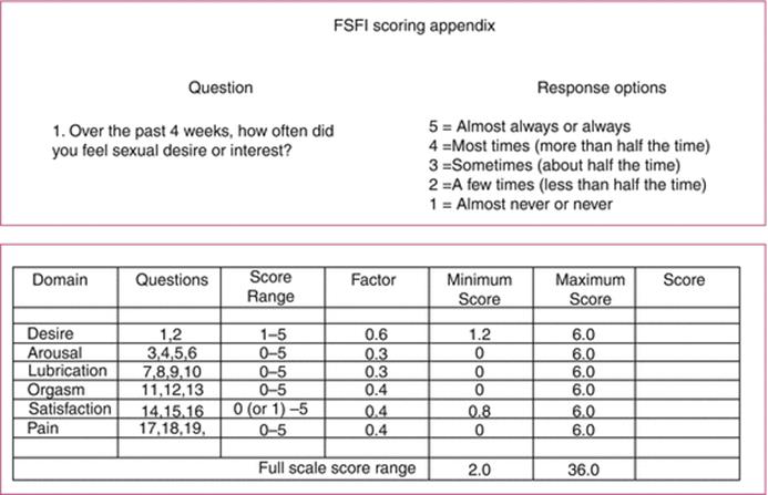
Fig. 7.81
FSFI scoring system
In conclusion questionnaires are a useful method in detecting FSD but it is important to underline that sexual function is affected by several factors, including couple relationship conflicts, socioeconomic level, sexual compatibility, and physical and psychiatric disorders of the couple. For these reasons, the importance of a multidisciplinary, collaborative approach is central in evaluating any sexual dysfunction, both organic and psychosocial issues must be considered, taking in consideration that whatever is the cause of a sexual dysfunction, it typically occurs in a relationship context.
Masked Stress Urinary Incontinence
Elisabetta Costantini18 , Antonio Carbone19 and Antonio Luigi Pastore19
(18)
Urology and Andrology Clinic, Department of Surgical and Biomedical Science, University of Perugia, Rome, Italy
(19)
Department of Sciences and Medico-Surgical Biotechnologies, Sapienza, University of Rome, Rome, Italy
Elisabetta Costantini
Email: ecostant@unipg.it
Stress urinary incontinence (SUI) is a common complaint in women with pelvic organ prolapse (POP). In fact, urinary incontinence is more common in women with prolapse than in women with normal vaginal support and it is usually associated with prolapse of the anterior vaginal wall. SUI and POP probably share some of the same etiological mechanisms.
The relation between POP and SUI is complex. Approximately 40 % of women with POP report symptoms of stress urinary incontinence while some women with POP experience SUI only with a pessary inserted, on provocative testing with reduction of the prolapse, up to half of patients with POP but without symptoms of SUI may demonstrate SUI. The reason seems to be linked to the fact that POP may actually function to kink the urethra, maintaining stress continence by causing urethral obstruction. After a successful POP surgery, the development of SUI postoperatively may result from relieving the urethral obstruction caused by prolapse, thereby unmasking a preexisting compromised urethral function. Urogynecologists have referred to this as an unmasking of occult or latent SUI.
Diagnostic Evaluation
In preoperatively continent woman with POP, the risk of “de novo” SUI is approximately estimated between 11 and 20 %, while in women with occult SUI the risk to develop postoperative SUI may even be as high as 80 %. Therefore it is important to assess SUI prior to POP surgery to adequately inform the patient and decide if additional surgical procedures are required to treat this condition. There is still controversy regarding the diagnostic evaluation of masked SUI. Physical examination and instrumental tests may be helpful to identify subjects with higher risk to develop SUI after POP surgery. Preoperative stress test after prolapse reduction, to mimic a repaired pelvic anatomy, may reveal the occult SUI and is often performed to predict whether a patient would benefit from a concomitant SUI procedure. Despite this, the incidence of de novo SUI after an open sacrocolpopexy without an anti-SUI procedure can be as high as 39 %; and a 25 % of de novo SUI has been reported in women who underwent mesh-augmented transvaginal POP repair without concomitant mid-urethral sling.
Urodynamic assessment after reducing POP may further contribute to show a masked SUI. Kleeman in 2006 reported that de novo SUI and urinary urgency developed only in 1.9 % of females submitted to POP surgery and previously evaluated by urodynamic with a negative stress test at cystometry. Therefore, clinicians should not base their surgical decision only on a negative report of SUI from females with POP, but they should address some clinical maneuvers or instrumental tests which can help them to unmask occult conditions.
Some of the questions that remain are what is the most effective way to look for occult SUI (which prolapse reduction method is ideal) and which specific algorithm to use.
Treatment Strategies
Up to date no general agreement exists concerning treatment strategies for women with POP and masked SUI. Some surgeons prefer to do a concomitant anti-incontinence surgical procedure if the patient has symptoms or any clinical evidence of SUI. Other surgeons prefer to correct the vaginal prolapse first and evaluate afterward whether an additional anti-incontinence procedure for unmasked SUI is indicated.
The advantage of combining POP and SUI surgery is that fewer patients may report SUI following such combination. However, this combined surgical strategy may be associated with an increased risk of complications, of which the development of bladder outlet obstruction, responsible for overactive bladder symptoms and retention, is the most important. Furthermore, combining the two procedures may be unnecessary, as performing only POP surgery may cure SUI in about 40 % of patients.
To date data on the treatment of masked SUI remain controversial. Studies report different results not only because of the different preoperative assessment of this condition but also for the different surgical procedures used for POP repair. The prevalence of occult SUI was 27 % in the CARE trial and 33 % in the OPUS trial. The Colpopexy and Urinary Reduction Efforts (CARE) trial demonstrated that the postoperative risk of stress incontinence in stress continent women undergoing open abdominal sacrocolpopexy could be substantially reduced by the addition of a Burch colposuspension. Other RCT studies however did not confirm these results. The Outcomes Following Vaginal Prolapse Repair and Mid Urethral Sling (OPUS) trial reached similar conclusions following vaginal prolapse repair and mid-urethral sling, with lower rates of postoperative SUI after combination surgery (Table 7.10).
Table 7.10
De novo stress urinary incontinence asymptomatic for SUI preoperatively: with occult SUI versus without occult SUI
|
Study |
Outcome |
Postoperative SUI in women with masked SUI, % (n/N) |
Postoperative SUI in women without masked SUI |
||
|
Combination surgery |
Control |
Combination surgery |
Control |
||
|
CARE trial |
Objective SUI at 3 months |
32 (12/38) |
58 (23/40) |
21 (22/106) |
38 (41/109) |
|
Sokol et al. |
Objective SUI at 24 months |
14 (6/43) |
48 (16/39) |
NA |
NA |
|
OPUS trial |
UI at 12 months |
35 (19/54) |
60 (34/57) |
28 (30/107) |
41 (46/113) |
|
Total |
Objective SUI |
22 (18/81) |
52 (41/79) |
||
|
NNT (ARR) |
Objective SUI |
3.3 (30 %) |
NA |
||
ARR absolute risk of retention, NA not available, NNT number needed to treat
Sacrocolpopexy and transvaginal surgery have not been compared in a randomized trial evaluating this specific outcome, however it has been suggested that the transvaginal and transabdominal routes are related to different changes in dynamics between the anterior vaginal wall and the urethrovesical junction and, therefore, may be associated with different incidences of postoperative SUI in women with masked SUI.
All the available data did not still reduce the debate whether POP surgery repair should be combined or not with anti-incontinence surgery. If SUI has been documented in a woman preoperatively, then the benefits of a concomitant procedure to prevent SUI will probably outweigh the risks; if not, the risk–benefit ratio is less predictable. After a correct clinical assessment, the decision to perform – or not to perform – prophylactic surgery should be based on goals and desires of the patient, the skill and experience of the surgeon, and the risks and potential benefits for a particular patient. Going to the evidence as reported by the Cochrane Collaboration Group in 2013, concomitant continence surgery at the time of POP surgery in continent women does not reduce the rate of de novo SUI, while in women with preoperative occult SUI incontinence, the rate of de novo SUI may be reduced by concomitant anti-incontinence procedure, even if approximately 20 % of women will be prevented and 80 % will have an unnecessary procedure. It is not possible to conclude that one single treatment strategy would be correct for every woman. The two options, combined POP repair and anti-incontinence procedure and wait and see strategy, should be discussed with each woman. Future research in this field should be aimed at identifying preoperative tests that could select women that are likely to be cured of SUI by prolapse surgery alone and postpone incontinence surgery until it is demonstrated that such an operation is really needed. Obviously, it has to be considered that further evaluation of these issues is necessary especially if new and larger randomized clinical trials will be developed, in which the benefit needs to be balanced against adverse effects, including the costs for the public health-care systems, which can vary between the different countries.
Neurogenic Stress Urinary Incontinence
Antonio Carbone20 and Giovanni Palleschi20
(20)
Department of Sciences and Medico-Surgical Biotechnologies, Sapienza, University of Rome, Rome, Italy
Definition
Independently from the specific cause (inflammatory, degenerative, vascular, traumatic, iatrogenic, neoplastic), neurogenic stress urinary incontinence (SUI) is secondary to striated urethral sphincter deficiency as a consequence of a lesion which involves regions of the central and/or peripheral nervous system controlling this muscle through the pudendal nerve (Fig. 7.82) (erroneously positioned at the end of “mesh complications”). Patients with neurogenic SUI very often present also storage symptoms such as urinary urgency, frequency, and urgency incontinence, suggestive for a neurogenic detrusor overactivity (NDO) or low bladder compliance. In some cases, these symptoms may be masked by a severe SUI. Therefore, these subjects have to be carefully evaluated before planning a therapeutic protocol (Fig. 7.83), especially if surgical, to avoid failure and to prevent complications to the upper urinary tract function. In fact, in patients with mixed symptoms (urgency incontinence + stress urinary incontinence), the first approach aims to treat storage disorder and as second step SUI is treated if still necessary. In patients with severe NDO, major consideration is to be reserved to the risk of severe damage to upper urinary tract (i.e., vesicoureteral reflux) if bladder alteration is not treated before SUI surgery which may further contribute to increase endovesical pressures during filling phase, especially in case of bladder hyperactivity. As a further consideration, neurogenic implication of SUI should be considered in females with previous pregnancy and vaginal childbirth which represents an important risk factor for SUI. These conditions may be responsible for compression and stretching of the pudendal nerve contributing to the development of SUI. There is evidence that women with SUI diagnosed by urodynamics also demonstrate pudendal nerve dysfunction as evidenced by abnormal nerve conduction velocity and external urethral sphincter electromyography. Increased pudendal nerve terminal motor latency, an indicator of nerve damage or dysfunction, is correlated with vaginal delivery, advanced age, and SUI. Additionally, the external anal sphincter in these women showed abnormal fiber densities, suggesting partial reinnervation occurred following pudendal nerve injury. The underestimation of these findings in women with SUI categorized as “non-neurogenic” probably could explain unsatisfying outcomes of treatment.

Fig. 7.82
The image shows the neurological control of the bladder and the innervation of striated urethral sphincter provided by pudendal nerve (From O’Rahilli, Capernter, Nueller and Svenson: Basic Human Anatomy)
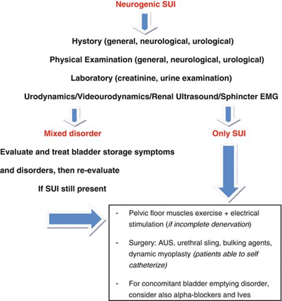
Fig. 7.83
The diagnostic and therapeutic flow chart for Neurogenic Stress Urinary Incontinence
Patient’s Assessment
History must provide information related to pregnancy and vaginal childbirth, previous surgery, ongoing and pharmacologic treatment. Neurological diagnosis and onset of neurological and urological symptoms have to be assessed. General, vaginal, and neurological physical examination are mandatory, particularly aimed to evaluate the presence of a concomitant genital prolapse. Urodynamic study combined with sphincteric/pelvic floor electromyography is recommended to support the neurogenic origin of SUI, establish disorder severity, assess the presence of associated bladder disorders (either of filling or voiding phase), and control the outcomes treatment. For these purposes, imaging techniques, especially cystography or videourodynamic, may improve diagnostic power and accuracy.
Treatment
In presence of neurogenic SUI, a specialized management is required. Conservative treatment with rehabilitation (pelvic floor muscle exercise) and electrical stimulation are not indicated for subjects affected by a complete denervation. Subjects with partial denervation might benefit from long-term protocols of electrical stimulation administered by a vaginal device because in these cases patients could contribute with their voluntary activity to the rehabilitation program. However, there are no studies reporting data from randomized trials. Pharmacotherapy is not recommended. Although the efficacy of various drugs has been explored to treat neurogenic SUI (adrenergic alpha-blockers, beta-3-agonists, antidepressants), no study has been yet published. For this reason, many patients with sphincteric incompetence secondary to neurogenic conditions may be considered for surgical treatment. As previously reported, combined dysfunction of bladder filling phase, as detrusor overactivity and/or low compliance, must be conservatively treated before a surgical approach for neurogenic SUI. In fact, the minimal criteria which make a patient eligible for surgical treatment of neurogenic SUI are: intrinsic sphincteric deficiency, underactive detrusor or well-controlled overactive detrusor, absence of urethral injury or pathological urethral condition (stricture), no evidence of vesicoureteral reflux, and well-stabilized neurological disease. Artificial sphincter implantation is considered one of the best surgical choices and it provides satisfying outcomes also at long-term follow-up (up to 10 years). This procedure is more used in males than in women; the long-term results are satisfying, even though there is a higher complication rate such as urethral erosion. As an alternative to artificial sphincter, good results are reported with the use of sub-urethral slings. In fact, European Urological Association Guidelines suggest that in female patients with neurogenic SUI who are able to self-catheterize, placement of an autologous urethral sling is the first choice with a high grade of recommendation (3A) while in male patients artificial urinary sphincter (AUS) is the best suggested treatment (grade of recommendation: 3A). However, all of the published studies are case series characterized by poor cohorts and without long-term data. Furthermore the studies are often not comparable because they include different surgical devices with different pre- and postoperative evaluations. Encouraging results come from the use of the dynamic myoplasty with gracilis muscle, but the experience is limited and it does not allow to consider this technique as a standard approach. Various authors report experience with bulking agents. This approach presents a large success variability in non-neurogenic population; therefore, in neurogenic cohorts its efficacy is even more unpredictable. Patients with coexisting voiding disorders can be managed by alpha-blockers and self-intermittent catheterism. Endovesical electrical stimulation (IVES) is also considered a therapeutic option although data are absolutely preliminary and controversial.
Further Reading
1.
Moen M, Noone M, Vassallo B. Anterior colporrhaphy: why surgeon performance is paramount. Int Urogynecol J. 2014;25(7):857–62. doi:10.1007/s00192-014-2345-6. Epub 2014 Mar 7. PubMed.CrossRefPubMed
2.
Turgal M, Sivaslioglu A, Yildiz A, Dolen I. Anatomical and functional assessment of anterior colporrhaphy versus polypropylene mesh surgery in cystocele treatment. Eur J Obstet Gynecol Reprod Biol. 2013;170(2):555–8. doi:10.1016/j.ejogrb.2013.07.014. Epub 2013 Aug 2. PubMed.CrossRefPubMed
3.
Lensen EJ, van den Berg-van Erp SH, Stoutjesdijk JA, Hasaart TH, Withagen MI, Kluivers KB, Dietz V, Vierhout ME. Does the method of dissecting in anterior colporraphy lead to a difference in thickness of removed vaginal tissue? Eur J Obstet Gynecol Reprod Biol. 2013;168(1):112–6. doi:10.1016/j.ejogrb.2012.12.028. Epub 2013 Jan 21. PubMed.CrossRefPubMed
4.
Baessler K, Maher C. Pelvic organ prolapse surgery and bladder function. Int Urogynecol J. 2013;24(11):1843–52. doi:10.1007/s00192-013-2175-y. Review. PubMed.CrossRefPubMed
5.
Ek M, Tegerstedt G, Falconer C, Kjaeldgaard A, Rezapour M, Rudnicki M, Altman D. Urodynamic assessment of anterior vaginal wall surgery: a randomized comparison between colporraphy and transvaginal mesh. Neurourol Urodyn. 2010;29(4):527–31. doi:10.1002/nau.20811. PubMed.PubMed
6.
Gynaecology by ten teachers 19th ed Dutta-Gynecology Bonney Gynaecological Surgery 11E.
7.
Diwan A, Rardin CR, Kohli N. Uterine preservation during surgery for uterovaginal prolapsed: a review. Int Urogynecol J. 2004;15:286–92.
8.
Zucchi A, Lazzeri M, Porena M, Mearini L, Costantini E. Uterus preservation in pelvic organ prolapse surgery. Nat Rev Urol. 2010;7:626–33. doi:10.1038/nrurol.2010.164.CrossRefPubMed
9.
Alkış I, Karaman E, Han A, Gülaç B, Ark HC. The outcome of Manchester-Fotergill operation for uterine decensus repair: a single center experience. Arch Gynecol Obstet. 2014;290(2):309–14. doi:10.1007/s00404-014-3200-1. Epub 2014 Mar 18. PubMed.CrossRefPubMed
10.
Thys SD, Coolen A, Martens IR, Oosterbaan HP, Roovers J, Mol B, Bongers MY. A comparison of long-term outcome between Manchester Fothergill and vaginal hysterectomy as treatment for uterine descent. Int Urogynecol J. 2011;22(9):1171–8. doi:10.1007/s00192-011-1422-3. Epub 2011 Apr 12. PubMed.CrossRefPubMed
11.
Maher C, Feiner B, Baessler K, Schmid C. Surgical management of pelvic organ prolapse in women. Cochrane Database Syst Rev. 2013;(4):CD004014. doi:10.1002/14651858.CD004014.
12.
Lensen EJ, Withagen MI, Kluivers KB, Milani AL, Vierhout ME. Surgical treatment of pelvic organ prolapse: a historical review with emphasis on the anterior compartment. Int Urogynecol J. 2013;24(10):1593–602. doi:10.1007/s00192-013-2074-2. Epub 2013 Mar 15. Review. PubMed.CrossRefPubMed
13.
van Geelen JM, Dwyer PL. Where to for pelvic organ prolapse treatment after the FDA pronouncements? A systematic review of the recent literature. Int Urogynecol J. 2013;24(5):707–18. doi:10.1007/s00192-012-2025-3. Epub 2013.CrossRefPubMed
14.
Dos Reis Brandão da Silveira S, Haddad JM, de Jármy-Di Bella ZI, Nastri F, Kawabata MG, da Silva Carramão S, Rodrigues CA, Baracat EC, Auge AP. Multicenter, randomized trial comparing native vaginal tissue repair and synthetic mesh repair for genital prolapse surgical treatment. Int Urogynecol J. 2015;26(3):335–42.CrossRefPubMed
15.
Chesson R, Hallner B. Why complex pelvic organ prolapse should be repaired vaginally. Curr Opin Urol. 2013;23(4):312–6. Review. PubMed.CrossRefPubMed
16.
Bugge C, Adams EJ, Gopinath D, Reid F. Pessaries (mechanical devices) for pelvic organ prolapse in women. Cochrane Database Syst Rev. 2013;(2):CD004010. doi:10.1002/14651858.CD004010.pub3. Review. PubMed.
17.
Tenfelde S, Tell D, Thomas TN, Kenton K. Quality of life in women who use pessaries for longer than 12 months. Female Pelvic Med Reconstr Surg. 2015;21:146–9. [Epub ahead of print] PubMed.CrossRefPubMed
18.
Jones KA, Harmanli O. Pessary use in pelvic organ prolapse and urinary incontinence. Rev Obstet Gynecol. 2010;3(1):3–9. PubMed PMID: 20508777; PubMed Central PMCID: PMC2876320.PubMedCentralPubMed
19.
Wiegersma M, Panman CM, Kollen BJ, Vermeulen KM, Schram AJ, Messelink EJ, Berger MY, Lisman-Van Leeuwen Y, Dekker JH. Pelvic floor muscle training versus watchful waiting or pessary treatment for pelvic organ prolapse (POPPS): design and participant baseline characteristics of two parallel pragmatic randomized controlled trials in primary care. Maturitas. 2014;77(2):168–73. doi:10.1016/j.maturitas.2013.10.014. Epub 2013 Oct 31. PubMed.CrossRefPubMed
20.
Alperin M, Khan A, Dubina E, Tarnay C, Wu N, Pashos CL, Anger JT. Patterns of pessary care and outcomes for medicare beneficiaries with pelvic organ prolapse. Female Pelvic Med Reconstr Surg. 2013;19(3):142–7. doi:10.1097/SPV.0b013e31827e857c. PubMed PMID: 23611931; PubMed Central PMCID: PMC3635496.PubMedCentralCrossRefPubMed
21.
Bump RC, Mattiasson A, Bø K, Brubaker LP, DeLancey JO, Klarskov P, Shull BL, Smith AR. The standardization of terminology of female pelvic organ prolapse and pelvic floor dysfunction. Am J Obstet Gynecol. 1996;175(1):10–7. PubMed.CrossRefPubMed
22.
Hall AF, Theofrastous JP, Cundiff GW, Harris RL, Hamilton LF, Swift SE, Bump RC. Interobserver and intraobserver reliability of the proposed International Continence Society, Society of Gynecologic Surgeons, and American Urogynecologic Society pelvic organ prolapse classification system. Am J Obstet Gynecol. 1996;175(6):1467–70; discussion 1470–1. PubMed.CrossRefPubMed
23.
Abrams P, Andersson KE, Birder L, Brubaker L, Cardozo L, Chapple C, Cottenden A, Davila W, de Ridder D, Dmochowski R, Drake M, Dubeau C, Fry C, Hanno P, Smith JH, Herschorn S, Hosker G, Kelleher C, Koelbl H, Khoury S, Madoff R, Milsom I, Moore K, Newman D, Nitti V, Norton C, Nygaard I, Payne C, Smith A, Staskin D, Tekgul S, Thuroff J, Tubaro A, Vodusek D, Wein A, Wyndaele JJ, Members of Committees; Fourth International Consultation on Incontinence. Fourth International Consultation on Incontinence Recommendations of the International Scientific Committee: Evaluation and treatment of urinary incontinence, pelvic organ prolapse, and fecal incontinence. Neurourol Urodyn. 2010;29(1):213–40. doi:10.1002/nau.20870. Review. PubMed.CrossRefPubMed
24.
Riss P, Dwyer PL. The POP-Q classification system: looking back and looking forward. Int Urogynecol J. 2014;25(4):439–40. doi:10.1007/s00192-013-2311-8.PubMed. Barber MD, Lambers A. Visco Obstet Gynecol. 2000; 96(1):18–22.
25.
Swift S, Morris S, McKinnie V, Freeman R, Petri E, Scotti RJ, Dwyer P. Validation of a simplified technique for using the POPQ pelvic organ prolapse classification system. Int Urogynecol J Pelvic Floor Dysfunct. 2006;17(6):615–20. Epub 2006 Apr 6. PubMed.CrossRefPubMed
26.
Maher C, Feiner B, Baessler K, Schmid C. Surgical management of pelvic organ prolapse in women. Cochrane Database Syst Rev. 2013;(4):CD004014. doi:10.1002/14651858.CD004014.pub5. Review. PubMed.
27.
Wan OY, Cheung RY, Chan SS, Chung TK. Risk of malignancy in women who underwent hysterectomy for uterine prolapse. Aust N Z J Obstet Gynaecol. 2013;53(2):190–6. doi:10.1111/ajo.12033. Epub 2013 Jan 15. PubMed.CrossRefPubMed
28.
Costantini E, Zucchi A, Lazzeri M, Del Zingaro M, Vianello A, Porena M. Managing mesh erosion after abdominal pelvic organ prolapse repair: ten years’ experience in a single center. Urol Int. 2011;86(4):419–23. doi:10.1159/000324243. Epub 2011 Mar 30. PubMed.CrossRefPubMed
29.
Matthews CA. Robot-assisted laparoscopic colposacropexy and cervicosacropexy with the da Vinci® surgical system. Surg Technol Int. 2010;20:232–7. PubMed.PubMed
30.
Barber MD, Maher C. Epidemiology and outcome assessment of pelvic organ prolapse. Int Urogynecol J. 2013;24(11):1783–90.CrossRefPubMed
31.
Cabral PUL, Canário ACG, Spyrides MHC, Uchôa SAC, Eleutério J, Gonçalves AK. Determinants of sexual dysfunction among middle-aged women. Int J Gynaecol Obstet. 2013;120(3):271–4.CrossRefPubMed
32.
Filocamo MT, Serati M, Li Marzi V, Costantini E, et al. The female sexual function index (FSFI): linguistic validation of the Italian Version. J Sex Med. 2014;11:447–53.CrossRefPubMed
33.
Kammerer-Doak D. Assessment of sexual function in women with pelvic floor dysfunction. Int Urogynecol J. 2009;20 Suppl 1:S45–50.CrossRef
34.
US Food and Drug Administration. FDA safety communication: update on serious complications associated with transvaginal placement of surgical mesh for pelvic organ prolapse. 2011. Available at: http://www.fda.gov/MedicalDevices/Safety/AlertsandNotices/ucm262435.htm. Accessed 31 July 2012.
35.
Barber MD. Surgical techniques for removing problematic mesh. Clin Obstet Gynecol. 2013;56:289–302.CrossRefPubMed
36.
Skoczylas LC, Shepherd JP, et al. Managing mesh exposure following vaginal prolapse repair: a decision analysis comparing conservative versus surgical treatment. Int J Urogynecol J. 2013;24:119–25.CrossRef
37.
Abbott S, Unger CA, Evans JM, Jallad K, Mishra K, Karram MM, Iglesia CB, Rardin CR, Barber MD. Evaluation and management of complications from synthetic mesh after pelvic reconstructive surgery: a multicenter study. Am J Obstet Gynecol. 2014;210(2):163.e1–8. http://www.ncbi.nlm.nih.gov/pubmed/24126300.CrossRef
38.
van der Ploeg JM, van der Steen A, Oude Rengerink K, van der Vaart CH, Roovers JP. Prolapse surgery with or without stress incontinence surgery for pelvic organ prolapse: a systematic review and meta-analysis of randomised trials. BJOG. 2014;121(5):537–47.CrossRefPubMed
39.
Costantini E, Lazzeri M, Bini V, Del Zingaro M, Zucchi A, Porena M. Pelvic organ prolapse repair with and without prophylactic concomitant burch colposuspension in continent women: a randomized, controlled trial with 8-year followup. J Urol. 2011;185(6):2236–40. Epub 2011 Apr 16.CrossRefPubMed
40.
Visco AG, Brubaker L, Nygaard I, et al. The role of preoperative urodynamic testing in stress-continent women undergoing sacrocolpopexy: the Colpopexy and Urinary Reduction Efforts (CARE) randomized surgical trial. Int Urogynecol J Pelvic Floor Dysfunct. 2008;19:607–14.PubMedCentralCrossRefPubMed
41.
Wei J, Nygaard I, Richter H, Nager C, Barber MD, Kenton K. A midurethral sling to reduce incontinence after vaginal prolapse repair. N Engl J Med. 2012;366(25):2358–67.PubMedCentralCrossRefPubMed
42.
Borstad E, Abdelnoor M, Staff AC, Kulseng-Hanssen S. Surgical strategies for women with pelvic organ prolapse and urinary stress incontinence. Int Urogynecol J. 2010;21:179–86.CrossRefPubMed
43.
Abrams P, Cardozo L, Khoury S, Wein A, editors. Incontinence. 4th ed. Plymouth: Health Publication Ltd; 2009.
44.
Wyndaele JJ. Correlation between clinical neurological data and urodynamics function in spinal cord injured patients. Spinal Cord. 1997;35:213.CrossRefPubMed
45.
Sakakibara R, Hattori T, Uchiyama T, Yamanishi T, Ito H, Ito K. Neurologic failures of the external urethral sphincter closure and relaxation; a videourodynamic study. Auton Neurosci. 2001;86:208.CrossRefPubMed
46.
Groen J, Pannek J, Castro Diaz D, Del Popolo G, Gross T, Hamid R, Karsenty G, Kessler TM, Schneider M, ‘t Hoen L, Blok B. Summary of European Association of Urology (EAU) Guidelines on NeuroUrology. Eur Urol 2015 pii: S0302–2838(15)00740–X. doi: 10.1016/j.eururo.2015.07.071. [Epub ahead of print]
47.
Barber MD, Brubaker L, Burgio KL, Richter HE, Nygaard I, Weidner AC, Menefee SA, Lukacz ES, Norton P, Schaffer J, Nguyen JN, Borello-France D, Goode PS, Jakus-Waldman S, Spino C, Warren LK, Gantz MG, Meikle SF, Eunice Kennedy Shriver National Institute of Child Health and Human Development Pelvic Floor Disorders Network. Comparison of 2 transvaginal surgical approaches and perioperative behavioral therapy for apical vaginal prolapse: the OPTIMAL randomized trial. JAMA. 2014;311(10):1023–34.PubMedCentralCrossRefPubMed
48.
Barber MD, Maher C. Apical prolapse. Int Urogynecol J. 2013;24(11):1815–33.CrossRefPubMed
49.
Souviat C, Bricou A, Porcher R, Demaria F, Fritel X, Benifla JL, Pigné A. Long-term functional stability of sacrospinous ligament-fixation repair of pelvic organ prolapse. J Obstet Gynaecol. 2012;32(8):781–5.CrossRefPubMed
50.
Maher C, Feiner B, Baessler K, Schmid C. Surgical management of pelvic organ prolapse in women. Cochrane Database Syst Rev. 2013;(4):CD004014.
51.
Nicolau-Toulouse V, Tiwari P, Lee T, Cundiff GW, Geoffrion R. Does bilateral sacrospinous fixation with synthetic mesh recreate nulliparous pelvic anatomy? An MRI evaluation. Female Pelvic Med Reconstr Surg. 2014;20(4):222–7. doi:10.1097/SPV.0000000000000066. PubMed.CrossRefPubMed