
Q. A fit 75-year-old man presents for transurethral resection of the prostate (TURP). Preoperative assessment revealed good effort tolerance, no symptoms of cardiac failure and all investigations were normal. He undergoes spinal anaesthetic after a preload of 500 mL saline, and is given oxygen via Hudson mask. Surgery begins after the block was confirmed at the T8 level. At 60 minutes into the procedure, the patient complains of nausea and was given ondansetron. HR: 106 bpm, BP: normal. Then, 15 minutes later, he becomes anxious, pulls off the oxygen mask off and tries to get off the operating table. What is the probable diagnosis and how does it occur?
A. I would strongly suspect the diagnosis is likely to be transurethral resection (TUR) syndrome.
TUR syndrome is a multifactorial syndrome, which arises from absorption of large volumes of irrigation fluid (1.5% glycine), typically during TURP. Although it is commonly thought to be due to dilutional hyponatraemia exclusively, fluid overload and effects of glycine toxicity contribute significantly to the pathophysiology of this condition.
Q. What is the incidence of TUR syndrome?
A. Classical studies quote an incidence of 0.5%-2.0% following TURP [3,4]. Recent contemporary studies suggest an even lower incidence, based on technological evolution. Interestingly, TUR syndrome may also be associated with transurethral resection of bladder tumour (TURBT) and percutaneous nephrolithotomy (PCNL).
Q. What concentration of glycine is used in resection, and what is its osmolality?
A. Typically, 1.5% glycine is used. It is an inhibitory amino acid, and a non-electrolyte solution, with an osmolality of 200 mOsomol/L - hence, it is hypotonic, with respect to plasma.
Q. How is glycine handled in the body?
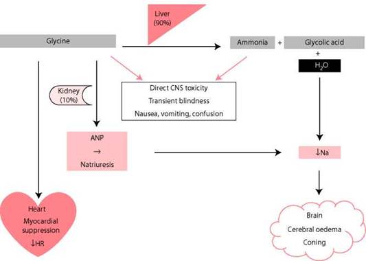
Figure 7.4 Handling and toxic effects of glycine.
A. Figure 7.4 shows handling and toxic effects of glycine. In a typical TURP, there is approximately 20 mL/min fluid absorption and thus during a 60 minute resection one would anticipate 1.2 L of glycine solution being absorbed. Absorption of 1.5% glycine solution occurs directly (and thus immediately) into the periprostatic venous plexus, as well as indirectly (resulting in delayed absorption) from the perivesical and retroperitoneal spaces. This amount of absorbed hypotonic fluid is relatively easily dealt with in a normal individual, with 90% of glycine being metabolised to ammonia, glycolic acid and water, by the liver, and the remaining 10% being metabolised by the kidney.
Q. Can you explain the symptoms of TUR syndrome?
A. TUR syndrome is a multifactorial syndrome, which arises from absorption of large volumes of 1.5% glycine solution, resulting in dilutional hyponatraemia, fluid overload and effects of glycine toxicity. Knowledge of these three factors allows an understanding of the clinical symptoms of TUR syndrome (Figure 7.4).
The dilutional hyponatraemia results in osmotic shift of water from plasma into the brain. Symptoms are generally dependent on sodium concentration, resulting in cerebral herniation and death, if left untreated (Table 7.1).
Furthermore, glycine induces an osmotic diuresis, which results in absolute losses of sodium from the body, and this can be further exacerbated by the release of atrial natriuretic peptide, which promotes natriuresis (Figure 7.4).
Table 7.1 Symptoms associated with dilutional hyponatraemia
|
Na concentration (mmol/L) |
Symptoms |
|
130-135 |
Asymptomatic |
|
120-130 |
Restlessness Confusion |
|
115-120 |
Nausea |
|
<115 |
Seizures Coma |
With fluid overload patients initially develop hypertension, shortness of breath, chest pain and cyanosis, from resultant pulmonary oedema and cardiac failure. Later clinical features include bradycardia and a marked decrease in systolic arterial pressure.
Glycine is an inhibitory neurotransmitter in the retina, present at a concentration of 400 mol/L in humans. An excess amount slows down the transmission of impulses from the retina to the cerebral cortex, with prolongation of visual evoked potentials and deterioration of vision occurring after absorption of as little as a few hundred millilitres of glycine. Thus, clinically, if the patient is under spinal anaesthesia, he may report seeing flashing lights. Prickling sensations and facial warmth are also early signs of glycine absorption. At higher concentrations, glycine results in bradycardia due to direct and indirect cardiotoxic effects. Late clinical features include hypotension and coma.
Q. How would you manage TUR syndrome?
A. I conveniently divide the management of TUR syndrome into prevention, recognition and definitive treatment.
Prevention - Initially, I diagnose and treat any pre-existing hyponatraemia, before considering the patient for TURP. Second, I identify putative risk factors for TUR syndrome. In the American Urological Association (AUA) co-operative study, of immediate and postoperative complications of almost 4,000 patients from 13 institutions, Mebust et al. [3] identified significant differences in TUR syndrome when time of resection and size of gland were assessed. Among patients with a resection time of >90 minutes, the incidence of TUR syndrome was 2.0% compared to 0.7% where there was a shorter resection time (p < .01) [3]. Similarly, a statistically significant difference was noted in patients with glands >45 g (incidence 1.5%) as opposed to <45 g (incidence 0.8%) [3]. It would There fore, at first glance, seem logical to suggest standard TURP be avoided if the operative time would exceed 90 minutes or if the prostate gland size was larger than 45 g. However, in my practice, and in contemporary practice in the United Kingdom, an operative time of 60 minutes is usually standard, and open prostatectomy is only usually performed for gland sizes >100 g.
Other potential risk factors, such as height of irrigation fluid and intravesical pressure, race and age have also been suggested, but the evidence for these is less robust. Despite this, in my practice, I use the Iglesias continuous flow resectoscope, avoid aggressive resection near the capsule and try to complete the TURP as soon as the capsule is breached. In addition, if a prolonged procedure is inevitable, I request the anaesthetist to administer furosemide prophylactically, to off-load the excess fluid that may be absorbed. More recently, other preventative strategies have included the use of bipolar or laser resection with normal saline irrigation, as well as the use of 5% glucose as an irrigation solution in a randomised, prospective trial.
Recognition - I aim to perform TURP with the patient awake, under spinal anaesthesia, as several of the clinical factors described above will become apparent if TUR syndrome develops. With general anaesthetic, however, hypertension, from fluid overload, may be the only early warning sign, often detected by the anaesthetist. Arrhythmias, hypotension and decreased oxygen saturation are usually late features. Although not universally used,
I am aware that 1% ethanol in the irrigant can be a useful strategy for detection, as it allows breath alcohol levels to be checked by a breathalyser, allowing an estimate of the volume of excess fluid that has been absorbed. Furthermore, the use of weighing machines being added to the ordinary operating table has also been reported as a technique for measuring fluid overload.
Definitive Treatment - In mild cases of established TUR syndrome, supportive management along with a period of watchful waiting is often sufficient. In my practice, the serum sodium and electrolytes are checked, but the result is not awaited prior to commencing medical treatment, in the form of the loop diuretic furosemide. (ttis drug results in relative loss of more water than sodium thus decreasing fluid overload and also increasing serum sodium levels.) Typically, a dose of 40 mg is given intravenously. More diuretic may be warranted depending on the serum sodium levels, as slower absorption from the retroperitoneal or perivesical space occurs (an alternative to furosemide given by many is mannitol). Concurrently, I ensure that I quickly control any haemorrhage and finish the operation as soon as possible. In addition, I think it is essential to have early input from the intensive care team, in all cases of TUR syndrome.
Severe cases occur due to lack of recognition or inadequate early treatment of mild cases.
In these cases it is extremely vital that the high-dependency unit/intensive therapy unit (HDU/ITU) team are called early. Using this multidisciplinary approach, a central line and invasive arterial monitoring are usually performed as well as transferring the patient to HDU/ITU, when stable. Clearly, in extreme cases the patient may need to be intubated and ventilated. Furthermore, the intensive care environment is useful for the small minority of patients who have a dangerously low serum sodium, which requires correction with hypertonic saline solution. A correction of 1 mmo1/1 per hour is recommended to avoid the devastating complication of rapid correction of hyponatraemia, resulting in central pontine myelinolysis.
Typically, no specific treatment of hyperglycineaemia/hyperammonaemia is required as patients usually recover within 12-24 hours with general supportive care. Furthermore, depending on the blood loss, blood transfusion may also be required.
Q. A 65-year-old man with recurrent acute urinary retention undergoes TURP. Prostate volume was 70 mL and during the procedure, it was noted that the prostate was extremely vascular. A large perforation of the surgical capsule was made on the left side, but otherwise the procedure was performed uneventfully. Post-operatively, in the recovery room, you are called because the catheter is draining dark red urine and the patient is pale, drowsy and looking unwell. HR is noted to be 120/min and BP = 80/55 mm Hg. How will you manage this patient?
A. I would consider this a urological emergency and see the patient immediately, myself, without delay, as I suspect that he may have uncontrolled haemorrhage after TURP.
Normally 2%-5% of patients require blood transfusion after TURP and although venous ooze usually settles with conservative management, arterial bleeding may be present and necessitate early return to theatre. Importantly, several manoeuvres described below often need to be performed in quick succession or simultaneously and early help should be enlisted from HDU/ITU.
Initially, using basic principles of Advanced Trauma and Life Support (ATLS), I would resuscitate this patient, with the close involvement of my HDU/ITU anaesthetic colleagues.
I would administer 100% high flow oxygen and give good analgesia. In addition, I would draw blood to check the full blood count (FBC) (Hb), clotting, urea and electrolytes (U+E) (Creatinine) and cross-match four units of blood.
Urologically, I would first check to see if the catheter is blocked and that the irrigation is running adequately. If clot retention is present, I would immediately perform a bladder washout, to remove all clots. If this does not improve the situation, or clot retention is not present, I would inflate the catheter balloon to 50 mL and maintain in-line traction with the irrigation running on maximum flow. There is no recognised time limit for traction on the catheter, although clearly, the longer this is applied, in some cases many hours, the higher is the risk of future contracture due to bladder neck ischaemia. I usually apply traction for 20-30 minutes and release for 5 minutes. Traction can then be reapplied at a later stage if further bleeding occurs.
In the meantime, I would give blood transfusion and correct any clotting abnormalities, as required. If the traction does not stabilise the situation and if there is ongoing bleeding, persistent hypotension, persistent clot retention or excessive blood transfusion requirement, the patient needs urgent return to theatre, which I will organise.
In theatre, initially, clot evacuation, endoscopic washout and careful diathermy to bleeding points is performed. In my practice, I perform a thorough washout with a resectoscope (26 or 28 Fr) and Ellick evacuator, using the diathermy loop to dislodge any organised clot (without current). Use of a bladder syringe attached to the end of the resectoscope can aid the evacuation of clot resistant to washout with the Ellick evacuator. If arterial bleeding is present, it can be difficult to detect in the presence of hypotension. Careful observation with low pressure irrigation can sometimes help.
If there is ongoing bleeding despite this, then the patient needs open surgical exploration and packing of the prostatic fossa.
Alternatively, if facilities are available, super-selective internal iliac artery embolisation may be performed. The procedure can be performed under local anaesthesia and may be a safer option in frail elderly patients.
Q. How do you perform open surgical exploration of the prostatic fossa?
A. In my practice, I perform a Pfannenstiel incision and the bladder is opened to pack the prostatic fossa (around the urethral catheter) with swabs through a transvesical approach.
The packs are left in place for 48 hours to tamponade any bleeding points and subsequently removed in theatre, with the definitive closure of the anterior abdominal wall.