
Acute testicular pain, complications of TURP, sepsis and penile fracture Iqbal S Shergill and Basharat Jameel
Q. A 15-year-old boy (Figure 7.1) is referred to you with acute onset right testicular pain. What is the differential diagnosis?

Figure 7.1 Image of a young boy presenting with acute onset right testicular pain.
A. Differential diagnosis includes testicular torsion, epididymo-orchitis, testicular trauma, torted hydatid of Morgagni or mumps orchitis (idiopathic scrotal oedema if under 10 years of age).
Q. How would you assess this patient?
A. I would consider this a urological emergency and see the patient immediately myself, without delay. I would take a history, examine the patient, arrange further investigations (if required) and institute a management plan as appropriate.
Q. What features would be suggestive of testicular torsion?
A. Testicular torsion is a clinical diagnosis. However, even with a high index of suspicion, the definitive diagnosis can only be made at emergency surgical exploration.
Clinical features of testicular torsion are as follows:
History
Pain is of acute onset
Mainly testicular pain
Occasionally radiating to groin, abdomen or thigh
History of previous testicular pain (intermittent torsion) may be common
Examination
Acutely tender and swollen testicle
Horizontal lie (bell-clapper deformity)
High riding testis in scrotum (Figure 7.1)
Absent cremasteric reflex
Mild fever and erythema of the scrotal skin (late signs)
Investigations
Urinalysis is usually normal
Doppler ultrasound scan (USS) (only in equivocal cases) may show poor or absent blood flow
Q. When does torsion typically occur and what is the difference between intravaginal and extra-vaginal torsion?
A. Testicular torsion can occur at any age but commonly the incidence has a bimodal distribution [1], with the main peak around puberty (12-18 years) and a smaller peak in the first year of life (Figure 7.2).
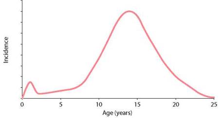
Figure 7.2 The bimodal incidence of testicular torsion.
Intravaginal torsion is the most common form of testicular torsion seen in adolescents and adults and is due to a congenital high investment of the tunica vaginalis on the cord, resulting in a horizontally lying testis, and it produces the ‘bell clapper’ deformity. This anomaly allows the testis and cord to rotate more readily than a normal testis. The bell clapper deformity is often bilateral, with a significant risk of torsion to the contralateral testis.
Extravaginal torsion is most commonly seen in the first year of life. It can occur both pre and post-natally. The attachment between the tunica vaginalis and the scrotum is loose, i.e. there is incomplete fixation of the gubernaculum to the scrotal wall, resulting in the entire testis and tunica vaginalis twisting in a vertical axis on the spermatic cord.
Q. Are there any investigations which can diagnose testicular torsion in certain patients?
A. Testicular torsion is a clinical diagnosis and the gold standard management for suspected testicular torsion is urgent surgical exploration of the scrotum.
However, in cases where clinical features are equivocal and urgent scrotal exploration is not indicated on clinical grounds, colour Doppler USS may be used to aid diagnosis. Poor arterial blood flow signal in the testicular artery to the testicle suggests a diagnosis of torsion. This technique is operator dependent, with studies demonstrating 85%-90% sensitivity and 75%-95% specificity. Radionuclide imaging has been proposed to be of high sensitivity (87%-98%) and specificity (100%), however it is time consuming and as yet has no place in the clinical assessment of an acute scrotum. The most important point to bear in mind is that the use of radiological investigations must not unnecessarily delay definitive surgical treatment.
The gold standard management for suspected testicular torsion is urgent surgical exploration of the scrotum.
Q. If you think the patient has testicular torsion, how quickly should you perform the operation?
A. Salvage rates directly correlate with the number of hours after the onset of pain (Figure 7.3). Hence, I try to operate as soon as is possible.
Figure 7.3 is a graph showing that salvage rates correlate with number of hours after onset of pain.
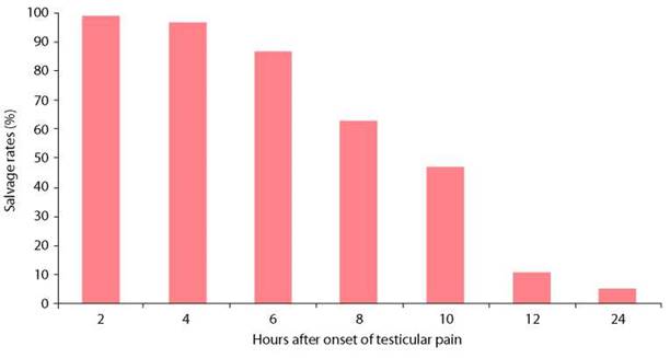
Figure 7.3 Salvage rates according to number of hours after onset of pain.
Q. The patient appears to have a clinical diagnosis of testicular torsion. What are the key features in the pre-operative consent for emergency scrotal exploration?
A. The informed consent for emergency scrotal exploration would involve a description of the procedure, discussion of alternative treatments and an explanation of potential complications. The following points would need to be raised [2]:
Bilateral testicular fixation (orchidopexy) in the case of torsion, where testis is viable Orchidectomy in the case of torsion, where testis is not viable, with orchidopexy of contralateral testicle
No fixation in the case where no torsion is found
If orchidopexy is performed, non-absorbable sutures may be palpable
Risk of haematoma, which may require surgical exploration
Risk of wound infection or orchitis
Long-term risks of testicular atrophy
No guarantee of fertility
Small risk of future torsion despite fixation
Q. What incision do you use? Describe your technique of fixation.
A. At scrotal exploration, although various skin incisions can be employed, including transverse, bilateral vertical and oblique, I use the midline incision through the median raphe.
The layers of the scrotum (skin, dartos, external spermatic fascia, cremasteric fascia, internal spermatic fascia, tunica vaginalis) are divided. The affected testis is delivered and inspected. Testicular torsion occurs inwards and towards the midline and in a case of torsion, the testis is initially untwisted. The testis is then wrapped in a warm saline-soaked swab and the anaesthetist supplies 100% oxygen, via the endotracheal tube.
If the testis is viable, I perform an orchidopexy using the three-point fixation technique. The testis is fixed medially, laterally and infero-anteriorly to the scrotal wall using nonabsorbable sutures (typically 3/0 or 4/0 Prolene). If the viability of the testis is questionable,
I make a small stab incision through the tunica albuginea to assess for evidence of viability through signs of bleeding. If the testicle is not salvageable, I perform an orchidectomy.
In a case of confirmed testicular torsion, I explore the contralateral testis, through the same incision, and perform a prophylactic three-point orchidopexy, to prevent future torsion on that side. This is supported by reports of contralateral torsion following unilateral orchidopexy and a 40% incidence of anatomical abnormalities predisposing to torsion in the contralateral testis. If an appendix testis is found at operation, I remove it to prevent future torsion of appendix testis mimicking testicular torsion.
Additional procedures have been proposed, namely eversion of the tunica vaginalis at the time of surgical exploration to prevent future re-torsion, as well as the use of a sub-dartos pouch.
In the case where no torsion is found, I do not carry out orchidopexy, due to the potential complications of needle trauma (including breach of the blood-testis barrier). The testis should be replaced intact. In addition, I do not perform contralateral exploration.
Q. Are you aware of any potential complications of testicular torsion?
A. Misdiagnosis, especially as an epididymo-orchitis, is the most common problem. If a patient shows no improvement, despite 48-72 hours of antibiotic therapy, the diagnosis of testicular torsion (dead testis) should be considered. An infarcted testis left in the scrotum, may result in abscess or sinus formation. The potential long-term complication of this event is the formation of antisperm antibodies, causing infertility in the contralateral testis. Other long-term complications include future torsion in a testis that has undergone previous inadequate prophylactic fixation. This risk may be minimised by performing orchidopexy in the contralateral testis when torsion is found. In the case where orchidectomy is carried out, a testicular prosthesis insertion may be considered, in the future, to improve cosmetic outcome and psychological recovery. In my practice, I do not insert the prosthesis at the time of emergency exploration, through the scrotal route, due to the significant risk of erosion.