
Davendra M Sharma, Iqbal S Shergill and Manit Arya
Q. A 26-year-old motorcyclist, travelling at 20 mph, collides with a car. The motorcyclist is thrown from his bike. You are called to accident and emergency (A&E) because he complains of left flank pain. His pulse is 90 beats/minute, blood pressure (BP) 120/80 mm Hg, respiratory rate (RR) 16, oxygen saturation 99%. How would you assess this patient?
A. As with all trauma cases, advanced trauma life support (ATLS) principles are strictly
followed. It is important to have a multidisciplinary approach, consulting with emergency, orthopaedic, general surgical and any other speciality doctors if necessary (standard answer to trauma question).
Traumatic injuries remain an important cause of mortality and morbidity in the civilian population. Trauma may be generally classified as due to blunt or penetrating injury. Blunt trauma, which is much more common in the United Kingdom, may result in significant genitourinary injury. Urologists are expected to understand the principles of trauma management and safely manage these patients.
Q. Your colleagues have ‘cleared’ this patient of other major injuries. What specific information would you like to have to assess him urologically?
A. I would take a focussed history and relevant examination (standard answer). It is important to elucidate the following points from the history:
1. The mechanism of injury
2. Whether there is any previous urological or renal history
3. Whether the patient has noticed any blood in the urine
Q. What examination findings are important?
A.
Extent and location of any bruising and tenderness.
Haematuria (macroscopic in adults or dipstick/macroscopic in children) - particularly in first sample following accident. I am aware that microscopic analysis (at least five red blood cells per high power field) is more reliable in assessing the extent of haemorrhage than dipstick testing. However, this is very rarely used in daily clinical practice.
Blood pressure (BP) - has there been a significant drop in BP, i.e. systolic <90 mm Hg, at any time since the accident?
Q. This patient has dipstick haematuria. What would you do?
A. If the patient has had a measured (even single) drop in his systolic BP (<90 mm Hg), then an urgent contrast spiral computed tomography (CT) scan is arranged. If there is no significant drop in BP recorded, the patient is admitted for analgesia, 24 hours of observation and repeat full blood count (FBC).
Q. In a stable adult patient with dipstick haematuria, are there any factors that would lower your threshold for imaging, and why?
A. Yes. If the mechanism of injury is that of a rapid deceleration injury or a fall from a
significant height, imaging is recommended. These patients may have a pelviureteric junction (PUJ) disruption or vascular injury which may not result in haematuria or a fall in BP. In fact haematuria (microscopic and macroscopic) may be absent in up to 40% of renal injuries and 25% of pedicle injuries.
Q. What are the indications for imaging (CT with contrast) in a stable patient following renal trauma as suggested by the European Association of Urology (EAU)?
A.
Penetrating trauma
Deceleration injuries (or other ‘significant’ mechanism of injury, e.g. fall from a considerable height)
Blunt trauma in adults associated with a systolic BP <90 mm Hg at any time following the injury
Blunt trauma in adults associated with frank haematuria (thus blunt trauma associated with dipstick/microscopic haematuria in adults is not an indication for imaging providing it is not a deceleration injury and BP has been stable)
Blunt trauma in children associated with dipstick/microscopic or frank haematuria - in children, a lower threshold for imaging (ideally spiral CT with contrast) is required as hypotension is a late manifestation of hypovolaemia
Q. What are the objectives of radiographic imaging in renal trauma and which modality best delivers these objectives?
A.
1. Accurately stage the injury
2. Document contralateral renal function
3. Recognise pre-existing renal pathology
4. Identify injuries to other organs
A contrast spiral CT scan is superior to all other forms of imaging in trauma. As well as delivering the objectives above, it is quick and now familiar to most surgeons. Ultrasound resolution is inferior to CT but it may be useful in the follow-up of renal injury.
Q. How is the CT scan performed and why?
A. Spiral CT scan with contrast:
1. Arterial and/or portal venous phase - Demonstrates vascular and parenchymal injury as well as haematoma
2. Delayed images (10-20 minutes) - PUJ or collecting system injury. May be omitted if, on the early phase scan, the kidneys are normal, and there is no perinephric, retroperitoneal, pelvic or perivesical fluid present.
Q. What do Figures 8.1 and 8.2 show? How would you stage the injury and what are the different grades? What are the essential components of the staging system?
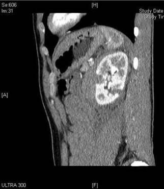
Figure 8.1 Sagittal contrast CT demonstrating grade 3 renal trauma.
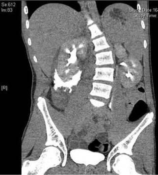
Figure 8.2 Coronal contrast CT demonstrating grade 4 renal trauma.
A. The AAST (American Association for the Surgery of Trauma) grading system is based primarily on the CT findings (Table 8.1). The essential components describe the presence of a renal haematoma, injury to the renal parenchyma, collecting system or the vasculature.
Table 8.1 The American Association for the Surgery ofTrauma (AAST) renal trauma severity scale
|
Grade (AAST) |
Type |
Description |
|
I |
Contusion |
|
|
Haematoma |
Subcapsular not expanding with no parenchymal laceration |
|
|
II |
Haematoma |
Not expanding perirenal haematoma confined to renal retroperitoneum |
|
Laceration |
< 1 cm parenchymal depth of renal cortex with no urinary extravasation |
|
|
III |
Laceration |
> 1 cm parenchymal depth of renal cortex with no urinary extravasation |
|
IV |
Laceration |
Parenchymal laceration through corticomedullary junction extending into collecting system |
|
Vascular |
Segmental renal artery or vein injury with contained haematoma, or partial vessel laceration; or vessel thrombosis |
|
|
V |
Laceration |
Completely shattered kidney |
|
Vascular |
Avulsion of renal pedicle or hilum which devascularises kidney |
Q. What proportion of renal trauma is due to blunt trauma?
A. In the United Kingdom the majority (over 95%) of injuries are due to blunt trauma. In South Africa and territories affected by violent conflict, the incidence of penetrating trauma is much higher.
Q. You are called to theatre later that night by the on-call trauma team. Another blunt trauma patient was taken to the emergency theatre as he was not able to maintain his BP despite fluid boluses. No imaging was performed prior to laparotomy, which reveals normal viscera and no intraperitoneal blood. There is, however, a large left retroperitoneal haematoma. What is your next step?
A. Arrange a one-shot on-table intravenous urography (IVU).
Q. Why do a one-shot IVU and how would this be performed?
A. Most importantly, it shows whether there are two functioning kidneys. Delayed contrast excretion or extravasation may also be detected. A normal film may obviate the need for renal exploration.
A one-shot IVU depends on a rapid bolus administration of 2 mL/kg contrast (e.g. Omnipaque). A single portable plain abdominal x-ray is performed at 10 minutes. Fluoroscopy (C-arm) results in poor images and should be avoided.
Q. What are the absolute indications for exploring the kidney in trauma?
A.
1. Persistent life-threatening blood loss believed to stem from renal injury
2. Renal pedicle avulsion (grade 5 injury) which is suspected clinically, by imaging or by the observation of an expanding pulsatile retroperitoneal haematoma at laparotomy
3. Penetrating renal trauma (most cases)
Q. What are the principles to exploring a kidney in trauma?
A.
A midline generous laparotomy incision from sternum to pubis.
The small bowel is lifted out of the peritoneal cavity in order to expose the retroperitoneum.
Incise peritoneum over aorta above the inferior mesenteric artery and dissect alongside it superiorly up to the left renal vein (if a large perirenal haematoma obscures the site for this incision then make incision medial to inferior mesenteric vein). The left and right renal arteries are then easily identified. Vessel loops can then be placed around the renal artery and vein thus establishing early vascular control.
The colon can now be reflected thus exposing the kidney (or haematoma).
If nephrectomy is avoided then renal tissue is preserved by controlling bleeding and debriding all non-viable tissue.
Q. How would you deal with a non-expanding, non-pulsatile haematoma at laparotomy?
A. In this situation haematoma should be left alone. This is so as exploration increases the probability of loss of the kidney because of bleeding, which can be controlled only by nephrectomy.
Q. What proportion of patients suffering blunt trauma to the kidneys require surgical intervention?
A. Less than 5%.
Q. What is the role of angioembolisation?
A. Selective renal artery angioembolisation is increasingly used to successfully manage stable patients with haemorrhage following blunt and penetrating trauma.
Q. What are the potential complications of conservatively managed renal trauma and how are they managed?
A. Early:
Secondary haemorrhage requiring radiological or surgical intervention.
Urinary extravasation leading to urinoma (or if superimposed infection to perinephric abscess formation). Radiological drainage with or without internal stenting may be necessary.
Infection, e.g. perinephric abscess or systemic sepsis.
Vascular complications including arterio-venous fistula (AVF) or pseudoaneurysm formation.
Late:
Hypertension - Re-evaluate to look for renal artery thrombosis, subcapsular haematoma, extensive fibrosis (Page kidney is hypertension due to scar formation) or AVF.
Renal insufficiency - Follow-up DMSA scan to look for significant functional loss.
Calculus formation and chronic pyelonephritis.
It is recommended that higher-grade renal injuries (4 and 5) are imaged by CT 48-72 hours after injury to look for complications such as secondary haemorrhage or urinoma formation.
Q. How are renovascular injuries managed?
A.
Arterial - Irreversible damage ensues after 2-6 hours so most kidneys will not function following arterial injury. Reconstruction should be attempted in solitary kidneys, bilateral renal injury or if diagnosed very quickly. Incomplete injury, e.g. intimal flaps, can be managed conservatively. Segmental arterial injuries are managed with angioembolisation. Endovascular techniques are also described for main artery and branch injuries and may take the primary role in the future.
Hypertension develops in a small subset of patients with major arterial injury - elective nephrectomy may be necessary in these cases.
Venous injuries are rare and difficult to identify - Avulsion from the inferior vena cava (IVC) following blunt trauma requires urgent laparotomy, IVC repair and nephrectomy. The left renal vein may be tied leaving the kidney to drain from the gonadal and adrenal veins. Penetrating injuries should be repaired.
Q. What does the image show in Figure 8.3?
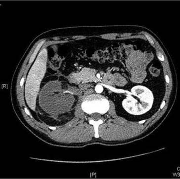
Figure 8.3
A. Figure 8.3 is a contrast CT scan (arterial phase) showing right arterial injury with absence of right nephrogram.
Q. Figure 8.4 is an investigation of this previously discussed patient 3 months later. What does this demonstrate?
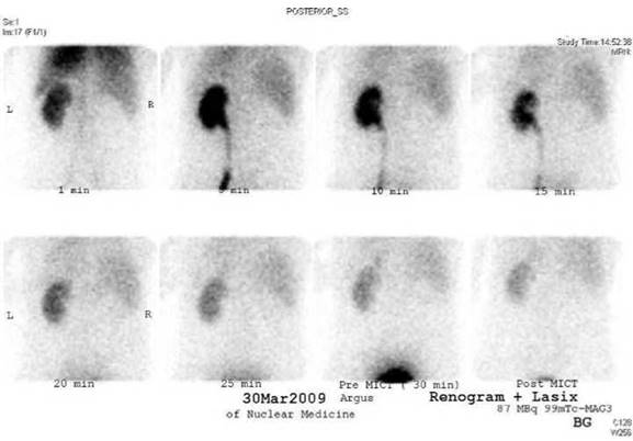
Figure 8.4
A. Figure 8.4 is a 99mTc MAG-3 renogram showing absence of right renal function.
Q. Can you summarise the management of blunt renal trauma?
A. I use the management algorithm in Figure 8.5, which is based on the EAU guidelines, for managing blunt renal trauma.
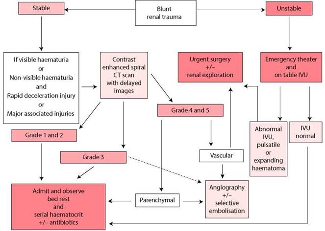
Figure 8.5 Management of blunt renal trauma in adults (EAU guidelines).
Q. Can you summarise the management of penetrating renal trauma?
A. I use the management algorithm in Figure 8.6, which is based on the EAU guidelines, for managing penetrating renal trauma.