
Q. A 47-year-old man is in the accident and emergency (A&E) with high fever and severe right loin pain. How will you manage this case?
A. This is a urological emergency and I would see the patient myself, in A&E without delay.
On arrival, I would resuscitate him using basic principles of advanced life support. I would administer 100% high-flow oxygen, insert two large-bore Venflons, taking blood for FBC, U&Es, C-reactive protein (CRP), clotting and blood cultures. In addition, I would ensure that he is catheterised and a catheter specimen of urine (or a prior MSU) is sent for urine dipstick and subsequent microscopy and C and S. The patient should be provided with adequate analgesia.
I would then take a general and focused urological history asking specifically for a history of dysuria, haematuria, recent urological intervention or a prior history of stones. I would also ask questions pertaining to a non-urological cause (gastrointestinal or respiratory symptoms). It is important to establish the patient’s immune status (diabetes, steroid use, HIV).
Upon examination, I would record the patient’s mental state, pulse, blood pressure (BP), temperature, respiration and O2 saturation. A full and thorough examination is then required to look for a septic focus including chest, abdomen, loins, genitalia and rectal examination (prostatic tenderness).
After ensuring that he is appropriately resuscitated with IV fluids, I would commence the patient on broad-spectrum IV antibiotics. In my practice, I initially give gentamicin (3-7 mg/kg) together with a second-generation cephalosporin. Subsequent antibiotic treatment is reviewed according to culture results, microbiology advice and the clinical situation. Clearly, these patients are admitted for inpatient parenteral antibiotic treatment and observation. In my practice, I seek the early assistance of the microbiologist and if necessary high-dependency unit/intensive therapy unit (HDU/ITU) anaesthetic colleagues, to help in the multidisciplinary management of these patients.
Q. What is systemic inflammatory response syndrome (SIRS)?
A. Response to a variety of infectious (sepsis) or non-infectious (burns, pancreatitis) stimuli.
Two of the criteria listed in Table 9.3 are required.
Table 9.3 Criteria for diagnosis of systemic inflammatory response syndrome
|
T° |
>38° or <36° |
|
HR |
>90/min |
|
RR |
>20/min or paCO2 <32 mm Hg (<4.3 kPa) or need for mechanical ventilation |
|
WCC |
<4000 or > 12,000 cells/mm3 |
Q. What is the definition of sepsis?
A. Sepsis is defined as proven infection causing SIRS.
Q. What is the definition of severe sepsis?
A. Severe sepsis is defined as sepsis with evidence of organ dysfunction, e.g. confusion, lactic acidosis and oliguria.
Q. What is the definition of septic shock?
A. Septic shock is defined as sepsis-induced hypotension (systolic BP <90 mm Hg) persisting despite adequate fluid resuscitation.
Q. What is the definition of refractory septic shock?
A. Refractory septic shock is defined as septic shock lasting for >1 hour and not responding to fluid administration or pharmacological intervention.
Q. What is your understanding of the pathogenesis of sepsis?
A. The major pathogenesis of sepsis is secondary to the presence of endotoxins released by Gram-negative bacteria in the circulation. This in turn results in a cascade of events resulting in release of mediators such as TNFa, interleukins (IL-2, IL-6, IL-8) from target cells (e.g. neutrophils, macrophages, lymphocytes and plasma cells) and activation of the kinin, complement and fibrinolytic systems. These events result in widespread microvascular injury, tissue ischaemia and clinical manifestations of sepsis.
Q. Unfortunately, in spite of your initial management the patient’s BP is falling. What would you now do?
A. I would acknowledge that this patient is very unwell. In order to manage this patient
effectively, the further treatment of this patient should take place in an ITU setting and will include consideration of vasopressors, inotropes, steroids and recombinant human activated protein C. Once the patient is more haemodynamically stable, radiological investigations should be organised to identify the source of sepsis and steps taken to eradicate it.
Q. What imaging would you like to arrange?
A. I would initially request an ultrasound scan of the renal tract.
Q. Ultrasound shows strong focal echoes in the right renal parenchyma without acoustic shadowing. What would you do now?
A. This is not diagnostic, although hydronephrosis (infected obstructed kidney) has been excluded, I would There fore organise a CT scan of the abdomen with contrast, providing there are no contraindications (such as renal failure).
Q. This is the CT scan (Figure 9.2). Can you describe this? What is the likely diagnosis?
A. Figure 9.2 is a CT scan of abdomen showing air fluid level and debris in the right renal pelvis suggestive of an emphysematous pyelonephritis (EPN).
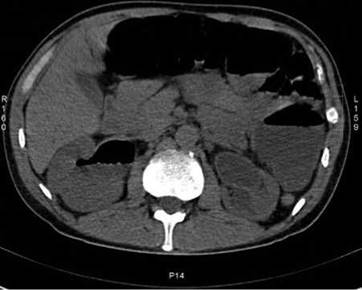
Figure 9.2 Sagittal CT scan of the abdomen.
Q. What is EPN?
A. It is an acute necrotizing parenchymal and perirenal infection caused by gas-forming organisms. It commonly occurs in diabetics. Other associated conditions may be urinary tract obstruction, stones and impaired immunity.
Q. What is this condition commonly caused by?
A. The most common organisms isolated include E. coli and Klebsiella. However, it may also be associated with Proteus, Pseudomonas and Streptococcus infection.
Q. What is the pathogenesis of EPN?
A. Unfortunately, this is a poorly understood process. The possible mechanisms leading to accumulation of gas include a mixed acid fermentation of glucose by bacteria and an overwhelming local inflammation coupled with slow transport of the end products due to diabetic microangiopathy. The analysis of accumulated gas has shown to include nitrogen, hydrogen, carbon dioxide and oxygen.
Q. Are you aware of any different types of EPN and what is the significance?
A. EPN may be classified on the basis of the CT appearance into type I and type II. Type I is classically characterised by gross parenchymal destruction with either absence of fluid collection or presence of streaky or mottled gas radiating from the medulla to the cortex. Type II EPN is associated with a confined, bubbly or loculated intrarenal gas pattern, presence of renal or perirenal fluid or gas within the collecting system. The differentiation is important as type I EPN is associated with a higher mortality rate of up to 60% versus around 20% for type II.
Q. Four days after treatment in the ITU, the patient is still not better. What can you do next?
A. In this patient, the CT does not show any evidence of obstruction or a large perirenal fluid collection; There fore, the next step would be a nephrectomy. The functional status of the contralateral kidney will determine the involvement of nephrologists. In the presence of obstruction or perirenal fluid collection, percutaneous drainage may stabilize the patient, allowing complete urological workup and deferred nephrectomy.
Q. Figure 9.3 is a CT scan from a different patient whose symptoms have not resolved with conservative treatment. What does it show?
A. This is a CT scan with contrast showing a large right-sided collection in and around the kidney. The collection distorts and enlarges the renal contour, infiltrates perinephric fat and extends into the psoas muscle. The normal renal collecting system fat has been obliterated by the process. This is highly indicative of a perinephric abscess.
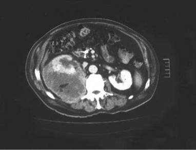
Figure 9.3 Cross sectional slice of CT scan of the abdomen at the level of the kidneys.
Q. What is your management plan?
A. Surgical drainage, or nephrectomy if the kidney is non-functioning or severely infected, is the classic treatment. However, more recently renal ultrasound and CT make percutaneous aspiration and drainage of small perirenal collections possible. In this case, I would consider percutaneous drainage to be contraindicated as it is a large abscess cavity which is likely to be filled with thick, purulent fluid. Moreover, percutaneous drainage may be more appropriate if this was a single kidney or single functioning kidney.
Q. How would you differentiate a case of acute pyelonephritis clinically from a perinephric abscess?
A. The literature suggests that most patients with uncomplicated pyelonephritis are symptomatic for less than 5 days before hospitalization, whereas most with perinephric abscesses are symptomatic for longer than 5 days. Moreover, patients with acute pyelonephritis do not usually remain febrile for longer than 4 days once appropriate antimicrobial agents are started. In contrast, patients with a perinephric abscess often have fevers for on average 7 days. tterefore, a perinephric abscess should be suspected in patients with abdominal pain or a flank mass with a persistent fever after 4 days of antimicrobial therapy.
Q. What is a perinephric abscess caused by?
A. The most common causes of a perinephric abscess are secondary to the rupture of an acute cortical abscess into the perinephric space or from haematogenous spread from other sites of infection (e.g. skin). When patients develop an obstructed infected kidney (pyonephrosis) secondary to an obstructing stone, they are at high risk of developing a perinephric abscess if the obstruction is not relived early. This is especially true in patients with immunosuppression and diabetes mellitus (30% of patients). Rarer causes include secondary infection of a perirenal haematoma or infection from nearby structures (bowel perforation, Crohn’s disease, osteomyelitis from the thoracolumbar spine).
Q. Figure 9.4 is a CT scan from a different patient with a history of fever, rigors and right flank pain. Can you describe the findings and what is the likely diagnosis?

Figure 9.4 Sagittal CT scan of the abdomen demonstrating the renal tract.
A. This demonstrates a coronal reconstruction of a CT scan of the abdomen showing an expanded non-enhancing right kidney invading into the surrounding tissues and a stone within it suggesting a xanthogranulomatous pyelonephritis (XGP).
Q. What is XGP?
A. It is a severe chronic renal infection resulting in diffuse parenchymal destruction and an enlarged non-functioning kidney usually associated with calculi.
Q. What are the common organisms?
A. XGP is most commonly associated with infection with either E. coli or Proteus species. However, Pseudomonas and Klebsiella infection is also frequently demonstrated.
Q. What is the pathology?
A. Under the microscope, the pathology seen in cases of XGP is described as diffuse infiltration of inflammatory cells, e.g. lymphocytes, giant cells and plasma cells within the kidney.
The characteristic finding is the presence of xanthoma cells, which are lipid-laden foamy macrophages.
Q. What is the usual course of events in this condition?
A. If a patient is systemically unwell, the patient is usually admitted to the HDU/ITU and requires intensive treatment for sepsis. On subsequent imaging, the distinction from cancer may be difficult and a nephrectomy is usually performed. If nuclear medicine function scans such as dimercapto-succinic acid are performed these often demonstrate that the kidney is associated with very poor or no function. Moreover, the formal diagnosis is often delayed until the post-operative histology is available.
Q. How will you perform a nephrectomy in this patient?
A. Performing a nephrectomy in a patient with XGP is usually technically very challenging as there is usually extension of the inflammatory process into the retroperitoneum. Furthermore, the inflammation results in tissue planes being very stuck and difficult to dissect. As a result, I believe that an open nephrectomy is more suitable than a laparoscopic approach as there is a high risk of injury to the blood vessels, bowel and other neighbouring organs due to the difficult tissue planes.