
Q. Please describe the motor innervation to the lower urinary tract.
A. The lower urinary tract (LUT) receives innervation from both the parasympathetic and the sympathetic branches of the autonomic nervous system. The parasympathetic preganglionic fibres are located in S2-4 spinal segments and these synapse with postganglionic cell bodies lying within the detrusor muscle. These parasympathetic nerves provide cholinergic excitatory input to bladder smooth muscle resulting in detrusor contraction. However, parasympathetic innervation of the outflow tract exerts an inhibitory effect resulting in relaxation of the bladder neck and urethra.
The sympathetic cell bodies are located in spinal segments T10-T12 and L1-L2. The preganglionic fibres synapse with postganglionic fibres in the hypogastric plexus. The predominant effect of the sympathetic innervation is inhibition of the parasympathetic pathways thus providing an inhibitory control on detrusor contraction. Additionally, sympathetic innervation results in contraction of the outflow tract (in males by stimulating contraction of the pre-prostatic sphincter and in females there is some, although relatively sparse, sympathetic innervation to the bladder neck).
The somatic nerve supply to the pelvic floor musculature and the external urethral rhabdosphincter originates from S2-S4 and is conveyed peripherally via the pudendal nerves. The cell bodies of the axons that innervate the external urethral rhabdosphincter lie in a distinct, medially placed motor nucleus at the same spinal level, called Onuf’s nucleus.
Q. What is the sensory innervation of the bladder?
A. Sensory nerves have been identified in the suburothelial layer as well as in the detrusor muscle. This suburothelial plexus is particularly prominent at the bladder neck and relatively sparse at the dome of the bladder.
Sensations of bladder fullness are conveyed to the spinal cord in the pelvic and hypogastric nerves. The afferent components of these nerves contain myelinated (A8) and unmyelinated (C) axons. While the A6 fibres respond to passive distension and active contraction and thus convey information about bladder filling, the C-fibres respond primarily to noxious stimuli such as chemical irritation of the urothelium or cooling. The cell bodies of both these classes of axons are located in the dorsal root ganglia (DRG) at the level of S2-S3 and T11-L2 spinal segments. Bladder afferent activity enters the spinal cord through the dorsal horn and ascends rostrally to higher brain centres involved in bladder control, i.e. The pontine micturition centre and then on to cerebral cortex.
Afferent fibres originating from the trigone and urethra run in the hypogastric and pudendal nerves, respectively.
Q. Please describe the micturition cycle.
A. The micturition cycle comprises filling and voiding phases:
During the filling phase the intravesical pressure is kept low by the phenomenon of receptive relaxation. The extent to which a change in volume occurs as related to a change in pressure is known as bladder compliance (i.e. compliance = change in volume/ change in pressure). Factors which contribute to bladder compliance are the vesicoelastic properties of the bladder and also the ability of detrusor smooth muscle cells to increase in length without significant increase in tension.
During bladder filling, afferent activity from stretch receptors passes to the pons and cerebral cortex. If voiding is not to be initiated, activity within the external urethral rhabdosphincter is increased. Additionally, central inhibition decreases parasympathetic activity to the detrusor. Detrusor contraction is prevented by the ‘gating mechanism’. This is inhibitory influence of interneurons via the sympathetics to prevent the transmission of afferent activity. This prevents the transmission of activity from preganglionic to postganglionic parasympathetic efferent neurons.
When appropriate, voiding is initiated. The voiding phase begins first with relaxation of the external urethral sphincter followed by contraction of the detrusor muscle. Micturition is coordinated by Barrington’s nucleus in the pons. The afferents from the bladder travel via parasympathetic nerves to the periaqueductal gray matter (PAG) in the pons. The PAG and cerebral cortical areas decide if it is appropriate to void. If so, the pontine micturition centre relays impulses resulting in external sphincter relaxation, and urine enters the posterior urethra. It also sends direct signals to the detrusor parasympathetics to initiate contraction.
If this co-ordination is lost as in a suprasacral type of SCI, then the patient develops detrusor sphincter dyssynergia (i.e. uncoordinated contraction of the detrusor and external sphincter).
Q. What are the main urological characteristics of suprapontine, suprasacral and conus (S1- S5)/cauda equina/peripheral nerve (lower motor neurone) lesions?
A.
Suprapontine lesions (e.g. cerebrovascular accident [CVA], Parkinson’s disease) - In these the micturition reflexes are intact. Following a CVA, voiding at inappropriate times occurs (although voiding itself is normal) in addition to detrusor overactivity (latter also occurs in Parkinson’s disease). These are ‘safe’ low-pressure bladders.
Suprasacral spinal cord injuries/lesions (i.e. lesions between the pons and spinal cord segment L5) - These are characterised by neurogenic detrusor overactivity (NDO), detrusor sphincter dyssynergia (DSD; synchronous contraction of the detrusor and external urethral sphincter) and low-compliance bladders. In lesions above T6 autonomic dysreflexia may be a significant problem (see later discussion). Unlike suprapontine and conus/cauda equine/ peripheral nerve lower motor lesions, suprasacral injuries can result in ‘unsafe’ high- pressure bladders, as upper tract damage can result (due to DSD and low compliance).
Conus (S1-S5)/cauda equina/peripheral nerve lesions - These tend to lead to a lower motor neurone type of injury and are characterised by an acontractile (areflexic) bladder with urethral sphincter weakness (leading to stress incontinence) (possible low compliance may also occur). The bladders tend to be ‘safe’ low-pressure bladders.
Q. What is detrusor sphincter dyssynergia (DSD)?
A. It is defined as involuntary contraction of the urethral and/or periurethral striated muscle simultaneously with a detrusor contraction. This usually is specific to a suprasacral neurological disorder.
Q. What do you know about detrusor leak point pressure (DLPP) and abdominal leak point pressure (ALPP)?
A.
The term DLPP must only be used in those with a neurological disorder/injury, i.e. those with a neuropathic bladder. DLPP is the lowest detrusor pressure at which urine leakage occurs in the absence of either a detrusor contraction or increased abdominal pressure. McGuire observed, in spina bifida patients, that if the DLPP is greater than 40 cm H2O then there is a significant risk of damage to the upper tracts [11].
The ALPP (also called the Valsalva leak point pressure) is terminology used in nonneuropathic females as related to stress incontinence. It is the intravesical pressure at which urine leakage occurs due to increased abdominal pressure in the absence of a detrusor contraction. If the ALPP is <60 cm H2O then stress incontinence is likely to be due to intrinsic sphincter deficiency. If the ALPP is >90 cm H2O, stress incontinence is likely to be due to urethral hypermobility. An ALPP between 60 and 90 cm H2O is an equivocal result. If the ALPP is >150 cm H2O, then the urethra is unlikely to be the cause of urinary incontinence.
Q. A 28-year-old man sustained a T5 spinal cord injury (SCI) a year ago. He can walk and has been emptying his bladder by strain voiding and complains of recurrent UTIs and urinary leakages. What type of injury has he got?
A. He has sustained a suprasacral type of injury and is likely to have neurogenic detrusor
overactivity, poorly sustained bladder contractions, DSD, low bladder compliance and reflex bladder voiding. As the lesion is above T6 he may also suffer from autonomic dysreflexia (see later discussion).
Q. What is the most important investigation in this patient, which will help you in management?
A. Video-urodynamic studies.
Q. What are your indications for urodynamics in general?
A. These include the following:
Patients with persistent LUTs after appropriate therapy
Patients with previous failed incontinence surgery
Patients with mixed urinary symptoms ± incontinence
Any patient with suspicion of neurological disease and urinary symptoms
Patients in whom potential therapy may be hazardous
Children with complex voiding dysfunction
Q. What do Figure 11.6a and b show?
A. Figure 11.6a demonstrates a classical DSD trace with a saw-toothed appearance on the Pdet line (also sustained detrusor contraction lasting for more than 5 minutes, with Pdet pressures of 80-90 cm H2O).
Figure 11.6b is cystography performed during a VCMG investigation. It shows a hold-up of contrast at the level of the external urethral sphincter, typical of DSD.
Q. How would you treat this patient?
A. The aim is to achieve low-pressure storage and complete bladder emptying while promoting continence.
I would be guided by the urodynamics result but assuming that this patient with T6 SCI has NDO and DSD, I would start him on anticholinergic medication and institute a program of clean intermittent self-catheterisation (CISC). He will be closely monitored and will undergo ultrasound scan of the kidneys and repeat urodynamics in 3-6 months’ time to ensure the bladder pressures have come down. Assuming that there are no problems, i.e. UTIs or problems with CISC, he will be reviewed on an annual basis.
Q. The previously discussed patient returns and states he does not like performing CISC and would like to know about other options. Could you explain these to him?
A. Aside from CISC and the Credé manoeuvre (that he was practicing) the other options include
Behavioural and timed voiding - This is not suitable for him.
Intradetrusor botulinum toxin injections - This would, however, be unsuitable as he would continue to have to perform CISC.
Indwelling catheter (suprapubic or urethral) - Not a good option for him as he is very young and mobile.
Urethral stents or external sphincterotomy (with a subsequent convene sheath) - Both of these treat DSD (the urethral stent is placed across the external urethral sphincter thus holding it open) but the patient will be completely incontinent afterwards. Thus as he is young and mobile these are not a good option for him.
Augmentation cystoplasty (see preceding section on Overactive Bladder) with or without a Mitrofanoff (catheterisable channel) - However, following this procedure he will almost certainly need to perform CISC.
Sacral anterior nerve root stimulator (SARS) with dorsal rhizotomy - Not suitable for him as he is walking and has an incomplete SCI. This is an option for wheelchair-bound patients with a complete SCI.
* A video-urodynamic study (videocystometrogram [VCMG] - synchronous cystography and cystometry recordings) rather than a CMG is more appropriate in these cases, as this allows a combined evaluation of the anatomy and function of the lower urinary tract.
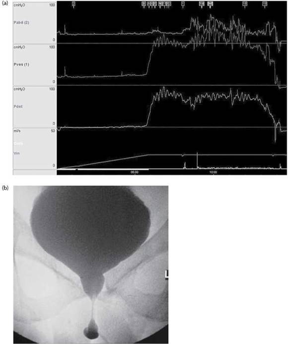
Figure 11.6 (a) Urodynamics trace. The ‘saw-tooth’ appearance on the detrusor pressure line is classically seen with detrusor-sphincter-dyssynergia (DSD). (b) Cystography at the same time as the VCMG demonstrates a hold up of contrast at the level of the external urethral sphincter, typical of DSD.
Q. What are the advantages and disadvantages of a SARS? (This is currently combined with a sacral dorsal rhizotomy.)
A. The benefits include the following:
Abolition of reflex bladder
Increased bladder capacity
Abolition of autonomic dysreflexia
Improved bowel management
However the disadvantages are as follows:
Stress incontinence
Loss of reflex erections
Loss of reflex ejaculation
Loss of reflex defaecation
Q. What are the complications of long-term catheters?
A.
Recurrent UTIs
Blockages
Need for regular changes
Stones
Risk of cancer
Q. What are the complications of urethral stents to treat DSD in his case?
A. He will be completely incontinent (as the urethral stent is placed across the external urethral sphincter to hold it open) and will have to wear a sheath with risk of detachment. The urethral stents can migrate, block or become encrusted. They are generally reserved for immobile patients as is external sphincterotomy, which is irreversible.
Q. What is the success rate of botulinum toxin injections in NDO?
A. It is 70%-90% with effects lasting an average of 9 months.
Q. What is autonomic dysreflexia (AD)?
A. This is a medical emergency. It occurs only in patients with a SCI above T6 level (the sympathetic outflow). It is more common in cervical injuries compared to thoracic. It is a result of acute massive disordered autonomic (primarily sympathetic) response to specific stimuli. If left untreated it can lead to convulsions, cerebral haemorrhage and death.
Q. What are the causes of AD?
A. Autonomic dysreflexia can be triggered by many potential causes. Essentially any noxious stimuli below the level of SCI can cause AD. Bladder distension/irritation is responsible in 75%-85% of cases. The second most common cause is bowel distension, usually due to faecal impaction which accounts for 13%-19% of cases [12]. Other causes may include urological interventions (e.g. catheterisation, urodynamics, cystoscopy), UTI and urinary calculi. Non-urological causes include constipation, pressure sores, fractures, ingrown toenails and distal skin infections.
Q. What is the mechanism of AD?
A. Any of the above trigger conditions can cause sympathetic discharges, these results in reflex vasoconstriction leading to systemic hypertension. The carotid body detects this rise in blood pressure, reflex vagal discharges cause vasodilatation and bradycardia as a normal homeostatic response to try to lower blood pressure. However this neural signal cannot cross the level of the injury, vasoconstriction persists below the level of the lesion. The hypertension persists as the compensatory mechanisms are ineffective. Vasodilatation above the SCI level causes profuse sweating and flushing. Vasoconstriction below the level of the injury causes pale clammy skin.
Q. What are the symptoms and signs of AD?
A. A patient with autonomic dysreflexia may complain of headaches, blurred vision, nasal congestion and blotchy skin above the level of spinal cord injury.
Patients during an episode of autonomic dysreflexia may have
A significant rise in systolic and diastolic blood pressure
Profuse sweating above the level of the lesion, usually seen in the face, neck and shoulders
Flushing of the skin above the level of the lesion
Pale skin and goosebumps below the level of the lesion
In advanced untreated cases - this can result in convulsions, intracranial bleeds, hypertensive encephalopathy and ultimately death
Q. What is the treatment of AD?
A. AD is a life-threatening condition. Management includes
Prompt recognition of the condition
Identification and treatment of the precipitating cause (e.g. drainage of the bladder, evacuation of the bowel)
Sitting the patient upright (this induces an orthostatic hypotension allowing gravitational pooling of blood in the lower extremities causing a resultant drop in blood pressure) Loosening the patient’s clothing and any constrictive devices Administering sublingual glyceryl trinitrate (GTN)
Consider immediate-release nifedipine
Administering IV labetalol/phentolamine
Continuing to monitor the patient’s blood pressure after the episode to make sure that it has truly settled and not just dropped as a result of antihypertensive medication
Q. How would you manage a patient with a conus (S1-S5) SCI?
A. He has a generally safe bladder (see previous discussion) and the options include the following:
Behavioural and timed voiding
Emptying by straining if bladder has good capacity
CISC if incomplete voiding or complains of UTIs
If he complains of (stress) urinary incontinence then
Sheaths
Bulking agents
Tapes/slings
Artificial urinary sphincter (AUS)