
Q. A 43-year-old man is referred by his GP with a 2-month history of penile pain, penile deviation and a subcutaneous lump on the dorsum of the penis. How will you assess him?
A. I would take a detailed subjective history, including the duration of symptoms, the presence or absence of pain, degree of erectile function, amount of curvature (degree and direction), ability to penetrate and any previous treatment modalities. I would use a validated questionnaire such as the IIEF, to assess erectile function. I would ask specifically about any risk factors for ED, such as diabetes, hypertension and hyperlipidaemia and for a past history of penile trauma.
In the physical examination, I would note the size and location of the plaque/mass, the presence or absence of a foreskin and whether there were any signs of previous trauma. I would specifically measure the penile length in both the stretched and flaccid state. Unless the patient has brought in a good-quality digital photograph showing the degree and direction of the curvature, I would perform an artificial erection using an intracavernosal injection of PGE-1. I would then perform an examination of the extremities to identify any co-existing Dupuytren’s contracture.
Q. What is the diagnosis in Figure 13.9?
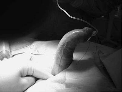
Figure 13.9
A. Figure 13.9 shows an image of a penile curvature due to Peyronie’s disease.
Q. What are the cardinal features of this disease?
A. It is characterized by the development of a fibrous plaque or scar tissue within the tunica albuginea of the penis. It can present with one or a combination of symptoms, such as penile curvature, indentation, buckling, penile pain and penile shortening. It can also result in erectile dysfunction (ED) due to the altered hemodynamics of cavernosal blood flow.
Q. What is it caused by?
A. It is not known as to the exact underlying cause. However, the most common hypothesis is that recurrent micro-trauma of the tunica albuginea during sexual intercourse leads to a small subtunical bleed that activates the processes of wound healing and the eventual development of a fibrotic plaque. Transforming growth factor beta (TGF-(31) seems to have an important role in this process as it is overexpressed in the plaque.
Q. Is Peyronie’s known to be associated with any other diseases?
A. Up to 30%-40% of men with Peyronie’s disease will also have a Dupuytren’s contracture.
Also associated with Peyronie’s disease are plantar fascial contracture (Ledderhose’s disease), tympanosclerosis and post-penile trauma.
Q. Which cytokines are implicated in the pathogenesis of Peyronie’s disease?
A. There appears to be an imbalance between pro- and antifibrotic cytokines. Research suggests an overexpression of TGF-|31 in penile plaques. Furthermore, fibrin and plasminogen activator inhibitor-1 (PAI-1) levels have also been shown to be increased in these plaques.
Q. How common is this condition and what is the natural history?
A. It has a prevalence of 0.4%-3.2% and usually occurs in men between 40 and 70 years of age. Managed conservatively 14% of patients have complete, spontaneous resolution and 40% of patients experience progression of the disease within 1 year.
Q. What disease stages are you aware of?
A. ttere are two stages of the disease. Initially, a third of patients present with painful erections during the acute phase. This period can also be characterized by worsening deformity of the penis. The second stage of PD is characterized by a stabilization of the deformity. Pains with erections generally subside as the chronic phase begins.
Q. Does the patient require any further evaluation?
A. If the history and examination are characteristic then no further evaluation is required. However, objective evaluation may include penile Doppler ultrasonography after the administration of an intracavernosal injection of a vasoactive agent to stimulate an erection. This test is also useful in cases where patients have erectile dysfunction with Peyronie’s disease and also if surgery using grafts is to be performed. A magnetic resonance imaging (MRI) scan of the penis with an artificial erection can also demonstrate the presence of hourglass deformities and any hinge defects that may lead to buckling with axial loading and allows visualisation of the amount of muscle within the corpus cavernosum.
Q. Are there any medical treatments available?
A. ttere are several options available for the medical management of Peyronie’s disease including oral medical therapy, topical and intralesional injection therapy, extracorporeal shockwave therapy (ESWL) and iontophoresis. Many of these therapies have not undergone rigorous evaluation in controlled clinical trials with most of the relevant studies being retrospective and provide little more than anecdotal evidence to support the use of the agents studied.
the oral medical therapies that have been investigated include vitamin E, tamoxifen, para-aminobenzoate (POTABA), colchicines, L-arginine and pentoxifylline. Intralesional agents include steroids, verapamil, interferons and more recently collagenase (collagenase clostridium histolyticum [Xiapex]).
Q. The patient says that he has already had ESWL for renal stones in the past. He was impressed with the results of this. He would like to have ESWL for Peyronie’s. Why was ESWL used for Peyronie’s and what are the success rates?
A. ESWL was used because it was thought that initiating an inflammatory reaction through direct damage to the plaque would result in plaque resorption. However, multiple randomized studies designed to evaluate the efficacy of shockwave therapy have failed to show any improvements in curvature or plaque size. Furthermore NICE guidelines in the United Kingdom do not recommend it unless the patient has been informed of the low success rate and appropriate arrangements for audit and research are in place.
Q. He decides on no treatment for the present time. The patient returns 9 months later. The pain has settled. The curvature is stable at 45° dorsally. What is important in the history?
A. Whether the patient has ED and whether he can have penetrative sexual intercourse.
Q. He is unable to penetrate and has no ED. What are his options?
A. His options are either to treat conservatively by trying a vacuum erection device or traction therapy, injection therapy with Xiapex or undergoing a surgical penile straightening procedure.
Q. Which patients should undergo surgery for Peyronie’s disease (PD)?
A. Surgical treatment is indicated in patients with stable PD and erections which allow penetrative intercourse with or without pharmacological agents. Problematic angulations or a hinge deformity causing difficult penetration are indications for surgery.
Q. Which surgical straightening procedures are you aware of?
A. The surgical treatment involves either penile shortening or potentially penile lengthening procedures. Penile shortening procedures involve plication techniques, such as the Nesbit procedure or the 16 dot plication. Penile lengthening procedures involve excision or incision of the Peyronie’s plaque with the subsequent defect being filled by a graft.
Q. Which patients are candidates for a plication technique?
A. Penile plication procedures are typically employed in patients with normal erectile function and a mild-to-moderate curvature (<60°). Also, these patients should have no hourglass deformities or destabilizing hinge effect.
Q. What types of plication technique are you aware of?
A. Penile plication involves shortening the convex side of the penis. Patients need to be thoroughly counselled regarding the expected postoperative penile shortening.
There are several methods for performing the plication.
1. Nesbit - Involves excising an elliptical portion of the tunica albuginea on the convex side. Success rate - 82% penis straight, all had penile shortening and 1.2% developed postoperative ED [5].
2. Yachia procedure - No excision of tunica, multiple longitudinal incisions in the tunica closed horizontally straightening the penis.
3. Essed and Schroeder - No excision or incision of the tunica albuginea. Plicating sutures are simply placed on the convex side of the curvature.
Q. He is concerned about penile shortening. What else is available?
A. An alternative to plication procedures involves extending or lengthening the concave side of the curvature. This is generally performed by incising the plaque with placement of graft over the defect in the tunica albuginea. Regardless of the shape of the incision, the goal is to completely relax the tunica and to cover the tunical defect with graft material.
Q. Should the plaque be incised or excised?
A. It is not recommended that the plaque be completely excised as this may compromise the veno-occlusive mechanism and increase the risk for postoperative ED.
Q. How would you counsel them with respect to a plaque incision and grafting procedure?
A. After making sure that the operation is appropriate for them
I would inform the patient of the success rate (straight penis 86%) and of any important potential risks (bleeding, bruising, infection, need for a circumcision, loss of length >1 cm in 26%, erectile dysfunction 15%, glans hypoaesthesia). Some surgeons recommend penile traction devices postoperatively, as well as penile rehabilitation with PDE-5 inhibitors.
Q. What types of graft have been used?
A. They can be divided into autografts (tunica vaginalis, fascia lata), allografts (cadaveric pericardium), xenografts (porcine small intestine submucosa) and synthetic grafts (Dacron mesh, polytetrafluoroethylene). No graft has been shown to be superior. For example the results using fascia lata graft are similar to that with autologous vein.
Q. By the time he returns for surgery he now complains of severe ED. Will you still go ahead with surgery?
A. After counselling, I would cancel his operation and then discuss alternative options. If he responds to pharmacotherapies (oral or intracavernosal) then a plication procedure can be performed provided that the patient accepts that pharmacotherapy is likely to be required post-operatively. However in the presence of severe ED unresponsive to pharmacotherapy then the option of a penile prosthesis insertion should be discussed. This is indicated for patients with Peyronie’s and concomitant severe ED.
Q. Do you know how to straighten the penis with severe angulations of a patient who is undergoing inflatable penile prosthesis insertion?
A. After insertion and inflation of the penile prosthesis the angulation is checked. If this is mild (<30°) then the curvature will resolve as the device is cycled over a 6-month period. For more severe dorsal angulations a modelling technique can be used which involves bending the penis in the opposite direction to the curvature. If this fails then the Peyronie’s plaque can be excised and a graft sutured into the defect or multiple relaxing incisions into the plaque can be performed without grafting.