
Q. A 53-year-old male patient with an 8 mm mid-ureteric stone and JJ stent in situ presents for ureteroscopy. Can you tell me what energy source you would use to fragment this stone?
A. I would use a Ho:YAG (Holmium:Yttrium-Aluminium-Garnet) LASER. LASER is an acronym for Light Amplification by Stimulated Emission of Radiation. LASER is formed by applying energy to a lasing medium; a process known as ‘pumping’. The energy may be light, chemical or even another LASER, and the medium may be solid, liquid or a gas. The LASER chamber itself is fully reflective apart from an aperture at one end that is able to let light escape when it reaches a certain intensity. A photon is released from an atom within the medium when it is excited by the energy applied, in a process known as spontaneous emission. These photons are reflected internally and collide with atoms already in an excited state, leading to the release of more photons from the medium, a process known as stimulated emission. The light is There fore amplified and a state known as ‘population inversion’ occurs, whereby more light is released than is absorbed.
LASER light has three characteristics: it is monochromatic (single wavelength and There fore the same colour), collimated (waves are parallel) and coherent (waves are in phase).
The wavelength of Holmium LASER is 2140 nm, and it is There fore invisible. A secondary red aiming beam is utilised. The depth of penetration is 0.4 mm. LASER works primarily via a photoexcitation/photothermal effect (i.e. heat production). Different LASER fibres sizes exist. The 200 ^m fibres should be used with the flexible ureterorenoscope and 365 ^m fibres (or less) with the semi-rigid ureteroscope.
Q. What alternative forms of energy could you use to fragment a stone?
A. The lithoclast is a contact-type intracorporeal lithotripter and is used in rigid endoscopes. Pneumatically generated energy (compressed air) fires a projectile in the handpiece of the lithoclast into a probe; the kinetic energy is directly transmitted to the calculus (the probe must be in contact with the stone to fragment it). The probe tends to ‘bounce’ off the wall of the ureter minimising trauma and is considered a safe modality in the ureter (however ureteric perforation may still occur if used without care). A disadvantage of the device is retrograde propulsion of the stone into the renal pelvis. Lithoclast energy may be combined with ultrasonic energy and suction for use in percutaneous nephrolithotomy (PCNL).
The Swiss Lithoclast Master is a popular device combining all three modalities (EMS, Switzerland). However, ultrasonic energy should be avoided in the ureter because of thermal side effects (i.e. high temperature at tip of the ultrasound probe). EHL is a further option for stone fragmentation (see next question).
Q. How does EHL work?
A. An underwater spark plug is generated by applying voltage/current to two concentric electrodes with different voltage polarities, which are 1 mm apart and separated by insulation. This electrically generated spark at the tip of the probe results in momentary heat in a localised area and a small amount of fluid surrounding the electrode is vaporised forming a gas bubble. Subsequent expansion and collapse of the gas bubble generates a hydraulic shockwave in 1/800 second, which impacts on the stone. Collapse of the cavitation bubble can be symmetrical (»1 mm from stone) or asymmetrical (»3 mm from stone). The symmetrical aspect results in the production of a strong secondary shockwave, while the asymmetrical part results in the formation of high-speed microjets. Both these then result in stone breakage in a similar mechanism to ESWL (see previous discussion).
The probe should be placed on or not more than 1 mm from the stone. EHL is delivered using a flexible probe (via cystoscope) and is generally used to fragment bladder stones. EHL should never be used in the ureter as it may result in ureteric perforation.
Q. How does ultrasound lithotripsy work?
A. Ultrasound waves, produced by an ultrasound generator, are transmitted down a hollow probe resulting in vibration of the probe tip. This vibration, when in contact with the stone, produces a drilling or grinding action leading to stone fragmentation. Ultrasound is used in PCNL, often in combination with lithoclast - the hollow ultrasound probe allows suction of stone fragments. Additionally, this energy form is used for disintegration of bladder stones. As it is a rigid probe it is used with rigid endoscopes only.
Note that ultrasound must not be used in the ureter as vibration of the tip results in high temperatures and thus there is a significant risk of ureteric perforation.
Q. What other types of LASER are used in contemporary urology?
A. LASERs commonly used in contemporary urology include Holmium, Nd:YAG, Greenlight, Thulium and Diode. Their use is determined by the target tissues, which absorb lights of different wavelengths. The properties of the most commonly used LASERs in urology are shown in Table 14.3.
Diode LASERs require a much smaller box size and are more energy efficient than others and can usually be operated from a standard power outlet.
Table 14.3 LASERS in urology
|
Active crystal |
Abbreviation |
Wavelength (nm) |
Uses |
|
Holmium |
Ho:YAG |
2140 |
• Ablation, resection or enucleation of prostate (Ho^P/HoLRP/HoLEP) • Resection or ablation of TCC • Fragmentation of urinary tract calculi |
|
Neodymium |
Nd:YAG |
1064 |
• Coagulation of prostate tissue • Ablation of TCC |
|
Kalium titanyl phosphate (Greenlight) |
KTP:Nd:YAG |
532 |
• Absorbed by haemoglobin, can be used to vaporise prostate tissue |
|
Lithium borate (Greenlight) |
LBO:Nd:YAG |
532 |
|
|
Thulium |
Tm:YAG |
2013 |
• Vaporisation or enucleation of prostate • Ablation of TCC |
|
Diode LASERs |
830-1470 |
• Vaporisation of prostate or TCC |
Greenlight LASER has a wavelength of 532 nm, which is absorbed by haemoglobin at a penetration of 0.8 mm. It can be used to vaporise prostate tissue.
Nd:YAG has a wavelength of 1064 nm and is absorbed by water and haemoglobin. It penetrates tissue up to 10 mm and can be used to coagulate prostate tissue or ablate TCC.
Holmium:YAG LASER has a wavelength of 2140 nm and is absorbed by water. It penetrates tissue up to 0.4 mm and can be used to ablate, resect or enucleate the prostate (HoLAP/ HoLRP/HoLEP), to resect or ablate TCC or to fragment urinary tract calculi.
Thulium has a wavelength of 2000 nm and is absorbed by water with a tissue penetration of 0.25 mm. It can be used to vaporise or enucleate prostate or to ablate TCC.
Diode LASERs have wavelengths ranging from 830 to 1470 nm and are There fore absorbed by both water and haemoglobin. Use of them causes vaporisation, which can be used in both prostate and TCC.
Q. How do you ensure LASER safety in theatre?
A. LASER presents a significant hazard to both the patient and anyone present in the operating theatre. The main risks are of burns to the skin and to the eyes. The various properties of different LASERs mean that they pose slightly different risks, e.g. Nd:YAG has a tissue penetration depth of 10 mm and can cause retinal injuries whereas LASERs that penetrate more superficially may cause corneal injuries. While everyone present should take a degree of responsibility for LASER safety, the LASER operator and the surgeon are chiefly responsible and both should have had up-to-date, certified training in the safe use of LASER. General precautions include minimising the number of staff in theatre and locking and utilising warning signs at the theatre doors. Within the theatre windows must be covered and all surfaces should have non-reflective coatings. All present, including the patient, should wear LASER safety goggles that are specific to the particular wavelength of that LASER and the skin of sedated or anaesthetised patients must be carefully draped. The LASER pedal should have a guard to prevent inadvertent activation and fastidious care should be taken to ensure that the LASER is placed on standby whenever it is not in use. The use of LASER in close proximity to oxygen poses a risk of LASER fire and significant burns and great care must be taken.
Q. What is the device in Figure 14.4a and b and how does it work?
A. The Swiss Lithoclast Master has both a lithoclast and ultrasound probe and is used for PCNLs. During PCNL the lithoclast is combined with the hollow ultrasound probe, the latter of which is able to suck up stone fragments.
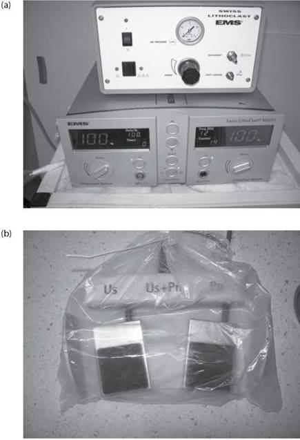
Figure 14.4 (a) The generator for the Swiss Lithoclast Master (b) The foot pedals used to activate the Swiss Lithoclast Master.