
Q. What is the machine shown in Figure 14.5 and how does it work?
A. Figure 14.5 is a picture of an ultrasound machine. This can be used as either a diagnostic or therapeutic tool in medicine. High-frequency sound waves are produced by the passage of current through a piezoelectric transducer, and subsequently focused. Medical ultrasound waves vary from 2 to 18 MHz. Lower frequencies are used to look at ‘deeper’ tissues, as the attenuation of sound waves is greater at higher frequencies. For example a transrectal ultrasound (TRUS) probe works at about 7 MHz, and transabdominal ultrasound works at around 3.5 MHz. Ultrasound waves pass into the body via an interface comprising the soft rubber coating on the transducer and gel. The sound waves are deflected back to the transducer, depending on an appropriate density change within the tissues. Large density changes (e.g. fluid and stone) produce a greater ‘echo’, and the time the waves take to come back to the transducer can determine the depth of the tissue.
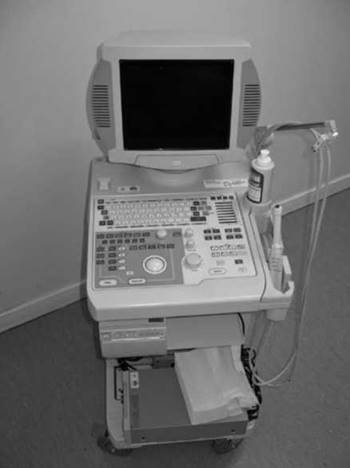
Figure 14.5
Q. What are the main therapeutic applications of ultrasound?
A. The main current therapeutic applications of ultrasound are lithotripsy (extracorporeal, during percutaneous nephrolithotomy and intracorporeal) and high-intensity focused ultrasound (HIFU), which is used in the treatment of prostate cancer. Ultrasound as a modality can guide other therapies such as prostate brachytherapy, cryotherapy and ESWL.
Q. What are the general contraindications to administration of intravenous contrast media?
A. The Royal College of Radiologists state that increased risk of adverse reactions may be seen in patients with a previous contrast reaction, asthma, renal impairment, diabetes mellitus and metformin therapy. In patients with normal renal function, there is no need to stop metformin. If renal function is impaired, metformin may be stopped for 48 hours following contrast administration. Untreated hyperthyroidism and myelomatosis are contraindications to contrast use. Nephrogenic systemic fibrosis is a rare complication seen after administration of gadolinium-based contrast agents in patients with severe renal impairment.
Q. A 64-year-old male is referred for an MRI following the diagnosis of prostate cancer.
He has previously had intracranial surgery following a stroke and works as an electrical engineer. What would be your concerns?
A. In this particular case, the concerns would be that the patient has an intracranial clip (for an aneurysm, e.g.), or may have an intra-ocular ferrous foreign body (secondary to his job). Imaging should not be performed on patients with intra-cranial clips, unless one is absolutely certain that they are MRI compatible. Patients who may have metal foreign bodies in their eyes should have radiographs of their orbits performed prior to MRI scanning. Radiographs can pick up objects greater or equal to 0.1 mm in size, and ferrous foreign bodies are thought not to be dangerous below this size.
Other implanted devices that are contraindicated include ICDs, pacemakers, cochlear implants, dental implants, neurostimulators, ocular implants and tissue expanders and prosthetic heart valves (depending on type).
Extra-cranial surgical clips, e.g. following abdominal surgery, are generally encased in fibrous tissue, however they may cause artefact, and scanning should be deferred for 6 weeks post-operatively.
Q. Briefly describe the physics behind MRI. What is the difference between T1 and T2 images?
A. MRI utilises the nuclei of hydrogen atoms (protons). The protons usually spin in a random fashion, however on entering the MRI scanner they align with the magnetic field in the longitudinal plane (the magnet in an MRI scanner is always ‘on’!), and produce a secondary spin (precession) at the same frequency, which will vary according to the strength of the magnet. A radiofrequency (RF) pulse is applied, which gives the nuclei the energy to move out of alignment and into the transverse plane, and to process in phase with one another. When this pulse is removed the atoms release their energy in two ways. First, energy is released back into the surrounding environment causing magnetic movements to relax and realign back into the longitudinal plane, a process referred to as T1 relaxation. Second, nuclei then lose their precessional coherence and dephase, due to energy loss between adjacent nuclei, and this is referred to as T2 decay. The release of energy is picked up in the transverse plane as an electrical voltage by a receiver coil, and this is the MR signal.
T1 relaxation occurs more rapidly in fat, as the size of the molecules enables them to give energy back to the environment more quickly. This means that there is a greater degree of transverse magnetisation following the next RF pulse, resulting in a very bright signal from fat on Tl-weighted images, while fluid remains dark. These scans are excellent for viewing anatomy due to the good tissue differentiation.
T2-weighted images rely on the process of T2 decay, which occurs more slowly in water, and There fore maintains transverse magnetisation for longer resulting in a higher signal. Consequently water has a very bright signal on these images, producing a scan which is more useful for demonstrating pathology.
Q. What is nephrogenic systemic fibrosis? How may this be related to MRI investigations?
A. Nephrogenic systemic fibrosis (NSF) is a condition of unknown cause that affects patients with renal disease. It causes collagen deposition and tightening of the skin of the extremities and sometimes trunk. It can be fatal, and 5% of patients develop the fulminant form. Causes of death are related to respiratory complications, clotting abnormalities and fractures/ falls, among others. There is no consistently successful treatment for NSF, although various strategies such as steroids, plasmapheresis and renal transplant have all been used.
Recent reports have linked the use of gadolinium-containing contrast agents to the development of NSF in patients with renal impairment. Gadolinium-containing contrast is used with great caution in patients with a GFR <60 mL/min/1.73 m2, including dialysis patients. The Royal College of Radiologists have recommended that if patients must receive these agents they should be specific ‘highly stable’ agents, and that the use should not be repeated within 7 days. The smallest dose possible should be used. Specific agents (Omniscan, Magnevist and OptiMARK) should not be used.
Q. You have requested a MAG-3 scan on a 29-year-old male you suspect has a pelviureteric junction obstruction. What is MAG-3, how is it handled by the kidney and what should the patient know prior to the test?
A. MAG-3 stands for mercaptoacetyltriglycine. MAG-3 is attached to the radioactive tracer Technetium-99m, an isotope with a short half-life (approximately 6 hours) that is used for other nuclear medicine scans, such as dimercapto-succinic acid (DMSA). MAG-3 is principally excreted by tubular secretion (90%), although approximately 10% is filtered at the glomerulus. Radioactivity is recorded via a gamma-camera (as with DMSA).
The patient will be asked to attend the nuclear medicine unit, and before the investigation the patient will have to empty his bladder. Usual medications should not be stopped, and the patient should be well hydrated. Children should not be brought along for the scan due to the potential radiation risk. A cannula is inserted, and a diuretic is injected (usually 15 minutes prior to the test, although protocols vary). The patient does not need to undress, although metal objects should be removed. The patient then sits on a chair while the MAG-3 is injected through the cannula. The patient then has to sit still for approximately 20 minutes while images are recorded. The patient is asked to keep well hydrated after the test, with no specific instructions otherwise. (Please also refer to Chapter 6.)
Q. A 2-year-old girl requires a MAG-3 scan to investigate a unilateral hydronephrosis. The parents are concerned about the process surrounding the scan and the risk of radiation. How would you reassure them?
A. Although the investigation is associated with radiation exposure, the overall dose is low (approximately 0.7 microSv). This is equivalent to about 4 months of background radiation. As a comparison, air travel (at 26,000 feet) provides approximately 3 microSv per hour at temperate latitudes, and approximately 1 microSv per hour around the equator. There fore no investigation involving radiation is entirely without risk; however the benefits of the investigation need to be weighed against the risks.
Children should eat and drink as normal before the scan and not stop any regular medications. The child should attend the ward in a well-hydrated state, and the paediatrician will insert a cannula after the application of anaesthetic cream. Occasionally the child may need some sedation. Diuretic may be injected prior to the isotope injection. The child must lie on a bed for approximately 20 minutes. The child does not need to be undressed, but will have to remove metal objects. After the scan the child should be kept well hydrated and empty the bladder regularly. (Please also refer to Chapter 6.)
Q. You have requested a DMSA scan on a 34-year-old female to look for the presence of renal scarring suggested on an ultrasound scan. What is DMSA, and how is the scan performed?
A. DMSA stands for dimercapto-succinic acid. It is attached to the radio-tracer Technetium-99m (see previous discussion). DMSA is a cortical-scanning agent that localises in the proximal tubule. It is minimally excreted, and its presence is a reflection of functioning renal tissue and nephrons.
An important difference between the ‘patient experience’ of DMSA versus MAG-3 is that patients may be in the hospital for many hours (during a DMSA renogram). If females suspect that they may be pregnant they should inform the department before attending, and should not be accompanied by children. A cannula is inserted into the patient, and the isotope injected. The static images are taken after an interval of approximately 2-4 hours post-injection. Patients are not required to undress, but metal objects should be removed. During the scan itself they will have to lie still on a couch. The gamma ‘camera’ is placed close to the kidneys but not touching the patient. After the scan patients are asked to keep well hydrated and empty their bladders regularly. (Please also refer to Chapter 6.)
Q. What steps are applied to ensure x-ray safety in theatres?
A. First, it is important that the surgeon has an understanding of the equipment in use and an up-to-date knowledge of radiation protection issues. The image intensifier should be operated by a trained radiographer and ideally the surgeon should have received specialised training from a medical physicist in protection aspects of fluoroscopy. It is important that all fluoroscopy equipment is periodically tested and maintained to ensure it is functioning correctly.
The main source of radiation to staff is scattered radiation from the x-ray tube, which in urological procedures is usually the end of the C-arm situated under the patient’s bed. Keeping the tube as close as possible to the table minimises radiation scatter. Staff should stand as far from the x-ray tube as possible to reduce their radiation exposure. Personal protective equipment should be worn by everyone in the operating theatre, with the exception of the patient. Lead aprons are the most effective and may reduce the dose received by around 90%. A lead apron with 0.35 mm lead thickness equivalence should be sufficient for most fluoroscopic procedures. A 0.25 mm apron may suffice for low work load fluoroscopy. Other equipment, such as thyroid shields and lead glass eyewear should be available and worn, especially when staff are exposed to regular and long fluoroscopy times. A personal radiation dosimeter should be worn at all times that fluoroscopy is in use.
For each individual case, the exposure of patients and staff to radiation must be reviewed and justified. Female patients of childbearing age must have a pregnancy test prior to leaving the ward. While fluoroscopic screening is in progress the theatre doors should be closed and a warning sign displayed. A red light at the theatre entrance indicates that fluoroscopy is in progress. The rationale for each image taken should be considered and rationalised in accordance with the ALARA (as little as is reasonably appropriate) principle. Intermittent screening, as opposed to continuous is preferred. Hands (patient or staff) should be kept out of the primary beam unless unavoidable for clinical reasons as the automatic exposure control system will trigger an increase in exposure to maintain image quality. An alarm can be set on the x-ray machine, which sounds when preset radiation dose limit is reached.