
Andrei Purysko and Michael A. Bolen
Despite continuing advances that have greatly expanded the ability to assess cardiovascular anatomy noninvasively through an array of imaging modalities, chest radiography (CR) remains a very useful part of the initial cardiovascular evaluation. CR is widely available, can be acquired rapidly and inexpensively, and has a low associated ionizing radiation dose. With an organized approach toward interpretation, and insight into the strengths and limitations of CR, the cardiologist can maximize the use of this tool in clinical practice. This chapter focuses on the most important components of the CR with regard to cardiovascular disease, namely:
1. Pulmonary vascular patterns
2. Cardiomediastinal silhouette
3. Calcification patterns
The following radiographs illustrate the appearance of a variety of cardiac abnormalities. Whenever possible, tables are included to delineate the differential diagnoses for representative chest radiographic findings.
BASIC APPROACH AND PROJECTIONS
Understanding the strengths and weaknesses of CR techniques is helpful in both the selection and interpretation of exam. Standard CR techniques include erect posteroanterior (PA) and lateral projections. In patients unable to stand for an erect PA, an anteroposterior (AP) radiograph can be performed. Additional specialized CR techniques exist, though their utilization is limited with widespread availability of tomographic imaging. The erect PA and lateral technique hold several advantages, as AP technique will magnify car- diomediastinal structures, and the ability to interpret pulmonary vasculature correctly is limited in supine patients. Additionally, more standardized techniques of acquisition are often used with PA radiographs, allowing for more reliable comparison between radiographs. Benefits of portable radiographs include the speed and convenience of obtaining a radiograph without transporting a patient.
Interpretation of radiographs can be aided by an ordered approach. Identification of the patient’s name and date of the examination is a straightforward but important step and can save further confusion or wasted time when checked first. Visible medical devices (catheters, drains, and other support devices) should be assessed to confirm position and any associated complication. When prior CRs are available, they should be assessed concurrently to help detect new abnormalities or to confirm the stability of a long-standing finding.
The three aforementioned components, pulmonary vasculature, cardiomediastinal silhouette, and calcifications, can then be reviewed in any order. Changes in each of these central components are a direct manifestation of the underlying cardiovascular disorder; and taking all three of these components into account ensures an appropriate, systematic approach to the standard CR. Other elements of CR interpretation are important (such as appropriate technique, assessment of pulmonary parenchyma) and have important clinical implications; however, we focus our discussion toward the elements most helpful in depiction of cardiovascular disease.
PULMONARY VASCULAR PATTERNS
Pulmonary Vascular Patterns in Normal and Various Cardiovascular Disease States
1. Normal
 In normal patients, the vessels in the upper zones are thinner than those at a similar distance from the hilum in the lower zones (normal ratio 1:3)
Pulmonary artery (PA)/pulmonic valve obstruction (no shunt or pulmonary hypertension)
Aorta/aortic valve (AV) obstructions (no shunt, left ventricular compromise, mitral valve disease)
Insignificant left-to-right shunt: Qp/Qs< 1.5
2. Increased (overcirculation)
Significant left-to-right shunt: Qp/Qs>1.5 (acyanotic)
Atrial septal defect (ASD)
Ventricular septal defect (VSD)
Patent ductus arteriosus
Partial anomalous pulmonary venous return
3. Decreased (undercirculation)
Right-to-left shunt
Tetralogy of Fallot
Ebstein anomaly with ASD
Pulmonary oligemia
Ebstein anomaly (severe ± ASD)
4. Admixture shunt (no pulmonic stenosis or atresia/cyanotic)
Transposition complexes
Truncus arteriosis
Univentricular heart (single ventricle)
Total anomalous pulmonary venous return: types I and II
Tricuspid atresia + VSD
5. High-output states
Anemia
Pregnancy
Decreased Pulmonary Blood Flow (Fig. 8.1)
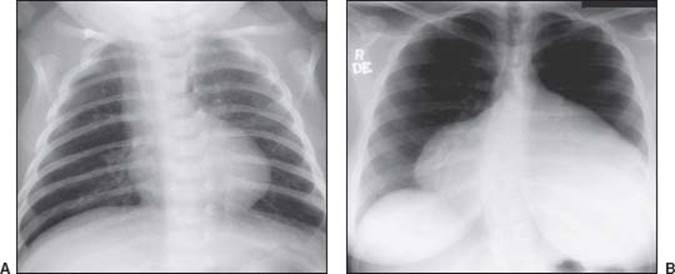
FIGURE 8.1 Decreased pulmonary blood flow. A: Tetralogy of Fallot. B: Ebstein anomaly.
Increased Pulmonary Blood Flow (Fig. 8.2)
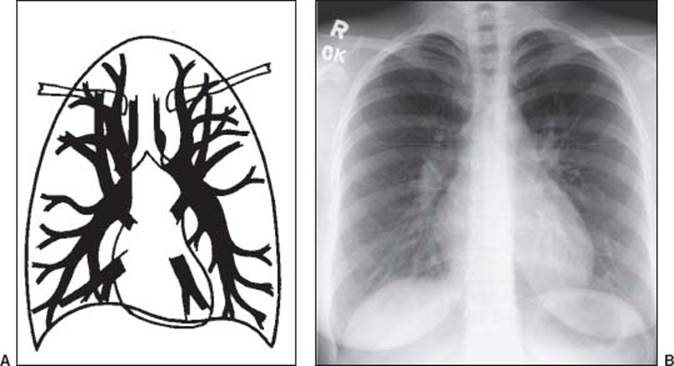
FIGURE 8.2 Increased pulmonary blood flow. A: Increased: balanced (overcirculation). B: ASD
Increased distributed (Fig 8.3)

FIGURE 8.3 Increased: redistributed. A: Pulmonary venous hypertension (PVH). B: Hypertrophic cardiomyopathy (HCM) PVH.
Pulmonary Arterial Hypertension (PAH)
Causes of PAH
 Precapillary: pulmonary hypertension (both primary and pulmonary hypertension associated with hepatic disease, drugs/toxins, HIV), congenital cardiovascular disease, chronic thromboembolism, chronic alveolar hypoxia (COPD, interstitial lung disease, hypoventilation)
Postcapillary: left sided cardiovascular disease, mitral stenosis, aortic valve disease, cardiac tumors, extrinsic pulmonary venous compression, fibrosing mediastinitis, pulmonary venoocclusive disease
Increased: Central (Fig. 8.4)
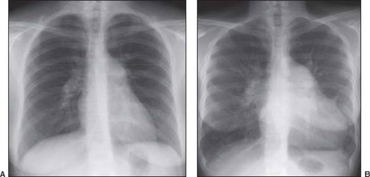
FIGURE 8.4 A: Idiopathic (pre-capillary) PAH; B: Post-capillary PAH.
Pulmonary Venous Hypertension
Stages of PVH
Stage 1: Pulmonary vascular redistribution
PCWP: Acute, 13 to 18 mm Hg; chronic, 18 to 22 mm Hg
Stage 2: Redistribution + interstitial pulmonary edema
PCWP: Acute, 18 to 25 mm Hg; chronic, 23 to 30 mm Hg
Stage 3: Redistribution + interstitial and alveolar pulmonary edema
PCWP: Acute, >25 mm Hg; chronic, >30 mm Hg>
Causes of PVH
Pulmonary venoocclusive disease
Pulmonary vein stenosis (postradiation therapy, post–atrial fibrillation ablation)
Left atrial/left ventricular obstruction
Myxoma or other tumors
Mitral valve disease
Mitral stenosis
Mitral regurgitation
Left ventricular compromise
Dilated cardiomyopathy
Acute or chronic myocardial ischemic disease
Restrictive cardiomyopathy
Pericardial disease
Constrictive pericarditis
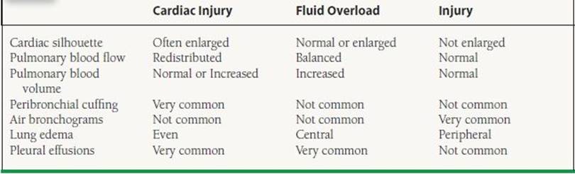
Pulmonary Edema Patterns
Increased hydrostatic pressure gradient
Increased capillary permeability
Decreased osmotic pressure gradient
Lymphatic incompetence
TABLE
8.1 Radiographic Features of Different Types of Pulmonary Edema
CARDIOMEDIASTINAL SILHOUETTE PATTERNS
Normal landmarks
Central
Tracheobronchial tree–PA relationship
Early branching to middle lobe on right
Right PA descends anterior to bronchus
Left PA passes over and descends posterior to bronchus
Right
Right chambers
Ascending aorta
Superior vena cava/azygos vein
Left
Aortic “knob” (distal arch + isthmus)
Descending aorta
Main PA
Descending aorta
Left atrium
Left ventricle
Normal Cardiac Silhouette (Fig. 8.5)
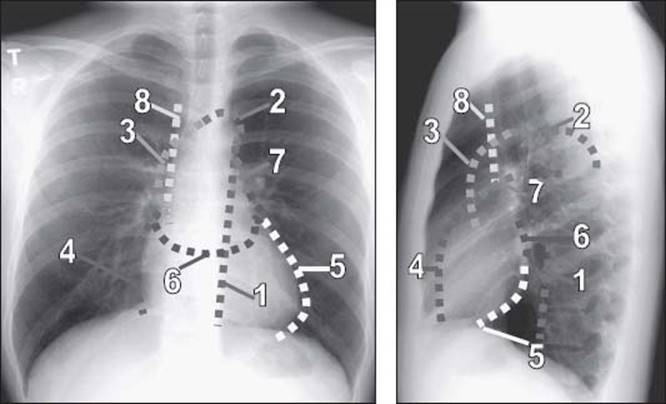
FIGURE 8.5 1, descending aorta; 2, aortic knob (distal arch and isthmus); 3, ascending aorta and proximal arch; 4, right ventricle; 5, left ventricle; 6, left atrium; 7, right atrium; 8, superior vena cava and azygous vein.
Normal Size
The most commonly used parameter for assessment of cardiac size is the cardiothoracic ratio, which corresponds to the maximum transverse diameter of the cardiac silhouette in relation to thoracic width. The accepted upper limit of normal is 50% in adults.
It should be emphasized that evaluation of individual cardiac chambers with CR is not reliable, with the occasional exception of left atrial dilation, which can be associated with a double density projecting over the right heart border as well as splayed central bronchi.
Interval changes in the size of the cardiac silhouette are of clinical interest, although this is always subject to variability in technique between CRs.
TABLE
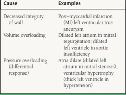
8.2 Enlargement of Cardiovascular Structure: Basic Causes
Pectus Excavatum (Fig. 8.6)
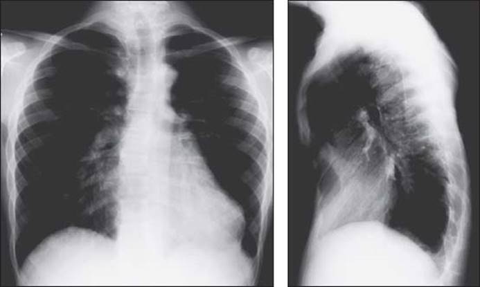
FIGURE 8.6 Pectus excavatum.
Findings
1. Pectus excavatum configuration of ribs
Straight or upsloping posterior ribs
Sharply downsloping anterior rib
2. Heart usually displaced to the left; right heart border not visible
Pericardial Cyst (Fig. 8.7)
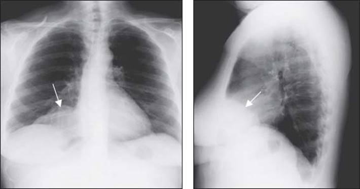
FIGURE 8.7 Pericardial cyst. Findings
Findings
1. The arrows mark the pericardial cyst
Aortic Stenosis (Fig. 8.8)
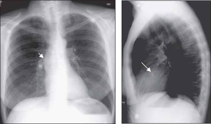
FIGURE 8.8 Aortic stenosis.
Findings
1. Calcified AV (long arrow)
2. Dilated ascending aorta beyond stenotic AV (short arrow)
3. Normal cardiac silhouette (because only pressure and not volume overload present)
Pseudocoarctation (Fig. 8.9)
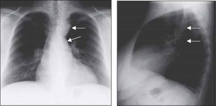
FIGURE 8.9 Pseudocoarctation.
Findings
1. “Double left aortic arch sign” (arrows mark the “2” arches)
2. Congenital elongation of the thoracic aorta associated with “kinking” of the aorta at the relatively fixed ligamentum arteriosum. There is no physiologic obstruction, so there is an absence of collateral vessel development and subsequent rib notching.
Coarctation (Fig. 8.10)

FIGURE 8.10 Coarctation.
Findings
1. Number “3” sign (short arrows)
2. Rib notching (long arrows) on the inferior portion of the posterior ribs (third to ninth) from pressure erosion by dilated intercostal arteries that serve as collateral blood flow between the internal mammary arteries and the descending aorta
Valvar Pulmonic Stenosis (Fig. 8.11)
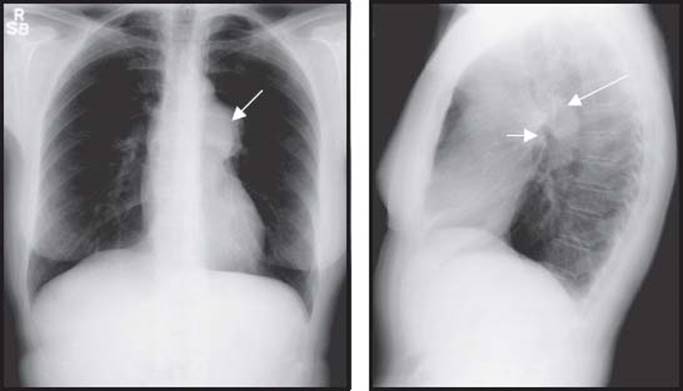
FIGURE 8.11 Valvar pulmonic stenosis.
Findings
1. Enlarged main PA, the degree of this enlargement does not predict the severity of stenosis
2. Selective enlargement of the left PA (long arrow) with normal-sized right PA (short arrow)
3. Normal cardiac silhouette and normal to decreased pulmonary vascular markings, depending on the degree of stenosis
Mitral Stenosis (Fig. 8.12)
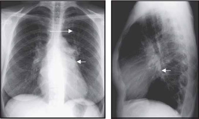
FIGURE 8.12 Mitral stenosis.
Findings
1. Increased pulmonary vascularity (long arrow)
2. Increased left atrial size (short arrows)
3. Normal-sized left ventricle
Mitral Regurgitation (Fig. 8.13)
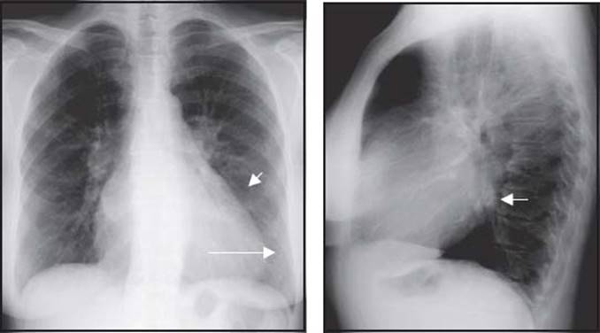
FIGURE 8.13 Mitral regurgitation.
Findings
1. Increased pulmonary vascularity
2. Increased left atrial size (short arrows)
3. Increased left ventricle size secondary to volume overload (long arrow)
CALCIFICATION PATTERNS
Cardiac
Left atrium
Mitral annulus
Mitral valve
Aortic valve
Coronary artery
Myocardium (post-MI)
Paracardiac
Pericardium
Thoracic aorta
Calcific Constrictive Pericarditis (Fig. 8.14)
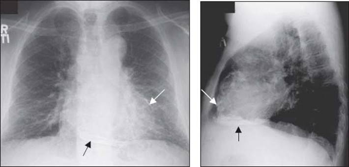
FIGURE 8.14 Calcific constrictive pericarditis.
Findings
1. Extensive pericardial calcification (arrows)
2. Calcifications along atrial surfaces suggest pericardium rather than myocardium origin.
Left Atrial Wall Calcification (Fig. 8.15)
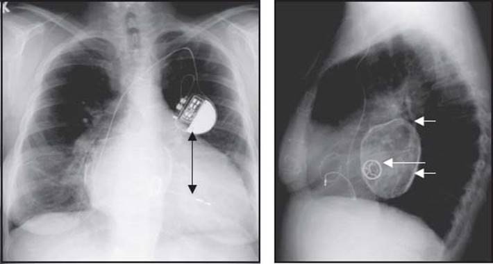
FIGURE 8.15 Left atrial wall calcification.
Findings
1. Severe diffuse calcification of the left atrium (short arrow)
2. Bjork–Shiley valve in the mitral position (long arrow)
3. Single right ventricular (RV)-lead pacemaker (black arrows)
Mitral Annular Calcification (Fig. 8.16)
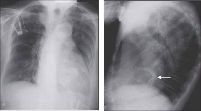
FIGURE 8.16 Mitral annular calcification.
Findings
1. Extensive mitral annulus calcification, seen best on the lateral radiograph as a large backward “C”
Calcified Post-MI Left Ventricular True Aneursym (Fig. 8.17)
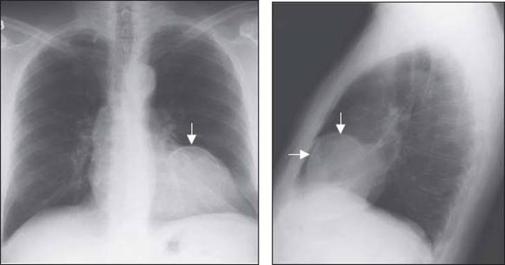
FIGURE 8.17 Calcified post-MI left ventricular true aneursym.
Findings
1. Calcified left ventricular aneurysm (arrows)
Coronary Artery Calcification (Fig. 8.18)

FIGURE 8.18 Coronary artery calcification.
Findings
1. Extensive coronary calcification of the left anterior descending artery (arrow), best seen on the lateral radiograph.
ACKNOWLEDGMENTS
The authors acknowledge Drs. Ross Downey, Richard White, and Richard Krasuski for their contributions to the first edition of this chapter.
QUESTIONS AND ANSWERS
Questions
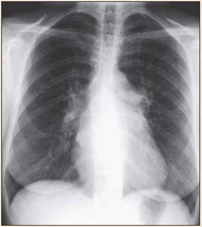
1. Ms. G. is a 28-year-old woman who immigrated to the United States from South America approximately 4 years ago. She has been told in the past that she has a heart murmur and recently noted the onset of exertional dyspnea. Her electrocardiogram (ECG) is notable for atrial fibrillation, and examination reveals a diastolic murmur best heard at the apex. Her chest radiograph is displayed below. Her atrial fibrillation is most likely secondary to:
a. Atrial myxomab.
b. Increased transmitral gradient
c. Idiopathic “primary” pulmonary hypertensiond.
d. “Lone” atrial fibrillatione.
e. Advanced left ventricular dysfunction
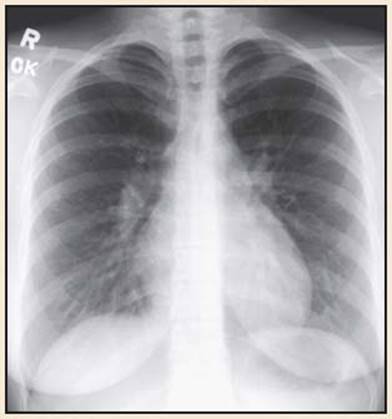
2. Mr. S. is a 38-year-old man with no prior medical history. Recently he has noticed a progressive reduction in his exercise tolerance. On examination he has a soft systolic murmur at the upper sternal border and a split second heart sound that does not appear to change with respiration. His chest radiograph is displayed below. The most likely explanation for these findings is:
a. Paroxysmal atrial fibrillation
b. Undiagnosed pulmonary stenosis
c. Undiagnosed ASD
d. Undiagnosed primary pulmonary hypertension
e. Left ventricular dysfunction with mitral regurgitation
3. Ms. B. is a 74-year-old woman who immigrated to the United States from Southwest Asia 6 years ago. For the last 2 years she has noted progressive fatigue and lower-extremity edema. Her chest radiograph is displayed below. A right heart catheterization in this patient would be expected to show all of the following characteristics except:

a. Elevated pulmonary capillary wedge pressure
b. Diastolic equalization of pressures
c. Elevated RV pressure
d. A significant step-up in oxygen saturations
e. Normal to decreased cardiac output
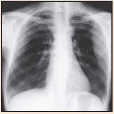
4. Mr. N. is a 32-year-old man with recently diagnosed hypertension that has been refractory to medical therapy. His exam is notable for a loud systolic murmur and weak peripheral pulses. His chest radiograph is shown below. The most appropriate surgical intervention for this patient is:
a. Resection and end-to-end anastomosis
b. Fontan procedure
c. Glenn shunt
d. Mitral valve repair
e. AV replacement
Answers
1. Answer B: Using the organized approach to radiograph interpretation, the pulmonary vascular pattern shows evidence of vascular redistribution suggestive of pulmonary venous hypertension. The left atrium is enlarged and the left ventricle is normal in size, suggesting possible mitral valve pathology. No chamber calcification is present to assist in the diagnosis. The case vignette describes a classical presentation for mitral stenosis. Though persistent inflammation may contribute to atrial fibrillation in this disorder, the primary mechanism in mitral stenosis is still felt to be elevated atrial pressure resulting in stretching of the atrial myocardium.
2. Answer C: Using the organized approach to radiograph interpretation, the pulmonary vascular pattern shows balanced overcirculation, and the pulmonary arteries are quite prominent. The right ventricle appears mildly enlarged. No significant calcification is present. The case vignette is notable for the physical exam, which is consistent with an ASD. Although this patient is certainly at increased risk for atrial fibrillation because of his congenital lesion, it is not the primary explanation for the findings. In pulmonic stenosis an oligemic pulmonary blood flow pattern is normally seen, and in left ventricular dysfunction with mitral regurgitation one expects to see a large left ventricle with pulmonary vascular redistribution suggestive of pulmonary venous hypertension. Primary pulmonary hypertension does not account for the physical findings in this case; additionally, a loud pulmonic closure sound would usually be present.
3. Answer D: Using the organized approach to radiograph interpretation, the pulmonary vascular pattern shows redistribution and pulmonary venous engorgement suggestive of elevated left heart filling pressure. There is chamber enlargement of both ventricles. The most notable feature of this radiograph, however, is the pericardial calcification, which appears circumferential. The vignette describes a case of constrictive pericarditis, possibly from old tuberculous infection. In this condition, one expects elevated filling pressure and a prominent Kussmaul sign (failure of the jugular venous pressure to drop with inspiration). A pericardial knock is often present as well. A step-up in oxygen saturations (suggestive of an intracardiac shunt) would not be expected in this patient.
4. Answer A: Using the organized approach to radiograph interpretation, the pulmonary vascular pattern does not appear to be particularly prominent. The chambers of the heart also do not appear to be abnormal in size. There is, however, significant rib notching. This is due to the prominent collaterals that develop to bypass the circulation and allow adequate blood flow to reach the periphery. The presence of these collaterals combined with the case history suggests the aortic coarctation. The classical surgical correction of this abnormality is resection and end-to-end anastomosis of the aorta. Recently, percutaneous techniques have become more popular, not only for postoperative recurrence of coarctation, but also as primary therapy.